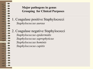
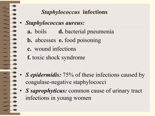
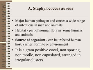
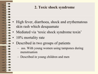
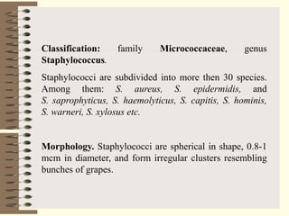
Gram positive bacteria
Lecturer Mr. matekwa
Gram positive bacteria
• Cocci:Staphylococcus, Streptococcus, Enterococcus.
• Rods:Bacillus, Corynebacterium,Nocardia, Clostridi
um, Actinobacteria and Listeria.
• In Gram-positive bacteria, the S-layer is attached to
the peptidoglycan layer.
• Gram +ve bacteria have teichoic acids in the cell
wall.
• Gram +ve: retain the crystal violet dye due to a thick
layer of peptidoglycan in the cell wall that encases
their cell membrane hence retains the stain.
• N.B. In Gram-ve bacteria, the S-layer is directly
attached to the outer membrane.
Gram-positive Bacteria Gram-negative Bacteria
Gram reaction
Retain crystal violet dye and stain dark
violet or purple
Can be decolourized to accept counter stain
(Safranin or Fuchsine); stain red or pink,.
Peptidoglycan layer Thick (multilayered) Thin (single-layered)
Teichoic acids Present in many Absent
Periplasmic space Absent present
Outer membrane Absent Present
Lipopolysaccharide (LPS)
content
Virtually none High
Lipid and lipoprotein
content
Low (acid-fast bacteria have lipids linked
to peptidoglycan)
High (due to presence of outer membrane)
Flagellar structure 2 rings in basal body 4 rings in basal body
Toxins produced Primarily Exotoxins Primarily Endotoxins
Resistance to physical
disruption
High Low
Inhibition by basic dyes High Low
Susceptibility to anionic
detergents
High Low
Resistance to sodium azide High Low
Resistance to drying High Low
Cell wall composition
Cell wall is 100-120 Armstrong thick,
single layered. Lipid content of cell wall is
low; Murein content is 70-80% (Higher).
The cell wall is 70-120 Armstrong thick,
two layered. The lipid content is 20-30%
(High), whereas Murein content is 10-20%
(Low).
Antibiotic Resistance More Susceptible to antibiotics More Resistant to antibiotics.
Gram + cocci
Catalase test
Coagulase +
Staph. aureus
Coagulase test
Coagulase -
Staph. epidermitis
Catalase +
Streptococci
Catalase +
Staphyllococci
Differentiating Gram positive
Staphylococcus
-By alexender Ogstein in 1880, he identified cluster
forming microorganisms
-Its from a two greek words i.e (staphyle- a bunch of
grapes and kokkos- grain or berry)
- They are facultative anaerobes, gram positive, colonizes
the skin and also found as normal flora of other sites like
the upper respiratory tract.
Staphylococci characteristics
• Staphyloccocci -from Greek “stapyle” (bunch of grapes)
• Gram positive cocci (single, in pairs, or irregular grape-
like clusters)
• 0.5-1.5µm in diameter
• Non Motile
• Non spore-forming and non flagellated
• Facultative anaerobes
• Hardy organisms surviving many non physiologic
conditions .e.g. High salt tolerance (up to 10% NaCl),
drying, heat (they withstand 50 °C for 30 minutes)
• Catalase positive (usually)
• Capsule variable - usually negative
• Colonies: round, smooth, raised, and glistening
• Rapidly develop resistance to many antimicrobial agents
Major pathogens in genus
Grouping for Clinical Purposes
1. Coagulase positive Staphylococci
Staphylococcus aureus
2. Coagulase negative Staphylococci
Staphylococcus epidermidis
Staphylococcus saprophyticus
Staphylococcus hominis
Staphyloccocus capitis
STAPHYLOCOCCUS :
• Three common species are:
1. Staphylococcus epidermidis
2. Staphylococcus saprophyticus
• Staphylococcus aureus: most pathogenic species with virulence due
to: Coagulase- clots plasma Toxins-necrosis of the skin;
Hemolysins- damages RBC Enterotoxin- food poisoning and
Leukocidins: kill WBC
Staphylococcus infections
• Staphylococcus aureus:
a. boils d. bacterial pneumonia
b. abcesses e. food poisoning
c. wound infections
f. toxic shock syndrome
• S epidermidis: 75% of these infections caused by
coagulase-negative staphylococci
• S saprophyticus: common cause of urinary tract
infections in young women
11
Staphylococcus aureus
• Grows in large, round, opaque colonies
• Optimum temperature of 37oC
– Facultative anaerobe
• Withstands high salt, extremes in pH, and
high temperatures
• Produces many virulence factors
A. Staphylococcus aureus
• Major human pathogen and causes a wide range
of infections in man and animals
• Habitat - part of normal flora in some humans
and animals
• Source of organism - can be infected human
host, carrier, formite or environment
• It is a gram positive cocci, non sporing,
non motile, non capuslated, arranged in
irregular clusters
13
Epidemiology and Pathogenesis
• Present in most environments frequented by
humans
• Readily isolated from fomites
• Carriage rate for healthy adults is 20-60%
• Carriage is mostly in anterior nares, skin,
nasopharynx, intestine
• Predisposition to infection include: poor hygiene
and nutrition, tissue injury, preexisting primary
infection, diabetes, immunodeficiency
• Increase in community acquired methicillin
resistance - MRSA
14
Virulence factors of S. aureus
Enzymes:
• Coagulase – coagulates plasma and blood;
produced by 97% of human isolates; diagnostic
• Hyaluronidase – digests connective tissue
• Staphylokinase – digests blood clots
• DNase – digests DNA
• Lipases – digest oils; enhances colonization on skin
• Penicillinase – inactivates penicillin
15
Virulence factors of S. aureus
Staphylococal Toxins:
• Hemolysins (α, β, γ, δ) – lyse red blood cells
• Leukocidin – lyses neutrophils and macrophages
• Enterotoxin – induce gastrointestinal distress and
food poisoning
• Toxic shock syndrome toxin (TSST) – induces
fever, vomiting, shock, systemic organ damage,
tumors and necrosis
• Epidermolytic toxins- cause blistering, most
common dramatic manifestation of epidermolytic
toxins , scalded skin, skin infections, painful rash
Sources of infection
Infected lesions
-Pus, dried exudates from infected wounds,
burns, infected skin lesions and in sputum
coughed from lungs of patients with
bronchopneumonia.
-Direct contact mode of spread may also
occur
-Food handlers may introduce enterotoxins
producing food poisoning strains into food
Cont..
Healthy carries
-Staphyloccoccus aureus grows harmlessly in
moist skin and nostrils
-Organisms spread from these areas into the
environment through; hands, handkachiefs,
clothing, dust
- Some carries called shedders , disseminate
large no of staphylococci
-Babies with the staphylococcus aureaus in
the nose and skin can transmit it to the
nursing mothers who develop mastitis
Cont..
Animals
- Animals disseminate staphylococcus
aureus and cause human infection eg. Milk
from a cow with mastitis, causing
staphylococcal food poisoning
Mode of infection
-May be exogenous (from an external source)
or endogenous ( from carriage site or
minor lesions in patients own body)
- Body surfaces of humans and animals are
the main reservoir
19
Clinical Concerns and Treatment:
• 95% have penicillinase and are resistant to
penicillin and ampicillin
• MRSA – methicillin-resistant S. aureus –
carry multiple resistance
Prevention of staph infections:
• Universal precautions by healthcare
providers to prevent nosocomial infections
• Hygiene and cleansing
Natural history of disease
• Many neonates, children, adults -intermittently
colonised by S. aureus
• Usual sites - skin, nasopharynx, perineum
• Breach in mucosal barriers - can enter
underlying tissue
• Characteristic abscesses
• Disease due to toxin production
DISEASES
• Due to direct effect
of organism
– Local lesions of
skin
– Deep abscesses
– Systemic
infections
• Toxin mediated
– Food poisoning
– toxic shock
syndrome
– Scalded skin
syndrome
SKIN LESIONS
• Boils
• Styes
• Furuncles(infection of hair follicle)
• Carbancles (infection of several hair follicles)
• Wound infections(progressive appearance of
swelling and pain in a surgical wound after about
2 days from the surgery)
• Impetigo (skin lesion with blisters that break and
become covered with crusting exudate)
DEEP ABSCESSSES
• Can be single or multiple
• Breast abscess can occur in 1-3% of
nursing mothers in puerperiem
• Can produce mild to severe disease
• Other sites - kidney, brain from septic foci
in blood
Systemic Infections
• 1. With obvious focus
– Osteomyelitis, septic arthritis
• 2. No obvious focus
• heart (infective endocarditis)
• Brain(brain abscesses)
• 3. Ass. With predisposing factors
– multiple abscesses, septicaemia(IV drug
users)
– Staphylococcal pneumonia (Post viral)
B. TOXIN MEDIATED
DISEASES
• 1. Staphylococcal food poisoning
– Due to production of entero toxins
– heat stable entero toxin acts on gut
– produces severe vomiting following a very
short incubation period
– Resolves on its own within about 24 hours
2. Toxic shock syndrome
• High fever, diarrhoea, shock and erythematous
skin rash which desquamate
• Mediated via ‘toxic shock syndrome toxin’
• 10% mortality rate
• Described in two groups of patients
– ass. With young women using tampones during
menstruation
– Described in young children and men
3. Scalded skin syndrome
• Disease of young children
• Mediated through minor Staphylococcal infection
by ‘epidermolytic toxin’ producing strains
• Mild erythema and blistering of skin followed by
shedding of sheets of epidermis
• Children are otherwise healthy and most
eventually recover
Classification: family Micrococcaceae, genus
Staphylococcus.
Staphylococci are subdivided into more then 30 species.
Among them: S. aureus, S. epidermidis, and
S. saprophyticus, S. haemolyticus, S. capitis, S. hominis,
S. warneri, S. xylosus etc.
Morphology. Staphylococci are spherical in shape, 0.8-1
mcm in diameter, and form irregular clusters resembling
bunches of grapes.
Laboratory diagnosis.
Pus – from abscesses, wounds, burns etc
Sputum- from patients wit peumonia
Faeces or vomit- suspected food poisoning
Blood – patients with bacteraemia
Mid stream urine – patients with cystitis
Anterior nasal and perineal swab
The material is examined for the presence of pathogenic
staphylococci.
Treatment.
Staphylococcal diseases are treated with antibiotics
(penicillin, phenoxymethylpenicillin, tetracycline,
gramicidin, etc.), sulphonamides (norsulphazol, sulphazol,
etc.), and antistaphylococcal gamma-globulin.
Coagulase negative staphylococci
- Is a large group of related species found on the surface of
healthy individuals and rarely cause infections. i.e
 staphylococcus Epidermatis – cause wound infection,
sinus infection, endocarditis , and inflamation
 Staphylococcus saprophyticus – urinary tract infection
 Staphylococcus hominis – infect people with weak
immunity
 Staphylococcus capitis – causes endocarditis
-Are opportunistic pathogens that cause infections in immuno-
compromised patients
-Colonize medical devices and can cause problems in cardiac
surgery

Staphylococci.pptx

  • 1.
  • 2.
    Gram positive bacteria •Cocci:Staphylococcus, Streptococcus, Enterococcus. • Rods:Bacillus, Corynebacterium,Nocardia, Clostridi um, Actinobacteria and Listeria. • In Gram-positive bacteria, the S-layer is attached to the peptidoglycan layer. • Gram +ve bacteria have teichoic acids in the cell wall. • Gram +ve: retain the crystal violet dye due to a thick layer of peptidoglycan in the cell wall that encases their cell membrane hence retains the stain. • N.B. In Gram-ve bacteria, the S-layer is directly attached to the outer membrane.
  • 4.
    Gram-positive Bacteria Gram-negativeBacteria Gram reaction Retain crystal violet dye and stain dark violet or purple Can be decolourized to accept counter stain (Safranin or Fuchsine); stain red or pink,. Peptidoglycan layer Thick (multilayered) Thin (single-layered) Teichoic acids Present in many Absent Periplasmic space Absent present Outer membrane Absent Present Lipopolysaccharide (LPS) content Virtually none High Lipid and lipoprotein content Low (acid-fast bacteria have lipids linked to peptidoglycan) High (due to presence of outer membrane) Flagellar structure 2 rings in basal body 4 rings in basal body Toxins produced Primarily Exotoxins Primarily Endotoxins Resistance to physical disruption High Low Inhibition by basic dyes High Low Susceptibility to anionic detergents High Low Resistance to sodium azide High Low Resistance to drying High Low Cell wall composition Cell wall is 100-120 Armstrong thick, single layered. Lipid content of cell wall is low; Murein content is 70-80% (Higher). The cell wall is 70-120 Armstrong thick, two layered. The lipid content is 20-30% (High), whereas Murein content is 10-20% (Low). Antibiotic Resistance More Susceptible to antibiotics More Resistant to antibiotics.
  • 5.
    Gram + cocci Catalasetest Coagulase + Staph. aureus Coagulase test Coagulase - Staph. epidermitis Catalase + Streptococci Catalase + Staphyllococci Differentiating Gram positive
  • 6.
    Staphylococcus -By alexender Ogsteinin 1880, he identified cluster forming microorganisms -Its from a two greek words i.e (staphyle- a bunch of grapes and kokkos- grain or berry) - They are facultative anaerobes, gram positive, colonizes the skin and also found as normal flora of other sites like the upper respiratory tract.
  • 7.
    Staphylococci characteristics • Staphyloccocci-from Greek “stapyle” (bunch of grapes) • Gram positive cocci (single, in pairs, or irregular grape- like clusters) • 0.5-1.5µm in diameter • Non Motile • Non spore-forming and non flagellated • Facultative anaerobes • Hardy organisms surviving many non physiologic conditions .e.g. High salt tolerance (up to 10% NaCl), drying, heat (they withstand 50 °C for 30 minutes) • Catalase positive (usually) • Capsule variable - usually negative • Colonies: round, smooth, raised, and glistening • Rapidly develop resistance to many antimicrobial agents
  • 8.
    Major pathogens ingenus Grouping for Clinical Purposes 1. Coagulase positive Staphylococci Staphylococcus aureus 2. Coagulase negative Staphylococci Staphylococcus epidermidis Staphylococcus saprophyticus Staphylococcus hominis Staphyloccocus capitis
  • 9.
    STAPHYLOCOCCUS : • Threecommon species are: 1. Staphylococcus epidermidis 2. Staphylococcus saprophyticus • Staphylococcus aureus: most pathogenic species with virulence due to: Coagulase- clots plasma Toxins-necrosis of the skin; Hemolysins- damages RBC Enterotoxin- food poisoning and Leukocidins: kill WBC
  • 10.
    Staphylococcus infections • Staphylococcusaureus: a. boils d. bacterial pneumonia b. abcesses e. food poisoning c. wound infections f. toxic shock syndrome • S epidermidis: 75% of these infections caused by coagulase-negative staphylococci • S saprophyticus: common cause of urinary tract infections in young women
  • 11.
    11 Staphylococcus aureus • Growsin large, round, opaque colonies • Optimum temperature of 37oC – Facultative anaerobe • Withstands high salt, extremes in pH, and high temperatures • Produces many virulence factors
  • 12.
    A. Staphylococcus aureus •Major human pathogen and causes a wide range of infections in man and animals • Habitat - part of normal flora in some humans and animals • Source of organism - can be infected human host, carrier, formite or environment • It is a gram positive cocci, non sporing, non motile, non capuslated, arranged in irregular clusters
  • 13.
    13 Epidemiology and Pathogenesis •Present in most environments frequented by humans • Readily isolated from fomites • Carriage rate for healthy adults is 20-60% • Carriage is mostly in anterior nares, skin, nasopharynx, intestine • Predisposition to infection include: poor hygiene and nutrition, tissue injury, preexisting primary infection, diabetes, immunodeficiency • Increase in community acquired methicillin resistance - MRSA
  • 14.
    14 Virulence factors ofS. aureus Enzymes: • Coagulase – coagulates plasma and blood; produced by 97% of human isolates; diagnostic • Hyaluronidase – digests connective tissue • Staphylokinase – digests blood clots • DNase – digests DNA • Lipases – digest oils; enhances colonization on skin • Penicillinase – inactivates penicillin
  • 15.
    15 Virulence factors ofS. aureus Staphylococal Toxins: • Hemolysins (α, β, γ, δ) – lyse red blood cells • Leukocidin – lyses neutrophils and macrophages • Enterotoxin – induce gastrointestinal distress and food poisoning • Toxic shock syndrome toxin (TSST) – induces fever, vomiting, shock, systemic organ damage, tumors and necrosis • Epidermolytic toxins- cause blistering, most common dramatic manifestation of epidermolytic toxins , scalded skin, skin infections, painful rash
  • 16.
    Sources of infection Infectedlesions -Pus, dried exudates from infected wounds, burns, infected skin lesions and in sputum coughed from lungs of patients with bronchopneumonia. -Direct contact mode of spread may also occur -Food handlers may introduce enterotoxins producing food poisoning strains into food
  • 17.
    Cont.. Healthy carries -Staphyloccoccus aureusgrows harmlessly in moist skin and nostrils -Organisms spread from these areas into the environment through; hands, handkachiefs, clothing, dust - Some carries called shedders , disseminate large no of staphylococci -Babies with the staphylococcus aureaus in the nose and skin can transmit it to the nursing mothers who develop mastitis
  • 18.
    Cont.. Animals - Animals disseminatestaphylococcus aureus and cause human infection eg. Milk from a cow with mastitis, causing staphylococcal food poisoning Mode of infection -May be exogenous (from an external source) or endogenous ( from carriage site or minor lesions in patients own body) - Body surfaces of humans and animals are the main reservoir
  • 19.
    19 Clinical Concerns andTreatment: • 95% have penicillinase and are resistant to penicillin and ampicillin • MRSA – methicillin-resistant S. aureus – carry multiple resistance Prevention of staph infections: • Universal precautions by healthcare providers to prevent nosocomial infections • Hygiene and cleansing
  • 20.
    Natural history ofdisease • Many neonates, children, adults -intermittently colonised by S. aureus • Usual sites - skin, nasopharynx, perineum • Breach in mucosal barriers - can enter underlying tissue • Characteristic abscesses • Disease due to toxin production
  • 21.
    DISEASES • Due todirect effect of organism – Local lesions of skin – Deep abscesses – Systemic infections • Toxin mediated – Food poisoning – toxic shock syndrome – Scalded skin syndrome
  • 22.
    SKIN LESIONS • Boils •Styes • Furuncles(infection of hair follicle) • Carbancles (infection of several hair follicles) • Wound infections(progressive appearance of swelling and pain in a surgical wound after about 2 days from the surgery) • Impetigo (skin lesion with blisters that break and become covered with crusting exudate)
  • 23.
    DEEP ABSCESSSES • Canbe single or multiple • Breast abscess can occur in 1-3% of nursing mothers in puerperiem • Can produce mild to severe disease • Other sites - kidney, brain from septic foci in blood
  • 24.
    Systemic Infections • 1.With obvious focus – Osteomyelitis, septic arthritis • 2. No obvious focus • heart (infective endocarditis) • Brain(brain abscesses) • 3. Ass. With predisposing factors – multiple abscesses, septicaemia(IV drug users) – Staphylococcal pneumonia (Post viral)
  • 25.
    B. TOXIN MEDIATED DISEASES •1. Staphylococcal food poisoning – Due to production of entero toxins – heat stable entero toxin acts on gut – produces severe vomiting following a very short incubation period – Resolves on its own within about 24 hours
  • 26.
    2. Toxic shocksyndrome • High fever, diarrhoea, shock and erythematous skin rash which desquamate • Mediated via ‘toxic shock syndrome toxin’ • 10% mortality rate • Described in two groups of patients – ass. With young women using tampones during menstruation – Described in young children and men
  • 27.
    3. Scalded skinsyndrome • Disease of young children • Mediated through minor Staphylococcal infection by ‘epidermolytic toxin’ producing strains • Mild erythema and blistering of skin followed by shedding of sheets of epidermis • Children are otherwise healthy and most eventually recover
  • 28.
    Classification: family Micrococcaceae,genus Staphylococcus. Staphylococci are subdivided into more then 30 species. Among them: S. aureus, S. epidermidis, and S. saprophyticus, S. haemolyticus, S. capitis, S. hominis, S. warneri, S. xylosus etc. Morphology. Staphylococci are spherical in shape, 0.8-1 mcm in diameter, and form irregular clusters resembling bunches of grapes.
  • 29.
    Laboratory diagnosis. Pus –from abscesses, wounds, burns etc Sputum- from patients wit peumonia Faeces or vomit- suspected food poisoning Blood – patients with bacteraemia Mid stream urine – patients with cystitis Anterior nasal and perineal swab The material is examined for the presence of pathogenic staphylococci.
  • 30.
    Treatment. Staphylococcal diseases aretreated with antibiotics (penicillin, phenoxymethylpenicillin, tetracycline, gramicidin, etc.), sulphonamides (norsulphazol, sulphazol, etc.), and antistaphylococcal gamma-globulin.
  • 31.
    Coagulase negative staphylococci -Is a large group of related species found on the surface of healthy individuals and rarely cause infections. i.e  staphylococcus Epidermatis – cause wound infection, sinus infection, endocarditis , and inflamation  Staphylococcus saprophyticus – urinary tract infection  Staphylococcus hominis – infect people with weak immunity  Staphylococcus capitis – causes endocarditis -Are opportunistic pathogens that cause infections in immuno- compromised patients -Colonize medical devices and can cause problems in cardiac surgery