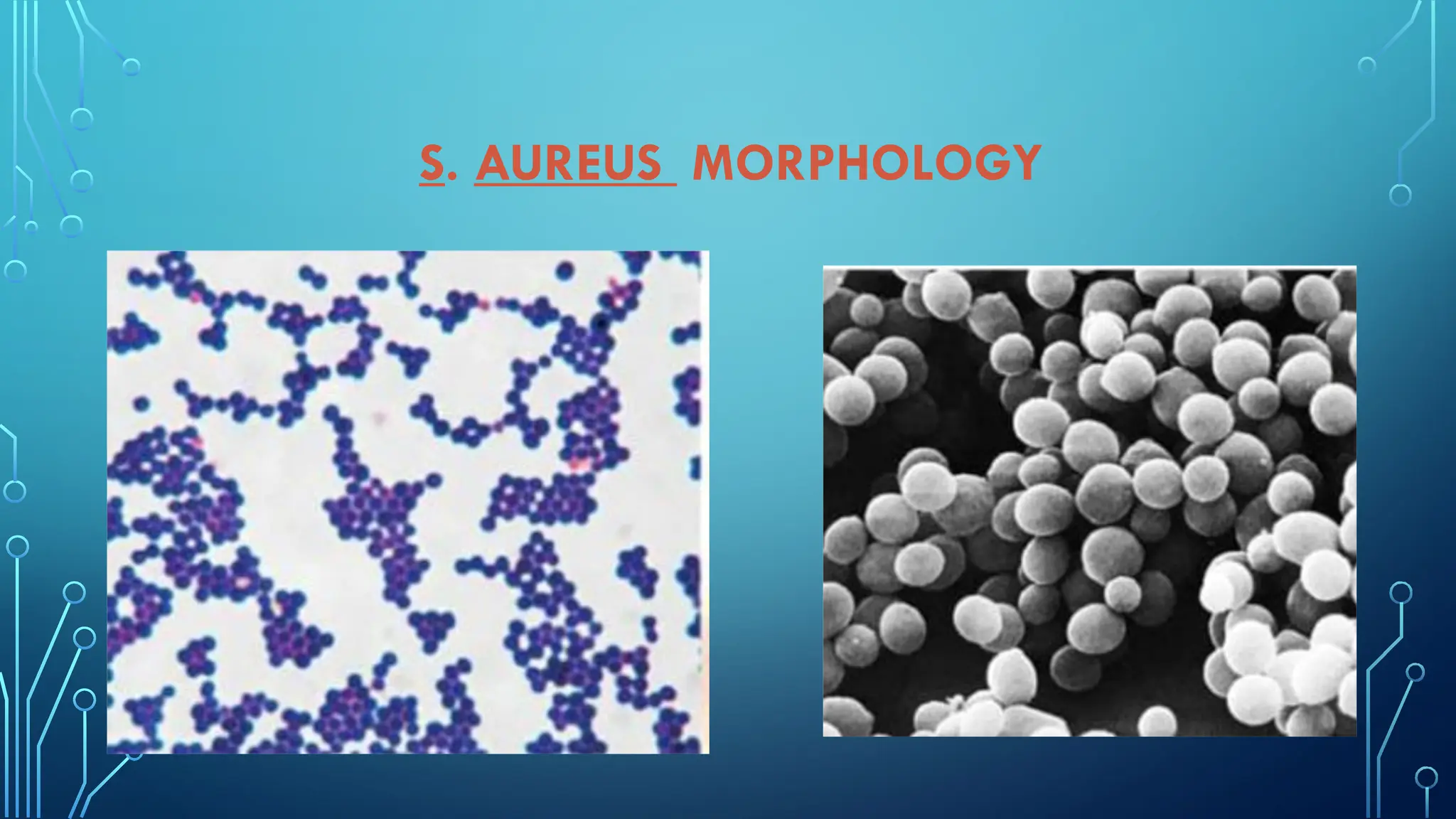
VIVEKANANDHA
ARTS AND SCIENCE COLLEGE FOR WOMEN
. VEERACHIPALAYAM-637303, SANKAGIRI, SALEM DT, TAMILNADU, INDIA
AFFILIATED TO PERIYAR UNIVERSITY, SALEM
. DEPARTMENT OF MICROBIOLOGY
. SUBJECT : MEDICAL BACTERIOLOGY
TITLE : STAPHYLOCOCCUS .AUREUS
SUBJECT INCHARGE:
Dr.R.MYTHILI RAVICHANDRAN,
ASSISTANT PROFESSOR,
HEAD OF THE DEPARTMENT,
DEPARTMENT OF MICROBIOLOGY.
SUBMITTED BY :
Ms . M . ARTHI,
lll. Bsc., MICROBIOLOGY,
DEPARTMENT OF MICROBIOLOGY.
STAPHYLOCOCCUS AUREUS
SYNOPSIS :
• Introduction
• Morphology
• History
• Virulence factors
• Pathogenesis
• Clinical syndrome
• Culture characteristics
• Lab diagnosis
• Mode of transmission
• Treatment
• Prevention
• reference
INTRODUCTION
STAPHYLOCOCCUS
• Gram positive cocci, belongs to Family Micrococcaceae.
• Aerobic , some facultative anaerobes.
• Arrangement in clusters ( like grapes ), pairs or tetrads,
• Catalase positive.
• S. aureus is the most important human pathogen, others human pathogens are
CONS (Coagulase negative Staphylococci) which include S. saprophyticus, S.
epidermidis, S. hominis etc.
• Staphylococcus are capable of acquiring resistance to antibiotics,
• Can cause serious clinical and epidemiological problems.
Staphylococcus aureus
Morphology
• Non motile and non spore forming
•Gram positive cocci
•Size is 0.7 to 1.2mm in diameter
•Irregular clusters that resemble cluster of grapes.
S. AUREUS MORPHOLOGY
HISTORY
• Robert Koch (1878)- first to see staphylococci in
pus specimen
• Louis Pasteur (1880)- first to cultivate in liquid
medium
• Sir Alexander Ongston (1881)- named the
bacteria as “ Staphylococcus”
VIRULENCE FACTORS
CELL WALL
ASSOCIATED
STRUCTURES
• Peptidoglycan
• Capsule
• Protein A
• Clumping factor
(bound Coagulase)
EXTRA CELLULAR
TOXINS
• Haemolysin
• Leukocidin
• Enterotoxin
• TSST
• Exfoliatin toxin
COAGULASE
• Staphylokinase
• DNAase
• Phosphatase
• Lipase
• Phospholipase
• Hyaluronidase
• Serokinase
• Protease
PATHOGENESIS
• Cause localised lesions
• First multiply in tissues, produce toxins and stimulate inflammation.
• Adhere to damaged skin.
• Evade defense mechanism of host.
• Cause tissue damage and from abscesses, produce extra-cellular
enzymes and exotoxins.
Host immunity:
• No life long immunity.
• Repeated infections occur in susceptible host.
CLINICAL SYNDROME
• 2 types: inflammatory and toxin mediated staphylococcal disease.
INFLAMMATORY DISEASES:
• Impetigo, folliculitis, furuncle, carbuncle, surgical wound infection,
postpartum breast infection.
• Bacterimia (bacteria found in blood) and septicemia
( multiplication of bacteria produce toxins cause high rate of fever)
• Endocarditis.
• Osteomyelitis and arthritis.
• Deep seated abscess in any organ after bacterimia.
Toxin mediated staphylococcal disease:
1. Staphylococcal food poisoning:
• Caused by Enterotoxin.
• Milk, milk products, meat fish when kept at room temperature after
cooking, contaminants multiply and produce toxins.
• Virulent when release of interoleukins( L-1and L-2).
• Sudden symptoms after 2-6 hour of ingestion
• Nausea, vomiting, abdominal cramps, watery or bloody diarrhea.
2. Staphylococcal toxic shock syndrome (TSST):
• Caused by toxin shock syndrome toxin.
• Release of large amount of interleukin lL-1 and lL-2.
• Life threatening condition.
• Fever, vomiting, hypertension, myalgia( muscle pain) mucosal hyperemia,
erythematous rash ( redness due to increased blood flow).
3. Staphylococcal scaled skin syndrome (SSSS):
• Caused by exfoliative toxin
• Common in infants and children
• Outer layer of skin of epidermis is separated from underlying tissue.
• Appears as extensive bullae ( rupture and leave behind red, tender skin ).
STAPHYLOCOCCUS AUREUS
Culture characteristics:
•Grow readily on ordinary culture media.
•Optimum temperature -37°C
•Optimum pH – 7.4 – 7.6
•Aerobes and facultative anaerobes.
LAB DIAGNOSIS:
1. Specimens:
•Pus – from wound or abscess or burns
•Nasal Swab – from suspected carrier
•Food – to diagnose staphylococcal intoxication
•Blood- to diagnose endocarditis and bacteremia
•Sputum- to diagnose lower respiratory tract infection
2. Gram staining:
The diagnosis of staphylococcus aureus is suggested by the finding of gram
positive bacteria in clumps in the sample.
3. Culture:
on nutrient agar,
• Golden yellow and opaque colonies with smooth glistening surface,
1-2mm in diameter
On blood agar:
• Golden yellow colonies, surrounded by a clear zone of hemolysis ( beta
hemolysis), esp. When incubated in sheep blood agar in atmosphere of 20%
CO2
• On MacConkey agar:
• Smaller colonies than those on NA (0.1-0.5mm) and are pink coloured due to
lactose fermentation
On Mannitol salary agar:
S.aureus ferments mannitol and appear as yellow colonies
• MSA is a useful selective medium for recovering S. aureus from
faecal specimen, when investigating food poisoning
BIOCHEMICAL PROPERTIES
• Indole test – negative
• MR test – positive
• VP test – positive
• Urease test – positive
• Phosphatase – positive
• DNAase test – positive
• Coagulase test – positive
• Hydrolyse gelatin
• Reduces nitrate to nitrite
MODE OF TRANSMISSION
• Person with lesions
•Airborne droplets
•Asymptomatic carrier
•Cross infection
TREATMENT
PREVENTION
REFERENCE
• Ananthanarayan and Paniker’s text book of Microbiology
12 th edition
• Google images
THANK YOU 🤗

staphylococcus aureus from medical bacteriology

  • 1.
    VIVEKANANDHA ARTS AND SCIENCECOLLEGE FOR WOMEN . VEERACHIPALAYAM-637303, SANKAGIRI, SALEM DT, TAMILNADU, INDIA AFFILIATED TO PERIYAR UNIVERSITY, SALEM . DEPARTMENT OF MICROBIOLOGY . SUBJECT : MEDICAL BACTERIOLOGY TITLE : STAPHYLOCOCCUS .AUREUS SUBJECT INCHARGE: Dr.R.MYTHILI RAVICHANDRAN, ASSISTANT PROFESSOR, HEAD OF THE DEPARTMENT, DEPARTMENT OF MICROBIOLOGY. SUBMITTED BY : Ms . M . ARTHI, lll. Bsc., MICROBIOLOGY, DEPARTMENT OF MICROBIOLOGY.
  • 2.
    STAPHYLOCOCCUS AUREUS SYNOPSIS : •Introduction • Morphology • History • Virulence factors • Pathogenesis • Clinical syndrome • Culture characteristics • Lab diagnosis • Mode of transmission • Treatment • Prevention • reference
  • 3.
    INTRODUCTION STAPHYLOCOCCUS • Gram positivecocci, belongs to Family Micrococcaceae. • Aerobic , some facultative anaerobes. • Arrangement in clusters ( like grapes ), pairs or tetrads, • Catalase positive. • S. aureus is the most important human pathogen, others human pathogens are CONS (Coagulase negative Staphylococci) which include S. saprophyticus, S. epidermidis, S. hominis etc. • Staphylococcus are capable of acquiring resistance to antibiotics, • Can cause serious clinical and epidemiological problems.
  • 4.
    Staphylococcus aureus Morphology • Nonmotile and non spore forming •Gram positive cocci •Size is 0.7 to 1.2mm in diameter •Irregular clusters that resemble cluster of grapes.
  • 5.
  • 6.
    HISTORY • Robert Koch(1878)- first to see staphylococci in pus specimen • Louis Pasteur (1880)- first to cultivate in liquid medium • Sir Alexander Ongston (1881)- named the bacteria as “ Staphylococcus”
  • 7.
    VIRULENCE FACTORS CELL WALL ASSOCIATED STRUCTURES •Peptidoglycan • Capsule • Protein A • Clumping factor (bound Coagulase) EXTRA CELLULAR TOXINS • Haemolysin • Leukocidin • Enterotoxin • TSST • Exfoliatin toxin COAGULASE • Staphylokinase • DNAase • Phosphatase • Lipase • Phospholipase • Hyaluronidase • Serokinase • Protease
  • 8.
    PATHOGENESIS • Cause localisedlesions • First multiply in tissues, produce toxins and stimulate inflammation. • Adhere to damaged skin. • Evade defense mechanism of host. • Cause tissue damage and from abscesses, produce extra-cellular enzymes and exotoxins. Host immunity: • No life long immunity. • Repeated infections occur in susceptible host.
  • 9.
    CLINICAL SYNDROME • 2types: inflammatory and toxin mediated staphylococcal disease. INFLAMMATORY DISEASES: • Impetigo, folliculitis, furuncle, carbuncle, surgical wound infection, postpartum breast infection. • Bacterimia (bacteria found in blood) and septicemia ( multiplication of bacteria produce toxins cause high rate of fever) • Endocarditis. • Osteomyelitis and arthritis. • Deep seated abscess in any organ after bacterimia.
  • 11.
    Toxin mediated staphylococcaldisease: 1. Staphylococcal food poisoning: • Caused by Enterotoxin. • Milk, milk products, meat fish when kept at room temperature after cooking, contaminants multiply and produce toxins. • Virulent when release of interoleukins( L-1and L-2). • Sudden symptoms after 2-6 hour of ingestion • Nausea, vomiting, abdominal cramps, watery or bloody diarrhea.
  • 12.
    2. Staphylococcal toxicshock syndrome (TSST): • Caused by toxin shock syndrome toxin. • Release of large amount of interleukin lL-1 and lL-2. • Life threatening condition. • Fever, vomiting, hypertension, myalgia( muscle pain) mucosal hyperemia, erythematous rash ( redness due to increased blood flow). 3. Staphylococcal scaled skin syndrome (SSSS): • Caused by exfoliative toxin • Common in infants and children • Outer layer of skin of epidermis is separated from underlying tissue. • Appears as extensive bullae ( rupture and leave behind red, tender skin ).
  • 13.
    STAPHYLOCOCCUS AUREUS Culture characteristics: •Growreadily on ordinary culture media. •Optimum temperature -37°C •Optimum pH – 7.4 – 7.6 •Aerobes and facultative anaerobes.
  • 14.
    LAB DIAGNOSIS: 1. Specimens: •Pus– from wound or abscess or burns •Nasal Swab – from suspected carrier •Food – to diagnose staphylococcal intoxication •Blood- to diagnose endocarditis and bacteremia •Sputum- to diagnose lower respiratory tract infection
  • 15.
    2. Gram staining: Thediagnosis of staphylococcus aureus is suggested by the finding of gram positive bacteria in clumps in the sample. 3. Culture: on nutrient agar, • Golden yellow and opaque colonies with smooth glistening surface, 1-2mm in diameter
  • 16.
    On blood agar: •Golden yellow colonies, surrounded by a clear zone of hemolysis ( beta hemolysis), esp. When incubated in sheep blood agar in atmosphere of 20% CO2 • On MacConkey agar: • Smaller colonies than those on NA (0.1-0.5mm) and are pink coloured due to lactose fermentation
  • 17.
    On Mannitol salaryagar: S.aureus ferments mannitol and appear as yellow colonies • MSA is a useful selective medium for recovering S. aureus from faecal specimen, when investigating food poisoning
  • 18.
    BIOCHEMICAL PROPERTIES • Indoletest – negative • MR test – positive • VP test – positive • Urease test – positive • Phosphatase – positive • DNAase test – positive • Coagulase test – positive • Hydrolyse gelatin • Reduces nitrate to nitrite
  • 19.
    MODE OF TRANSMISSION •Person with lesions •Airborne droplets •Asymptomatic carrier •Cross infection
  • 20.
  • 21.
  • 22.
    REFERENCE • Ananthanarayan andPaniker’s text book of Microbiology 12 th edition • Google images
  • 23.