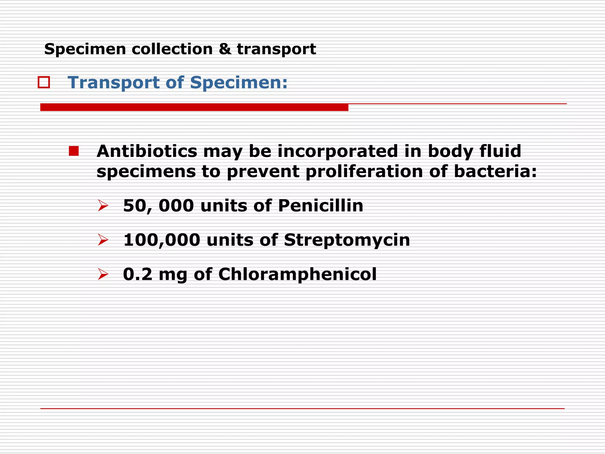
Downloaded 508 times


Direct microscopic examination of clinical specimens can provide a presumptive diagnosis of fungal infection by revealing the presence of fungal elements. Different stains and techniques are used to visualize fungi depending on the suspected infection. KOH wet mounts are useful for superficial mycoses while GMS, H&E and fluorescent antibody stains aid in diagnosis of deep mycoses from tissue biopsies and body fluids. Proper specimen collection and rapid microscopic evaluation can help initiate appropriate antifungal treatment.


