Downloaded 550 times
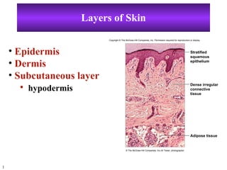


The document discusses the structure and functions of the skin and its layers. The skin consists of three main layers - the epidermis, dermis and subcutaneous layer. The epidermis is the outermost layer and provides protection against water loss and microorganisms. Below the epidermis lies the dermis, which contains hair follicles, sweat and sebaceous glands, nerves and blood vessels. The innermost layer, the subcutaneous layer, consists of adipose tissue and insulates the body.


![Chapt06 Holes Lecture Animation[1]](https://cdn.slidesharecdn.com/ss_thumbnails/chapt06holeslectureanimation1-091122122041-phpapp02-thumbnail.jpg?width=640&height=640&fit=bounds)


![Chapt06 Holes Lecture Animation[1]](https://cdn.slidesharecdn.com/ss_thumbnails/chapt06holeslectureanimation1-091122124045-phpapp01-thumbnail.jpg?width=640&height=640&fit=bounds)


