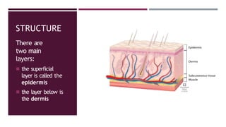
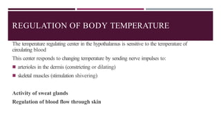
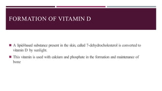
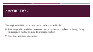
UNIT VII
INTEGUMENTARY SYSTEM
Daud Akhtar
OBJECTIVES
◾ By the end of this presentation, the learners will be able
to...
◾ Describe structure of the two layers of skin
◾ Epidermis
◾ Dermis
◾ Briefly discuss the structure & function of skin derivatives
◾ Sweat glands, Sebaceous glands, Hair & Nail
◾ Discuss the following functions of
skin
◾ Protection,Thermoregulation, Sensation,
Absorption & Excretion
THE SKIN
The skin is the largest organ in the body.
The skin completely covers the body and is continuous with the
membranes lining the body orifices.
◾ The skin has a surface area of about 1.5-2 square meters in adults
◾ It varies in thickness, being thickest on the palms of the hands and soles of the feet
◾ Integumentary” means a body’s outer covering
STRUCTURE
There are
two main
layers:
◾ the superficial
layer is called the
epidermis
◾ the layer below is
the dermis
EPIDERMIS
Your epidermis is the top layer of the skin that you can see and touch. Keratin, a protein
inside skin cells, makes up the skin cells and, along with other proteins, sticks together to
form this layer. The epidermis:
Acts as a protective barrier: The epidermis keeps bacteria and germs from entering your
body and bloodstream and causing infections. It also protects against rain, sun and other
elements.
Makes new skin: The epidermis continually makes new skin cells. These new cells replace
the approximately 40,000 old skin cells that your body sheds every day.
EPIDERMIS
You have new skin every 30 days.
Protects your body: Langerhans cells in the epidermis are part of the body’s immune
system. They help fight off germs and infections.
Provides skin colour: The epidermis contains melanin, the pigment that gives skin its
colour. The amount of melanin you have determines the colour of your skin, hair and eyes.
People who make more melanin have darker skin and may tan more quickly.
EPIDERMIS
The cells of the epidermis originate in the basal layer, which is made up of
cuboidal nucleated, highly active epithelial cells that are constantly dividing
◾ By the time they reach the skin surface, they are flat, thin, non-nucleated, dead cells, or
squames, in which the cytoplasm has been replaced by the fibrous protein keratin
◾ Complete replacement of the epidermis takes about a month
DERMIS
The dermis is tough and elastic.
It is formed from connective tissue, and the matrix contains collagen fibers
interlaced with elastic fibers.
◾ The subcutaneous layer, containing areolar tissue and varying amounts of
adipose (fat) tissue, lies under the dermis
◾ Structures found in the dermis are:
◾ Small blood and lymph vessels
◾ Sensory nerve endings
◾ Sweat glands and their ducts
◾ Hairs, arrector pili muscles and sebaceous glands
Blood and Lymph Vessels
Arterioles form a fine network with capillary
branches supplying sweat glands, sebaceous
glands, hair follicles and the dermis
Lymph vessels also form a network
throughout the dermis
DERMIS
DERMIS
Sensory Nerve Endings
◾ Sensory receptors sensitive to touch,
temperature, pressure and pain are widely
distributed in the dermis
◾ The skin is an important sensory
organ through which individuals
receive information about their
environment
DERMIS
Sweat Glands
These are widely distributed throughout the skin and are most numerous in the palms of the
hands, soles of the feet, axillae and groins
They are formed from epithelial cells
The bodies of the glands lie coiled in the subcutaneous tissue
There are two types of sweat gland:
Eccrine Glands
Apocrine Glands
DERMIS
s
Hairs
These grow outwards from hair follicle
At the base of the follicle is a cluster of
cells called the hair papilla or bulb
Arrector Pili
These are little bundles of smooth
muscle fibers attached to the hair
follicles.
Contraction of the muscle fibers makes the
hair stand erect and raises the skin around
the hair
DERMIS
Sebaceous Glands
Sebaceous glands consist of secretory epithelial cells derived from the same tissue as
the hair follicles.
They secrete an oily antimicrobial substance, sebum into the hair follicles
Sebum keeps the hair soft and pliable, and gives it a shiny appearance
It waterproofs the skin and acts as a bactericidal and fungicidal agent, preventing
infection.
DERMIS
Nails
Derived from the same cells as epidermis and hair, these are
hard, keratin plates that protect the tips of the fingers and toes.
◾ The root of the nail is embedded in the skin and covered
by the cuticle, which forms the hemispherical pale area
called the lunula
◾ The nail plate is the exposed part that has grown out
from the nail bed. the basal layer of the epidermis.
HYPODERMIS
• The bottom layer of skin, or hypodermis, is the fatty layer. The hypodermis:
• Cushions muscles and bones: Fat in the hypodermis protects muscles and bones
from injuries when you fall or are in an accident.
• Has connective tissue: This tissue connects layers of skin to muscles and bones.
• Helps the nerves and blood vessels: Nerves and blood vessels in the dermis
(middle layer) get larger in the hypodermis. These nerves and blood vessels
branch out to connect the hypodermis to the rest of the body.
• Regulates body temperature: Fat in the hypodermis keeps you from getting too
cold or hot.
FUNCTIONS
Skin has the following functions:
◾ Protection
◾ Regulation of body temperature
◾ Formation of Vitamin D
◾ Cutaneous sensation
◾ Absorption
◾ Excretion
PROTECTION
The skin forms a relatively waterproof layer, provided mainly by its keratinized
epithelium
As an important non-specific defense mechanism, it acts as a barrier against:
◾ invasion by microorganisms
◾ Chemicals
◾ physical agents, e.g., mild trauma, ultraviolet light
◾ dehydration
PROTECTION
◾ The epidermis contains specialized immune cells called dendritic (Langerhans) cells,
which are a type of fixed macrophage
◾ Abundant sensory nerve endings in the dermis enable perception, discrimination and location
of internal and external stimuli
◾ The pigment melanin protects against harmful ultraviolet rays in sunlight
REGULATION OF BODY TEMPERATURE
Core body temperature remains constant at around 36.8°C across a wide range of
environmental temperatures.
To maintain this constant temperature, a negative feedback system regulates the balance between
heat produced in the body and heat lost to the environment.
Heat Loss
Most heat loss from the body occurs through the skin.
Heat loss through the skin is affected by the difference between body and environmental
temperatures, the amount of the body surface exposed and the type of clothes worn.
REGULATION OF
BODY TEMPERATURE
Mechanism of Heat Loss
There are four mechanisms of heat
loss:
◾ Radiation
◾ Evaporation
◾ Convection
◾ Conduction
REGULATION OF BODY TEMPERATURE
The temperature regulating center in the hypothalamus is sensitive to the temperature of
circulating blood
This center responds to changing temperature by sending nerve impulses to:
◾ arterioles in the dermis (constricting or dilating)
◾ skeletal muscles (stimulation shivering)
Activity of sweat glands
Regulation of blood flow through skin
FORMATION OF VITAMIN D
◾ A lipid-based substance present in the skin, called 7-dehydrocholesterol is converted to
vitamin D by sunlight.
◾ This vitamin is used with calcium and phosphate in the formation and maintenance of
bone
CUTANEOUS SENSATION
◾ Sensory receptors in the dermis can be sensitive to touch, pressure, temperature or
pain
◾ Stimulation generates impulses in sensory nerves, which are transmitted to the cerebral
cortex
◾ Some areas have more abundant sensory receptors than others, causing them to be especially
sensitive, e.g., the lips and fingertips
ABSORPTION
This property is limited but substances that can be absorbed include:
◾ Some drugs when applied as transdermal patches, e.g., hormone replacement therapy during
the menopause, nicotine as an aid to smoking cessation
◾ Some toxic chemicals, e.g., mercury.
EXCRETION
The skin is a minor excretory organ for some substances, including:
◾ Sodium chloride in sweat; excess sweating may lead to low blood sodium levels
(hyponatremia)
◾ Urea, especially when kidney function is impaired
THANK
YOU!
WHAT QUESTIONS DO YOU
HAVE…
REFERENCES
Waugh, A. and Grant, A., 2014. Ross & Wilson Anatomy & Physiology in
Health and Illness. 13th ed.

UNIT 7 - The Integumentary System bsn.pptx

  • 1.
  • 2.
    OBJECTIVES ◾ By theend of this presentation, the learners will be able to... ◾ Describe structure of the two layers of skin ◾ Epidermis ◾ Dermis ◾ Briefly discuss the structure & function of skin derivatives ◾ Sweat glands, Sebaceous glands, Hair & Nail ◾ Discuss the following functions of skin ◾ Protection,Thermoregulation, Sensation, Absorption & Excretion
  • 3.
    THE SKIN The skinis the largest organ in the body. The skin completely covers the body and is continuous with the membranes lining the body orifices. ◾ The skin has a surface area of about 1.5-2 square meters in adults ◾ It varies in thickness, being thickest on the palms of the hands and soles of the feet ◾ Integumentary” means a body’s outer covering
  • 4.
    STRUCTURE There are two main layers: ◾the superficial layer is called the epidermis ◾ the layer below is the dermis
  • 5.
    EPIDERMIS Your epidermis isthe top layer of the skin that you can see and touch. Keratin, a protein inside skin cells, makes up the skin cells and, along with other proteins, sticks together to form this layer. The epidermis: Acts as a protective barrier: The epidermis keeps bacteria and germs from entering your body and bloodstream and causing infections. It also protects against rain, sun and other elements. Makes new skin: The epidermis continually makes new skin cells. These new cells replace the approximately 40,000 old skin cells that your body sheds every day.
  • 6.
    EPIDERMIS You have newskin every 30 days. Protects your body: Langerhans cells in the epidermis are part of the body’s immune system. They help fight off germs and infections. Provides skin colour: The epidermis contains melanin, the pigment that gives skin its colour. The amount of melanin you have determines the colour of your skin, hair and eyes. People who make more melanin have darker skin and may tan more quickly.
  • 7.
    EPIDERMIS The cells ofthe epidermis originate in the basal layer, which is made up of cuboidal nucleated, highly active epithelial cells that are constantly dividing ◾ By the time they reach the skin surface, they are flat, thin, non-nucleated, dead cells, or squames, in which the cytoplasm has been replaced by the fibrous protein keratin ◾ Complete replacement of the epidermis takes about a month
  • 8.
    DERMIS The dermis istough and elastic. It is formed from connective tissue, and the matrix contains collagen fibers interlaced with elastic fibers. ◾ The subcutaneous layer, containing areolar tissue and varying amounts of adipose (fat) tissue, lies under the dermis ◾ Structures found in the dermis are: ◾ Small blood and lymph vessels ◾ Sensory nerve endings ◾ Sweat glands and their ducts ◾ Hairs, arrector pili muscles and sebaceous glands
  • 9.
    Blood and LymphVessels Arterioles form a fine network with capillary branches supplying sweat glands, sebaceous glands, hair follicles and the dermis Lymph vessels also form a network throughout the dermis DERMIS
  • 10.
    DERMIS Sensory Nerve Endings ◾Sensory receptors sensitive to touch, temperature, pressure and pain are widely distributed in the dermis ◾ The skin is an important sensory organ through which individuals receive information about their environment
  • 11.
    DERMIS Sweat Glands These arewidely distributed throughout the skin and are most numerous in the palms of the hands, soles of the feet, axillae and groins They are formed from epithelial cells The bodies of the glands lie coiled in the subcutaneous tissue There are two types of sweat gland: Eccrine Glands Apocrine Glands
  • 12.
    DERMIS s Hairs These grow outwardsfrom hair follicle At the base of the follicle is a cluster of cells called the hair papilla or bulb Arrector Pili These are little bundles of smooth muscle fibers attached to the hair follicles. Contraction of the muscle fibers makes the hair stand erect and raises the skin around the hair
  • 13.
    DERMIS Sebaceous Glands Sebaceous glandsconsist of secretory epithelial cells derived from the same tissue as the hair follicles. They secrete an oily antimicrobial substance, sebum into the hair follicles Sebum keeps the hair soft and pliable, and gives it a shiny appearance It waterproofs the skin and acts as a bactericidal and fungicidal agent, preventing infection.
  • 14.
    DERMIS Nails Derived from thesame cells as epidermis and hair, these are hard, keratin plates that protect the tips of the fingers and toes. ◾ The root of the nail is embedded in the skin and covered by the cuticle, which forms the hemispherical pale area called the lunula ◾ The nail plate is the exposed part that has grown out from the nail bed. the basal layer of the epidermis.
  • 15.
    HYPODERMIS • The bottomlayer of skin, or hypodermis, is the fatty layer. The hypodermis: • Cushions muscles and bones: Fat in the hypodermis protects muscles and bones from injuries when you fall or are in an accident. • Has connective tissue: This tissue connects layers of skin to muscles and bones. • Helps the nerves and blood vessels: Nerves and blood vessels in the dermis (middle layer) get larger in the hypodermis. These nerves and blood vessels branch out to connect the hypodermis to the rest of the body. • Regulates body temperature: Fat in the hypodermis keeps you from getting too cold or hot.
  • 16.
    FUNCTIONS Skin has thefollowing functions: ◾ Protection ◾ Regulation of body temperature ◾ Formation of Vitamin D ◾ Cutaneous sensation ◾ Absorption ◾ Excretion
  • 17.
    PROTECTION The skin formsa relatively waterproof layer, provided mainly by its keratinized epithelium As an important non-specific defense mechanism, it acts as a barrier against: ◾ invasion by microorganisms ◾ Chemicals ◾ physical agents, e.g., mild trauma, ultraviolet light ◾ dehydration
  • 18.
    PROTECTION ◾ The epidermiscontains specialized immune cells called dendritic (Langerhans) cells, which are a type of fixed macrophage ◾ Abundant sensory nerve endings in the dermis enable perception, discrimination and location of internal and external stimuli ◾ The pigment melanin protects against harmful ultraviolet rays in sunlight
  • 19.
    REGULATION OF BODYTEMPERATURE Core body temperature remains constant at around 36.8°C across a wide range of environmental temperatures. To maintain this constant temperature, a negative feedback system regulates the balance between heat produced in the body and heat lost to the environment. Heat Loss Most heat loss from the body occurs through the skin. Heat loss through the skin is affected by the difference between body and environmental temperatures, the amount of the body surface exposed and the type of clothes worn.
  • 20.
    REGULATION OF BODY TEMPERATURE Mechanismof Heat Loss There are four mechanisms of heat loss: ◾ Radiation ◾ Evaporation ◾ Convection ◾ Conduction
  • 22.
    REGULATION OF BODYTEMPERATURE The temperature regulating center in the hypothalamus is sensitive to the temperature of circulating blood This center responds to changing temperature by sending nerve impulses to: ◾ arterioles in the dermis (constricting or dilating) ◾ skeletal muscles (stimulation shivering) Activity of sweat glands Regulation of blood flow through skin
  • 23.
    FORMATION OF VITAMIND ◾ A lipid-based substance present in the skin, called 7-dehydrocholesterol is converted to vitamin D by sunlight. ◾ This vitamin is used with calcium and phosphate in the formation and maintenance of bone
  • 24.
    CUTANEOUS SENSATION ◾ Sensoryreceptors in the dermis can be sensitive to touch, pressure, temperature or pain ◾ Stimulation generates impulses in sensory nerves, which are transmitted to the cerebral cortex ◾ Some areas have more abundant sensory receptors than others, causing them to be especially sensitive, e.g., the lips and fingertips
  • 25.
    ABSORPTION This property islimited but substances that can be absorbed include: ◾ Some drugs when applied as transdermal patches, e.g., hormone replacement therapy during the menopause, nicotine as an aid to smoking cessation ◾ Some toxic chemicals, e.g., mercury.
  • 26.
    EXCRETION The skin isa minor excretory organ for some substances, including: ◾ Sodium chloride in sweat; excess sweating may lead to low blood sodium levels (hyponatremia) ◾ Urea, especially when kidney function is impaired
  • 27.
  • 28.
    REFERENCES Waugh, A. andGrant, A., 2014. Ross & Wilson Anatomy & Physiology in Health and Illness. 13th ed.