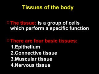
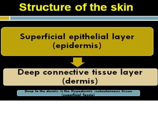
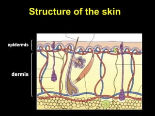
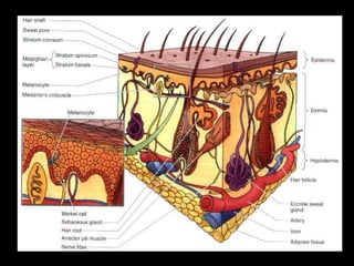
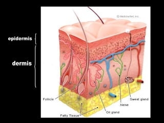
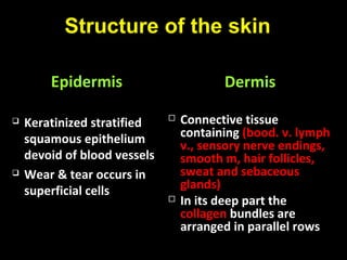
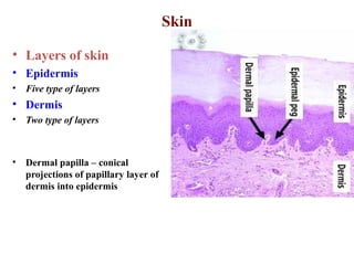
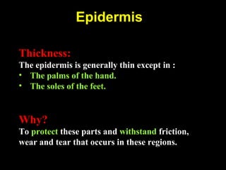
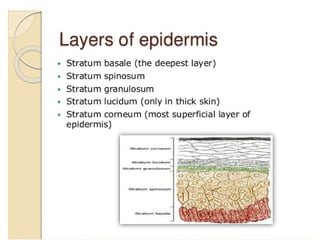
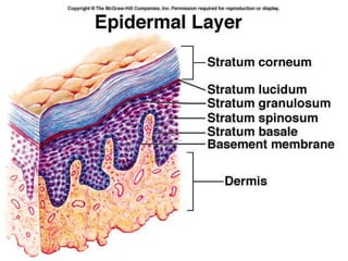
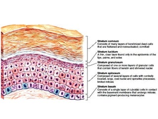
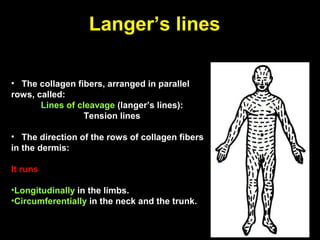
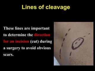
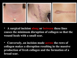
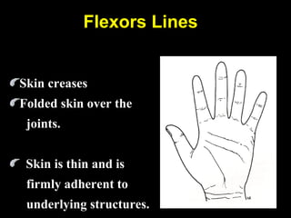
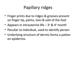
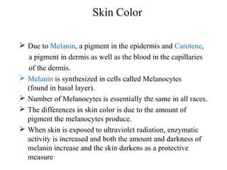
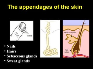
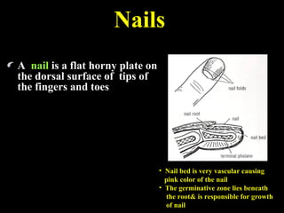
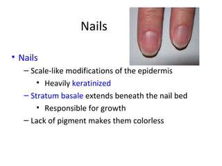
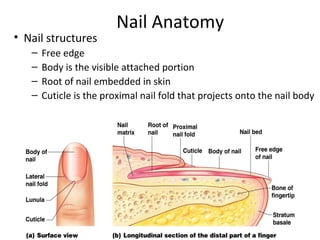
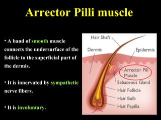
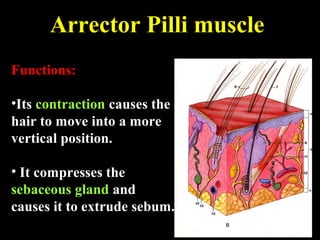
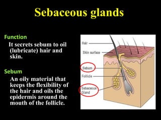
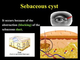
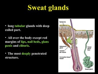
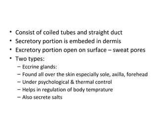
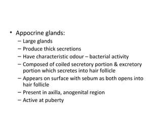
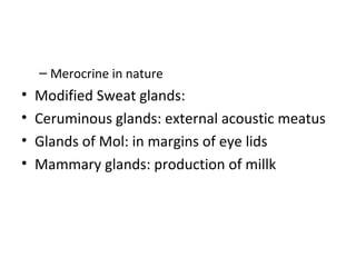
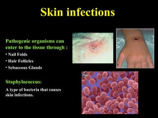
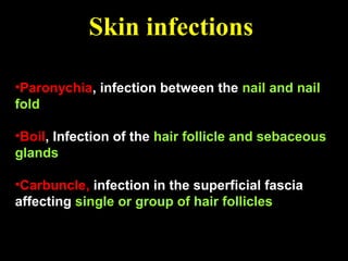
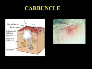
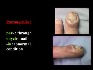
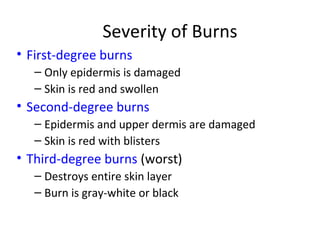
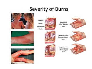
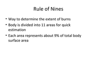
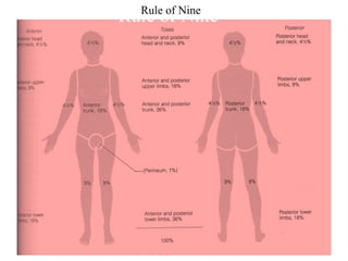
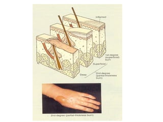
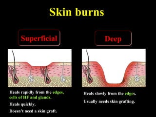
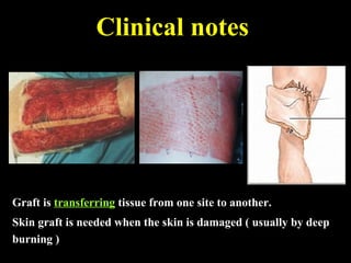
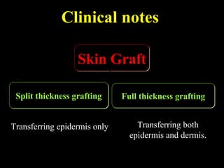
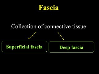
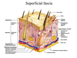
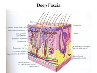
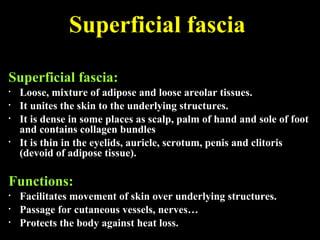
Integumentary System
Dr. Ammarah Naseer
Tissues of the body
 The tissue: is a group of cells
which perform a specific function
 There are four basic tissues:
1.Epithelium
2.Connective tissue
3.Muscular tissue
4.Nervous tissue
Structure of the skin
EpidermisEpidermis
 Keratinized stratifiedKeratinized stratified
squamous epitheliumsquamous epithelium
devoid of blood vesselsdevoid of blood vessels
 Wear & tear occurs inWear & tear occurs in
superficial cellssuperficial cells
DermisDermis
 Connective tissueConnective tissue
containingcontaining (bood. v. lymph(bood. v. lymph
v., sensory nerve endings,v., sensory nerve endings,
smooth m, hair follicles,smooth m, hair follicles,
sweat and sebaceoussweat and sebaceous
glands)glands)
 In its deep part theIn its deep part the
collagencollagen bundles arebundles are
arranged in parallel rowsarranged in parallel rows
Skin
‱ Layers of skin
‱ Epidermis
‱ Five type of layers
‱ Dermis
‱ Two type of layers
‱ Dermal papilla – conical
projections of papillary layer of
dermis into epidermis
Epidermis
Thickness:
The epidermis is generally thin except in :
‱ The palms of the hand.
‱ The soles of the feet.
Why?
To protect these parts and withstand friction,
wear and tear that occurs in these regions.
Dermis
‱ Corium
‱ Vascular layer of skin
‱ Made up of connective tissue
‱ Two layers:
– Superficial – papillary layer
‱ Thin
‱ 20%
‱ Contains loosely arrange collagen fibers
‱ Reticular layer:
– Thick
– 80%
– Composed of thick collagen bundles running
parallel to skin
Functions of the Skin
‱ 1-Protection
‱ ïź abrasion, invasion, water loss, UV protection
‱ 2-Vitamin D synthesis
‱ ïź epidermal keratinocytes when exposed to UV
light
‱ ïź helps maintain health of skeleton by increasing
absorption of Ca2+
‱ 3-Sensation
‱ ïź receptors for heat, cold, touch, pressure,
vibration and pain
‱ 4- Thermoregulation
‱ ïź thermo receptors and sweat glands
‱ ïź hypothalamus controls cutaneous arteries
and sweat glands to retain or dissipate heat
‱ 5- Psychological and social functions
‱ ïź appearance and social acceptance
‱ ïź facial expression and nonverbal
communication
Langer’s lines
‱ The collagen fibers, arranged in parallel
rows, called:
Lines of cleavage (langer’s lines):
Tension lines
‱ The direction of the rows of collagen fibers
in the dermis:
It runs
‱Longitudinally in the limbs.
‱Circumferentially in the neck and the trunk.
Lines of cleavage
These lines are important
to determine the direction
for an incision (cut) during
a surgery to avoid obvious
scars.
‱ A surgical incision along or between these lines
causes the minimum disruption of collagen so that the
wound heals with a small scar.
‱ Conversely, an incision made across the rows of
collagen makes a disruption resulting in the massive
production of fresh collagen and the formation of a
broad scar.
Flexors Lines
Skin creases
Folded skin over the
joints.
Skin is thin and is
firmly adherent to
underlying structures.
Papillary ridges
‱ Finger prints due to ridges & grooves present
on finger tip, palms, toes & sole of the foot
‱ Appears in intrauterine life – 3rd
& 4th
month
‱ Peculiar to individual, used to identify person
‱ Underlying structure of dermis forms a patten
on epidermis
Skin Color
 Due to Melanin, a pigment in the epidermis and Carotene,
a pigment in dermis as well as the blood in the capillaries
of the dermis.
 Melanin is synthesized in cells called Melanocytes
(found in basal layer).
 Number of Melanocytes is essentially the same in all races.
 The differences in skin color is due to the amount of
pigment the melanocytes produce.
 When skin is exposed to ultraviolet radiation, enzymatic
activity is increased and both the amount and darkness of
melanin increase and the skin darkens as a protective
measure
‱ Nails
‱ Hairs
‱ Sebaceous glands
‱ Sweat glands
The appendages of the skin
A nail is a flat horny plate on
the dorsal surface of tips of
the fingers and toes
‱ Nail bed is very vascular causing
pink color of the nail
‱ The germinative zone lies beneath
the root& is responsible for growth
of nail
Nails
Nails
‱ Nails
– Scale-like modifications of the epidermis
‱ Heavily keratinized
– Stratum basale extends beneath the nail bed
‱ Responsible for growth
– Lack of pigment makes them colorless
Nail Anatomy
‱ Nail structures
– Free edge
– Body is the visible attached portion
– Root of nail embedded in skin
– Cuticle is the proximal nail fold that projects onto the nail body
‱ Nail Plate: visible part
‱ Nail root: hidden part lying in groove
‱ Nail folds: skin fold bounding nail grooves
‱ Nail bed: beneath nail bed is stratified squamous
epithelium called nail bed
‱ Nail matrix: beneath proximal portion of nail fold
is epithelium from which nail grows (0.5
mm/week)
‱ Hyponychium: thickened epidermis under free
edge of nail
Hair
‱ Hair
– Keratinous filaments derived from invagination of
germinative layer of epidermis into dermis
– Produced by hair follicle which are made of hard
keratinized epithelial cells
– Melanocytes provide pigment for hair color
Structure of Hair Follicle
Cover whole surface of
the body except some
areas as lips, palms, soles,
reproductive organs.
Hairs
Consist of 2 parts:
Shaft: free end
Root: embeded part
Hairs
Root is surrounded by hair follicle
Hair follicles: invaginations of the
epidermis into the dermis, the
hair grows out of these follicles
(hair shaft).
Hair bulb: the expanded extremity
of the follicle, concaved at the
end (located deep in the
dermis).
Hair papilla: a vascular connective
tissue that occupies the
concavity of the bulb.
‱ A band of smooth muscle
connects the undersurface of the
follicle to the superficial part of
the dermis.
‱ It is innervated by sympathetic
nerve fibers.
‱ It is involuntary.
Arrector Pilli muscle
Functions:
‱Its contraction causes the
hair to move into a more
vertical position.
‱ It compresses the
sebaceous gland and
causes it to extrude sebum.
Arrector Pilli muscle
Hair Structures
‱ Associated hair structures
– Hair follicle
‱ Dermal and epidermal sheath
surround hair root
– Arrector pili muscle
‱ Smooth muscle
‱ Pulls hairs upright when cold or
frightened
– Sebaceous gland
Function
It secrets sebum to oil
(lubricate) hair and
skin.
Sebum
An oily material that
keeps the flexibility of
the hair and oils the
epidermis around the
mouth of the follicle.
Sebaceous glands
It occurs because of the
obstruction (blocking) of the
sebaceous duct.
Sebaceous cyst
‱ long tubular glands with deep
coiled part.
‱ All over the body except red
margins of lips, nail beds, glans
penis and clitoris.
‱ The most deeply penetrated
structure.
Sweat glands
‱ Consist of coiled tubes and straight duct
‱ Secretory portion is embeded in dermis
‱ Excretory portion open on surface – sweat pores
‱ Two types:
– Eccrine glands:
– Found all over the skin especially sole, axilla, forehead
– Under psychological & thermal control
– Helps in regulation of body temprature
– Also secrete salts
‱ Appocrine glands:
– Large glands
– Produce thick secretions
– Have characteristic odour – bacterial activity
– Composed of coiled secretory portion & excretory
portion which secretes into hair follicle
– Appears on surface with sebum as both opens into
hair follicle
– Present in axilla, anogenital region
– Active at puberty
– Merocrine in nature
‱ Modified Sweat glands:
‱ Ceruminous glands: external acoustic meatus
‱ Glands of Mol: in margins of eye lids
‱ Mammary glands: production of millk
Skin infections
Pathogenic organisms can
enter to the tissue through :
‱ Nail Folds
‱ Hair Follicles
‱ Sebaceous Glands
Staphylococcus:
A type of bacteria that causes
skin infections.
Skin infections
‱Paronychia, infection between the nail and nail
fold
‱Boil, Infection of the hair follicle and sebaceous
glands
‱Carbuncle, infection in the superficial fascia
affecting single or group of hair follicles
CARBUNCLE
Par/onych/ia :
par- : through
onych- :nail
-ia :abnormal
condition
Severity of Burns
‱ First-degree burns
– Only epidermis is damaged
– Skin is red and swollen
‱ Second-degree burns
– Epidermis and upper dermis are damaged
– Skin is red with blisters
‱ Third-degree burns (worst)
– Destroys entire skin layer
– Burn is gray-white or black
Severity of Burns
Rule of Nines
‱ Way to determine the extent of burns
‱ Body is divided into 11 areas for quick
estimation
‱ Each area represents about 9% of total body
surface area
Rule of NineRule of Nine
Skin burns
DeepDeepSuperficialSuperficial
Heals slowly from the edges.
Usually needs skin grafting.
Heals rapidly from the edges,
cells of HF and glands.
Heals quickly.
Doesn’t need a skin graft.
Clinical notes
Graft is transferring tissue from one site to another.
Skin graft is needed when the skin is damaged ( usually by deep
burning )
Clinical notes
Full thickness graftingFull thickness graftingSplit thickness graftingSplit thickness grafting
Transferring epidermis only Transferring both
epidermis and dermis.
Skin GraftSkin Graft
Fascia
Collection of connective tissueCollection of connective tissue
Deep fasciaDeep fasciaSuperficial fasciaSuperficial fascia
Superficial fascia
Deep Fascia
Superficial fascia
Superficial fascia:
‱ Loose, mixture of adipose and loose areolar tissues.
‱ It unites the skin to the underlying structures.
‱ It is dense in some places as scalp, palm of hand and sole of foot
and contains collagen bundles
‱ It is thin in the eyelids, auricle, scrotum, penis and clitoris
(devoid of adipose tissue).
Functions:
‱ Facilitates movement of skin over underlying structures.
‱ Passage for cutaneous vessels, nerves

‱ Protects the body against heat loss.
Superficial fascia
It isIt is more densemore dense than superficial fasciathan superficial fascia
Collagenous bundles are moreCollagenous bundles are more
compact and more regularly arrangedcompact and more regularly arranged
It is usually present in the form ofIt is usually present in the form of
membranesmembranes
Deep fascia
A. Intermuscular
septa
lie between muscleslie between muscles
dividing the limb intodividing the limb into
compartmentscompartments
Examples of deep fascia
B. Investing fascia
‱ Covers the surfaces of musclesCovers the surfaces of muscles
‱ In theIn the neckneck: it forms well-defined layers,: it forms well-defined layers,
bounds fascial spaces so limits spread ofbounds fascial spaces so limits spread of
infection or determine the path of infectioninfection or determine the path of infection
‱ In theIn the abdomenabdomen: it is thin: it is thin
‱ In theIn the limbslimbs: forms a definite sheath around the: forms a definite sheath around the
musclesmuscles
Examples of deep fascia
Localized thickening of deep fascia around joints, hold the tendons
in place, prevent bowstringing of tendons
Examples of deep fascia
C. Retinacula
Skin and fascia

Skin and fascia

  • 1.
  • 3.
    Tissues of thebody  The tissue: is a group of cells which perform a specific function  There are four basic tissues: 1.Epithelium 2.Connective tissue 3.Muscular tissue 4.Nervous tissue
  • 6.
  • 9.
    Structure of theskin EpidermisEpidermis  Keratinized stratifiedKeratinized stratified squamous epitheliumsquamous epithelium devoid of blood vesselsdevoid of blood vessels  Wear & tear occurs inWear & tear occurs in superficial cellssuperficial cells DermisDermis  Connective tissueConnective tissue containingcontaining (bood. v. lymph(bood. v. lymph v., sensory nerve endings,v., sensory nerve endings, smooth m, hair follicles,smooth m, hair follicles, sweat and sebaceoussweat and sebaceous glands)glands)  In its deep part theIn its deep part the collagencollagen bundles arebundles are arranged in parallel rowsarranged in parallel rows
  • 10.
    Skin ‱ Layers ofskin ‱ Epidermis ‱ Five type of layers ‱ Dermis ‱ Two type of layers ‱ Dermal papilla – conical projections of papillary layer of dermis into epidermis
  • 11.
    Epidermis Thickness: The epidermis isgenerally thin except in : ‱ The palms of the hand. ‱ The soles of the feet. Why? To protect these parts and withstand friction, wear and tear that occurs in these regions.
  • 15.
    Dermis ‱ Corium ‱ Vascularlayer of skin ‱ Made up of connective tissue ‱ Two layers: – Superficial – papillary layer ‱ Thin ‱ 20% ‱ Contains loosely arrange collagen fibers
  • 16.
    ‱ Reticular layer: –Thick – 80% – Composed of thick collagen bundles running parallel to skin
  • 17.
    Functions of theSkin ‱ 1-Protection ‱ ïź abrasion, invasion, water loss, UV protection ‱ 2-Vitamin D synthesis ‱ ïź epidermal keratinocytes when exposed to UV light ‱ ïź helps maintain health of skeleton by increasing absorption of Ca2+ ‱ 3-Sensation ‱ ïź receptors for heat, cold, touch, pressure, vibration and pain
  • 18.
    ‱ 4- Thermoregulation â€ąïź thermo receptors and sweat glands ‱ ïź hypothalamus controls cutaneous arteries and sweat glands to retain or dissipate heat ‱ 5- Psychological and social functions ‱ ïź appearance and social acceptance ‱ ïź facial expression and nonverbal communication
  • 19.
    Langer’s lines ‱ Thecollagen fibers, arranged in parallel rows, called: Lines of cleavage (langer’s lines): Tension lines ‱ The direction of the rows of collagen fibers in the dermis: It runs ‱Longitudinally in the limbs. ‱Circumferentially in the neck and the trunk.
  • 20.
    Lines of cleavage Theselines are important to determine the direction for an incision (cut) during a surgery to avoid obvious scars.
  • 21.
    ‱ A surgicalincision along or between these lines causes the minimum disruption of collagen so that the wound heals with a small scar. ‱ Conversely, an incision made across the rows of collagen makes a disruption resulting in the massive production of fresh collagen and the formation of a broad scar.
  • 22.
    Flexors Lines Skin creases Foldedskin over the joints. Skin is thin and is firmly adherent to underlying structures.
  • 23.
    Papillary ridges ‱ Fingerprints due to ridges & grooves present on finger tip, palms, toes & sole of the foot ‱ Appears in intrauterine life – 3rd & 4th month ‱ Peculiar to individual, used to identify person ‱ Underlying structure of dermis forms a patten on epidermis
  • 24.
    Skin Color  Dueto Melanin, a pigment in the epidermis and Carotene, a pigment in dermis as well as the blood in the capillaries of the dermis.  Melanin is synthesized in cells called Melanocytes (found in basal layer).  Number of Melanocytes is essentially the same in all races.  The differences in skin color is due to the amount of pigment the melanocytes produce.  When skin is exposed to ultraviolet radiation, enzymatic activity is increased and both the amount and darkness of melanin increase and the skin darkens as a protective measure
  • 25.
    ‱ Nails ‱ Hairs ‱Sebaceous glands ‱ Sweat glands The appendages of the skin
  • 26.
    A nail isa flat horny plate on the dorsal surface of tips of the fingers and toes ‱ Nail bed is very vascular causing pink color of the nail ‱ The germinative zone lies beneath the root& is responsible for growth of nail Nails
  • 27.
    Nails ‱ Nails – Scale-likemodifications of the epidermis ‱ Heavily keratinized – Stratum basale extends beneath the nail bed ‱ Responsible for growth – Lack of pigment makes them colorless
  • 28.
    Nail Anatomy ‱ Nailstructures – Free edge – Body is the visible attached portion – Root of nail embedded in skin – Cuticle is the proximal nail fold that projects onto the nail body
  • 29.
    ‱ Nail Plate:visible part ‱ Nail root: hidden part lying in groove ‱ Nail folds: skin fold bounding nail grooves ‱ Nail bed: beneath nail bed is stratified squamous epithelium called nail bed ‱ Nail matrix: beneath proximal portion of nail fold is epithelium from which nail grows (0.5 mm/week) ‱ Hyponychium: thickened epidermis under free edge of nail
  • 30.
    Hair ‱ Hair – Keratinousfilaments derived from invagination of germinative layer of epidermis into dermis – Produced by hair follicle which are made of hard keratinized epithelial cells – Melanocytes provide pigment for hair color
  • 31.
  • 33.
    Cover whole surfaceof the body except some areas as lips, palms, soles, reproductive organs. Hairs Consist of 2 parts: Shaft: free end Root: embeded part
  • 34.
    Hairs Root is surroundedby hair follicle Hair follicles: invaginations of the epidermis into the dermis, the hair grows out of these follicles (hair shaft). Hair bulb: the expanded extremity of the follicle, concaved at the end (located deep in the dermis). Hair papilla: a vascular connective tissue that occupies the concavity of the bulb.
  • 35.
    ‱ A bandof smooth muscle connects the undersurface of the follicle to the superficial part of the dermis. ‱ It is innervated by sympathetic nerve fibers. ‱ It is involuntary. Arrector Pilli muscle
  • 36.
    Functions: ‱Its contraction causesthe hair to move into a more vertical position. ‱ It compresses the sebaceous gland and causes it to extrude sebum. Arrector Pilli muscle
  • 37.
    Hair Structures ‱ Associatedhair structures – Hair follicle ‱ Dermal and epidermal sheath surround hair root – Arrector pili muscle ‱ Smooth muscle ‱ Pulls hairs upright when cold or frightened – Sebaceous gland
  • 38.
    Function It secrets sebumto oil (lubricate) hair and skin. Sebum An oily material that keeps the flexibility of the hair and oils the epidermis around the mouth of the follicle. Sebaceous glands
  • 39.
    It occurs becauseof the obstruction (blocking) of the sebaceous duct. Sebaceous cyst
  • 40.
    ‱ long tubularglands with deep coiled part. ‱ All over the body except red margins of lips, nail beds, glans penis and clitoris. ‱ The most deeply penetrated structure. Sweat glands
  • 41.
    ‱ Consist ofcoiled tubes and straight duct ‱ Secretory portion is embeded in dermis ‱ Excretory portion open on surface – sweat pores ‱ Two types: – Eccrine glands: – Found all over the skin especially sole, axilla, forehead – Under psychological & thermal control – Helps in regulation of body temprature – Also secrete salts
  • 42.
    ‱ Appocrine glands: –Large glands – Produce thick secretions – Have characteristic odour – bacterial activity – Composed of coiled secretory portion & excretory portion which secretes into hair follicle – Appears on surface with sebum as both opens into hair follicle – Present in axilla, anogenital region – Active at puberty
  • 43.
    – Merocrine innature ‱ Modified Sweat glands: ‱ Ceruminous glands: external acoustic meatus ‱ Glands of Mol: in margins of eye lids ‱ Mammary glands: production of millk
  • 44.
    Skin infections Pathogenic organismscan enter to the tissue through : ‱ Nail Folds ‱ Hair Follicles ‱ Sebaceous Glands Staphylococcus: A type of bacteria that causes skin infections.
  • 45.
    Skin infections ‱Paronychia, infectionbetween the nail and nail fold ‱Boil, Infection of the hair follicle and sebaceous glands ‱Carbuncle, infection in the superficial fascia affecting single or group of hair follicles
  • 46.
  • 47.
    Par/onych/ia : par- :through onych- :nail -ia :abnormal condition
  • 48.
    Severity of Burns ‱First-degree burns – Only epidermis is damaged – Skin is red and swollen ‱ Second-degree burns – Epidermis and upper dermis are damaged – Skin is red with blisters ‱ Third-degree burns (worst) – Destroys entire skin layer – Burn is gray-white or black
  • 49.
  • 50.
    Rule of Nines ‱Way to determine the extent of burns ‱ Body is divided into 11 areas for quick estimation ‱ Each area represents about 9% of total body surface area
  • 51.
  • 53.
    Skin burns DeepDeepSuperficialSuperficial Heals slowlyfrom the edges. Usually needs skin grafting. Heals rapidly from the edges, cells of HF and glands. Heals quickly. Doesn’t need a skin graft.
  • 54.
    Clinical notes Graft istransferring tissue from one site to another. Skin graft is needed when the skin is damaged ( usually by deep burning )
  • 55.
    Clinical notes Full thicknessgraftingFull thickness graftingSplit thickness graftingSplit thickness grafting Transferring epidermis only Transferring both epidermis and dermis. Skin GraftSkin Graft
  • 57.
    Fascia Collection of connectivetissueCollection of connective tissue Deep fasciaDeep fasciaSuperficial fasciaSuperficial fascia
  • 58.
  • 59.
  • 60.
    Superficial fascia Superficial fascia: ‱Loose, mixture of adipose and loose areolar tissues. ‱ It unites the skin to the underlying structures. ‱ It is dense in some places as scalp, palm of hand and sole of foot and contains collagen bundles ‱ It is thin in the eyelids, auricle, scrotum, penis and clitoris (devoid of adipose tissue). Functions: ‱ Facilitates movement of skin over underlying structures. ‱ Passage for cutaneous vessels, nerves
 ‱ Protects the body against heat loss.
  • 61.
  • 62.
    It isIt ismore densemore dense than superficial fasciathan superficial fascia Collagenous bundles are moreCollagenous bundles are more compact and more regularly arrangedcompact and more regularly arranged It is usually present in the form ofIt is usually present in the form of membranesmembranes Deep fascia
  • 63.
    A. Intermuscular septa lie betweenmuscleslie between muscles dividing the limb intodividing the limb into compartmentscompartments Examples of deep fascia
  • 64.
    B. Investing fascia ‱Covers the surfaces of musclesCovers the surfaces of muscles ‱ In theIn the neckneck: it forms well-defined layers,: it forms well-defined layers, bounds fascial spaces so limits spread ofbounds fascial spaces so limits spread of infection or determine the path of infectioninfection or determine the path of infection ‱ In theIn the abdomenabdomen: it is thin: it is thin ‱ In theIn the limbslimbs: forms a definite sheath around the: forms a definite sheath around the musclesmuscles Examples of deep fascia
  • 66.
    Localized thickening ofdeep fascia around joints, hold the tendons in place, prevent bowstringing of tendons Examples of deep fascia C. Retinacula

Editor's Notes

  • #6 Epidermis – ectoderm, dermis - mesoderm
  • #7 Thickness of skin 0.5mm – 3mm
  • #20 Correspond to the natural orientation of collagen fibers in dermis Linea gravidarum : pregnancy stretch marks due sudden weight gain
  • #21 Too much stretching of skin damages collagen fibers in dermis
  • #23 Visible on wrists, soles, fingers & toes
  • #34 Growth is controlled by hormones Life span 4 months – 4 years
  • #35 Hair follicle surrounded by keratinocytes and melanocytes Hair grow at hair bulb Growth 0.3mm/day , 1.5/2.2 mm/week
  • #38 Hirsutisum, alopecia Malnourished hai – thin, dry, Hypothyroidsm – coarse & dry