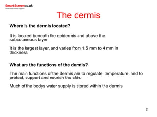
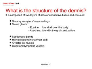
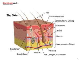
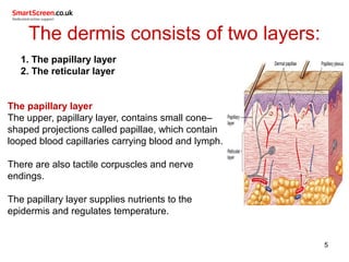
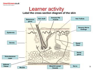
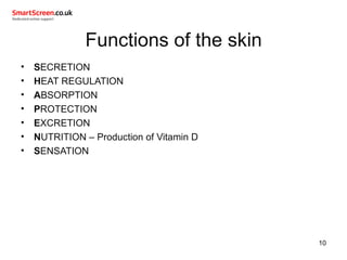
The skin
 The dermis and appendages
 The subcutaneous layer
 Functions of the skin
The dermis
Where is the dermis located?
It is located beneath the epidermis and above the
subcutaneous layer
It is the largest layer, and varies from 1.5 mm to 4 mm in
thickness
What are the functions of the dermis?
The main functions of the dermis are to regulate temperature, and to
protect, support and nourish the skin.
Much of the bodys water supply is stored within the dermis
2
What is the structure of the dermis?
It is composed of two layers of areolar connective tissue and contains:
Sensory receptors/nerve endings
Sweat glands:
- Eccrine: found all over the body
- Apocrine: found in the groin and axillae
Sebaceous glands
Hair follicles/hair shaft/hair bulb
Arrector pili muscle
Blood and lymphatic vessels
Handout 17
4
The dermis consists of two layers:
1. 1. The papillary layer
2. 2. The reticular layer
The papillary layer
The upper, papillary layer, contains small cone–
shaped projections called papillae, which contain
looped blood capillaries carrying blood and lymph.
There are also tactile corpuscles and nerve
endings.
The papillary layer supplies nutrients to the
epidermis and regulates temperature.
5
The reticular layer
The lower, reticular layer, is thicker and contains fibroblast cells, which
are responsible for the formation of collagen fibres and elastin.
Yellow elastin fibres give the skin its flexibility and elasticity.
White collagen fibres plump the skin and give strength and firmness.
Specialised cells are also found in the reticular layer:
Macrophages destroy bacteria and tissue debris
Mast cells secrete histamine, which causes vasodilation, and
heparin, which stops blood clotting (anti-coagulant).
The reticular layer supports other components of the skin, such as hair
follicles, sweat glands, and sebaceous glands.
Handout 17
Nerve endings of the dermis
 Meissner's corpuscles are for light touch and lie immediately
beneath the basement membrane of the epidermis
 Pacinian corpuscles are for deep pressure, and lie deeper in the
dermis
Free nerve endings are for pain and temperature, and these
endings lie at the superficial aspect of the dermis
7
Subcutaneous layer (hypodermis)
The deepest layer lying directly above the muscles of the body and
below the dermis of the skin.
It is made up of two main types of connective tissue:
Adipose tissue forms a network of fat cells providing the body
with insulation, as well as acting as a source of energy
Areolar tissue gives strength to protect underlying structures.
Elasticity to cope with increases in size, and support for blood
vessels and nerve endings found in this layer
8
Learner activity
Label the cross section diagram of the skin
9
Arrector Pili
muscle
Sweat
Duct
Sensory Nerve
Endings
Hair shaft
Adipose
Tissue
Subcutaneous
Layer
Dermis
Epidermis
Sweat
gland
Blood & Lymph
Vessels
Nerve
Sebaceous
gland
Hair Follicle
Muscle
Functions of the skin
• SECRETION
• HEAT REGULATION
• ABSORPTION
• PROTECTION
• EXCRETION
• NUTRITION – Production of Vitamin D
• SENSATION
10
Any questions?

Smartscreen skin 2

  • 1.
    The skin  Thedermis and appendages  The subcutaneous layer  Functions of the skin
  • 2.
    The dermis Where isthe dermis located? It is located beneath the epidermis and above the subcutaneous layer It is the largest layer, and varies from 1.5 mm to 4 mm in thickness What are the functions of the dermis? The main functions of the dermis are to regulate temperature, and to protect, support and nourish the skin. Much of the bodys water supply is stored within the dermis 2
  • 3.
    What is thestructure of the dermis? It is composed of two layers of areolar connective tissue and contains: Sensory receptors/nerve endings Sweat glands: - Eccrine: found all over the body - Apocrine: found in the groin and axillae Sebaceous glands Hair follicles/hair shaft/hair bulb Arrector pili muscle Blood and lymphatic vessels Handout 17
  • 4.
  • 5.
    The dermis consistsof two layers: 1. 1. The papillary layer 2. 2. The reticular layer The papillary layer The upper, papillary layer, contains small cone– shaped projections called papillae, which contain looped blood capillaries carrying blood and lymph. There are also tactile corpuscles and nerve endings. The papillary layer supplies nutrients to the epidermis and regulates temperature. 5
  • 6.
    The reticular layer Thelower, reticular layer, is thicker and contains fibroblast cells, which are responsible for the formation of collagen fibres and elastin. Yellow elastin fibres give the skin its flexibility and elasticity. White collagen fibres plump the skin and give strength and firmness. Specialised cells are also found in the reticular layer: Macrophages destroy bacteria and tissue debris Mast cells secrete histamine, which causes vasodilation, and heparin, which stops blood clotting (anti-coagulant). The reticular layer supports other components of the skin, such as hair follicles, sweat glands, and sebaceous glands. Handout 17
  • 7.
    Nerve endings ofthe dermis  Meissner's corpuscles are for light touch and lie immediately beneath the basement membrane of the epidermis  Pacinian corpuscles are for deep pressure, and lie deeper in the dermis Free nerve endings are for pain and temperature, and these endings lie at the superficial aspect of the dermis 7
  • 8.
    Subcutaneous layer (hypodermis) Thedeepest layer lying directly above the muscles of the body and below the dermis of the skin. It is made up of two main types of connective tissue: Adipose tissue forms a network of fat cells providing the body with insulation, as well as acting as a source of energy Areolar tissue gives strength to protect underlying structures. Elasticity to cope with increases in size, and support for blood vessels and nerve endings found in this layer 8
  • 9.
    Learner activity Label thecross section diagram of the skin 9 Arrector Pili muscle Sweat Duct Sensory Nerve Endings Hair shaft Adipose Tissue Subcutaneous Layer Dermis Epidermis Sweat gland Blood & Lymph Vessels Nerve Sebaceous gland Hair Follicle Muscle
  • 10.
    Functions of theskin • SECRETION • HEAT REGULATION • ABSORPTION • PROTECTION • EXCRETION • NUTRITION – Production of Vitamin D • SENSATION 10
  • 11.