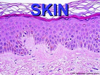
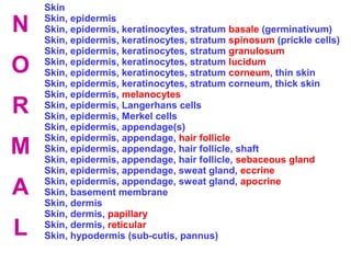
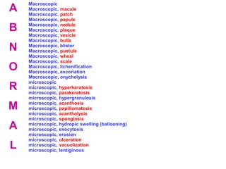
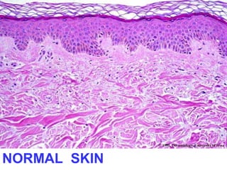
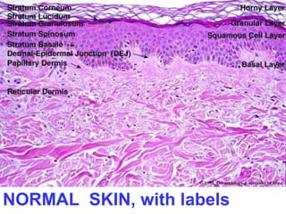
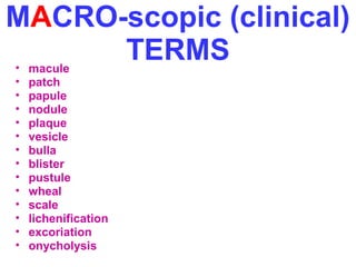
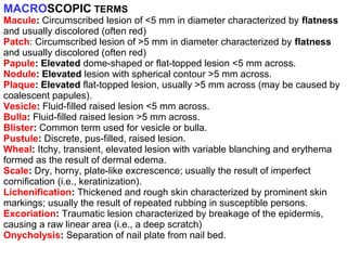
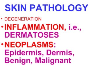
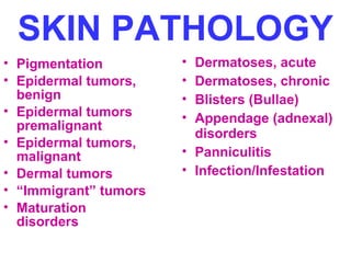
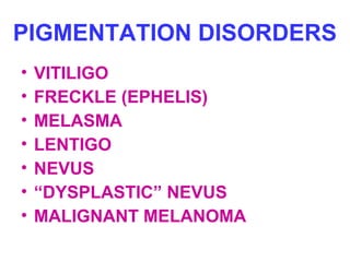
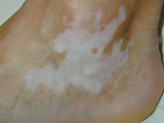
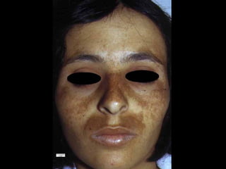
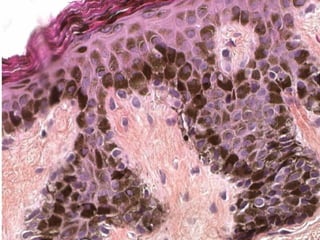
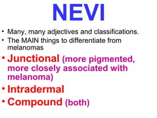
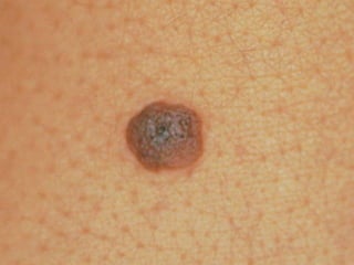
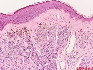
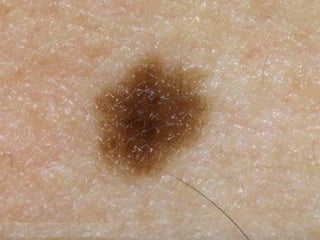
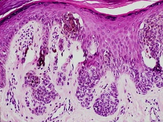
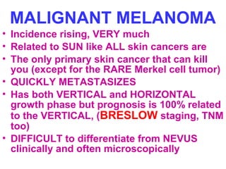
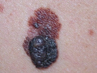
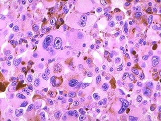
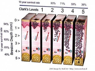
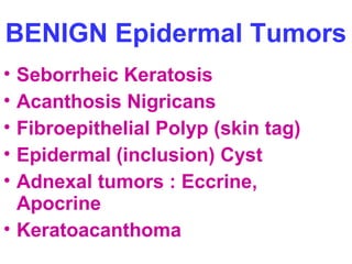
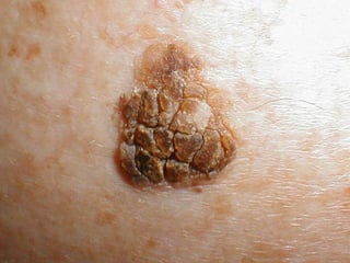
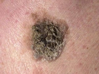
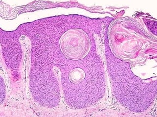
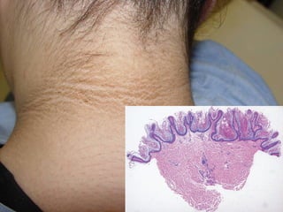
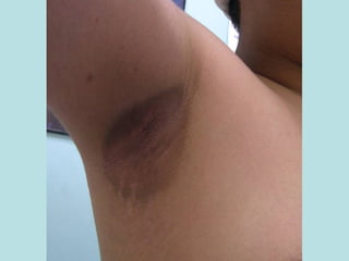
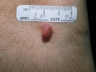
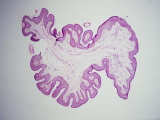
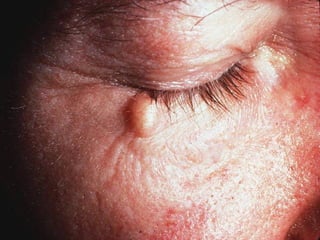
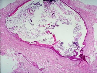
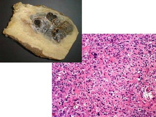
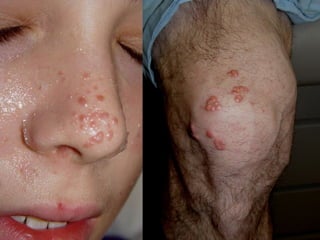
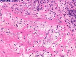
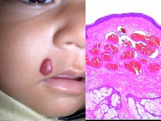
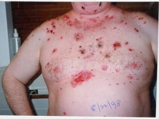
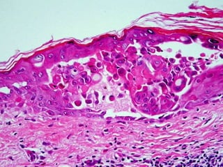
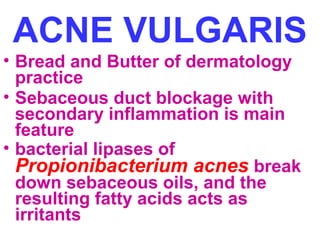
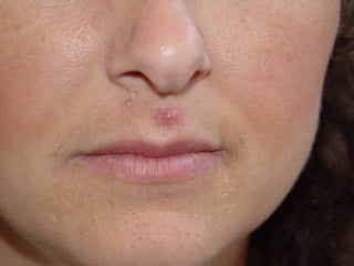
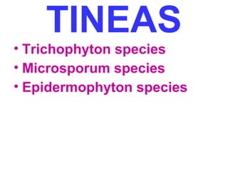
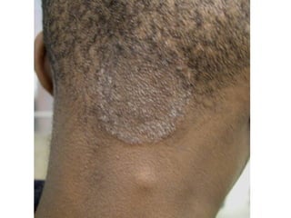
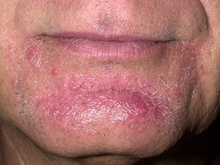
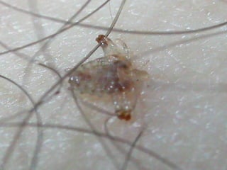
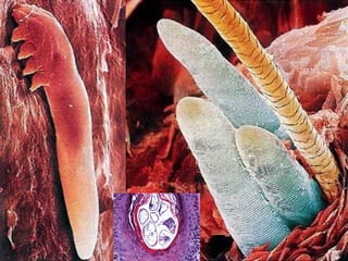
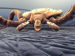
This document provides a comprehensive overview of normal and abnormal skin conditions organized into several sections. It begins by describing the normal anatomy and histology of skin. Macroscopic and microscopic terminology used to describe lesions is defined. The major sections that follow cover pigmentation disorders, epidermal and dermal neoplasms, inflammatory conditions, infections, and other abnormalities. Specific disorders are listed and briefly characterized within each section. The document serves as a reference for the clinical, histological, and pathological features of diverse skin diseases.
This document provides definitions and descriptions of terms related to normal skin anatomy and pathology. It begins with labeling diagrams of normal skin structures from the macroscopic to microscopic level. It then covers various skin conditions including pigmentation disorders, benign and malignant epidermal and dermal tumors, dermatoses, bullae, appendage disorders, panniculitis, and infections/infestations. For each condition, it lists key characteristics and histologic findings to aid in identification and diagnosis.
The document provides definitions and descriptions of various skin structures and conditions. It discusses the layers of the epidermis and dermis, as well as skin appendages. Common skin lesions, neoplasms, infections, and inflammatory conditions are defined at both the macroscopic and microscopic level, including examples like nevi, melanoma, psoriasis, and dermatitis. Images are included to illustrate examples like nevi subtypes, basal cell carcinoma, actinic keratosis, and dermatofibroma.
Chromoblastomycosis is a chronic fungal infection of the skin caused by certain dematiaceous fungi that form characteristic dark-colored sclerotic bodies in tissue. Microscopically, there is pseudoepitheliomatous hyperplasia and a mixed inflammatory infiltrate containing the sclerotic fungal bodies. Cultures grow darkly pigmented molds. Treatment involves thorough surgical excision or prolonged antifungal therapy.
This document summarizes various skin conditions and pathologies. It describes the types of cells found in the epidermis and dermis. It then lists and defines various skin lesions and rashes. It discusses acute inflammatory dermatoses like urticaria and eczema. It also mentions erythema multiforme and its causes. Finally, it outlines chronic inflammatory dermatoses, tumors of the skin epithelium and melanocytes, focusing on malignant melanoma.
This document provides an overview of common skin disorders, diseases, and conditions. It defines various primary lesions like macules, papules, pustules, and vesicles. It also covers secondary lesions, pigmentation disorders, inflammations, sebaceous and sudoriferous gland disorders, hypertrophies, skin cancers, contagious diseases, and acne. Key information includes definitions of medical terms, descriptions of common skin problems, and their identifying characteristics.
This document defines and describes various skin diseases and conditions through macroscopic and microscopic definitions. It discusses acute inflammatory dermatoses such as urticaria, acute eczematous dermatitis, erythema multiforme, erythema nodosum, and erythema induratum. It also covers chronic inflammatory dermatoses like psoriasis, lichen planus, lupus erythematosus, and acne vulgaris. For each condition, it provides details on presentation, histology, and clinical features.
This document summarizes the histology of normal skin and various skin diseases. It describes the five layers of normal skin (stratum basale, spinosum, granulosum, corneum, lucidum) and cell types in each layer. Definitions are provided for terms like hyperkeratosis and acanthosis. Histological features of actinic keratosis, keratoacanthoma, basal cell carcinoma, squamous cell carcinoma, malignant melanoma, and Kaposi sarcoma are outlined. Special histological subtypes and staining characteristics are also noted.
The document discusses common elementary lesions and infectious diseases of the skin. It describes the normal anatomy and histology of skin, then defines common macroscopic and microscopic skin lesions. It also covers frequent infectious diseases of the skin caused by viruses, bacteria, fungi and arthropods, including warts, molluscum contagiosum, herpes, impetigo, ringworm, scabies, and insect/ar tick bites.
The document discusses integumentary disorders and provides information on the anatomy and function of the skin. It describes the three layers of the skin - epidermis, dermis and subcutaneous tissue. It then discusses factors that influence skin integrity such as immobility, trauma, and malnutrition. Common dermatological terms and methods of describing skin lesions are also outlined. Finally, it provides an overview of common dermatological conditions like eczema, bacterial infections, fungal infections and viral infections.
The document discusses various disorders, conditions, and diseases that affect the integumentary system. It provides definitions and details on common skin issues like acne, psoriasis, eczema, contact dermatitis, athlete's foot, hives, rosacea, vitiligo, impetigo, boils, carbuncles, ringworm, warts, keloids, MRSA, gangrene, harlequin ichthyosis, scleroderma, alopecia areata, and burns. Treatment options are mentioned for some conditions. A wide range of both infectious and non-infectious skin problems are examined.
This document provides an overview of a dermatopathology course, including learning objectives, session details, sample exam questions, and study tips. It discusses key histopathological terms, classifications of skin diseases, common and rare skin conditions, and outlines the curriculum to be covered, including acute and chronic inflammatory diseases, infections, blistering diseases, and neoplastic conditions.
This document provides an overview of physical therapy for integumentary conditions. It describes the anatomy and physiology of the integumentary system and discusses common conditions treated by physical therapists, including vascular issues, trauma, and diseases. Examination principles and procedural interventions for various conditions are outlined, including wound management, scar management, and patient education.
This document provides an overview of common terminology used to describe diseases of the skin. It discusses terms used to describe the distribution, shape, color, type of lesions, secondary changes, epidermal changes, and histological changes that can be seen in various skin diseases. Some of the key terms mentioned include macule, papule, plaque, nodule, vesicle, bulla, pustule, abscess, desquamation, crusting, ulcer, erosion, fissure, atrophy, cicatrix, telangiectasis, acantholysis, acanthosis, basophilic degeneration, spongiosis, dyskeratosis, and hyperkeratosis. The document serves as
This document provides information about common skin conditions and diseases. It begins with an overview of the structure and function of skin, including its two main layers - the epidermis and dermis. It then discusses six common skin conditions in adults: acne, cellulitis, psoriasis, shingles, skin cancers, and vasculitis. Treatment options are provided for each condition. The document also summarizes six common skin conditions in children: chickenpox, eczema, Henoch–Schönlein purpura, impetigo, impetiginized eczema, and miliaria.
The document discusses various skin conditions including acute and chronic inflammatory diseases, infections, and neoplasms. It provides details on the pathogenesis, clinical features, and histopathology of conditions like urticaria, eczema, psoriasis, lichen planus, impetigo, fungal infections, viral infections including warts and molluscum, and acne. Case studies are presented to demonstrate clinical presentations.
Eczema is a skin inflammation characterized by itching and dryness. It is caused by both external (exogenous) factors like irritants or allergens as well as internal (endogenous) factors. The pathology involves inflammation and thickening of the epidermis and dermis. Clinically it presents as redness, scaling, excoriation and thickening of the skin (lichenification). It most commonly affects infants/children and the elderly. Treatment involves reducing inflammation and preventing infection through topical corticosteroids, moisturizers and oral antihistamines.
This document provides guidance on evaluating and managing dermatological conditions. It discusses the functions of skin, taking a thorough history, conducting a full examination of the skin and appendages, selecting appropriate investigations like skin scrapings or biopsies, and developing a treatment plan. Topical therapies are often first-line and include creams, ointments, or gels applied correctly based on the skin condition and amount of surface area involved. Close monitoring of the patient's response to treatment is important.
Skin cancer is a common, usually low-grade cancerous (malignant) growth of the skin. It starts from cells that begin as normal skin cells and transform into those with the potential to reproduce in an out-of-control manner. Unlike other cancers, the vast majority of skin cancers have no potential to spread to other parts of the body (metastasize) and threaten your life.
This document discusses various skin conditions and their associations with internal diseases and malignancies. It covers conditions like acanthosis nigricans seen in obesity/diabetes, Bazex syndrome seen with respiratory tract tumors, and dermatomyositis which has been linked to ovarian cancer. Skin findings in liver disease, renal disease, and malabsorption are also outlined. The document provides details on several paraneoplastic dermatoses and their linked malignancies. Common skin manifestations in diabetes like necrobiosis lipoidica and granuloma annulare are also summarized.
The Indian Dental Academy is the Leader in continuing dental education , training dentists in all aspects of dentistry and
offering a wide range of dental certified courses in different formats.for more details please visit
www.indiandentalacademy.com
The document provides details on various types of Kshudra Kusthas (minor skin diseases) described in Ayurveda and compares them to modern skin diseases. It describes 7 types of Kshudra Kusthas - Eka Kushta, Charmakyam, Kitibha Kushta, Vipadika, Alasaka, Dadru Mandala, and Charmadala. For each, it provides the dosha involvement, signs and symptoms. It then compares each to similar modern skin conditions such as psoriasis, scleroderma, tenia infections, herpes, impetigo, and provides details on symptoms and characteristics.
Basic Pathological Reactions of the Skin - Dr Zainab Almossalliaskadermatologist
This document summarizes the basic pathological reactions of the skin. It describes the different tissue compartments that make up the skin, including the epidermis, dermal-epidermal junction, papillary dermis, deep dermis, and subcutaneous tissue. It then discusses various pathological processes that can disturb the structure and function of these compartments, such as disturbances in epidermal cell kinetics, differentiation, and cohesion. Specific diseases are provided as clinical correlations for each pathological reaction described. The summary focuses on providing a high-level overview of the key tissue compartments involved and some examples of related pathological processes and diseases.
Anatomy of skin, Lichen planus, Dermatitis, Koebner phenomenon, collagen defects and elastin defects have been mentioned in details with various images to help u in understanding it well.
skin is an organ where internal disorders are manifested. some are early signs, some are late signs, some may be the only manifestation. they can result in diagnostic dilemma.
The document describes and classifies various skin lesions. It defines 26 different lesions including macules, papules, plaques, vesicles, bullae, pustules, cysts, nodules and wheals as primary skin lesions. Secondary skin lesions include scales, crusts, fissures, erosions, ulcers, lichenification and atrophy. Special skin lesions include telangiectasia, phlebectasia, burrows and comedones. Vasculopathies like petechiae, purpura and ecchymosis are also defined. Iris-like lesions are used to describe erythema multiforme. Each lesion is concisely defined and an example is provided.
The skin is the largest organ composed of three layers: the epidermis, dermis, and subcutis. The epidermis is stratified squamous epithelium consisting of keratinocytes, melanocytes, Langerhans cells, and Merkel cells. It has several layers including the stratum basale, stratum spinosum, stratum granulosum, stratum lucidum, and stratum corneum. Keratinocytes in the basal layer contain organelles and produce keratin intermediate filaments called tonofilaments. As keratinocytes differentiate and move upwards, they lose organelles and accumulate keratohyline granules containing proteins like filaggrin. The fully differentiated ker
The document discusses the structure and functions of the skin and its layers. The skin consists of three main layers - the epidermis, dermis and subcutaneous layer. The epidermis is the outermost layer and provides protection against water loss and microorganisms. Below the epidermis lies the dermis, which contains hair follicles, sweat and sebaceous glands, nerves and blood vessels. The innermost layer, the subcutaneous layer, consists of adipose tissue and insulates the body.
The skin has three main layers - the epidermis, dermis, and subcutaneous layer. The epidermis is made of epithelial tissue in 5 layers (stratum basale, stratum spinosum, stratum granulosum, stratum lucidum, stratum corneum) and contains four main cell types. The dermis is made of connective tissue and contains hair follicles, sebaceous glands, sweat glands, and nerves. The skin provides protection from external factors, regulates body temperature, and serves important sensory functions.
The skin is composed of three layers: the epidermis, dermis, and hypodermis. The epidermis contains keratinocytes and dendritic cells. It has multiple layers including the basal, spinous, granular, clear, and horny layers. The dermis contains fibrous and cellular connective tissue with nerve and vascular networks. The hypodermis contains adipose tissue and larger blood vessels and nerves. The skin has several functions including serving as a barrier, regulating temperature, sensation, vitamin D synthesis, and as a blood reservoir. Primary skin lesions include macules, papules, vesicles, pustules, nodules, and plaques. Secondary lesions develop from primary lesions and include scales
The skin has three main layers - the epidermis, dermis and subcutaneous tissue. The epidermis is the outermost layer and contains no blood vessels. It has multiple layers that cells move through as they are replaced every 28 days. The dermis lies below and contains collagen, elastin and blood vessels that provide structure and nourishment. It has two layers - a papillary layer with nerve endings and a reticular layer with hair follicles and glands. The deepest layer, subcutaneous tissue, contains fat and connects the dermis to underlying structures. Together these layers contain millions of cells, yards of blood vessels and nerves, and various sensory receptors and appendages like hair and glands.
This document provides an overview of normal skin histology and dermatopathology terms. It describes the three layers of normal skin - epidermis, dermis, and hypodermis. Key cell types like keratinocytes, melanocytes, and components of the epidermis and dermis are defined. Common microscopic skin findings are explained, such as hyperkeratosis, parakeratosis, spongiosis. Important dermatological conditions involving pigmentation, epidermal and dermal tumors, and inflammatory diseases are also highlighted.
This document summarizes several pre-malignant and malignant epidermal tumors and acute inflammatory dermatoses. It describes actinic keratosis as a premalignant skin lesion caused by UV damage that can progress to squamous cell carcinoma. Squamous cell carcinoma is described as arising from UV damage or other causes and often presents as an ulcer. Basal cell carcinoma is summarized as the most common skin cancer, locally aggressive but non-metastatic, appearing on sun-exposed skin. Urticaria, eczema, and erythema multiforme represent acute inflammatory dermatoses and their histological features are outlined.
This document discusses the structure and functions of skin. It provides details on the layers of the epidermis and dermis. Various skin appendages like hair follicles and sebaceous glands are also mentioned. The document then discusses several common dermatological conditions like psoriasis, lichen planus, bacterial and fungal infections, blistering disorders, and tumors of the skin. Macroscopic and microscopic features of these conditions are described along with their pathogenesis and clinical presentation. Common benign tumors of the skin like seborrheic keratosis and premalignant lesions like actinic keratosis are also summarized.
This document discusses various microscopic terms and classifications related to skin disorders. It defines terms like hyperkeratosis, dyskeratosis, acanthosis, and spongiosis. It also classifies skin disorders into categories such as inflammatory dermatoses (e.g. urticaria, eczema, psoriasis), infectious dermatoses (e.g. impetigo, verrucae), and neoplasms (e.g. seborrheic keratosis, squamous cell carcinoma, basal cell carcinoma). Microscopic features of these disorders are described, including characteristic cellular changes, infiltrates, and growth patterns.
This document provides information on malignant skin lesions and skin cancers. It discusses the anatomy and function of skin, pre-malignant lesions like actinic keratosis, Bowen's disease and conditions that increase risk of skin cancers like neurofibromatosis. It then describes the most common skin cancers - basal cell carcinoma (BCC), squamous cell carcinoma (SCC), and malignant melanoma. For each cancer, it discusses epidemiology, etiology, clinical features, histopathology, prognosis and treatment options.
This document summarizes various fungal infections. It describes that most fungal infections are mild and involve superficial areas like skin, hair, and nails. These include pityriasis versicolor, tinea infections like corporis, cruris, pedis, capitis and barbae, onychomycosis, tinea nigra, and piedra. Some deeper infections in subcutaneous tissues are also described such as chromoblastomycosis, mycetoma, and sporotrichosis. Systemic fungal infections that spread throughout the body are also briefly mentioned. Key identifying features, causative organisms and histopathological findings of major fungal infections are provided.
This document provides information about dermatopathology and skin biopsy procedures. It discusses the role of dermatopathology in diagnosing diseases and identifying infectious agents. Five common skin biopsy techniques are described: punch biopsy, shave biopsy, incisional biopsy, complete excisional biopsy, and curettage biopsy. Guidance is given for handling biopsied skin samples. The document also contains a glossary of dermatopathology terms defined in multiple sections.
Cutaneous malignancies and related disorders.pptxAjilAntony10
The document discusses cutaneous malignancies like basal cell carcinoma (BCC), squamous cell carcinoma (SCC) and malignant melanoma. It covers their etiology, risk factors, clinical features, histopathological classification, staging and treatment options. The key points are: BCC is the most common type and arises from UV exposure. SCC usually occurs on sun-exposed skin and risk is higher with tanning bed use. Malignant melanoma is the most deadly but can be cured if detected early, using the ABCDE criteria to monitor moles. Wide local excision is the main treatment for all, with Mohs surgery preferred for certain types/locations.
Skin cancers or cutaneous malignancies including Basal cell carcinoma, Squamous cell carcinoma and Melanoma and with a brief introduction of skin as an organ itself.
This document summarizes Onchocerca volvulus, the filarial worm that causes river blindness. It discusses the worm's taxonomy, morphology, life cycle, epidemiology in tropical Africa and Americas, and the pathology and clinical manifestations of infection, including skin changes, blindness, and lymphadenitis. Diagnosis involves skin snips, eye exams, and nodule removal and examination. Treatment is annual ivermectin doses plus doxycycline to kill Wolbachia bacteria in the worms. Prevention includes ivermectin prophylaxis and avoiding areas where blackfly vectors breed and bite.
onchocerciasis, definition, onchocerca volvulus, epidemiology of onchocerciasis, management of onchocerciasis, prevention of onchocerciasis, pathology of onchocerciasis
This document provides an overview of the integumentary system, including the structure and function of the skin and its layers. It discusses the epidermis and dermis in depth, describing the cells that make up each layer. The document also covers skin appendages like hair follicles, sebaceous glands, and nails. Additionally, it summarizes the functions of the skin and describes common skin conditions like psoriasis, outlining its causes, types, symptoms, and diagnostic evaluation.
SKIN CONDITION presentation for medical students.pptxIbrahimKargbo13
This document provides an outline for a course on skin conditions. It begins by listing common skin conditions categorized by symptoms such as itching, pigmentation, crusts, vesicles, pustules, and macules. It then defines dermatology as the study of skin, nails, and hair and discusses the functions and facts about skin. The document outlines important aspects of dermatologic history taking and examination, including describing different types of skin lesions and how their configuration, texture, location, and distribution can provide clues to diagnosis.
Automated cell counter & its quality controlSaikat Mandal
This document discusses various premalignant lesions of the skin, including actinic keratosis, oral leukoplakia, Bowen's disease, erythroplasia of Queyrat, and bowenoid papulosis. It describes the clinical presentation, histological features, causative factors, and risk of progression to squamous cell carcinoma for each condition. The document focuses on describing the characteristic layers and cell types found in normal skin and how these features are altered in the various premalignant lesions.
This document discusses various premalignant lesions of the skin. It begins by describing the normal architecture of the skin, including the different layers of the epidermis. It then discusses several specific premalignant lesions in more detail, including actinic keratosis, oral leukoplakia, Bowen's disease, erythroplasia of Queyrat, and Bowenoid papulosis. It also briefly mentions arsenical keratosis, Marjolin's ulcer, Paget's disease, extramammary Paget's disease, and xeroderma pigmentosum. For each condition, it provides information on clinical presentation, histopathology, causative factors, and risk of progression to
Urticaria is a common skin disorder caused by localized mast cell degranulation, leading to itchy wheals that typically develop and fade within hours. Acute eczematous dermatitis presents as erythematous papules and plaques that can become crusted or scaled due to conditions like atopic dermatitis. Psoriasis is a chronic inflammatory disease characterized by well-demarcated salmon-colored plaques covered in silver scale. Lichen planus features purple, pruritic planar papules in a symmetric distribution. Common benign skin tumors include seborrheic keratoses, actinic keratoses, and melanocytic nevi, while squamous cell
This document provides an overview of common dermatologic conditions seen in young adults, including benign skin lesions such as warts, molluscum contagiosum, and dermatofibromas. It also discusses atopic dermatitis, contact dermatitis, seborrheic dermatitis, psoriasis, and other inflammatory and infectious skin disorders. Treatment options are outlined for each condition, with an emphasis on moisturization and topical corticosteroids as first-line therapies. Referral is indicated for severe or treatment-resistant cases.
Skin is the largest organ of the body, covering a surface area of about 1.5-2m2. It has three main layers - the epidermis, dermis, and hypodermis. The epidermis is avascular and provides a protective barrier, while the dermis contains blood vessels, hair follicles, and glands. The hypodermis connects the skin to underlying muscles and stores fat. Skin regulates temperature, protects the body, and enables sensation. Common skin diseases include acne, athlete's foot, dermatitis, psoriasis, and various forms of skin cancer caused by sun exposure.
Styles of Scientific Reasoning, Scientific Practices and Argument in Science ...Elsa von Licy
The document discusses various topics related to scientific reasoning, practices, and argumentation including different styles of scientific thinking, features of scientific knowledge, and teaching and learning science. It provides examples of "crazy ideas" in science that are now accepted, examines the role of argument in science, and outlines the scientific practices and central questions of science. It also discusses developing models, planning investigations, analyzing data, and constructing explanations as key scientific practices.
Anti-philosophy rejects traditional philosophy and logic, instead embracing creativity, spirituality, and personality. It considers philosophy to be dead, kept alive artificially by analytic philosophers. The document criticizes how philosophy is currently taught and argues it has become unproductive, replacing original aims with nonsense. Anti-philosophy's goal is not to destroy philosophy but to transform its current state and avoid fundamentalism in philosophy and science.
There is no_such_thing_as_a_social_science_introElsa von Licy
This document provides an introduction and overview of the arguments made in the book "There is No Such Thing as Social Science". It begins by stating the provocative title and questioning whether the authors will take it back or qualify their position.
It then outlines three ways the term "social science" could be used - referring to a scientific spirit of inquiry, a shared scientific method, or reducibility to natural sciences. The authors argue against the latter two, methodological and substantive reductionism.
The introduction discusses how opponents may accuse the authors of being a priori or anti-reductionist, but argues that those defending social science are actually being dogmatic by insisting it must follow a scientific model. It frames the debate as being
Rasamanikya is a excellent preparation in the field of Rasashastra, it is used in various Kushtha Roga, Shwasa, Vicharchika, Bhagandara, Vatarakta, and Phiranga Roga. In this article Preparation& Comparative analytical profile for both Formulationon i.e Rasamanikya prepared by Kushmanda swarasa & Churnodhaka Shodita Haratala. The study aims to provide insights into the comparative efficacy and analytical aspects of these formulations for enhanced therapeutic outcomes.
One health condition that is becoming more common day by day is diabetes.
According to research conducted by the National Family Health Survey of India, diabetic cases show a projection which might increase to 10.4% by 2030.
Muktapishti is a traditional Ayurvedic preparation made from Shoditha Mukta (Purified Pearl), is believed to help regulate thyroid function and reduce symptoms of hyperthyroidism due to its cooling and balancing properties. Clinical evidence on its efficacy remains limited, necessitating further research to validate its therapeutic benefits.
8 Surprising Reasons To Meditate 40 Minutes A Day That Can Change Your Life.pptxHolistified Wellness
We’re talking about Vedic Meditation, a form of meditation that has been around for at least 5,000 years. Back then, the people who lived in the Indus Valley, now known as India and Pakistan, practised meditation as a fundamental part of daily life. This knowledge that has given us yoga and Ayurveda, was known as Veda, hence the name Vedic. And though there are some written records, the practice has been passed down verbally from generation to generation.
TEST BANK For Community and Public Health Nursing: Evidence for Practice, 3rd...Donc Test
TEST BANK For Community and Public Health Nursing: Evidence for Practice, 3rd Edition by DeMarco, Walsh, Verified Chapters 1 - 25, Complete Newest Version TEST BANK For Community and Public Health Nursing: Evidence for Practice, 3rd Edition by DeMarco, Walsh, Verified Chapters 1 - 25, Complete Newest Version TEST BANK For Community and Public Health Nursing: Evidence for Practice, 3rd Edition by DeMarco, Walsh, Verified Chapters 1 - 25, Complete Newest Version Test Bank For Community and Public Health Nursing: Evidence for Practice 3rd Edition Pdf Chapters Download Test Bank For Community and Public Health Nursing: Evidence for Practice 3rd Edition Pdf Download Stuvia Test Bank For Community and Public Health Nursing: Evidence for Practice 3rd Edition Study Guide Test Bank For Community and Public Health Nursing: Evidence for Practice 3rd Edition Ebook Download Stuvia Test Bank For Community and Public Health Nursing: Evidence for Practice 3rd Edition Questions and Answers Quizlet Test Bank For Community and Public Health Nursing: Evidence for Practice 3rd Edition Studocu Test Bank For Community and Public Health Nursing: Evidence for Practice 3rd Edition Quizlet Test Bank For Community and Public Health Nursing: Evidence for Practice 3rd Edition Stuvia Community and Public Health Nursing: Evidence for Practice 3rd Edition Pdf Chapters Download Community and Public Health Nursing: Evidence for Practice 3rd Edition Pdf Download Course Hero Community and Public Health Nursing: Evidence for Practice 3rd Edition Answers Quizlet Community and Public Health Nursing: Evidence for Practice 3rd Edition Ebook Download Course hero Community and Public Health Nursing: Evidence for Practice 3rd Edition Questions and Answers Community and Public Health Nursing: Evidence for Practice 3rd Edition Studocu Community and Public Health Nursing: Evidence for Practice 3rd Edition Quizlet Community and Public Health Nursing: Evidence for Practice 3rd Edition Stuvia Community and Public Health Nursing: Evidence for Practice 3rd Edition Test Bank Pdf Chapters Download Community and Public Health Nursing: Evidence for Practice 3rd Edition Test Bank Pdf Download Stuvia Community and Public Health Nursing: Evidence for Practice 3rd Edition Test Bank Study Guide Questions and Answers Community and Public Health Nursing: Evidence for Practice 3rd Edition Test Bank Ebook Download Stuvia Community and Public Health Nursing: Evidence for Practice 3rd Edition Test Bank Questions Quizlet Community and Public Health Nursing: Evidence for Practice 3rd Edition Test Bank Studocu Community and Public Health Nursing: Evidence for Practice 3rd Edition Test Bank Quizlet Community and Public Health Nursing: Evidence for Practice 3rd Edition Test Bank Stuvia
Clinic ^%[+27633867063*Abortion Pills For Sale In Tembisa Central19various
Clinic ^%[+27633867063*Abortion Pills For Sale In Tembisa Central Clinic ^%[+27633867063*Abortion Pills For Sale In Tembisa CentralClinic ^%[+27633867063*Abortion Pills For Sale In Tembisa CentralClinic ^%[+27633867063*Abortion Pills For Sale In Tembisa CentralClinic ^%[+27633867063*Abortion Pills For Sale In Tembisa Central
These lecture slides, by Dr Sidra Arshad, offer a simplified look into the mechanisms involved in the regulation of respiration:
Learning objectives:
1. Describe the organisation of respiratory center
2. Describe the nervous control of inspiration and respiratory rhythm
3. Describe the functions of the dorsal and respiratory groups of neurons
4. Describe the influences of the Pneumotaxic and Apneustic centers
5. Explain the role of Hering-Breur inflation reflex in regulation of inspiration
6. Explain the role of central chemoreceptors in regulation of respiration
7. Explain the role of peripheral chemoreceptors in regulation of respiration
8. Explain the regulation of respiration during exercise
9. Integrate the respiratory regulatory mechanisms
10. Describe the Cheyne-Stokes breathing
Study Resources:
1. Chapter 42, Guyton and Hall Textbook of Medical Physiology, 14th edition
2. Chapter 36, Ganong’s Review of Medical Physiology, 26th edition
3. Chapter 13, Human Physiology by Lauralee Sherwood, 9th edition
Cell Therapy Expansion and Challenges in Autoimmune DiseaseHealth Advances
There is increasing confidence that cell therapies will soon play a role in the treatment of autoimmune disorders, but the extent of this impact remains to be seen. Early readouts on autologous CAR-Ts in lupus are encouraging, but manufacturing and cost limitations are likely to restrict access to highly refractory patients. Allogeneic CAR-Ts have the potential to broaden access to earlier lines of treatment due to their inherent cost benefits, however they will need to demonstrate comparable or improved efficacy to established modalities.
In addition to infrastructure and capacity constraints, CAR-Ts face a very different risk-benefit dynamic in autoimmune compared to oncology, highlighting the need for tolerable therapies with low adverse event risk. CAR-NK and Treg-based therapies are also being developed in certain autoimmune disorders and may demonstrate favorable safety profiles. Several novel non-cell therapies such as bispecific antibodies, nanobodies, and RNAi drugs, may also offer future alternative competitive solutions with variable value propositions.
Widespread adoption of cell therapies will not only require strong efficacy and safety data, but also adapted pricing and access strategies. At oncology-based price points, CAR-Ts are unlikely to achieve broad market access in autoimmune disorders, with eligible patient populations that are potentially orders of magnitude greater than the number of currently addressable cancer patients. Developers have made strides towards reducing cell therapy COGS while improving manufacturing efficiency, but payors will inevitably restrict access until more sustainable pricing is achieved.
Despite these headwinds, industry leaders and investors remain confident that cell therapies are poised to address significant unmet need in patients suffering from autoimmune disorders. However, the extent of this impact on the treatment landscape remains to be seen, as the industry rapidly approaches an inflection point.
11. MACROSCOPIC TERMS
Macule: Circumscribed lesion of <5 mm in diameter characterized by flatness
and usually discolored (often red)
Patch: Circumscribed lesion of >5 mm in diameter characterized by flatness
and usually discolored (often red)
Papule: Elevated dome-shaped or flat-topped lesion <5 mm across.
Nodule: Elevated lesion with spherical contour >5 mm across.
Plaque: Elevated flat-topped lesion, usually >5 mm across (may be caused by
coalescent papules).
Vesicle: Fluid-filled raised lesion <5 mm across.
Bulla: Fluid-filled raised lesion >5 mm across.
Blister: Common term used for vesicle or bulla.
Pustule: Discrete, pus-filled, raised lesion.
Wheal: Itchy, transient, elevated lesion with variable blanching and erythema
formed as the result of dermal edema.
Scale: Dry, horny, plate-like excrescence; usually the result of imperfect
cornification (i.e., keratinization).
Lichenification: Thickened and rough skin characterized by prominent skin
markings; usually the result of repeated rubbing in susceptible persons.
Excoriation: Traumatic lesion characterized by breakage of the epidermis,
causing a raw linear area (i.e., a deep scratch)
Onycholysis: Separation of nail plate from nail bed.
13. MICROSCOPIC TERMS
Hyperkeratosis: Thickening of the stratum corneum, often associated with a qualitative
abnormality of the keratin.
Parakeratosis: Modes of keratinization characterized by the retention of the nuclei in the
stratum corneum. On mucous membranes, parakeratosis is normal.
Hypergranulosis: Hyperplasia of the stratum granulosum, often due to intense rubbing.
Acanthosis: Diffuse epidermal hyperplasia.
Papillomatosis: Surface elevation caused by hyperplasia and enlargement of
contiguous dermal papillae.
Dyskeratosis: Abnormal keratinization occurring prematurely within individual cells or
groups of cells below the stratum granulosum. Generally the same as DYSPLASIA.
Acantholysis: Loss of intercellular connections resulting in loss of cohesion between
keratinocytes.
Spongiosis: Intercellular edema of the epidermis.
Hydropic swelling (ballooning): Intracellular edema of keratinocytes.
Exocytosis: Infiltration of the epidermis by inflammatory or circulating blood cells.
Erosion: Discontinuity of the skin exhibiting incomplete loss of the epidermis.
Ulceration: Discontinuity of the skin exhibiting complete loss of the epidermis and often
of portions of the dermis and even subcutaneous fat.
Vacuolization: Formation of vacuoles within or adjacent to cells; often refers to basal
cell-basement membrane zone area.
Lentiginous: Referring to a linear pattern of melanocyte proliferation within the
epidermal basal cell layer. Lentiginous melanocytic hyperplasia can occur as a reactive
change or as part of a neoplasm of melanocytes.
22. NEVI
• Many, many adjectives and classifications.
• The MAIN things to differentiate from
melanomas
• Junctional (more pigmented,
more closely associated with
melanoma)
• Intradermal
• Compound (both)
23.
24.
25.
26.
27. MALIGNANT MELANOMA
• Incidence rising, VERY much
• Related to SUN like ALL skin cancers are
• The only primary skin cancer that can kill
you (except for the RARE Merkel cell tumor)
• QUICKLY METASTASIZES
• Has both VERTICAL and HORIZONTAL
growth phase but prognosis is 100% related
to the VERTICAL, (BRESLOW staging, TNM
too)
• DIFFICULT to differentiate from NEVUS
clinically and often microscopically
45. PREMALIGNANT/MALIGNANT
• ACTINIC (Solar) KERATOSIS, i.e. precursor
to SCC
• SQUAMOUS CELL CARCINOMA,
squamous “pearls”, intercellular bridges
• BASAL CELL CARCINOMA, by far, MOST
COMMON, BLUE palisading nests
• MERKEL CELL CARCINOMA (TUMOR),
VERY MALIGNANT AND LETHAL, LOOK
LIKE SMALL CELL CA. OF LUNG
46. GENERAL COMMENTS
• BOTH SCC and BCC related to SUN (i.e.,
radiation) exposure. (as is MM also)
• SCC also related to As, carcinogens, chaw,
betel nut, HPV, familial, etc.
• BOTH SCC and BCC can do local damage
but very rarely metastasize or kill.
• MERKEL CELL tumors metastasize early
and extensively, like melanomas.
68. URTICARIA
• DERMAL EDEMA
• DILATATION of VASCULAR
SPACES
• EARLY PERIVASCULAR
CUFFING OF
INFLAMMATORY CELLS
69.
70. ECZEMA
(aka, acute eczematous dermatitis)
• A myriad of ACUTE
inflammatory disorders, with
allergic, drug related, sun
related etiologies
• The common histologic feature
is
SPONGIOSIS
71.
72.
73.
74.
75.
76. PSORIASIS
• 1-2% of USA
• Elbows, Knees
• Parakeratosis, generalized
epidermal hyperplasia,
elongation of the rete pegs,
extensive chronic inflammatory
cell infiltrates, “MUNRO”
intraepidermal microabscesses
88. ACNE VULGARIS
• Bread and Butter of dermatology
practice
• Sebaceous duct blockage with
secondary inflammation is main
feature
• bacterial lipases of
Propionibacterium acnes break
down sebaceous oils, and the
resulting fatty acids acts as
irritants
Basic microscopic and macroscopic descriptive terms, the essentials of describing skin lesions properly
Classifications and major examples of the classifications.
Histology review (no labels)
Histology review (labels)
Voila!
Appendages: hair follicles, sebaceous glands, sweat glands (eccrine, apocrine). Is this thick skin or thin skin?
These are just descriptive terms, NOT diagnoses.
Vitiligo
Freckles (ephelis)
Melasma, also called “mask of pregnancy”
Lentigo, (plural: lentigenes), is generally considered a brown pigmented spot on the skin. It is a harmless (benign) hyperplasia of melanocytes which is linear in its spread
Lentigo. Would lentigo be palpable? Ans: NO
Intradermal nevus
Intradermal nevus.
Note the lack of “junctional” activity.
Junctional nevus
Junctional nevus. Why is this called “Junctional”?
What would a “compound” nevus be? Ans: BOTH junctional and intradermal.
Malignant melanomas are malignant proliferations of melanocytes.
What is the ABCDEEFG principle?
Asymmetry
Borders (irregular)
Color (variegated), and
Diameter (greater than 6 mm (0.24 in.), about the size of a pencil eraser)
Evolving over time
These classifications do not however apply to the most dangerous form of melanoma, nodular melanoma, which has its own classifications:
Elevated above the skin surface
Firm to the touch
Growing
Why do only idiots learn the ABCDEEFG acronym? Ans: Because all these features are already basic in understanding malignancy.
What kind of malignant cell would have pigment IN it? What could this pigment be?
Clark vs. Breslow (better) classification
Seborrheic keratosis
Seborrheic keratosis, a bit more pigmented than the previous one, pigmentation is very common in ALL types of benign keratoses.
Squamous “horn cysts” in seborrheic keratosis
Acanthosis nigricans, often associated with diabetes mellitus
Fibroepithelial polyp, or “skin tag”
Fibroepithelial polyp, or “skin tag”.
Would you call this a papilloma? Why, or why not?
Epidermal inclusion cyst, the overlying skin looks normal.
Epidermal inclusion cyst
Keratoacanthoma, the MAIN lesion to differentiate from squamous cell carcinoma
Keratoacanthoma, the MAIN lesion to differentiate from squamous cell carcinoma. What is a collarette?
Is a collarette the classical feature which differentiates KAs from SCCs? Ans: YES
Actinic keratosis
Actinic keratosis vs. squamous cell carcinoma
Squamous cell carcinoma, infiltrating. What is squamous cell carcinoma-in-situ usually called? Ans: Bowen’s disease.
Is there a collarette here? Ans: NO
Squamous cell carcinoma, infiltrating. Note the “pearls”. Does the presence of pearls make this well differentiated? Ans: Yes.
Squamous dysplasia, perhaps actinic keratosis, or something leading into squamous cell carcinoma.
By far, the commonest malignancy of skin, BCC, i.e., Basal Cell Carcinoma, typical appearance.
By far, the commonest malignancy of skin, BCC, i.e., Basal Cell Carcinoma, typical appearance. Note the PERIPHERAL PALISADING!!!
PALISADING!!!
PALISADING!!!
PALISADING!!!
Merkel cell tumor, very highly malignant RARE and usually fatal, looks EXACTLY like a small cell carcinoma of the lung? Ans: yes.
Note the LACK of the term “Fibroma”. Why” Ans: Because tumors of pure “fibroblasts” of the dermis are, somehow, rare.
Benign fibrous histiocytoma, or dermatofibroma
Large fibrous histiocytoma, perhaps a dermatofibrosarcoma protuberans?
Malignant fibrous histiocytoma.
Xanthomas filled with cholesterol and lipids, to give the “foamy” appearance.
Xanthoma filled with cholesterol and lipids, to give the “foamy” appearance. Would you suspect these are associated, often, with hypercholesterolemia? Ans: YES
Hemangioma, often a congenital “birth mark”, which can regress significantly with aging. A red lesion which “blanches” when you put pressure on it, is always suspected to be a vascular tumor.
Kaposi’s sarcoma
Ichthyosis, usually genetic. Do you see the lamellae? Would you guess the term “lamellar” ichthiosis is often used? Ans: Yes
What is the Greek word for fish?
Inflammatory skin diseases are called dermatoses. Emphasis will NOT be on etiology, chiefly because most are unknown and are classically described as being related to a host of many things, like drugs, sun, allergies, etc.
While the neoplastic skin diseases can often be easily diagnosed by slide only, the dermatoses can rarely be diagnosed by slide only.
The diagnosis of inflammatory skin diseases requires SEEING THE PATIENT!!!
Acute and chronic, often refer to clinical features, because acute and chronic inflammatory cells, i.e., polys and monos respectively, do not often apply here.
Urticaria = hives = wheals = Type I Hypersensitivity = triggered off by perivascular mast cells releasing histamine
Is urticaria the classic skin response to type 1 hypersensitivity? Ans: YES
Often the word or classification “atopic” precedes the word “eczema”, implying an allergic etiology
(Atopic) Eczema
Eczema with spongiosis. Spongiosis: = Intercellular edema of the epidermis.
Pustules can be thought of as extreme “spongiosis”
Pustules, ulcerated.
Pustules, like vesicles and bullae, have an “evolution” of clinical and histologic appearances, generally following the acute__>chronic inflammatory evolution.
Erythema multiforme is a skin condition of unknown cause, possibly mediated by deposition of immune complex (mostly IgM) in the superficial microvasculature of the skin and oral mucous membrane that usually follows an infection or drug exposure. It is a common disorder, with peak incidence in the second and third decades of life. This severe form may be related to Stevens-Johnson syndrome.
Does this look like extreme urticaria?
Name the FOUR histopathologic findings in most of the forms of psoriasis: 1,2,3,4
1) Parakeratosis, 4) hyperplasia, 3) rete peg elongation, 4) MUNRO abscesses.
Please note that the classical diseases which cause “pigmentary incontinence” are the lichenoid disorders.
Note the pigmented cells are MELANOPHAGES, not MELANOCYTES.
Possibly the commonest skin disease you will see every day, so I’m giving you 5 classic views.
Do you think stasis dermatitis is commonest in the areas of tissues often most compromised by atherosclerosis?
Know the various types of “bullous” diseases. What is a bulla? What is acantholysis? Bulla: Fluid-filled raised lesion greater than 5 mm across.
Acantholysis: Loss of intercellular connections resulting in loss of cohesion between keratinocytes.
Pemphigus, fresh bullae
Pemphigus, ruptured, scabbed bullae
Acantholysis in the bullous family of diseases. Notice that the “seperation” can be within the acanthocytes, i.e., the stratum spinosum, or at the dermal-epidermal junction. So would you imagine many of the bullous disorders are diseases of basement membrane and tonofibrils (i.e., desmosomes), and may be autoimmune?
So is acne chiefly a folliculitis? Ans: Yes!
In about 30-50% of cases, the cause of E.N. is unknown. EN may be associated with a wide variety of diseases, including infections (e.g., TB, Strep, Mycoplasma, Yersinia, and EBV), coccidiomycosis, sarcoid, IBD, autoimmune disorders , pregnancy, medications (sulfas, oral contraceptives, bromides), and cancer. Do you see a lot of granulomatous diseases here? Ans: YES.
E.I. is OBSCURE, RARE, and mainly in WOMEN, also felt to be related to TB.
A panniculitis is a primary inflammation of the subdermal connective tissues, i.e., the hypodermis, or subcutis.
How many viruses besides HPV can you think of which cause squamous proliferations in ANY part of the body? Ans: Not many.
Papillomatous epidermal hyperplasia is the most consistent feature of verrucae (warts), Also note the “hypergranulosis”.
“hypergranulosis” “hypergranulosis” “hypergranulosis”
Molluscum contagiosum, a pox virus
Molluscum contagiosum.
Some things in pathology can only best be described by pictures, not words.
Impetigo, caused by staph and strep, usually in small kids.
Ringworm of scalp, Tinea capitis
Tinea barbae
Ringworm of the body, Tinea corporis
Tinea cruris, or jock itch
Athlete’s foot, or tinea pedis.
Is this interspace the most common place for tinea pedis?
Why? If your patient has a gangrenous toe, which one is most likely?
Onychomycosis (Note the LACK of the word tinea)
PAS stain of hyphae, probably scrapings.
PAS stain of hyphae, probably a histologic slice, NOT scrapings.
Scabies in it’s most common location
Body lice (pediculosis)
Pubic louse (phthirus pubis)
Demodex follicularis, a mite larva, notice how it likes to share a hair follicle with a hair shaft.
Why is this an arachnid, and not an insect? Ans: 8 legs.