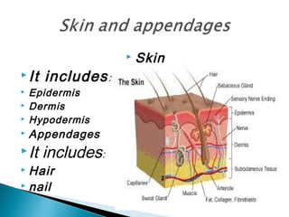
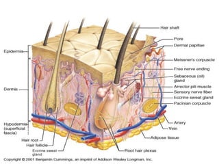
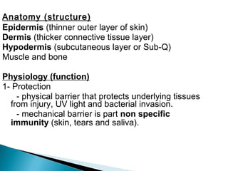
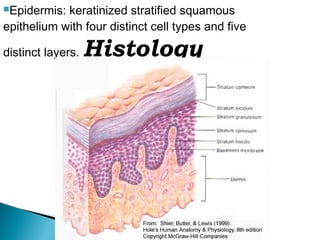
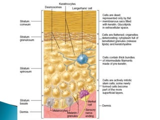
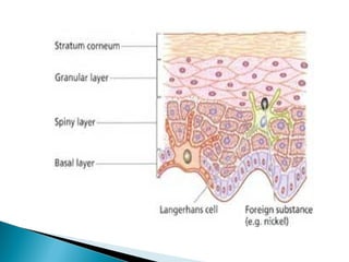
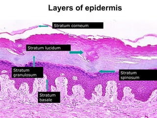
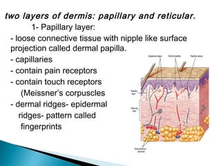
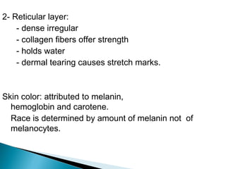
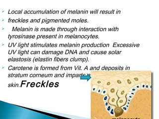
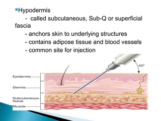
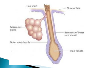
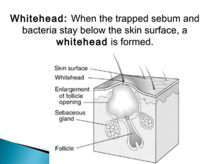
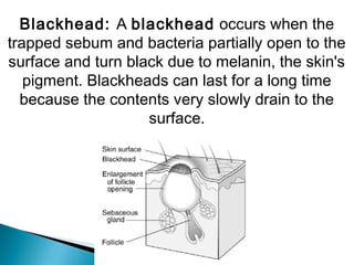
Pankaj Sharma
 Skin
It includes:
 Epidermis
 Dermis
 Hypodermis
 Appendages
It includes:
 Hair
 nail
Anatomy (structure)
Epidermis (thinner outer layer of skin)
Dermis (thicker connective tissue layer)
Hypodermis (subcutaneous layer or Sub-Q)
Muscle and bone
Physiology (function)
1- Protection
- physical barrier that protects underlying tissues
from injury, UV light and bacterial invasion.
- mechanical barrier is part non specific
immunity (skin, tears and saliva).
2- Regulation of body temperature
- high temperature or strenuous exercise; sweat
is evaporated from the skin surface to cool it
down.
- vasodilation (increases blood flow) and
vasoconstriction (decrease in blood flow)
regulates body temp.
3-Sensation
- nerve endings and receptor cells that detect
stimuli to temp., pain, pressure and touch.
4- Excretion
- sweat removes water and small amounts of
salt, uric acid and ammonia from the body surface
5- Blood reservoir
- dermis houses an extensive network of blood
vessels carrying 8-10% of total blood flow in a
resting adult.
6- Synthesis of Vitamin D (cholecalciferol)
-UV rays in sunlight stimulate the production of
Vit. D. Enzymes in the kidney and liver modify
and convert to final form; calcitriol (most active
form of Vit. D.) Calcitriol aids in absorption of
calcium from foods and is considered a hormone.
Epidermis: keratinized stratified squamous
epithelium with four distinct cell types and five
distinct layers. Histology
Cells in the epidermis:
 keratinoytes
 melanocytes
 Merkel cells
 Langerhans’ cells
1- Keratinocytes: most abundant
 produce keratin (fibrous protein)
 protective; waterproofing the skin
 continuous mitosis
 form in the deepest layer called the
stratum basale
 cells push their way up to the surface where
they are dead cells filled with keratin; will
slough off. Regenerates every 25-45 days.

2- Melanocytes:
 cells produce brownish/black pigment
 called melanin (8% of epidermal cells).
 melanin accumulates in melanosomes
 and transported along dendrites of
the melanocytes to keratinocytes.
 melanin also accumulates on the
superficial aspect of the keratinocyte shielding
its nucleus from harmful UV light.
 lack of melanin: albino
3- Merkel cells:
 stratum basale.
 epidermis of hairless skin.
 make contact with a sensory neuron ending.
called a Merkel disc (touch).
4- Langerhans’ cells:
 star-shaped cells arising from bone marrow
that migrate to epidermis.
 epidermal dendritic cells (macrophages)
 interact with a WBC called a T- helper cell
 easily damaged by UV light.
Stratum corneum
Stratum lucidum
Stratum
spinosum
Stratum
granulosum
Stratum
basale
Layers of epidermis
5 layers of the epidermis:
1- Stratum corneum (horny layer)
- layer has many rows of dead cells filled with
keratin
- continuously shed and replaced (desquamation)
- effective barrier against light, heat and
bacteria
- 20-30 cell layers thick
- dandruff and flakes
- 40 lbs. of skin flakes in a lifetime (dust mites!)
2- Stratum lucidum
 seen in thick skin of the palms and soles of
feet.
 3-5 rows of clear flat dead cells.
 keratohyalin (precursor) to keratin.
3- Stratum granulosum
 3-5 rows of flattened cells
 nuclei of cells flatten out
 organelles disintegrate cells eventually die
 keratohyalin granules (darkly stained)
accumulate
 lamellated granules secrete glycolipids into
extracellular spaces to slow water loss in the
epidermis
4- Stratum spinosum: “spiny layer”
 8-10 rows of polyhedral (many sided) cells
 appearance of prickly spines.
 shrink when prepared for slide
 melanin granules and Langerhans’ cell
predominate.
5- Stratum basale: deepest epidermal layer
- attached to dermis
- single row of cells
- mostly columnar keratinocytes
- with rapid mitotic division
- stratum germinativum
- contain merkel cells and melanocytes
- 10-25%
Dermis:
 flexible and strong connective tissue
 elastic, reticular and collagen fibers.
 cells: fibroblasts, macrophages (WBC),
 mast cells (histamine).
 nerves, blood and lymphatic vessels.
 oil and sweat glands originate.

two layers of dermis: papillary and reticular.
1- Papillary layer:
- loose connective tissue with nipple like surface
projection called dermal papilla.
- capillaries
- contain pain receptors
- contain touch receptors
(Meissner’s corpuscles
- dermal ridges- epidermal
ridges- pattern called
fingerprints
2- Reticular layer:
- dense irregular
- collagen fibers offer strength
- holds water
- dermal tearing causes stretch marks.
Skin color: attributed to melanin,
hemoglobin and carotene.
Race is determined by amount of melanin not of
melanocytes.
 Local accumulation of melanin will result in
 freckles and pigmented moles.
 Melanin is made through interaction with
tyrosinase present in melanocytes.
 UV light stimulates melanin production Excessive
UV light can damage DNA and cause solar
elastosis (elastin fibers clump).
 Carotene is formed from Vit. A and deposits in
stratum corneum and imparts an orange tone to
skin.Freckles
Hemoglobin (blood) will impart pinkish tones to skin.
Blushing
1- Redness (erythema) - reddened skin,
embarrassment, fever, hypertension,
inflammation, or allergy
2- Pallor/blanching - pale skin, emotional distress or
anemia, low blood pressure
3- Jaundice - liver disease, bile deposited in tissue
4- Bronzing - bronze coloration (Addison's disease)
hypofunction of adrenal cortex
5- Black & blue - bruises, escaped blood clots in
tissue spaces (clotted blood masses =
hematomas)
Hypodermis
- called subcutaneous, Sub-Q or superficial
fascia
- anchors skin to underlying structures
- contains adipose tissue and blood vessels
- common site for injection
Glands:
Two types of glands exist in the skin.
- Sebaceous glands (oil glands)
- Sudoriferous glands (sweat glands)
Sebaceous glands: (holocrine glands)
- connected to hair follicle
- not found on palms and soles of feet
- secretes sebum (fats, cholesterol and
proteins
- keep hair from drying out, keeps skin moist
- whiteheads, blackheads and acne
Whitehead: When the trapped sebum and
bacteria stay below the skin surface, a
whitehead is formed.
Blackhead: A blackhead occurs when the
trapped sebum and bacteria partially open to the
surface and turn black due to melanin, the skin's
pigment. Blackheads can last for a long time
because the contents very slowly drain to the
surface.
Sudoriferous glands(sweat gland): exocrine
glands
- millions located throughout the skin
- two types:eccrine and apocrine
- eccrine: more common (merocrine)
 originate in subQ layer.
 duct empties on skin surface.
 palms and soles of feet.
 sweat is watery (99% H20).
 sweating regulated by
sympathetic nervous system.
- apocrine: axillary and pubic region
- duct empties onto hair follicle
- viscous fluid
- causes body odor (“b-o “) when
bacteria break it down
Nails:
Produced by cells in the epidermis
 Nail plate (body): visible portion.
 Nail root: located under cuticle.
 Lunula: half moon crescent shaped
 white portion under cuticle.
 Nail bed: located under nail plate.
 Hypoxia: decr. oxygen in blood, nail bed
will turn blue- cyanosis.
Nerve endings: of skin
 Exteroceptors (stimulus outside of body).
 Pacinian (lamellated) corpuscles: deep pressure and
stretch.
 Meissner’s (tactile) corpuscles: light touch, vibration and
discriminative touch.
 free (naked) nerve endings: nociceptors (pain) and
thermoreceptors ( hot –deep and cold- surface).
 Pacinian corpusclePacinian corpuscle
Macule – flat spot on skin with color (freckle)
Wheal – round and temp. elevation of skin (hives)
Papule - solid elevated area, epidermal and
papillary (insect bite)
Nodule - large papules extending into subcutaneous
layer (cyst)
Vesicle - papule with fluid core (varicella zoster
virus)
Pustule - papule with pus core (acne)
Erosion - ruptured vesicle (ulcer)
Xeroderma - "dry skin"
Hemangiomas - benign tumor in the dermis
(capillary and cavernous)
skin ppt

skin ppt

  • 1.
  • 2.
     Skin It includes: Epidermis  Dermis  Hypodermis  Appendages It includes:  Hair  nail
  • 4.
    Anatomy (structure) Epidermis (thinnerouter layer of skin) Dermis (thicker connective tissue layer) Hypodermis (subcutaneous layer or Sub-Q) Muscle and bone Physiology (function) 1- Protection - physical barrier that protects underlying tissues from injury, UV light and bacterial invasion. - mechanical barrier is part non specific immunity (skin, tears and saliva).
  • 5.
    2- Regulation ofbody temperature - high temperature or strenuous exercise; sweat is evaporated from the skin surface to cool it down. - vasodilation (increases blood flow) and vasoconstriction (decrease in blood flow) regulates body temp. 3-Sensation - nerve endings and receptor cells that detect stimuli to temp., pain, pressure and touch.
  • 6.
    4- Excretion - sweatremoves water and small amounts of salt, uric acid and ammonia from the body surface 5- Blood reservoir - dermis houses an extensive network of blood vessels carrying 8-10% of total blood flow in a resting adult. 6- Synthesis of Vitamin D (cholecalciferol) -UV rays in sunlight stimulate the production of Vit. D. Enzymes in the kidney and liver modify and convert to final form; calcitriol (most active form of Vit. D.) Calcitriol aids in absorption of calcium from foods and is considered a hormone.
  • 7.
    Epidermis: keratinized stratifiedsquamous epithelium with four distinct cell types and five distinct layers. Histology
  • 9.
    Cells in theepidermis:  keratinoytes  melanocytes  Merkel cells  Langerhans’ cells 1- Keratinocytes: most abundant  produce keratin (fibrous protein)  protective; waterproofing the skin  continuous mitosis  form in the deepest layer called the stratum basale  cells push their way up to the surface where they are dead cells filled with keratin; will slough off. Regenerates every 25-45 days. 
  • 11.
    2- Melanocytes:  cellsproduce brownish/black pigment  called melanin (8% of epidermal cells).  melanin accumulates in melanosomes  and transported along dendrites of the melanocytes to keratinocytes.  melanin also accumulates on the superficial aspect of the keratinocyte shielding its nucleus from harmful UV light.  lack of melanin: albino
  • 13.
    3- Merkel cells: stratum basale.  epidermis of hairless skin.  make contact with a sensory neuron ending. called a Merkel disc (touch). 4- Langerhans’ cells:  star-shaped cells arising from bone marrow that migrate to epidermis.  epidermal dendritic cells (macrophages)  interact with a WBC called a T- helper cell  easily damaged by UV light.
  • 15.
  • 16.
    5 layers ofthe epidermis: 1- Stratum corneum (horny layer) - layer has many rows of dead cells filled with keratin - continuously shed and replaced (desquamation) - effective barrier against light, heat and bacteria - 20-30 cell layers thick - dandruff and flakes - 40 lbs. of skin flakes in a lifetime (dust mites!)
  • 17.
    2- Stratum lucidum seen in thick skin of the palms and soles of feet.  3-5 rows of clear flat dead cells.  keratohyalin (precursor) to keratin. 3- Stratum granulosum  3-5 rows of flattened cells  nuclei of cells flatten out  organelles disintegrate cells eventually die  keratohyalin granules (darkly stained) accumulate  lamellated granules secrete glycolipids into extracellular spaces to slow water loss in the epidermis
  • 18.
    4- Stratum spinosum:“spiny layer”  8-10 rows of polyhedral (many sided) cells  appearance of prickly spines.  shrink when prepared for slide  melanin granules and Langerhans’ cell predominate.
  • 19.
    5- Stratum basale:deepest epidermal layer - attached to dermis - single row of cells - mostly columnar keratinocytes - with rapid mitotic division - stratum germinativum - contain merkel cells and melanocytes - 10-25%
  • 20.
    Dermis:  flexible andstrong connective tissue  elastic, reticular and collagen fibers.  cells: fibroblasts, macrophages (WBC),  mast cells (histamine).  nerves, blood and lymphatic vessels.  oil and sweat glands originate. 
  • 21.
    two layers ofdermis: papillary and reticular. 1- Papillary layer: - loose connective tissue with nipple like surface projection called dermal papilla. - capillaries - contain pain receptors - contain touch receptors (Meissner’s corpuscles - dermal ridges- epidermal ridges- pattern called fingerprints
  • 22.
    2- Reticular layer: -dense irregular - collagen fibers offer strength - holds water - dermal tearing causes stretch marks. Skin color: attributed to melanin, hemoglobin and carotene. Race is determined by amount of melanin not of melanocytes.
  • 23.
     Local accumulationof melanin will result in  freckles and pigmented moles.  Melanin is made through interaction with tyrosinase present in melanocytes.  UV light stimulates melanin production Excessive UV light can damage DNA and cause solar elastosis (elastin fibers clump).  Carotene is formed from Vit. A and deposits in stratum corneum and imparts an orange tone to skin.Freckles
  • 24.
    Hemoglobin (blood) willimpart pinkish tones to skin. Blushing 1- Redness (erythema) - reddened skin, embarrassment, fever, hypertension, inflammation, or allergy 2- Pallor/blanching - pale skin, emotional distress or anemia, low blood pressure 3- Jaundice - liver disease, bile deposited in tissue 4- Bronzing - bronze coloration (Addison's disease) hypofunction of adrenal cortex 5- Black & blue - bruises, escaped blood clots in tissue spaces (clotted blood masses = hematomas)
  • 25.
    Hypodermis - called subcutaneous,Sub-Q or superficial fascia - anchors skin to underlying structures - contains adipose tissue and blood vessels - common site for injection
  • 26.
    Glands: Two types ofglands exist in the skin. - Sebaceous glands (oil glands) - Sudoriferous glands (sweat glands) Sebaceous glands: (holocrine glands) - connected to hair follicle - not found on palms and soles of feet - secretes sebum (fats, cholesterol and proteins - keep hair from drying out, keeps skin moist - whiteheads, blackheads and acne
  • 28.
    Whitehead: When thetrapped sebum and bacteria stay below the skin surface, a whitehead is formed.
  • 29.
    Blackhead: A blackheadoccurs when the trapped sebum and bacteria partially open to the surface and turn black due to melanin, the skin's pigment. Blackheads can last for a long time because the contents very slowly drain to the surface.
  • 30.
    Sudoriferous glands(sweat gland):exocrine glands - millions located throughout the skin - two types:eccrine and apocrine - eccrine: more common (merocrine)  originate in subQ layer.  duct empties on skin surface.  palms and soles of feet.  sweat is watery (99% H20).  sweating regulated by sympathetic nervous system.
  • 32.
    - apocrine: axillaryand pubic region - duct empties onto hair follicle - viscous fluid - causes body odor (“b-o “) when bacteria break it down
  • 33.
    Nails: Produced by cellsin the epidermis  Nail plate (body): visible portion.  Nail root: located under cuticle.  Lunula: half moon crescent shaped  white portion under cuticle.  Nail bed: located under nail plate.  Hypoxia: decr. oxygen in blood, nail bed will turn blue- cyanosis.
  • 34.
    Nerve endings: ofskin  Exteroceptors (stimulus outside of body).  Pacinian (lamellated) corpuscles: deep pressure and stretch.  Meissner’s (tactile) corpuscles: light touch, vibration and discriminative touch.  free (naked) nerve endings: nociceptors (pain) and thermoreceptors ( hot –deep and cold- surface).  Pacinian corpusclePacinian corpuscle
  • 35.
    Macule – flatspot on skin with color (freckle) Wheal – round and temp. elevation of skin (hives) Papule - solid elevated area, epidermal and papillary (insect bite) Nodule - large papules extending into subcutaneous layer (cyst) Vesicle - papule with fluid core (varicella zoster virus) Pustule - papule with pus core (acne) Erosion - ruptured vesicle (ulcer) Xeroderma - "dry skin" Hemangiomas - benign tumor in the dermis (capillary and cavernous)