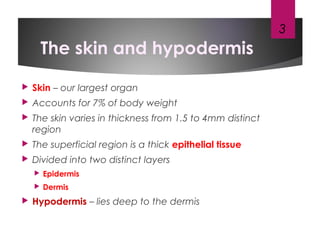
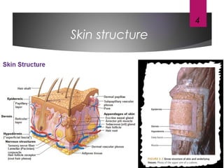
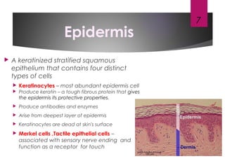
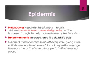
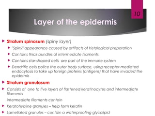
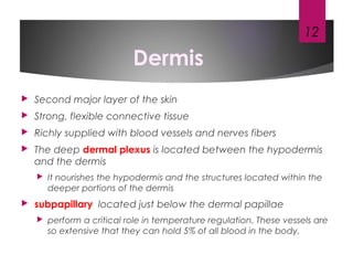
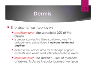
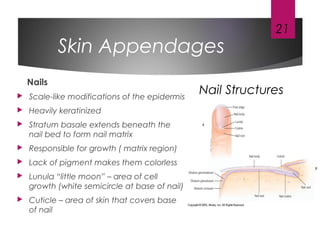
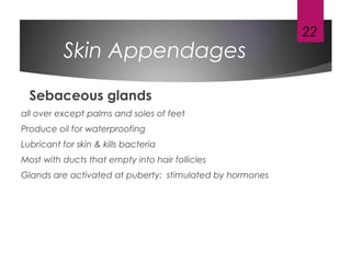
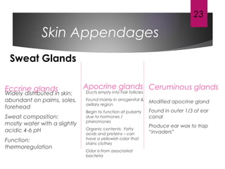
This document provides an overview of the structure and functions of the skin and its appendages. It notes that the skin is the largest organ and is composed of three main layers - the epidermis, dermis and hypodermis. The epidermis provides protection and waterproofing and has several layers including the stratum basale, stratum spinosum, stratum granulosum and stratum corneum. The dermis lies underneath and contains blood vessels, nerves and connective tissue. The deepest layer, the hypodermis, stores fat and anchors the skin. The document also describes skin appendages like hair, sebaceous glands, sweat glands and nails, and their structure and functions.
3. The skin and hypodermis
Skin – our largest organ
Accounts for 7% of body weight
The skin varies in thickness from 1.5 to 4mm distinct
region
The superficial region is a thick epithelial tissue
Divided into two distinct layers
Epidermis
Dermis
Hypodermis – lies deep to the dermis
3
5. Functions
Protection : Skin cushions and insulates
the deeper body organs
Protects the body from scrapes ,chemicals
and invading microorganisms
The epidermis is waterproof, preventing
unnecessary loss of water across the body
surface
produce pigment to protect the skin from
the harmful effects of ultraviolet (UV)
radiation
5
6. Function
Body temperature regulation :The skin’s rich
capillary networks and sweat glands
regulate the loss of heat from the body
Excreation : The skin acts as a miniature
excretory system when urea, salts, and
water are lost through sweat.
Production of vitamin : The epidermal cells
use UV radiation to synthesize vitamin D
Sensory reseption : touch, pressure,
temperature, and pain
6
7. Epidermis
A keratinized stratified squamous
epithelium that contains four distinct
types of cells
Keratinocytes – most abundant epidermis cell
Produce keratin – a tough fibrous protein that gives
the epidermis its protective properties.
Produce antibodies and enzymes
Arise from deepest layer of epidermis
Keratinocytes are dead at skin's surface
Merkel cells ,Tactile epithelial cells –
associated with sensory nerve ending and
function as a receptor for touch
7
8. Epidermis
Melanocytes – secrete the pigment melanin
Melanin is made in membrane-walled granules and then
transferred through the cell processes to nearby keratinocytes
Langerhans cells : macrophage like dendritic cells
Millions of these dead cells rub off every day, giving us an
entirely new epidermis every 35 to 45 days—the average
time from the birth of a keratinocyte to its final wearing
away.
8
9. Layer of the Epidermis
Stratum basale
Deepest layer of epidermis
Attached to underlying dermis
Also called the Stratum
germinativum
these Cells divide rapidly
Merkel cells – associated with
sensory nerve ending
Melanocytes – secrete the
pigment melanin
9
10. Layer of the epidermis
Stratum spinosum (spiny layer)
"Spiny" appearance caused by artifacts of histological preparation
Contains thick bundles of intermediate filaments
Contains star-shaped cells are part of the immune system
Dendritic cells police the outer body surface, using receptor-mediated
endocytosis to take up foreign proteins (antigens) that have invaded the
epidermis
Stratum granulosum
Consists of one to five layers of flattened keratinocytes and intermediate
filaments
intermediate filaments contain
Keratohyaline granules – help form keratin
Lamellated granules – contain a waterproofing glycolipid
10
11. Layer of the epidermis
Stratum lucidum (clear layer)
Occurs only in thick skin but not in thin skin
Consists of a few rows of flat, dead
keratinocytes
It is also named the transition zone
Stratum corneum (horny layer)
Thick layer of dead keratinocytes and
thickened plasma membranes
Protects skin against abrasion and
penetration
11
12. Dermis
Second major layer of the skin
Strong, flexible connective tissue
Richly supplied with blood vessels and nerves fibers
The deep dermal plexus is located between the hypodermis
and the dermis
It nourishes the hypodermis and the structures located within the
deeper portions of the dermis
subpapillary located just below the dermal papillae
perform a critical role in temperature regulation. These vessels are
so extensive that they can hold 5% of all blood in the body.
12
13. Dermis
The dermis has two layers
papillary layer the superficial 20% of the
dermis
is areolar connective tissue containing very thin
collagen and elastic fibers It includes the dermal
papillae
increase the surface area for exchange of gases,
nutrients, and waste products between these layers
reticular layer the deeper – 80% of thickness
of dermis. is dense irregular connective tissue
13
14. Hypodermis
Deep to the skin – also called superficial fascia
Contains areolar and adipose connective
tissues
Besides storing fat, the hypodermis anchors the
skin to the underlying structures
is also an insulator because fat is a poor
conductor of heat, it helps prevent heat loss
from the body
14
15. Skin color
Three pigments contribute to skin color
Melanin – most important pigment – made
from tyrosine
Carotene – yellowish pigment from carrots
and tomatoes
Hemoglobin – Caucasian skin contains little
melanin
Allows crimson color of blood to show through
15
16. Skin color
Melanin in granules passes from melanocytes (same number in all
races) to keratinocytes in stratum basale
Digested by lysosomes
Variations in color
Protection from UV light vs vitamin D?
Derived from epidermis but extend into dermis
Include
Hair and hair follicles
Sebaceous (oil) glands
Sweat (sudoriferous) glands
Nails
16
17. Skin Appendages
Hair
Minor protective functions (retain heat, decrease sunburn,
eyelashes protect eyes)
Structure
shaft – projects from skin
follicle – extends into dermis
root – lies within the follicle
bulb – contains CT, vessels and nerves
sebaceous gland – lubricates hair
arrector pili muscle – attached to follicle and contracts to move
hair (hair growth, goosebumps)
17
19. Skin Appendages
Hair Growth
influenced by: (in this order)
nutrition - main influence
hormones
blood flow
19
Hair Pigment
caused by proportions of 3 melanin types
• dark hair – true melanin
• blonde and red – melanin with iron and sulfur
• gray/white hair - melanin replaced by air bubbles in shaft
21. Skin Appendages
Nails
Scale-like modifications of the epidermis
Heavily keratinized
Stratum basale extends beneath the
nail bed to form nail matrix
Responsible for growth ( matrix region)
Lack of pigment makes them colorless
Lunula “little moon” – area of cell
growth (white semicircle at base of nail)
Cuticle – area of skin that covers base
of nail
21
Nail Structures
22. Skin Appendages
Sebaceous glands
all over except palms and soles of feet
Produce oil for waterproofing
Lubricant for skin & kills bacteria
Most with ducts that empty into hair follicles
Glands are activated at puberty: stimulated by hormones
22
23. Skin Appendages
Eccrine glands
Widely distributed in skin:
abundant on palms, soles,
forehead
Sweat composition:
mostly water with a slightly
acidic 4-6 pH
Function:
thermoregulation
Apocrine glands
Ducts empty into hair follicles
Found mainly in anogenital &
axillary region
Begin to function at puberty
due to hormones /
pheromones
Organic contents: Fatty
acids and proteins – can
have a yellowish color that
stains clothes
Odor is from associated
bacteria
Ceruminous glands
Modified apocrine gland
Found in outer 1/3 of ear
canal
Produce ear wax to trap
“invaders”
23
Sweat Glands