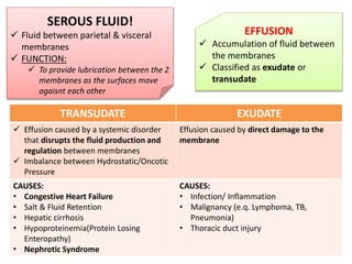
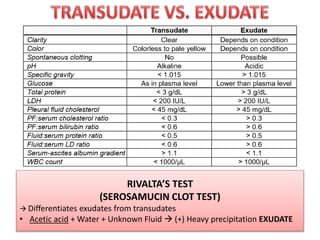
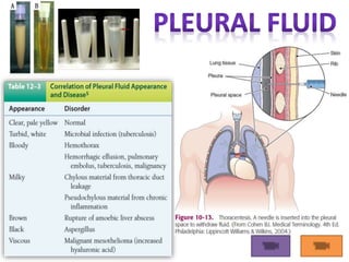
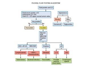
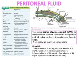
SEROUS FLUID!
 Fluid between parietal & visceral
membranes
 FUNCTION:
 To provide lubrication between the 2
membranes as the surfaces move
agaisnt each other
EFFUSION
 Accumulation of fluid between
the membranes
 Classified as exudate or
transudate
TRANSUDATE EXUDATE
 Effusion caused by a systemic disorder
that disrupts the fluid production and
regulation between membranes
 Imbalance between Hydrostatic/Oncotic
Pressure
Effusion caused by direct damage to the
membrane
CAUSES:
• Congestive Heart Failure
• Salt & Fluid Retention
• Hepatic cirrhosis
• Hypoproteinemia(Protein Losing
Enteropathy)
• Nephrotic Syndrome
CAUSES:
• Infection/ Inflammation
• Malignancy (e.q. Lymphoma, TB,
Pneumonia)
• Thoracic duct injury
RIVALTA’S TEST
(SEROSAMUCIN CLOT TEST)
 Differentiates exudates from transudates
• Acetic acid + Water + Unknown Fluid  (+) Heavy precipitation EXUDATE
3 P’S:
 NORMAL APPEARANCE  Clear, Pale yellow
Pleural Fluid = THORACENTESIS
Pericardial Fluid = PERICARDIOCENTESIS
Peritonial (Ascitic) Fluid = PARACENTESIS
Specimen is distributed in the following tubes:
EDTA  Cell counts and differential
STERILE HEPARIN TUBES  Microbiology &
Cytology
PLAIN/HEPARIN TUBES  Chemistry (Specimens
for pH must be maintained anaerobically in ice
TUMOR MARKER SIGNIFICANCE
CEA (Carcinoembryonic antigen) Colon Cancer
CA 125 Ovarian/ metastatic uterine cancer
CA 15-3, CA 549 Breast cancer
CYFRA 21-1 (CYtokeratin FRAgment) Lung cancer
Poorly differentiated
pleural fluid
adenocarcinoma showing
nuclear irregularities and
cytoplasmic vacuoles
(×500).
Metastatic breast
carcinoma cells in
pleural fluid. Notice the
hyperchromatic
nucleoli (×1000).
 only a small amount (10 to 50 mL)
of fluid is found between the
pericardial serous membranes.
TRANSUDATES : uremia,
hypothyroidism, and autoimmune
disorders
EXUDATES: Pericarditis, Malignancy &
Trauma
The serum-ascites albumin gradient (SAAG) is
recommended over the fluid:serum total protein
and LD ratios to detect transudates of hepatic
origin.
 >1.1 (TRANSUDATE) <1.1 (EXUDATE)
EXAMPLE:
1. Serum albumin of 3.8 mg/dL – fluid albumin of 1.2
mg/dL = gradient of 2.6 (transudate effusion)
2. Serum albumin of 3.8 mg/dL – fluid albumin of 3.0
mg/dL = gradient of 0.8 (exudate effusion)
PSOMMOMA BODIES
 Contain concentric striations of
collagen-like material
 Seen in Benign conditions and
associated with ovarian & thyroid
carcinomas
 Normal saline is
sometimes introduced
into the peritoneal cavity
as a lavage to detect
abdominal injuries that
have not yet resulted in
fluid accumulation
 is a sensitive test to
detect intra-abdominal
bleeding in blunt trauma
cases
 RBC counts greater than
100,000/mL are
indicative of blunt trauma
injuries
G CELLS GASTRIN
SECRETE
PARIETAL
CELLS
HCl
PEPSINOGEN PEPSIN
DIGEST
PROTEINS
CELLS OF THE
STOMACH
1. PARIETAL CELLS
 Produce HCl &
intrinsic factor
 Intrinsic
Factor
needed for Vit.
B12 absorption
2. CHIEF CELLS
 Produced
Pepsinogen
3. SPECIALIZED G
CELLS
 Produce
Gastrin
METHOD OF COLLECTION = ASPIRATION
GASTRIC TUBES:
1. LEVIN TUBE = pass through the nose
2. REHFUSS TUBE = passed through the
mouth
TYPE OF SPECIMEN DURATION OF COLLECTION
Basal Acid Output (BAO) 1 hour collection (consist of four 15-minute specimens,
but a simgle 1-hr can be used)
2 hour collection – for insulin hypoglycemia test
Maximum Acid Output (MAO) 1 hour collection (at 15-minute intervals) –when
Pentagastrin and Histamine are used
2 hour collection – for Insulin hypoglycemia test and
when Histalog is used
 BAO = Total gastric secretion during unstimulated, fasting state
 MAO= Total gastric secretion after gastric stimulation
GASTRIC STIMULANTS
TEST MEALS
a. Ewald’s= bread,weak
tea or water
b. Boa’s = oatmeal
c. Riegel’s = Beef steak
& mashed potato
d. Heckman’s = egg
albumin, water,
methylene blue
e. Dock’s= biscuit
f. Fischer’s = Ewald’s
meal + hamburg stock
g. Lavine’s = Ethyl
alcohol, Methylene Blue
h. Motor= Spinach or
raisins + water
i. Salzer = Beef,
lambchop, milk, rice,
egg
j. Stasis= rice, raisins
CHEMICAL STIMULANTS
a. Pentagastrin = most
preferred
b. Insulin = Assess
vagotomy procedure
c. Histalog (Betazole)
d. Histamine- gastric
stimulant
SHAM FEEDING FICTITIOUS FEEDING = SANDWICH
REPRESENTATIVE NORMAL AND ABNORMAL GASTRIC ANALYSIS RESULT
BAO (mEq/hr) MAO (mEq/hr) BAO/MAO
NORMAL 2.5 25 10%
PERNICIOUS ANEMIA 0 0 0
DUODENAL ULCER 5.0 30.0 17%
ZOLLINGER-ELLISON SYNDROME 18.0 25.0 72%
MACROSCOPIC EXAMINATION OF GASTRIC FLUID
COLOR SIGNIFICANCE
Pale gray with mucus Normal
Yellow-green Large amounts of bile
Red Small amount of fresh blood
Coffee ground Large amount of blood
VOLUME SIGNIFICANCE
Few mL to 50mL (ave; 30mL) Normal fasting specimen
>50 mL Abnormal fasting specimen
20-60mL up to 120mL After Ewald’s test meal
45-150mL After alcohol test meal or histamine stimulation
TERMINOLOGIES
TERM DEFINITION SIGNIFICANCE
EUCHLORHYDIA Normal Free HCl --
HYPERCHLORHYDIA Increased Free HCl Peptic ulcer
HYPOCHLORHYDIA
Gastric Fluid pH >3.5 but falls
after gastric stimuation
(Decreased free HCl)
Carcinoma of the
Stomach
ACHLORHYDIA
Gastric Fluid pH >3.5 and does
not fall even after gastric
stimulation (Absence of free HCl)
Pernicious Anemia
ANACIDITY
Failure to produce a pH <6.0
following gastric stimulation
Pernicious Anemia
QUALITATIVE TEST FOR FREE HCl
Dimethylaminnoazobenzol (+) cherr red
Gunzberg Reagent: Phloroglucin, Vanillin, Alcohol = (+) Purple red
Boas Reagent: Resorcinol, Cane Sugar, Alcohol= (+) Rose red
 Indicative of advanced gastric cancer/ gastric
stagnation
QUANTITATIVE TESTS FOR GASTRIC ACIDITY
Free HCl Total Acidity Combine HCl
(Bound to proteins)
Titrant Sodium hydroxide NaOH NaOH
pH indicator Dimethylaminoazobenzol
(Topfer’s reagent)
Phenolphthalein Sodium alizarin
End point Canary yellow Faint pink Violet
Normal value 25-500 50-750 10-150
TESTS FOR LACTIC ACID
TEST REAGENTS END POINT
Modified
Uffelmann’s
FeCl3 +
Phenol
YELLOW
Strauss FeCl3 + ehter YELLOW
Kelling’s FeCl3 YELLOW
DIAGNEX TUBELESS TEST
 Specimen = URINE
 PRINCIPLE:
 Azure A is given by Mouth
 Azure A is exchangedd from an
amberlite cation resin by H+ ions
from Hcl after caffeine stimulation.
The Azure A is rapidly absorbed
from the small intestine and
excreted in the urine
 The presence of Azure A in urine
indicates the presence of free HCl
List of References
Lillian Mundt & Kristy Shanahan, Graff’s
Textbook of Urinalysis and Body Fluids, 2nd Ed.
Susan Strassinger & Marjorie Di Lorenzo,
Urinalysis and Body Fluids, 5th & 6th Ed.
Erol Coderres,RMT-AUBF notes
Roderick Balce, RMT-CEU Professor AUBF
Notes

Serous fluid &amp; gastric fluid

  • 2.
    SEROUS FLUID!  Fluidbetween parietal & visceral membranes  FUNCTION:  To provide lubrication between the 2 membranes as the surfaces move agaisnt each other EFFUSION  Accumulation of fluid between the membranes  Classified as exudate or transudate TRANSUDATE EXUDATE  Effusion caused by a systemic disorder that disrupts the fluid production and regulation between membranes  Imbalance between Hydrostatic/Oncotic Pressure Effusion caused by direct damage to the membrane CAUSES: • Congestive Heart Failure • Salt & Fluid Retention • Hepatic cirrhosis • Hypoproteinemia(Protein Losing Enteropathy) • Nephrotic Syndrome CAUSES: • Infection/ Inflammation • Malignancy (e.q. Lymphoma, TB, Pneumonia) • Thoracic duct injury
  • 4.
    RIVALTA’S TEST (SEROSAMUCIN CLOTTEST)  Differentiates exudates from transudates • Acetic acid + Water + Unknown Fluid  (+) Heavy precipitation EXUDATE
  • 5.
    3 P’S:  NORMALAPPEARANCE  Clear, Pale yellow Pleural Fluid = THORACENTESIS Pericardial Fluid = PERICARDIOCENTESIS Peritonial (Ascitic) Fluid = PARACENTESIS Specimen is distributed in the following tubes: EDTA  Cell counts and differential STERILE HEPARIN TUBES  Microbiology & Cytology PLAIN/HEPARIN TUBES  Chemistry (Specimens for pH must be maintained anaerobically in ice
  • 8.
    TUMOR MARKER SIGNIFICANCE CEA(Carcinoembryonic antigen) Colon Cancer CA 125 Ovarian/ metastatic uterine cancer CA 15-3, CA 549 Breast cancer CYFRA 21-1 (CYtokeratin FRAgment) Lung cancer Poorly differentiated pleural fluid adenocarcinoma showing nuclear irregularities and cytoplasmic vacuoles (×500). Metastatic breast carcinoma cells in pleural fluid. Notice the hyperchromatic nucleoli (×1000).
  • 10.
     only asmall amount (10 to 50 mL) of fluid is found between the pericardial serous membranes. TRANSUDATES : uremia, hypothyroidism, and autoimmune disorders EXUDATES: Pericarditis, Malignancy & Trauma
  • 11.
    The serum-ascites albumingradient (SAAG) is recommended over the fluid:serum total protein and LD ratios to detect transudates of hepatic origin.  >1.1 (TRANSUDATE) <1.1 (EXUDATE) EXAMPLE: 1. Serum albumin of 3.8 mg/dL – fluid albumin of 1.2 mg/dL = gradient of 2.6 (transudate effusion) 2. Serum albumin of 3.8 mg/dL – fluid albumin of 3.0 mg/dL = gradient of 0.8 (exudate effusion)
  • 12.
    PSOMMOMA BODIES  Containconcentric striations of collagen-like material  Seen in Benign conditions and associated with ovarian & thyroid carcinomas  Normal saline is sometimes introduced into the peritoneal cavity as a lavage to detect abdominal injuries that have not yet resulted in fluid accumulation  is a sensitive test to detect intra-abdominal bleeding in blunt trauma cases  RBC counts greater than 100,000/mL are indicative of blunt trauma injuries
  • 13.
    G CELLS GASTRIN SECRETE PARIETAL CELLS HCl PEPSINOGENPEPSIN DIGEST PROTEINS CELLS OF THE STOMACH 1. PARIETAL CELLS  Produce HCl & intrinsic factor  Intrinsic Factor needed for Vit. B12 absorption 2. CHIEF CELLS  Produced Pepsinogen 3. SPECIALIZED G CELLS  Produce Gastrin
  • 14.
    METHOD OF COLLECTION= ASPIRATION GASTRIC TUBES: 1. LEVIN TUBE = pass through the nose 2. REHFUSS TUBE = passed through the mouth TYPE OF SPECIMEN DURATION OF COLLECTION Basal Acid Output (BAO) 1 hour collection (consist of four 15-minute specimens, but a simgle 1-hr can be used) 2 hour collection – for insulin hypoglycemia test Maximum Acid Output (MAO) 1 hour collection (at 15-minute intervals) –when Pentagastrin and Histamine are used 2 hour collection – for Insulin hypoglycemia test and when Histalog is used  BAO = Total gastric secretion during unstimulated, fasting state  MAO= Total gastric secretion after gastric stimulation
  • 15.
    GASTRIC STIMULANTS TEST MEALS a.Ewald’s= bread,weak tea or water b. Boa’s = oatmeal c. Riegel’s = Beef steak & mashed potato d. Heckman’s = egg albumin, water, methylene blue e. Dock’s= biscuit f. Fischer’s = Ewald’s meal + hamburg stock g. Lavine’s = Ethyl alcohol, Methylene Blue h. Motor= Spinach or raisins + water i. Salzer = Beef, lambchop, milk, rice, egg j. Stasis= rice, raisins CHEMICAL STIMULANTS a. Pentagastrin = most preferred b. Insulin = Assess vagotomy procedure c. Histalog (Betazole) d. Histamine- gastric stimulant SHAM FEEDING FICTITIOUS FEEDING = SANDWICH
  • 16.
    REPRESENTATIVE NORMAL ANDABNORMAL GASTRIC ANALYSIS RESULT BAO (mEq/hr) MAO (mEq/hr) BAO/MAO NORMAL 2.5 25 10% PERNICIOUS ANEMIA 0 0 0 DUODENAL ULCER 5.0 30.0 17% ZOLLINGER-ELLISON SYNDROME 18.0 25.0 72% MACROSCOPIC EXAMINATION OF GASTRIC FLUID COLOR SIGNIFICANCE Pale gray with mucus Normal Yellow-green Large amounts of bile Red Small amount of fresh blood Coffee ground Large amount of blood VOLUME SIGNIFICANCE Few mL to 50mL (ave; 30mL) Normal fasting specimen >50 mL Abnormal fasting specimen 20-60mL up to 120mL After Ewald’s test meal 45-150mL After alcohol test meal or histamine stimulation
  • 17.
    TERMINOLOGIES TERM DEFINITION SIGNIFICANCE EUCHLORHYDIANormal Free HCl -- HYPERCHLORHYDIA Increased Free HCl Peptic ulcer HYPOCHLORHYDIA Gastric Fluid pH >3.5 but falls after gastric stimuation (Decreased free HCl) Carcinoma of the Stomach ACHLORHYDIA Gastric Fluid pH >3.5 and does not fall even after gastric stimulation (Absence of free HCl) Pernicious Anemia ANACIDITY Failure to produce a pH <6.0 following gastric stimulation Pernicious Anemia QUALITATIVE TEST FOR FREE HCl Dimethylaminnoazobenzol (+) cherr red Gunzberg Reagent: Phloroglucin, Vanillin, Alcohol = (+) Purple red Boas Reagent: Resorcinol, Cane Sugar, Alcohol= (+) Rose red
  • 18.
     Indicative ofadvanced gastric cancer/ gastric stagnation QUANTITATIVE TESTS FOR GASTRIC ACIDITY Free HCl Total Acidity Combine HCl (Bound to proteins) Titrant Sodium hydroxide NaOH NaOH pH indicator Dimethylaminoazobenzol (Topfer’s reagent) Phenolphthalein Sodium alizarin End point Canary yellow Faint pink Violet Normal value 25-500 50-750 10-150 TESTS FOR LACTIC ACID TEST REAGENTS END POINT Modified Uffelmann’s FeCl3 + Phenol YELLOW Strauss FeCl3 + ehter YELLOW Kelling’s FeCl3 YELLOW DIAGNEX TUBELESS TEST  Specimen = URINE  PRINCIPLE:  Azure A is given by Mouth  Azure A is exchangedd from an amberlite cation resin by H+ ions from Hcl after caffeine stimulation. The Azure A is rapidly absorbed from the small intestine and excreted in the urine  The presence of Azure A in urine indicates the presence of free HCl
  • 19.
    List of References LillianMundt & Kristy Shanahan, Graff’s Textbook of Urinalysis and Body Fluids, 2nd Ed. Susan Strassinger & Marjorie Di Lorenzo, Urinalysis and Body Fluids, 5th & 6th Ed. Erol Coderres,RMT-AUBF notes Roderick Balce, RMT-CEU Professor AUBF Notes

Editor's Notes

  • #3 Hypoproteinemia- dec oncotic Congestive Heart Failure- increase hydrostatic Nephrotic Syndrome-dec oncotic
  • #8 CHYLOUS CAUSE THORACIC DUCT LEAKAGE PSEUDOCHYLOUS CAUSE CHRONIC INFLAMMATION OR LYMPHATIC OBSTRUCTION EOSINOPHILS – Pneumothorax, Hemothorax Increase plasma Cells - TB
  • #11 Pericardial effusions are all caused by damage to the mesothelium and not by mechanical factors. Therefore, pericardial effusions are usually always exudates. Like pleural fluid, WBC counts are of little clinical value, although a count of >1000 WBCs/µL with a high percentage of neutrophils can indicate bacterial endocarditis.
  • #14 Pepsinogen produced by the Chief cells
  • #15 1 hour collection-routinely performed
  • #16 Vagotomy is an essential component of surgical management of peptic (duodenal and gastric) ulcer disease (PUD). Vagotomy was once commonly performed to treat and prevent peptic ulcer disease