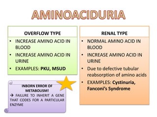
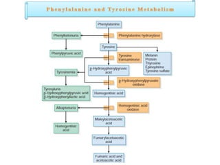
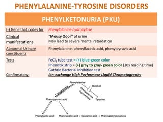
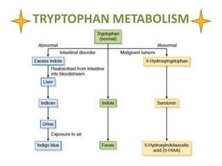
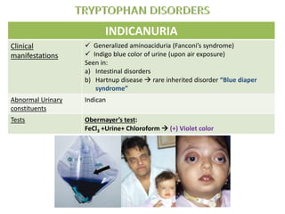
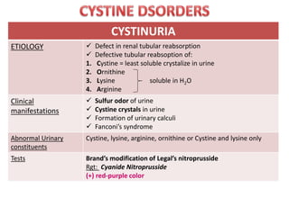
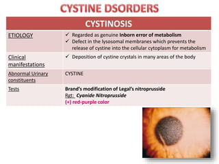
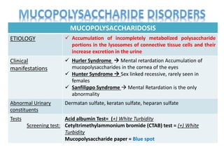
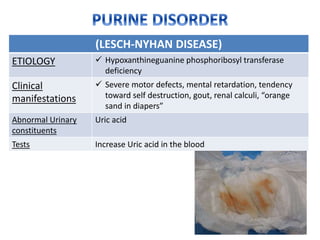
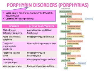
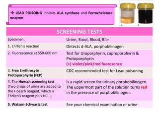
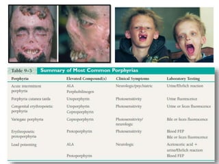
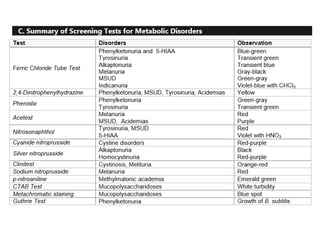
OVERFLOW TYPE
• INCREASE AMINO ACID IN
BLOOD
• INCREASE AMINO ACID IN
URINE
• EXAMPLES: PKU, MSUD
RENAL TYPE
• NORMAL AMINO ACID IN
BLOOD
• INCREASE AMINO ACID IN
URINE
• Due to defective tubular
reabsorption of amino acids
• EXAMPLES: Cystinuria,
Fanconi’s Syndrome
INBORN ERROR OF
METABOLISM!
 FAILURE TO INHERIT A GENE
THAT CODES FOR A PARTICULAR
ENZYME
PHENYLKETONURIA (PKU)
(-) Gene that codes for Phenylalanine hydroxylase
Clinical
manifestations
“Mousy Odor” of urine
May lead to severe mental retardation
Abnormal Urinary
constituents
Phenylalanine, phenyllacetic acid, phenylpyruvic acid
Tests
Confirmatory:
FeCl3 tube test = (+) blue-green color
Phenistix strip = (+) gray to gray- green color (30s reading time)
Guthrie Bacterial Inhibition test
Ion-exchange High Performace Liquid Chromatography
GUTHRIE BACTERIAL INHIBITION TEST!
sensitive to serum phenylalanine concentrations >4
mg/dL
Bacillus subtilis is cultured with beta2-thienylalanine
Beta2-thienylalanine inhibits the growht of B. subtilis
Phenylalanine counteracts the action of beta2-
thienylalanine
TYROSYLURIA
(-) Gene that codes for Type I Fumarylacetoacetate hydrolase (FAH)
Type 2Tyrosine aminotransferase
Type 3 p-hydroxyphenylpyruvic acid dioxygenase
Clinical
manifestations
Tyrosine and leucine crystals in urine; liver and renal disease
(temporary or permanent)
Rancid butter odor of urine
Abnormal Urinary
constituents
p-hydroxyphenylpyruvic acid and p-hydroxyphenyllactic acid
Tests
Confirmatory:
FeCl3 tube test = (+) transient green
Nitroso-naphthol= (+) orange-red
Chromatography, Quantitative Serum Assay of tyrosine
ALKAPTONURIA
(-) Gene that codes for Homogentisic acid oxidase
Clinical
manifestations
Dark blue to black pigmentation of cartilage and connective
tissues, liver and cardiac disorders
Urine darkens after becoming alkalne from staniding at room
temperature; OBSERVATION OF BLACK STAINED DIAPER IN
NEWBORN
Abnormal Urinary constituents Homogentisic acid
Tests
Confirmatory:
FeCl3 tube test = (+) transient Blue
Clinitest= (+) yellow precipitate
Alkalinization of fresh urine
Paper or thin-layer chromatography, Capillary electrophoresis
MELANURIA
(-) Gene that codes for --
Clinical
manifestations
Malignant melanoma
Due to overproliferation of melanocytes
Urine darkens upon air exposure
Abnormal Urinary
constituents
Melanin
Tests FeCl3 tube test = (+) Gray/Black ppt
Sodium nitroprusside test = (+) Red
Ehrlich test= (+) Red
MAPLE SYRUP URINE DISEASE (MSUD)
(-) Gene that codes for Deficiency in decarboxylases and other enzymes needed for the
conversion of ketoamino acids to fatty acids
Clinical
manifestations
Caramelized sugar/Maple syrup odor of urine, failure to thrive,
mental retardation
Leucine, Isoleucine & Valine in blood & urine
Abnormal Urinary
constituents
Ketoacids (α-ketoisovaleric; α-ketoisocaproic; α-keto,β-
methylvaleric)
Tests 2,4-Dinitrophenylhydrazine (DNPH) test
(+) Yellow turbidity/precipitate
ORGANIC ACIDEMIAS
(-) Gene that codes for 1. Isovaleric Acidemia= Isovaleryl CoA in the leucine pathway
2. & 3. Propionic and Methylmalonic acidemias = Error in the
metabolic pathway converting valine, isoleucine, threonine,
and methionine to succinyl CoA
Clinical
manifestations
1. Sweaty feet odor of urine
2. & 3  Early severe illness, vomiting, metabolic acidosis,
hypoglycemia, increased serum ammonia, ketonuria
Abnormal Urinary
constituents
Isovalerylglycine
Propionic and methylmalonic acid
Tests Methylmalonic acidemias :
P-nitroaniline test = (+) Emerald green
TRYPTOPHAN METABOLISM
INDICANURIA
Clinical
manifestations
 Generalized aminoaciduria (Fanconi’s syndrome)
 Indigo blue color of urine (upon air exposure)
Seen in:
a) Intestinal disorders
b) Hartnup disease  rare inherited disorder “Blue diaper
syndrome”
Abnormal Urinary
constituents
Indican
Tests Obermayer’s test:
FeCl3 +Urine+ Chloroform  (+) Violet color
ARGENTAFFINOMA
ETIOLOGY  Carcinoid tumors involving the argentaffin cells
(enterochromaffin) of the intestine
Clinical
manifestations
 Vascular and bronchospastic symptoms (carcinoid syndrome)
Abnormal Urinary
constituents
5-HIAA (>25 mg/24 hours)
Tests FeCl3 tube test = (+) Blue-green
Nitroso-naphthol w/ nitrous acid = (+) Violet
*Patient should avoid eating bananas, pineapples & tomatoes
CYSTINURIA
ETIOLOGY  Defect in renal tubular reabsorption
 Defective tubular reabsoption of:
1. Cystine = least soluble crystalize in urine
2. Ornithine
3. Lysine soluble in H2O
4. Arginine
Clinical
manifestations
 Sulfur odor of urine
 Cystine crystals in urine
 Formation of urinary calculi
 Fanconi’s syndrome
Abnormal Urinary
constituents
Cystine, lysine, arginine, ornithine or Cystine and lysine only
Tests Brand’s modification of Legal’s nitroprusside
Rgt: Cyanide Nitroprusside
(+) red-purple color
CYSTINOSIS
ETIOLOGY  Regarded as genuine Inborn error of metabolism
 Defect in the lysosomal membranes which prevents the
release of cystine into the cellular cytoplasm for metabolism
Clinical
manifestations
 Deposition of cystine crystals in many areas of the body
Abnormal Urinary
constituents
CYSTINE
Tests Brand’s modification of Legal’s nitroprusside
Rgt: Cyanide Nitroprusside
(+) red-purple color
HOMOCYSTINURA
ETIOLOGY  Cystathionine β-synthase deficiency which leads to defects
in the methionine metabolism thus increasing homocystine
throught the body
Clinical
manifestations
 Failure to thrive, cataracts, mental retardation,
thromboembolic problems
Abnormal Urinary
constituents
Homocystine and methionine
Tests Silver-nitroprusside test = (+) Red Color
MUCOPOLYSACCHARIDOSIS
ETIOLOGY  Accumulation of incompletely metabolized polysaccharide
portions in the lysosomes of connective tissue cells and their
increase excretion in the urine
Clinical
manifestations
 Hurler Syndrome  Mental retardation Accumulation of
mucopolysaccharides in the cornea of the eyes
 Hunter Syndrome  Sex linked recessive, rarely seen in
females
 Sanfilippo Syndrome  Mental Retardation is the only
abnormality
Abnormal Urinary
constituents
Dermatan sulfate, keratan sulfate, heparan sulfate
Tests
Screening test:
Acid albumin Test= (+) White Turbidity
Cetyltrimethylammonium bromide (CTAB) test = (+) White
Turbidity
Mucopolysaccharide paper = Blue spot
HUNTER SYNDROME
HURLER SYNDROME
SANFILIPPO SYNDROME
(LESCH-NYHAN DISEASE)
ETIOLOGY  Hypoxanthineguanine phosphoribosyl transferase
deficiency
Clinical
manifestations
 Severe motor defects, mental retardation, tendency
toward self destruction, gout, renal calculi, “orange
sand in diapers”
Abnormal Urinary
constituents
Uric acid
Tests Increase Uric acid in the blood
 Urine color = Red/Purple/burgundy-Red/Purplish
Red/Portwine
 Colorless in = Lead poisoning
DISORDER (-) GENE THAT CODES FOR
Ala hydratase
deficiency porphyria
Aminolevulinic acid (ALA)
Synthetase
Acute intermittent
porphyria
Uroporphyrinogen synthase
Congenital
erythropoietic
porphyria
Uroporphyrinogen cosynthase
Porphyria cutanea
tarda
Uroporphyrinogen
decarboxylase
Hereditary
coproporphyria
Coproporphyrinogen oxidase
Variegate porphyria Protoporphyrinogen oxidase
SCREENING TESTS
Specimen: Urine, Stool, Blood, Bile
1. Ehrlich’s reaction Detects d-ALA, porphobilinogen
2. Fluorescence at 550-600 nm Test for Uroporphyrin, coproporphyrin &
Protoporphyrin
(+) violet/pink/red fuoresence
3. Free Erythrocyte
Protoporphyrin (FEP)
CDC recommended test for Lead poisoning
4. The Hoesch screening test
(Two drops of urine are added to
the Hoesch reagent, which is
Ehrlich’s reagent plus HCl. )
is a rapid screen for urinary porphobilinogen.
The uppermost part of the solution turns red
in the presence of porphobilinogen.
5. Watson-Schwartz test See your chemical examination or urine
 LEAD POISOING inhibits ALA synthase and Ferrochelatase
enzyme
MARAMING
SALAMAT SA
PAKIKINIG!
List of References
Lillian Mundt & Kristy Shanahan, Graff’s Textbook
of Urinalysis and Body Fluids, 2nd Ed.
Susan Strassinger & Marjorie Di Lorenzo,
Urinalysis and Body Fluids, 5th & 6th Ed.
Erol Coderres,RMT-AUBF notes
Roderick Balce, RMT-CEU Professor AUBF Notes
Polansky, V.D., Quick Review Cards for MLS, 2nd Ed.
Hubbard, J.D., A Concise Review of Clinical
Laboratory Science, 2nd Ed.

Urine screening for metabolic disorders

  • 2.
    OVERFLOW TYPE • INCREASEAMINO ACID IN BLOOD • INCREASE AMINO ACID IN URINE • EXAMPLES: PKU, MSUD RENAL TYPE • NORMAL AMINO ACID IN BLOOD • INCREASE AMINO ACID IN URINE • Due to defective tubular reabsorption of amino acids • EXAMPLES: Cystinuria, Fanconi’s Syndrome INBORN ERROR OF METABOLISM!  FAILURE TO INHERIT A GENE THAT CODES FOR A PARTICULAR ENZYME
  • 4.
    PHENYLKETONURIA (PKU) (-) Genethat codes for Phenylalanine hydroxylase Clinical manifestations “Mousy Odor” of urine May lead to severe mental retardation Abnormal Urinary constituents Phenylalanine, phenyllacetic acid, phenylpyruvic acid Tests Confirmatory: FeCl3 tube test = (+) blue-green color Phenistix strip = (+) gray to gray- green color (30s reading time) Guthrie Bacterial Inhibition test Ion-exchange High Performace Liquid Chromatography
  • 5.
    GUTHRIE BACTERIAL INHIBITIONTEST! sensitive to serum phenylalanine concentrations >4 mg/dL Bacillus subtilis is cultured with beta2-thienylalanine Beta2-thienylalanine inhibits the growht of B. subtilis Phenylalanine counteracts the action of beta2- thienylalanine
  • 6.
    TYROSYLURIA (-) Gene thatcodes for Type I Fumarylacetoacetate hydrolase (FAH) Type 2Tyrosine aminotransferase Type 3 p-hydroxyphenylpyruvic acid dioxygenase Clinical manifestations Tyrosine and leucine crystals in urine; liver and renal disease (temporary or permanent) Rancid butter odor of urine Abnormal Urinary constituents p-hydroxyphenylpyruvic acid and p-hydroxyphenyllactic acid Tests Confirmatory: FeCl3 tube test = (+) transient green Nitroso-naphthol= (+) orange-red Chromatography, Quantitative Serum Assay of tyrosine
  • 7.
    ALKAPTONURIA (-) Gene thatcodes for Homogentisic acid oxidase Clinical manifestations Dark blue to black pigmentation of cartilage and connective tissues, liver and cardiac disorders Urine darkens after becoming alkalne from staniding at room temperature; OBSERVATION OF BLACK STAINED DIAPER IN NEWBORN Abnormal Urinary constituents Homogentisic acid Tests Confirmatory: FeCl3 tube test = (+) transient Blue Clinitest= (+) yellow precipitate Alkalinization of fresh urine Paper or thin-layer chromatography, Capillary electrophoresis
  • 8.
    MELANURIA (-) Gene thatcodes for -- Clinical manifestations Malignant melanoma Due to overproliferation of melanocytes Urine darkens upon air exposure Abnormal Urinary constituents Melanin Tests FeCl3 tube test = (+) Gray/Black ppt Sodium nitroprusside test = (+) Red Ehrlich test= (+) Red
  • 9.
    MAPLE SYRUP URINEDISEASE (MSUD) (-) Gene that codes for Deficiency in decarboxylases and other enzymes needed for the conversion of ketoamino acids to fatty acids Clinical manifestations Caramelized sugar/Maple syrup odor of urine, failure to thrive, mental retardation Leucine, Isoleucine & Valine in blood & urine Abnormal Urinary constituents Ketoacids (α-ketoisovaleric; α-ketoisocaproic; α-keto,β- methylvaleric) Tests 2,4-Dinitrophenylhydrazine (DNPH) test (+) Yellow turbidity/precipitate
  • 10.
    ORGANIC ACIDEMIAS (-) Genethat codes for 1. Isovaleric Acidemia= Isovaleryl CoA in the leucine pathway 2. & 3. Propionic and Methylmalonic acidemias = Error in the metabolic pathway converting valine, isoleucine, threonine, and methionine to succinyl CoA Clinical manifestations 1. Sweaty feet odor of urine 2. & 3  Early severe illness, vomiting, metabolic acidosis, hypoglycemia, increased serum ammonia, ketonuria Abnormal Urinary constituents Isovalerylglycine Propionic and methylmalonic acid Tests Methylmalonic acidemias : P-nitroaniline test = (+) Emerald green
  • 11.
  • 12.
    INDICANURIA Clinical manifestations  Generalized aminoaciduria(Fanconi’s syndrome)  Indigo blue color of urine (upon air exposure) Seen in: a) Intestinal disorders b) Hartnup disease  rare inherited disorder “Blue diaper syndrome” Abnormal Urinary constituents Indican Tests Obermayer’s test: FeCl3 +Urine+ Chloroform  (+) Violet color
  • 13.
    ARGENTAFFINOMA ETIOLOGY  Carcinoidtumors involving the argentaffin cells (enterochromaffin) of the intestine Clinical manifestations  Vascular and bronchospastic symptoms (carcinoid syndrome) Abnormal Urinary constituents 5-HIAA (>25 mg/24 hours) Tests FeCl3 tube test = (+) Blue-green Nitroso-naphthol w/ nitrous acid = (+) Violet *Patient should avoid eating bananas, pineapples & tomatoes
  • 14.
    CYSTINURIA ETIOLOGY  Defectin renal tubular reabsorption  Defective tubular reabsoption of: 1. Cystine = least soluble crystalize in urine 2. Ornithine 3. Lysine soluble in H2O 4. Arginine Clinical manifestations  Sulfur odor of urine  Cystine crystals in urine  Formation of urinary calculi  Fanconi’s syndrome Abnormal Urinary constituents Cystine, lysine, arginine, ornithine or Cystine and lysine only Tests Brand’s modification of Legal’s nitroprusside Rgt: Cyanide Nitroprusside (+) red-purple color
  • 15.
    CYSTINOSIS ETIOLOGY  Regardedas genuine Inborn error of metabolism  Defect in the lysosomal membranes which prevents the release of cystine into the cellular cytoplasm for metabolism Clinical manifestations  Deposition of cystine crystals in many areas of the body Abnormal Urinary constituents CYSTINE Tests Brand’s modification of Legal’s nitroprusside Rgt: Cyanide Nitroprusside (+) red-purple color
  • 16.
    HOMOCYSTINURA ETIOLOGY  Cystathionineβ-synthase deficiency which leads to defects in the methionine metabolism thus increasing homocystine throught the body Clinical manifestations  Failure to thrive, cataracts, mental retardation, thromboembolic problems Abnormal Urinary constituents Homocystine and methionine Tests Silver-nitroprusside test = (+) Red Color
  • 17.
    MUCOPOLYSACCHARIDOSIS ETIOLOGY  Accumulationof incompletely metabolized polysaccharide portions in the lysosomes of connective tissue cells and their increase excretion in the urine Clinical manifestations  Hurler Syndrome  Mental retardation Accumulation of mucopolysaccharides in the cornea of the eyes  Hunter Syndrome  Sex linked recessive, rarely seen in females  Sanfilippo Syndrome  Mental Retardation is the only abnormality Abnormal Urinary constituents Dermatan sulfate, keratan sulfate, heparan sulfate Tests Screening test: Acid albumin Test= (+) White Turbidity Cetyltrimethylammonium bromide (CTAB) test = (+) White Turbidity Mucopolysaccharide paper = Blue spot
  • 18.
  • 19.
    (LESCH-NYHAN DISEASE) ETIOLOGY Hypoxanthineguanine phosphoribosyl transferase deficiency Clinical manifestations  Severe motor defects, mental retardation, tendency toward self destruction, gout, renal calculi, “orange sand in diapers” Abnormal Urinary constituents Uric acid Tests Increase Uric acid in the blood
  • 20.
     Urine color= Red/Purple/burgundy-Red/Purplish Red/Portwine  Colorless in = Lead poisoning DISORDER (-) GENE THAT CODES FOR Ala hydratase deficiency porphyria Aminolevulinic acid (ALA) Synthetase Acute intermittent porphyria Uroporphyrinogen synthase Congenital erythropoietic porphyria Uroporphyrinogen cosynthase Porphyria cutanea tarda Uroporphyrinogen decarboxylase Hereditary coproporphyria Coproporphyrinogen oxidase Variegate porphyria Protoporphyrinogen oxidase
  • 21.
    SCREENING TESTS Specimen: Urine,Stool, Blood, Bile 1. Ehrlich’s reaction Detects d-ALA, porphobilinogen 2. Fluorescence at 550-600 nm Test for Uroporphyrin, coproporphyrin & Protoporphyrin (+) violet/pink/red fuoresence 3. Free Erythrocyte Protoporphyrin (FEP) CDC recommended test for Lead poisoning 4. The Hoesch screening test (Two drops of urine are added to the Hoesch reagent, which is Ehrlich’s reagent plus HCl. ) is a rapid screen for urinary porphobilinogen. The uppermost part of the solution turns red in the presence of porphobilinogen. 5. Watson-Schwartz test See your chemical examination or urine  LEAD POISOING inhibits ALA synthase and Ferrochelatase enzyme
  • 24.
  • 25.
    List of References LillianMundt & Kristy Shanahan, Graff’s Textbook of Urinalysis and Body Fluids, 2nd Ed. Susan Strassinger & Marjorie Di Lorenzo, Urinalysis and Body Fluids, 5th & 6th Ed. Erol Coderres,RMT-AUBF notes Roderick Balce, RMT-CEU Professor AUBF Notes Polansky, V.D., Quick Review Cards for MLS, 2nd Ed. Hubbard, J.D., A Concise Review of Clinical Laboratory Science, 2nd Ed.

Editor's Notes

  • #6 The blood-impregnated disks are then placed on culture media streaked with the bacterium Bacillus subtilis. If increased phenylalanine levels are present in the blood, phenylalanine counteracts the action of beta-2-thienylalanine, an inhibitor of B. subtilis that is present in the media, and growth will be observed around the paper disks.
  • #8 Akali-loving
  • #9 (-) melanin = albinism
  • #10 (-) melanin = albinism
  • #11 (-) melanin = albinism
  • #13 (-) melanin = albinism
  • #14 (-) melanin = albinism
  • #15 (-) melanin = albinism
  • #16 (-) melanin = albinism
  • #17 (-) melanin = albinism
  • #18 (-) melanin = albinism