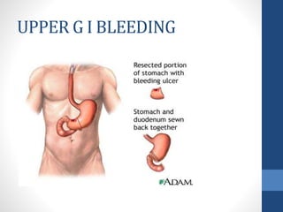
SCHOOL OF CLINICAL SCIENCES
MAKAMBO, MAKENI
GROUP 6
MODULE: INTERNAL MEDICINE
PRESENTATION
TOPIC: UPPER GI BLEEDING
Names of group members
1. Sewah Bangura 22008
2. Zainab Kadiatu Kamara 22045
3. Abdulai M.S. Mansaray 22063
4. Samuella Mary Kamara 22042
5. Santigie Aldo Bangura 22001
6. Memunatu A. Turay 22080
7. Memunatu Turay 22082
UPPER G I BLEEDING
OBJECTIVES
1. What is GI bleeding
2. Types of GI bleeding
3. Classification of GI bleeding
4. What is Upper GI bleeding, Prevalence, etiology and risk
factors
5. Clinical presentations
6. Clinical assessments
7. Lab diagnosis
8. Management
9. Medical and surgical therapy
10. Prevention and conclusion
INTRODUCTION
Gastrointestinal bleeding (GI bleeding) also known as
gastrointestinal hemorrhage, is all forms of bleeding in the
gastrointestinal tract, from the mouth to the rectum.
When there is significant blood loss over a short time,
symptoms may include
- vomiting of red blood
- vomiting of black blood
-bloody stool or black stool.
Types of GI bleeding
•Acute
•Chronic
•Occult
•Overt
•Obscure
CLASSIFICATION OF GI
BLEEDING
1. Upper GI bleeding
• Stem from issues with the esophagus,
stomach, and duodenum
2. Small bowel bleeding
• Stem from issues with your jejunum and
ileum
3.Lower GI bleeding
• Stem from issues with your colon,
rectum,and anus.
Upper GI bleeding
• Refer to the present of
bleeding in the upper
part of the
gastrointestinal tract,
which includes the
esophagus, stomach,
and duodenum (first
part of the small
intestine) .
Prevalence
•Upper gastrointestinal bleeding is
a common problem that is
estimated to occur in 80 – 150 out
of 100,000 people each year.
ETIOLOGY
 Esophageal causes:
 Esophageal varices
 Esophagitis
 Esophageal cancer
 Esophageal ulcers
 Mallory-Weiss tear
 Gastric causes:
 Gastric ulcer
 Gastric cancer
 Gastritis
 Gastric varices
 Dieulafoy's lesions
Duodenal causes:
 Duodenal ulcer
 Vascular malformation including
aorto-enteric fistulae
 Hematobilia, or bleeding from
the biliary tree
 Hemosuccus pancreaticus, or
bleeding from the pancreatic
duct
 Severe superior mesenteric
artery syndrome
RISK FACTORS
•1. NSAIDS use
•2. H.Pylori infection
•3. Increased age
Clinical presentation
Hematemesis
Melena
Hematochezia
Syncope
Dyspepsia
Epigastric pain
Heartburn
Diffuse abdominal pain
Dysphagia
Weight loss
Jaundice
Anemia
Orthostatic changes of BP and HR
SHOCK
CLINICAL ASSESSMENT
History
. Helpful to find out the site and cause
. History suggestive of acid peptic disease
. Alcoholic liver diseases / chronic hepatitis / cirrhosis
. History of anticoagulant / antiplatelets / NSAIDs / alcohol
binge intake / steroids
. History of coagulation disorder / blood dyscrasias
. History of epistaxis / hemoptysis to rule out the GI source of
bleeding
Patients of CVA, Burn, sepsis, Head trauma may have stress
ulcers .
ON EXAMINATION
Vitals
- pulse = Thready , BP= Orthostatic hypotension
Skin changes
- Cirrhosis – palmer erythema, spider angioma
- Bleeding diasthasis – purpura / echymosis
- Coagulation disorder – haemarthrosis, muscle haematoma
ENT – Look for clots (To rule out epistaxis P.N. bleed)
P/A
- Liver, spleen, caput medusa = cirrhosis
- Epigastric Tenderness = APD / ulcer
> Respiratory, CVS, CNS : For comorbid diseases
MONITORING
VITAL SIGNS :
• Continuous monitoring of vital signs such as heart rate, blood
pressure, respiratory rate, and saturation is essential to assess
the patient hemodynamic stability
• SIGNIFICANT CHANGES in this paramaters may indicate
ongoing bleeding or hemodynamic compromise .
HEMOGLOBIN AND HEMATOCRIT LEVEL
• Serial Measurement of these are crucial in evaluating the
extent of blood loss and monitoring response to treatment
URINE OUTPUT
• This is crucial to assess renal perfusion and detect any signs of
acute kidney injury.
Assessment of severity
Rockall Score
Lab Diagnosis:
• CBC with Platelet Count, and Differential
A complete blood count (CBC) is necessary to assess the
level of blood loss. CBC should be checked frequently(q4-6h)
during the first day.
• Hemoglobin Value, Type and Crossmatch Blood
The patient should be crossmatched for 2-6 units, based on
the rate of active bleeding.The hemoglobin level should be
monitored serially in order to follow the trend. An unstable
Hb level may signify ongoing hemorrhage requiring further
intervention.
• LFT- to detect underlying liver disease
• RFT- to detect underlying renal disease
• Calcium level- to detect
hyperparathyroidism and in monitoring
calcium in patients receiving multiple
transfusions of citrated blood
• Gastrin level
• The BUN-to- creatinine ratio increases with upper
gastrointestinal bleeding (UGIB). A ratio of greater than 36 in a
patient without renal insufficiency is suggestive of UGIB.
• The patient's prothrombin time (PT), activated partial
thromboplastin time, and International Normalized Ratio (INR)
should be checked to document the presence of a coagulopathy
•Prolongation of the PT based on an INR of more than 1.5 may indicate
moderate liver impairment.
•A fibrinogen level of less than 100 mg/dL also indicates advanced liver
disease with extremely poor synthetic function
• Initial diagnostic examination for all patients
presumed to have UGIB
• Endoscopy should be performed immediately
after endotracheal intubation (if indicated),
hemodynamic stabilization, and adequate
monitoring in an intensive care unit (ICU)
setting have been achieved.
Endoscopy :
• CHEST X-RAY-Chest radiographs should be
ordered to exclude aspiration pneumonia,
effusion, and esophageal perforation.
• Abdominal X-RAY- erect and supine films
should be ordered to exclude perforated
viscous and ileus.
Imaging :
• Computed tomography (CT) scanning and
ultrasonography may be indicated for the
evaluation of liver disease with cirrhosis,
cholecystitis with hemorrhage, pancreatitis
with pseudocyst and hemorrhage,
aortoenteric fistula, and other unusual
causes of upper GI hemorrhage.
• Nuclear medicine scans may be useful in
determining the area of active hemorrhage
Angiography
:
• Angiography may be useful if bleeding
persists and endoscopy fails to identify
a bleeding site.
• Angiography along with transcatheter
arterial embolization (TAE) should be
considered for all patients with a
known source of arterial UGIB that
does not respond to endoscopic
management, with active bleeding and
a negative endoscopy.
Nasogastric Lavage
A nasogastric tube is an important diagnostic tool.
This procedure may confirm recent bleeding
(coffee ground appearance), possible active
bleeding (red blood in the aspirate that does not
clear), or a lack of blood in the stomach (active
bleeding less likely but does not exclude an upper
GI lesion).
1. Better visualization during endoscopy
2. Give crude estimation of rapidity of bleeding
3. Prevent the development of Porto systemic encephalopathy in
cirrhosis
4. Increases PH of stomach, and hence, decreases clot
desolation due to gastric acid dilution
5. Tube placement can reduce the patient's need to vomit
 During gastric lavage use saline and not use large volume of to
avoid water intoxication.
 Gastric lavage should be done in alert and cooperative patient to
avoid bronco-pulmonary aspiration
BENEFITS OF LAVAGE :
Management
Priorities are:
1. Stabilize the patient: protect airway, restore
circulation.
2. Identify the source of bleeding.
3. Definitive treatment of the cause.
Resuscitation and initial management
 Protect airway: position the patient on side
 IV access: use 1-2 large bore cannula
Take blood for: Hb, PCV, PT and cross match
Restore the circulation: if pts haemodynamically
stable give N.S. infusion, if not give colloid
500ml/1hr and then crystalloid and continue
until blood is available.
o Transfuse blood for:
o Obvious massive blood loss
o Hematocrit < 25% with active bleeding
o Symptoms due to low hematocrit and hemoglobin
o Platelet transfusions should be offered to patients who are
actively bleeding and have a platelet count of <50000.
o Fresh frozen plasma should be used for patients who have
either a fibrinogen level of less than 1 g/litre, or (INR)
greater than 1.5 times normal.
o Over-transfusion may be as damaging as under-
transfusion.
Monitor urine output.
Watch for signs of fluid overload (raised
JVP, pul. edema, peripheral edema)
Commence IV PPI, omeprazole 80 mg iv
followed by 8mg/hr for 72 hrs.
Keep the pt nill by mouth for the
endoscopy
Surgical therapy:
The choice of operation depends on the site
and the bleeding lesions:
1. Duodenal ulcers are treated by under-
running with or without pyloro-plasty.
2. Gastric ulcers treated by under-running
(take a biopsy to exclude carcinoma).
3. Local excision or partial gastrectomy will
be required.
Complications
Can arise from treatments administered for
example:
Endoscopy:
1. Aspiration pneumonia
2. Perforation
3. Complications from coagulation, laser
treatments
Surgery:
1. Ileus
2. Sepsis
3. Wound problems
Prevention
The most important factor to consider is
treatment for H. pylori infection.
1st line therapy PPT
( omeprazole, lansoprazole, pantoprazole) +
two of these three AB
( clarithromycin, amoxicillin, metronidazole)
2nd line therapy - PPT
- bismuth
- metronidazole
- tetracycline
For 7 days
Conclusion
Clinical presentation
Hematemesis
Melena
Hematochezia
Syncope
Dyspepsia
Epigastric pain
Heartburn
Diffuse abdominal pain
Dysphagia
Weight loss
Jaundice
Anemia
Orthostatic changes of BP and HR
SHOCK
Prevention
The most important factor to consider is
treatment for H. pylori infection.
1st line therapy PPT
( omeprazole, lansoprazole, pantoprazole) +
two of these three AB
( clarithromycin, amoxicillin, metronidazole)
2nd line therapy - PPT
- bismuth
- metronidazole
- tetracycline
For 7 days
Thanks !!!!

G I BLEEDING.pptx school of clinical sci

  • 1.
    SCHOOL OF CLINICALSCIENCES MAKAMBO, MAKENI GROUP 6 MODULE: INTERNAL MEDICINE PRESENTATION TOPIC: UPPER GI BLEEDING
  • 2.
    Names of groupmembers 1. Sewah Bangura 22008 2. Zainab Kadiatu Kamara 22045 3. Abdulai M.S. Mansaray 22063 4. Samuella Mary Kamara 22042 5. Santigie Aldo Bangura 22001 6. Memunatu A. Turay 22080 7. Memunatu Turay 22082
  • 3.
    UPPER G IBLEEDING
  • 4.
    OBJECTIVES 1. What isGI bleeding 2. Types of GI bleeding 3. Classification of GI bleeding 4. What is Upper GI bleeding, Prevalence, etiology and risk factors 5. Clinical presentations 6. Clinical assessments 7. Lab diagnosis 8. Management 9. Medical and surgical therapy 10. Prevention and conclusion
  • 5.
    INTRODUCTION Gastrointestinal bleeding (GIbleeding) also known as gastrointestinal hemorrhage, is all forms of bleeding in the gastrointestinal tract, from the mouth to the rectum. When there is significant blood loss over a short time, symptoms may include - vomiting of red blood - vomiting of black blood -bloody stool or black stool.
  • 6.
    Types of GIbleeding •Acute •Chronic •Occult •Overt •Obscure
  • 7.
    CLASSIFICATION OF GI BLEEDING 1.Upper GI bleeding • Stem from issues with the esophagus, stomach, and duodenum 2. Small bowel bleeding • Stem from issues with your jejunum and ileum 3.Lower GI bleeding • Stem from issues with your colon, rectum,and anus.
  • 8.
    Upper GI bleeding •Refer to the present of bleeding in the upper part of the gastrointestinal tract, which includes the esophagus, stomach, and duodenum (first part of the small intestine) .
  • 9.
    Prevalence •Upper gastrointestinal bleedingis a common problem that is estimated to occur in 80 – 150 out of 100,000 people each year.
  • 10.
    ETIOLOGY  Esophageal causes: Esophageal varices  Esophagitis  Esophageal cancer  Esophageal ulcers  Mallory-Weiss tear  Gastric causes:  Gastric ulcer  Gastric cancer  Gastritis  Gastric varices  Dieulafoy's lesions Duodenal causes:  Duodenal ulcer  Vascular malformation including aorto-enteric fistulae  Hematobilia, or bleeding from the biliary tree  Hemosuccus pancreaticus, or bleeding from the pancreatic duct  Severe superior mesenteric artery syndrome
  • 11.
    RISK FACTORS •1. NSAIDSuse •2. H.Pylori infection •3. Increased age
  • 12.
    Clinical presentation Hematemesis Melena Hematochezia Syncope Dyspepsia Epigastric pain Heartburn Diffuseabdominal pain Dysphagia Weight loss Jaundice Anemia Orthostatic changes of BP and HR SHOCK
  • 13.
    CLINICAL ASSESSMENT History . Helpfulto find out the site and cause . History suggestive of acid peptic disease . Alcoholic liver diseases / chronic hepatitis / cirrhosis . History of anticoagulant / antiplatelets / NSAIDs / alcohol binge intake / steroids . History of coagulation disorder / blood dyscrasias . History of epistaxis / hemoptysis to rule out the GI source of bleeding Patients of CVA, Burn, sepsis, Head trauma may have stress ulcers .
  • 14.
    ON EXAMINATION Vitals - pulse= Thready , BP= Orthostatic hypotension Skin changes - Cirrhosis – palmer erythema, spider angioma - Bleeding diasthasis – purpura / echymosis - Coagulation disorder – haemarthrosis, muscle haematoma ENT – Look for clots (To rule out epistaxis P.N. bleed) P/A - Liver, spleen, caput medusa = cirrhosis - Epigastric Tenderness = APD / ulcer > Respiratory, CVS, CNS : For comorbid diseases
  • 15.
    MONITORING VITAL SIGNS : •Continuous monitoring of vital signs such as heart rate, blood pressure, respiratory rate, and saturation is essential to assess the patient hemodynamic stability • SIGNIFICANT CHANGES in this paramaters may indicate ongoing bleeding or hemodynamic compromise . HEMOGLOBIN AND HEMATOCRIT LEVEL • Serial Measurement of these are crucial in evaluating the extent of blood loss and monitoring response to treatment URINE OUTPUT • This is crucial to assess renal perfusion and detect any signs of acute kidney injury.
  • 16.
  • 17.
    Lab Diagnosis: • CBCwith Platelet Count, and Differential A complete blood count (CBC) is necessary to assess the level of blood loss. CBC should be checked frequently(q4-6h) during the first day. • Hemoglobin Value, Type and Crossmatch Blood The patient should be crossmatched for 2-6 units, based on the rate of active bleeding.The hemoglobin level should be monitored serially in order to follow the trend. An unstable Hb level may signify ongoing hemorrhage requiring further intervention.
  • 18.
    • LFT- todetect underlying liver disease • RFT- to detect underlying renal disease • Calcium level- to detect hyperparathyroidism and in monitoring calcium in patients receiving multiple transfusions of citrated blood • Gastrin level
  • 19.
    • The BUN-to-creatinine ratio increases with upper gastrointestinal bleeding (UGIB). A ratio of greater than 36 in a patient without renal insufficiency is suggestive of UGIB. • The patient's prothrombin time (PT), activated partial thromboplastin time, and International Normalized Ratio (INR) should be checked to document the presence of a coagulopathy •Prolongation of the PT based on an INR of more than 1.5 may indicate moderate liver impairment. •A fibrinogen level of less than 100 mg/dL also indicates advanced liver disease with extremely poor synthetic function
  • 20.
    • Initial diagnosticexamination for all patients presumed to have UGIB • Endoscopy should be performed immediately after endotracheal intubation (if indicated), hemodynamic stabilization, and adequate monitoring in an intensive care unit (ICU) setting have been achieved. Endoscopy :
  • 23.
    • CHEST X-RAY-Chestradiographs should be ordered to exclude aspiration pneumonia, effusion, and esophageal perforation. • Abdominal X-RAY- erect and supine films should be ordered to exclude perforated viscous and ileus. Imaging :
  • 24.
    • Computed tomography(CT) scanning and ultrasonography may be indicated for the evaluation of liver disease with cirrhosis, cholecystitis with hemorrhage, pancreatitis with pseudocyst and hemorrhage, aortoenteric fistula, and other unusual causes of upper GI hemorrhage. • Nuclear medicine scans may be useful in determining the area of active hemorrhage
  • 26.
    Angiography : • Angiography maybe useful if bleeding persists and endoscopy fails to identify a bleeding site. • Angiography along with transcatheter arterial embolization (TAE) should be considered for all patients with a known source of arterial UGIB that does not respond to endoscopic management, with active bleeding and a negative endoscopy.
  • 28.
    Nasogastric Lavage A nasogastrictube is an important diagnostic tool. This procedure may confirm recent bleeding (coffee ground appearance), possible active bleeding (red blood in the aspirate that does not clear), or a lack of blood in the stomach (active bleeding less likely but does not exclude an upper GI lesion).
  • 30.
    1. Better visualizationduring endoscopy 2. Give crude estimation of rapidity of bleeding 3. Prevent the development of Porto systemic encephalopathy in cirrhosis 4. Increases PH of stomach, and hence, decreases clot desolation due to gastric acid dilution 5. Tube placement can reduce the patient's need to vomit  During gastric lavage use saline and not use large volume of to avoid water intoxication.  Gastric lavage should be done in alert and cooperative patient to avoid bronco-pulmonary aspiration BENEFITS OF LAVAGE :
  • 31.
    Management Priorities are: 1. Stabilizethe patient: protect airway, restore circulation. 2. Identify the source of bleeding. 3. Definitive treatment of the cause. Resuscitation and initial management  Protect airway: position the patient on side  IV access: use 1-2 large bore cannula Take blood for: Hb, PCV, PT and cross match Restore the circulation: if pts haemodynamically stable give N.S. infusion, if not give colloid 500ml/1hr and then crystalloid and continue until blood is available.
  • 32.
    o Transfuse bloodfor: o Obvious massive blood loss o Hematocrit < 25% with active bleeding o Symptoms due to low hematocrit and hemoglobin o Platelet transfusions should be offered to patients who are actively bleeding and have a platelet count of <50000. o Fresh frozen plasma should be used for patients who have either a fibrinogen level of less than 1 g/litre, or (INR) greater than 1.5 times normal. o Over-transfusion may be as damaging as under- transfusion.
  • 33.
    Monitor urine output. Watchfor signs of fluid overload (raised JVP, pul. edema, peripheral edema) Commence IV PPI, omeprazole 80 mg iv followed by 8mg/hr for 72 hrs. Keep the pt nill by mouth for the endoscopy
  • 35.
    Surgical therapy: The choiceof operation depends on the site and the bleeding lesions: 1. Duodenal ulcers are treated by under- running with or without pyloro-plasty. 2. Gastric ulcers treated by under-running (take a biopsy to exclude carcinoma). 3. Local excision or partial gastrectomy will be required.
  • 36.
    Complications Can arise fromtreatments administered for example: Endoscopy: 1. Aspiration pneumonia 2. Perforation 3. Complications from coagulation, laser treatments Surgery: 1. Ileus 2. Sepsis 3. Wound problems
  • 37.
    Prevention The most importantfactor to consider is treatment for H. pylori infection. 1st line therapy PPT ( omeprazole, lansoprazole, pantoprazole) + two of these three AB ( clarithromycin, amoxicillin, metronidazole) 2nd line therapy - PPT - bismuth - metronidazole - tetracycline For 7 days
  • 38.
  • 39.
    Clinical presentation Hematemesis Melena Hematochezia Syncope Dyspepsia Epigastric pain Heartburn Diffuseabdominal pain Dysphagia Weight loss Jaundice Anemia Orthostatic changes of BP and HR SHOCK
  • 41.
    Prevention The most importantfactor to consider is treatment for H. pylori infection. 1st line therapy PPT ( omeprazole, lansoprazole, pantoprazole) + two of these three AB ( clarithromycin, amoxicillin, metronidazole) 2nd line therapy - PPT - bismuth - metronidazole - tetracycline For 7 days
  • 42.