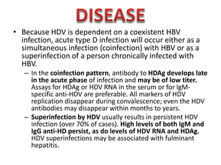
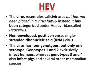
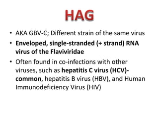
• Detected in some HBV infections.;The antigen is found
within certain HBsAg particles.
• In blood, HDV (delta agent) contains delta-Ag (HDAg)
surrounded by an HBsAg envelope. It has a particle size
of 35–37 nm and a buoyant density of 1.24–1.25 g/mL
in CsCl.
• The genome of HDV consists of single-stranded,
circular, negative-sense RNA, 1.7 kb in size.
• HDAg is the only protein coded for by HDV RNA and is
distinct from the antigenic determinants of HBV. HDV is
a defective virus that acquires an HBsAg coat for
transmission.
• One cannot become infected with hepatitis D unless
infected with HBV Chronic liver disease with cirrhosis
• Because HDV is dependent on a coexistent HBV
infection, acute type D infection will occur either as a
simultaneous infection (coinfection) with HBV or as a
superinfection of a person chronically infected with
HBV.
– In the coinfection pattern, antibody to HDAg develops late
in the acute phase of infection and may be of low titer.
Assays for HDAg or HDV RNA in the serum or for IgMspecific anti-HDV are preferable. All markers of HDV
replication disappear during convalescence; even the HDV
antibodies may disappear within months to years.
– Superinfection by HDV usually results in persistent HDV
infection (over 70% of cases). High levels of both IgM and
IgG anti-HD persist, as do levels of HDV RNA and HDAg.
HDV superinfections may be associated with fulminant
hepatitis.
• Hepatitis B vaccine, However, vaccination
does not protect hepatitis B carriers from
superinfection by HDV.
• The virus resembles caliciviruses but has not
been placed in a virus family instead it has
been categorized under Hepeviridaecalled
Hepevirus.
• Non-enveloped, positive-sense, singlestranded ribonucleic acid (RNA) virus
• The virus has four genotypes, but only one
serotype. Genotypes 1 and 2 exclusively
infect humans, whereas genotypes 3 and 4
also infect pigs and several other mammalian
species.
• Mainly through the faecal-oral route due to
faecal contamination of drinking water. Other
transmission routes have been identified,
which include:
– foodborne transmission from ingestion of
products derived from infected animals (pig,
deer);
– transfusion of infected blood products;
– Prenatal
• Replication of viral particles takes place in
intestinal mucosa cells, but primary in the
cytoplasm of hepatocytes
• HE is typically a self-limiting disease without
progression to chronic illness.
• Shares many clinical characteristics and
symptoms with hepatitis A
• RT-PCR (Reverse Transcriptase Polymerase Chain
Reaction)
– Molecular detection of HEV; First choice for RNA
viruses detection
• Enzyme immunoassay (EIA)
• Immunologic Diagnosis which is practical, highly sensitive and
inexpensive detection of Anti-HEV antibody.

• Immune fluorescence microscopy (IFE)
• IFE detects Ab that reacts against HEV Ag semiquantitatively.

• Virus isolation
• Immune electron microscopy (IEM)
• Ani-HEV Ab concentrations can be determined
semiquantitatively by rating the Ab coating
• No treatment involve-self limiting, no ati-viral
therapy available
• Travellers must drink purified water
• Avoid eating uncooked food
• Good sanitation
• No vaccine yet
• Hypothetical virus linked to hepatitis.
• An infection found in the Far East
• Consists of double-stranded DNA and
is substantially different from HAV
and HEV, both of which are RNA
based.
• AKA GBV-C; Different strain of the same virus
• Enveloped, single-stranded (+ strand) RNA
virus of the Flaviviridae
• Often found in co-infections with other
viruses, such as hepatitis C virus (HCV)common, hepatitis B virus (HBV), and Human
Immunodeficiency Virus (HIV)
• BLOOD TRANSFUSSION
• PATIENTS WITH HEMOPHILIA AT HIGH RISK
• PRENATAL
*PREVENTION IS JUST DOING THINGS THAT
AVOID THE RISK
REFERENCES
•
•
•
•
•
•

Alcamo’s Microbiology book
Bailey’s Scott
Jaweitz Microbiology book
WHO
CDC
Other internet links
Hepatitis type D, E, F G

Hepatitis type D, E, F G

  • 2.
    • Detected insome HBV infections.;The antigen is found within certain HBsAg particles. • In blood, HDV (delta agent) contains delta-Ag (HDAg) surrounded by an HBsAg envelope. It has a particle size of 35–37 nm and a buoyant density of 1.24–1.25 g/mL in CsCl. • The genome of HDV consists of single-stranded, circular, negative-sense RNA, 1.7 kb in size. • HDAg is the only protein coded for by HDV RNA and is distinct from the antigenic determinants of HBV. HDV is a defective virus that acquires an HBsAg coat for transmission. • One cannot become infected with hepatitis D unless infected with HBV Chronic liver disease with cirrhosis
  • 5.
    • Because HDVis dependent on a coexistent HBV infection, acute type D infection will occur either as a simultaneous infection (coinfection) with HBV or as a superinfection of a person chronically infected with HBV. – In the coinfection pattern, antibody to HDAg develops late in the acute phase of infection and may be of low titer. Assays for HDAg or HDV RNA in the serum or for IgMspecific anti-HDV are preferable. All markers of HDV replication disappear during convalescence; even the HDV antibodies may disappear within months to years. – Superinfection by HDV usually results in persistent HDV infection (over 70% of cases). High levels of both IgM and IgG anti-HD persist, as do levels of HDV RNA and HDAg. HDV superinfections may be associated with fulminant hepatitis.
  • 6.
    • Hepatitis Bvaccine, However, vaccination does not protect hepatitis B carriers from superinfection by HDV.
  • 8.
    • The virusresembles caliciviruses but has not been placed in a virus family instead it has been categorized under Hepeviridaecalled Hepevirus. • Non-enveloped, positive-sense, singlestranded ribonucleic acid (RNA) virus • The virus has four genotypes, but only one serotype. Genotypes 1 and 2 exclusively infect humans, whereas genotypes 3 and 4 also infect pigs and several other mammalian species.
  • 10.
    • Mainly throughthe faecal-oral route due to faecal contamination of drinking water. Other transmission routes have been identified, which include: – foodborne transmission from ingestion of products derived from infected animals (pig, deer); – transfusion of infected blood products; – Prenatal
  • 11.
    • Replication ofviral particles takes place in intestinal mucosa cells, but primary in the cytoplasm of hepatocytes • HE is typically a self-limiting disease without progression to chronic illness. • Shares many clinical characteristics and symptoms with hepatitis A
  • 12.
    • RT-PCR (ReverseTranscriptase Polymerase Chain Reaction) – Molecular detection of HEV; First choice for RNA viruses detection • Enzyme immunoassay (EIA) • Immunologic Diagnosis which is practical, highly sensitive and inexpensive detection of Anti-HEV antibody. • Immune fluorescence microscopy (IFE) • IFE detects Ab that reacts against HEV Ag semiquantitatively. • Virus isolation • Immune electron microscopy (IEM) • Ani-HEV Ab concentrations can be determined semiquantitatively by rating the Ab coating
  • 13.
    • No treatmentinvolve-self limiting, no ati-viral therapy available • Travellers must drink purified water • Avoid eating uncooked food • Good sanitation • No vaccine yet
  • 15.
    • Hypothetical viruslinked to hepatitis. • An infection found in the Far East • Consists of double-stranded DNA and is substantially different from HAV and HEV, both of which are RNA based.
  • 17.
    • AKA GBV-C;Different strain of the same virus • Enveloped, single-stranded (+ strand) RNA virus of the Flaviviridae • Often found in co-infections with other viruses, such as hepatitis C virus (HCV)common, hepatitis B virus (HBV), and Human Immunodeficiency Virus (HIV)
  • 19.
    • BLOOD TRANSFUSSION •PATIENTS WITH HEMOPHILIA AT HIGH RISK • PRENATAL *PREVENTION IS JUST DOING THINGS THAT AVOID THE RISK
  • 24.
    REFERENCES • • • • • • Alcamo’s Microbiology book Bailey’sScott Jaweitz Microbiology book WHO CDC Other internet links