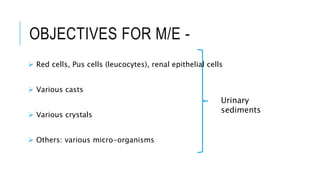
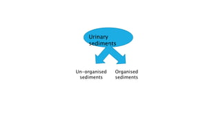
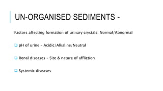
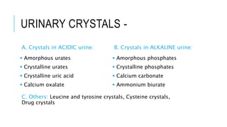
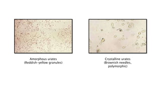
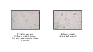
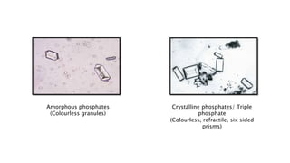
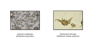
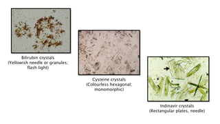
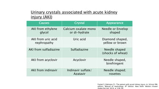
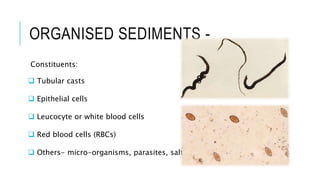
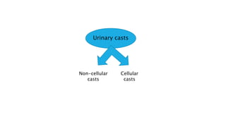
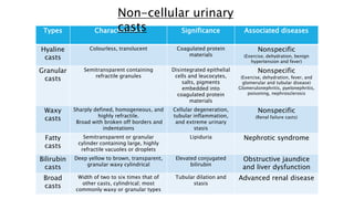
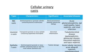
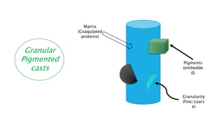
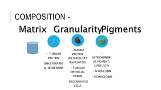
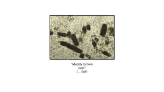
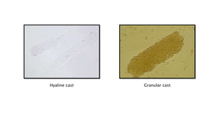
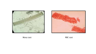
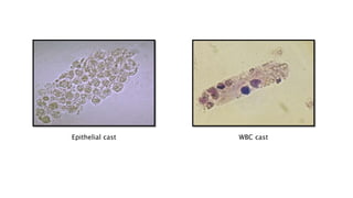
URINARY SEDIMENTS UNDER
MICROSCOPIC EXAMINATION
Presenter: Dr. ARNAB NANDY; MBBS, MD (Pediatrics)
1ST YEAR DrNB Trainee (Nephrology), MSHK, Kolkata
INTRODUCTION -
 Urine collection for microscopic examination:
 Timing of examination and preservation for transport:
 Processing before microscopic examination: 2000
rpm, 5 minutes
 Low and high power objective examination:
OBJECTIVES FOR M/E -
 Red cells, Pus cells (leucocytes), renal epithelial cells
 Various casts
 Various crystals
 Others: various micro-organisms
Urinary
sediments
Urinary
sediments
Un-organised
sediments
Organised
sediments
UN-ORGANISED SEDIMENTS -
Factors affecting formation of urinary crystals: Normal/Abnormal
 pH of urine – Acidic/Alkaline/Neutral
 Renal diseases – Site & nature of affliction
 Systemic diseases
URINARY CRYSTALS -
A. Crystals in ACIDIC urine:
 Amorphous urates
 Crystalline urates
 Crystalline uric acid
 Calcium oxalate
B. Crystals in ALKALINE urine:
 Amorphous phosphates
 Crystalline phosphates
 Calcium carbonate
 Ammonium biurate
C. Others: Leucine and tyrosine crystals, Cysteine crystals,
Drug crystals
Amorphous urates
(Reddish-yellow granules)
Crystalline urates
(Brownish needles,
polymorphic)
Crystalline uric acid
(Yellow or reddish brown;
Flat four sided, rhombic plates
or prisms)
Calcium oxalate
(Dumb-bell shaped)
Amorphous phosphates
(Colourless granules)
Crystalline phosphates/ Triple
phosphate
(Colourless, refractile, six sided
prisms)
Calcium carbonate
(Colourless granules)
Ammonium biurate
(Yellowish-brown spheres)
Bilirubin crystals
(Yellowish needle or granules;
flash light)
Cysteine crystals
(Colourless hexagonal;
monomorphic)
Indinavir crystals
(Rectangular plates, needle)
Causes Crystal Appearance
AKI from ethylene
glycol
Calcium oxalate mono
or di-hydrate
Needle or Envelop
shaped
AKI from uric acid
nephropathy
Uric acid Diamond shaped,
yellow or brown
AKI from sulfadiazine Sulfadiazine Needle shaped
(shocks of wheat)
AKI from acyclovir Acyclovir Needle shaped,
birefringent
AKI from indinavir Indinavir sulfate/
Azatavir
Needle shaped;
rosettes
Urinary crystals associated with acute kidney
injury (AKI)
Faubel S, Edelstein CL. The patient with acute kidney injury. In: Schrier RW,
editors. Manual of nephrology. 8th Edition. New Delhi: Wolters Kluwer
(India) Pvt Ltd; 2019. p. 228-30.
Shocks of
wheat
ORGANISED SEDIMENTS -
Constituents:
 Tubular casts
 Epithelial cells
 Leucocyte or white blood cells
 Red blood cells (RBCs)
 Others- micro-organisms, parasites, salts
Urinary casts
Non-cellular
casts
Cellular
casts
Types Characteristics Significance Associated diseases
Hyaline
casts
Colourless, translucent Coagulated protein
materials
Nonspecific
(Exercise, dehydration, benign
hypertension and fever)
Granular
casts
Semitransparent containing
refractile granules
Disintegrated epithelial
cells and leucocytes,
salts, pigments
embedded into
coagulated protein
materials
Nonspecific
(Exercise, dehydration, fever, and
glomerular and tubular disease)
Glomerulonephritis, pyelonephritis,
poisoning, nephrosclerosis
Waxy
casts
Sharply defined, homogeneous, and
highly refractile.
Broad with broken off borders and
indentations
Cellular degeneration,
tubular inflammation,
and extreme urinary
stasis
Nonspecific
(Renal failure casts)
Fatty
casts
Semitransparent or granular
cylinder containing large, highly
refractile vacuoles or droplets
Lipiduria Nephrotic syndrome
Bilirubin
casts
Deep yellow to brown, transparent,
granular waxy cylindrical
Elevated conjugated
bilirubin
Obstructive jaundice
and liver dysfunction
Broad
casts
Width of two to six times that of
other casts, cylindrical; most
commonly waxy or granular types
Tubular dilation and
stasis
Advanced renal disease
Non-cellular urinary
casts
Types Characteristics Significance Associated diseases
RBC
casts
Semitransparent or granular cylinder
containing distinct erythrocytes
Renal parenchymal
bleeding, glomerular
leakage
Acute
glomerulonephritis, IgA
nephropathy, lupus
nephritis, and renal
infarction
Leucocyt
e casts
Transparent granular or waxy cylinder
containing segmented neutrophils
Interstitial
inflammation
Tubulointerstitial
disease
(pyelonephritis),
glomerular disease,
interstitial nephritis,
and lupus nephritis
Epithelia
l casts
Semitransparent granular or waxy
cylinder containing intact or necrotic
renal tubular epithelial cells
Tubular damage Acute tubular necrosis,
acute allograft
rejection,
Cellular urinary
casts
Granular
Pigmented
casts
Matrix
(Coagulated
proteins)
Pigments
(embedde
d)
Granularity
(fine/coars
e)
COMPOSITION -
- TUBULAR
PROTEIN
(DEGENERATIO
N/SECRETION)
- PLASMA
PROTEIN
(FILTERED/EXT
RAVASATED)
- TUBULAR
EPITHELIAL
DEBRIS
-DEGENERATED
CELLS
-
MITOCHONDRI
AL PIGMENT/
LIPOFUSCIN
- MYOGLOBIN
- HEMOGLOBIN
‘Muddy brown
cast”
(…/lpf)
Hyaline cast Granular cast
Waxy cast RBC cast
Epithelial cast WBC cast
TAMM-HORSFALL
PROTEIN/UROMODULIN -
 Glycoprotein secreted by thick ascending loop (TAL)
 Matrix of renal casts and stones
 Protective role in acute tubular injury (ATI)
 Estimation of uromodulin (in urine and serum) and severity of ATI
 Uromodulin: Creatinine ratio in urine (lower the ratio, higher the
injury)
Opal SM. Innate Immunity and the Kidney. In: Editor(s): Ronco C, Bellomo R, Kellum JA, Ricci Z. Critical Care Nephrology. 3rd Edition. New
York: Elsevier; 2019. p. 476-82.
CLUES TO FIND-
 Physiological or pathological
 SITE of affliction
 DURATION of affliction
 NATURE of affliction
THANK YOU
arnabn.office@gmail.c
om

Urinary sediments under microscopic examination

  • 1.
    URINARY SEDIMENTS UNDER MICROSCOPICEXAMINATION Presenter: Dr. ARNAB NANDY; MBBS, MD (Pediatrics) 1ST YEAR DrNB Trainee (Nephrology), MSHK, Kolkata
  • 2.
    INTRODUCTION -  Urinecollection for microscopic examination:  Timing of examination and preservation for transport:  Processing before microscopic examination: 2000 rpm, 5 minutes  Low and high power objective examination:
  • 3.
    OBJECTIVES FOR M/E-  Red cells, Pus cells (leucocytes), renal epithelial cells  Various casts  Various crystals  Others: various micro-organisms Urinary sediments
  • 4.
  • 5.
    UN-ORGANISED SEDIMENTS - Factorsaffecting formation of urinary crystals: Normal/Abnormal  pH of urine – Acidic/Alkaline/Neutral  Renal diseases – Site & nature of affliction  Systemic diseases
  • 6.
    URINARY CRYSTALS - A.Crystals in ACIDIC urine:  Amorphous urates  Crystalline urates  Crystalline uric acid  Calcium oxalate B. Crystals in ALKALINE urine:  Amorphous phosphates  Crystalline phosphates  Calcium carbonate  Ammonium biurate C. Others: Leucine and tyrosine crystals, Cysteine crystals, Drug crystals
  • 7.
    Amorphous urates (Reddish-yellow granules) Crystallineurates (Brownish needles, polymorphic)
  • 8.
    Crystalline uric acid (Yellowor reddish brown; Flat four sided, rhombic plates or prisms) Calcium oxalate (Dumb-bell shaped)
  • 9.
    Amorphous phosphates (Colourless granules) Crystallinephosphates/ Triple phosphate (Colourless, refractile, six sided prisms)
  • 10.
    Calcium carbonate (Colourless granules) Ammoniumbiurate (Yellowish-brown spheres)
  • 11.
    Bilirubin crystals (Yellowish needleor granules; flash light) Cysteine crystals (Colourless hexagonal; monomorphic) Indinavir crystals (Rectangular plates, needle)
  • 12.
    Causes Crystal Appearance AKIfrom ethylene glycol Calcium oxalate mono or di-hydrate Needle or Envelop shaped AKI from uric acid nephropathy Uric acid Diamond shaped, yellow or brown AKI from sulfadiazine Sulfadiazine Needle shaped (shocks of wheat) AKI from acyclovir Acyclovir Needle shaped, birefringent AKI from indinavir Indinavir sulfate/ Azatavir Needle shaped; rosettes Urinary crystals associated with acute kidney injury (AKI) Faubel S, Edelstein CL. The patient with acute kidney injury. In: Schrier RW, editors. Manual of nephrology. 8th Edition. New Delhi: Wolters Kluwer (India) Pvt Ltd; 2019. p. 228-30.
  • 13.
  • 14.
    ORGANISED SEDIMENTS - Constituents: Tubular casts  Epithelial cells  Leucocyte or white blood cells  Red blood cells (RBCs)  Others- micro-organisms, parasites, salts
  • 15.
  • 16.
    Types Characteristics SignificanceAssociated diseases Hyaline casts Colourless, translucent Coagulated protein materials Nonspecific (Exercise, dehydration, benign hypertension and fever) Granular casts Semitransparent containing refractile granules Disintegrated epithelial cells and leucocytes, salts, pigments embedded into coagulated protein materials Nonspecific (Exercise, dehydration, fever, and glomerular and tubular disease) Glomerulonephritis, pyelonephritis, poisoning, nephrosclerosis Waxy casts Sharply defined, homogeneous, and highly refractile. Broad with broken off borders and indentations Cellular degeneration, tubular inflammation, and extreme urinary stasis Nonspecific (Renal failure casts) Fatty casts Semitransparent or granular cylinder containing large, highly refractile vacuoles or droplets Lipiduria Nephrotic syndrome Bilirubin casts Deep yellow to brown, transparent, granular waxy cylindrical Elevated conjugated bilirubin Obstructive jaundice and liver dysfunction Broad casts Width of two to six times that of other casts, cylindrical; most commonly waxy or granular types Tubular dilation and stasis Advanced renal disease Non-cellular urinary casts
  • 17.
    Types Characteristics SignificanceAssociated diseases RBC casts Semitransparent or granular cylinder containing distinct erythrocytes Renal parenchymal bleeding, glomerular leakage Acute glomerulonephritis, IgA nephropathy, lupus nephritis, and renal infarction Leucocyt e casts Transparent granular or waxy cylinder containing segmented neutrophils Interstitial inflammation Tubulointerstitial disease (pyelonephritis), glomerular disease, interstitial nephritis, and lupus nephritis Epithelia l casts Semitransparent granular or waxy cylinder containing intact or necrotic renal tubular epithelial cells Tubular damage Acute tubular necrosis, acute allograft rejection, Cellular urinary casts
  • 18.
  • 19.
    COMPOSITION - - TUBULAR PROTEIN (DEGENERATIO N/SECRETION) -PLASMA PROTEIN (FILTERED/EXT RAVASATED) - TUBULAR EPITHELIAL DEBRIS -DEGENERATED CELLS - MITOCHONDRI AL PIGMENT/ LIPOFUSCIN - MYOGLOBIN - HEMOGLOBIN
  • 20.
  • 21.
  • 22.
  • 23.
  • 24.
    TAMM-HORSFALL PROTEIN/UROMODULIN -  Glycoproteinsecreted by thick ascending loop (TAL)  Matrix of renal casts and stones  Protective role in acute tubular injury (ATI)  Estimation of uromodulin (in urine and serum) and severity of ATI  Uromodulin: Creatinine ratio in urine (lower the ratio, higher the injury) Opal SM. Innate Immunity and the Kidney. In: Editor(s): Ronco C, Bellomo R, Kellum JA, Ricci Z. Critical Care Nephrology. 3rd Edition. New York: Elsevier; 2019. p. 476-82.
  • 25.
    CLUES TO FIND- Physiological or pathological  SITE of affliction  DURATION of affliction  NATURE of affliction
  • 26.