SANJEEV KUMAR
B.Sc MLT PART-III
INTRODUCTION
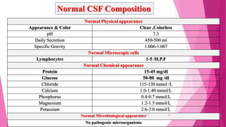
 Cerebrospinal fluid (CSF) is a clear, colourless fluid formed in the ventricles of
the brain
 Mainly by choroid plexus (meshwork of tiny small blood vessels in lateral third
and fourth ventricles).
 An ultrafiltrate of plasma.
 CSF is contained within the cerebral ventricles, the spinal canal and the
subarachnoid space (space between arachnoid externally and pia mater internally)
surrounding the brain and spinal cord.
 CSF is reabsorbed into the blood through the arachnoid villi of dural venous
sinuses.
31-05-2017 2
Cerebrospinal fluid (CSF) is present
within the subarachnoid space
surrounding the brain in the skull and
the spinal cord in the spinal column.
31-05-2017 3
Formation
 Secreted by choroid plexuses into
each ventricle.
 Choroid plexus are areas where the
lining wall of the ventricle is very
thin and has a profusion of
capillaries.
 Selective ultrafiltration of plasma
 Active secretion by epithelial
membranes.
 some originates from the
ependymal cells lining the
ventricles and from the brain
substance.
31-05-2017 6
CSF FORMATION
 Rate 0.35-0.40 ml/min OR
 500-600 ml/Day
 Total Volume:100-150ml & 10-60ml in New Born
 0.25% of total vol replace each minute
 Turn over time for total CSF vol  5-7 hours
 = 4 times / day
 40%-70% enters macroscopic spaces via CP
 30%-60% enters across ependyma and pia
31-05-2017 7
CIRCULATION OF CSF
 Circulation: CSF is mainly formed in choroid plexus of the lateral ventricle.
 CSF passes from the lateral ventricle to the third ventricle through the interventricular
foramen (foramen of Monro).
 From third ventricle it passes to the fourth ventricle through the cerebral aqueduct. The
circulation is aided by the arterial pulsations of the choroid plexuses.
 From the fourth ventricle (CSF) passes to the subarachnoid space around the brain and
spinal cord through the foramen of magendie and foramina of luschka.
31-05-2017 9
CIRCULATION OF CSF
Lateral ventricle
Foramen of Monro [Interventricular foramen]
Third ventricle:
Subarachnoid space of Brain and Spinal cord
Fourth ventricle:
Cerebral aqueduct
Foramen of megendie and formen of luschka
31-05-2017 10
FUNCTIONS OF CSF
A shock absorber
A mechanical buffer
Act as cushion between the brain and cranium
Act as a reservoir and regulates the contents of the cranium
Serves as a medium for nutritional exchange in CNS
Transport hormones and hormone releasing factors
Removes the metabolic waste products through absorption
31-05-2017 16
CLINICALAPPLICATION OF CSF
EXAMINATION
In the diagnosis of
a. Bacterial, viral or fungal meningitis.
b. Encephalitis.
c. Malignant infiltrates like in acute leukemia, lymphoma.
d. Subarachnoid hemorrhage.
e. Spinal canal blockage leading to elevated intracranial tension.
f. Sub acute sclerosing pan encephalitis (SSPE)
31-05-2017 17
Routine Laboratory CSF Analysis
Collection
 Lumbar puncture
 At interspace of vertebrate Lumber 3rd-4th or
4th-5th
 With complete aseptic techniques
 3-5 ml of CSF is collected
 It requires certain precautions and careful
technique to prevent the introduction of
infection or the damaging of neural tissue.
31-05-2017 18
Sampling
Collected CSF sample is immediately divided into three
tubes:
Tube 1: Chemical Investigation (kept in freezer till
performed)
Tube 2: Microbiology Investigation (kept in room
temperature)
Tube 3: Microscopic Investigation (Cellular Counting –
Differential)
Tube 4: Cytology and Special Studies
31-05-2017 20
Normal Physical appearance
Appearance & Color Clear ,Colorless
pH 7.3
Daily Secretion 450-500 ml
Specific Gravity 1.006-1.007
Normal Microscopic cells
Lymphocytes 1-5 /H.P.F
Normal Chemical appearance
Protein 15-45 mg/dl
Glucose 50-80 mg /dl
Chloride 115-130 mmol /L
Calcium 1.0-1.40 mmol/L
Phosphorus 0.4-0.7 mmol/L
Magnesium 1.2-1.5 mmol/L
Potassium 2.6-3.0 mmol/L
Normal Microbiological appearance
No pathogenic microorganisms
Normal CSF Composition
31-05-2017 21
PHYSICAL EXAMINATION OF CSF
Normal CSF: Clear & colorless
Viscosity: equal to water (increased with increased proteins)
Color and/or turbidity of CSF: observed only in pathological circumstances.
Turbid CSF
Bacteria
WBCs cells or pus cells: suggestive of a CNS infection (menigitis or encephalitis)
Blood : suggestive of hemorrhage: subarachnoid or artifactual traumatic tap.
Red & brown color :
Blood
Yellow colour
1- Jaundice (bilirubin in CSF)
2- Xanthochromia (hemoglobin breakdown pigments in CSF)
Xanthochromic CSF suggests that a subarachnoid hemorrhage has recently occurred
(at least within two hours prior to tapping).
The yellow color is due to bilirubin generated in the CNS by the breakdown of hemoglobin
released from RBC's. (so jaundice should be excluded).
31-05-2017 22
MICROSCOPIC EXAMINATION OF CSF
WBCs
Normal Total WBCs count: 1-5 lymphocytes /HPF
Normal differential WBCs Count: (in centrifuged cells)
- 62% lymphocytes
- 36% monocytes
- 2% neutrophils
Increased neutrophils: bacterial meningitis
Increased lymphocytes: aseptic and viral meningitis
RBCs
Normally CSF is blood free
RBCs in CSF: subarachnoid hemorrhage & malignancy
Artifact: traumatic tap (should be excluded)
(Traumatic tap bright red color RBCS in decreasing number as the
fluid is sampled) 31-05-2017 23
CHEMICAL EXAMINATION OF CSF
In addition to the major ions, CSF contains oxygen, sugars (e.g.
glucose, fructose), lactate, proteins (e.g. albumin, globulins) and
amino acids.
31-05-2017 24
ANALYSIS OF PROTEIN IN CSF
 Turbidimetric method
 Coomassie Brilliant Blue Method
 Pyrogallol Red Method
 Reverse Biuret method
31-05-2017 25
TURBIDIMETRIC METHOD
 Protein molecules which are normally soluble in solution can be made to precipitate by the
addition of 3% Trichloro acetic Acid(TCA).
 Protein precipitation causes the solution to become turbid.
 Thus the concentration of protein can be determined by measuring the degree of turbidity.
 The absorbance of turbidity is measured at 490nm.
 Precipitation can be achieved by sulfosalicylic acid and with sulfosalicylic acid in
combination with sodium sulphate/TCA.
Reagent
 3% TCA
 0.5% w/v stock protein standard
31-05-2017 26
S.No. Reagent Test Std Blank
1 CSF Sample 0.5ml - -
2 Std - 0.5ml -
3 D.W. - - 0.5ml
4 3% TCA 1.5ml 1.5ml 1.5ml
Working protein standard
Stock 0.1ml
Normal saline 10m
Procedure
Mix and keep at RT for 10-15 min.
Read at 490nm
31-05-2017 27
Calculation
𝐶𝑆𝐹 𝑃𝑟𝑜𝑡𝑒𝑖𝑛𝑠,
𝑔
𝑙
=
𝑇 − 𝐵
𝑆 − 𝐵
𝑥 𝑐𝑜𝑛𝑐. 𝑜𝑓 𝑠𝑡𝑑
COOMASSIE BRILLIANT BLUE(CBB)
PRINCIPLE: Dye-binding methods are based on the ability of proteins to
bind dyes such as Amido black 10B and Coomassie brilliant blue (CBB). The
dye-binding method is particularly used for assay of Total protein in CSF,
Urine.
This is simple and fast method but linearity is less. Leaves stain on glassware.
 Ability of proteins to bind with CBB G-250 & read at 700nm.
 Particularly for total protein in CSF and Urine.
 It leaves stain on the glassware. Imparts color to the cuvette also.
31-05-2017 28
PYROGALLOL RED
 This dye combined with the molybdate, when they combined with albumin
of CSF, they form a complex with albumin & give red color. Read at
470nm.
 This dye also react with basic amino acid & gives red purple color
31-05-2017 29
REVERSE BIURET METHOD
 A method of protein determination has been developed which combines the biuret
reaction and the copper(I)-bathocuproine chelate reaction.
 Protein in the specimen forms a C𝑢2+-protein chelate complex (biuret reaction)
during the first step.
 Excess C𝑢2+
is reduced to C𝑢+ by ascrobic acid, allowing the C𝑢+ to form a C𝑢+-
bathocuproine chelate complex during the second step.
 The amount of C𝑢+-bathocuproine chelate complex formed is inversely proportional
to the protein concentration.
 The sensitivity of this method was higher than that of the original Lowry, pyrogallol
red and commercially available Coomassie Brilliant Blue G.250 methods.
31-05-2017 30
Clinical Significance
INCREASED PROTEIN: CSF protein may rise to 500 mg/dl in
bacterial meningitis.
A more moderate increase (150-200 mg/dl) occurs in inflammatory
diseases of meninges (meningitis, encephalitis), intracranial tumors,
subarachnoid hemorrhage, and cerebral infarction.
A more severe increase occurs in the Guillain-Barre syndrome and acoustic
and spinal schwannoma.
31-05-2017 31
MULTIPLE SCLEROSIS
 CSF protein is normal or mildly increased.
 Increased IgG in CSF, but not in serum [IgG/albumin index normally
10:1].
 90% of MS patients have oligoclonal IgG bands in the CSF.
 Oligoclonal bands occur in the CSF only not in the serum.
 The CSF in MS often contains myelin fragments and myelin basic
protein (MBP).
 MBP can be detected by radioimmunoassay. MBP is not specific for
MS. It can appear in any condition causing brain necrosis, including
infarcts.
31-05-2017 32
PANDY'S METHOD
 Proteins (globulin and albumin) are precipitated by a saturated solution of phenol(7%) in
water.
 The reagent used is phenol (carbolic acid crystals dissolved in water) or, pyrogallic
acid or, cresol, usually termed as Pandy's reagent or Pandy's solution.
Procedure
 One drop of CSF sample (collected from the patient by lumbar puncture technique), is
added to about 1ml of Pandy's solution.
 The turbid appearance signifies the presence of elevated levels of globulin protein in the
CSF and is regarded as positive Pandy's reaction.
 The CSF from a normal adult shows no turbidity or precipitates and this is a negative
Pandy's reaction.
GLOBULIN
31-05-2017 33
NONNE-APELT TEST
 Half saturated ammonium sulphate are common use which
demonstrate the presence of an increase of globulin in CSF.
 Carefully layer 1ml clear CSF over 1ml of saturated solution of
ammonium sulphate.
 A thin white ring appearing at the junction of the liquids which
disappears on mixing indicates a 1+ reaction.
 Heavy cloudiness persisting after mixing is 4+ reaction.
31-05-2017 34
Analysis Of Protein Fractions: (Albumin & IgG)
 Albumin of CSF is obtained from blood by means of blood-
brain barrier (as it is produced solely by the liver).In cases of
increased permeability of BBB, albumin is increased in CSF.
 IgG of CSF can be obtained: from blood (By BBB) : increase in
cases of increase permeability of BBB & by local synthesis from plasma
cells within CSF (increased in cases of MS)
So, it is essential to determine the source of IgG
31-05-2017 35
FIRST: CHECK INTEGRITY OF BLOOD BRAIN BARRIER (BBB)
BY CSF / serum albumin index calculation
CSF serum albumin index = CSF albumin (mg/dl) / serum albumin (g/dl)
Index less than 9 indicates intact BBB (no increased permeability of BBB)
SECOND: CSF IgG INDEX IS CALCULATED
𝐼𝑔𝐺 𝐼𝑛𝑑𝑒𝑥 =
𝐶𝑆𝐹 𝐼𝑔𝐺 × 𝑆𝑒𝑟𝑢𝑚 𝐴𝑙𝑏𝑢𝑚𝑖𝑛
𝑆𝑒𝑟𝑢𝑚 𝐼𝑔𝐺 × 𝐶𝑆𝐹 𝐴𝑙𝑏𝑢𝑚𝑖𝑛
Normal : less than 0.7
Increased in cases of demyelinating diseases of CNS as : Multiple sclerosis (MS)
31-05-2017 36
Glucose Estimation
GOD-POD Method
 The enzyme glucose oxidase catalyses the oxidation of glucose to gluconic
acid and hydrogen peroxide.
 The enzyme peroxidase and a chromogenic oxygen acceptor, such as o-
dianisidine, results in the formation of a colored compound that is
measured
 Glucose oxidase is highly specific for β-d-glucose.
 36% and 64% of glucose in solution are in α- and β- forms, respectively.
 The second step, involving peroxidase, is much less specific than the
glucose oxidase reaction.
31-05-2017 37
Glucose+𝐻2 𝑂+ 𝑂2 GOD gluconic acid + 2𝐻2 𝑂2
𝐻2 𝑂2 POD 𝐻2 𝑂 + (O)
Phenol+(O) Quinone
Quinone + 4 amino antipyrine Quinone-imimne
(pink colur complex)
31-05-2017 38
 Chloride
ISE Method
Scale & Titration Method
31-05-2017 39
SCHALES AND SCHALES TITRATION
METHOD
PRINCIPLE: PFF of CSF is titrated with mercuric nitrate solution. Diphenyl
Carbazone is used as indicator. Chloride ions in CSF react with mercuric ions. At
the end point, mercuric ions in excess react with Diphenyl carbazone indicator to
blue-violet colored complex.
Specimen with low proteins content can be titrated directly without
removing proteins.
𝐻𝑔2+ ions have more affinity for chloride ions as compared to DPC .
31-05-2017 40
INCREASED CSF GLUCOSE IS OF NO CLINICAL
SIGNIFICANCE.
Causes of decreased CSF glucose
• Meningitis-Bacterial, fungal tubercular and syphilitic
meningitis.
• Tumors involving the meninges.
• Subarachnoid hemorrhage.
• Cerebral ameobiasis.
31-05-2017 41
Test Appearance Pressure WBC/μL Protein mg/dL Glucose mg/dL Chloride
Normal CSF Clear 90 – 180 mm 0-8 lymph. 15-45 50-80 115-130 mEq/L
Acute bacterial
meningitis
Turbid Increased 1000 -10000 100 – 500 < 40 Decreased
Viral meningitis Clear Normal to
moderate
increase
5-300, rarely
>1000
Normal to mild
increased
Normal Normal
Tubercular
meningitis
Slightly opaque
cobweb
formation
Increased/
decreased,
spinal block
100-600 mixed
or lymph.
50-300 due to
spinal block
Decreased Decreased
Fungal
meningitis
Clear Increased 40-400 mixed 50-300 Decreased Decreased
Acute syphilitic Clear Increased About 500
lymph
Increased but
<100
Normal Normal
Clinical Significance
31-05-2017 42
31-05-2017 43

Cerebrospinal fluid

  • 1.
  • 2.
    INTRODUCTION  Cerebrospinal fluid(CSF) is a clear, colourless fluid formed in the ventricles of the brain  Mainly by choroid plexus (meshwork of tiny small blood vessels in lateral third and fourth ventricles).  An ultrafiltrate of plasma.  CSF is contained within the cerebral ventricles, the spinal canal and the subarachnoid space (space between arachnoid externally and pia mater internally) surrounding the brain and spinal cord.  CSF is reabsorbed into the blood through the arachnoid villi of dural venous sinuses. 31-05-2017 2
  • 3.
    Cerebrospinal fluid (CSF)is present within the subarachnoid space surrounding the brain in the skull and the spinal cord in the spinal column. 31-05-2017 3
  • 4.
    Formation  Secreted bychoroid plexuses into each ventricle.  Choroid plexus are areas where the lining wall of the ventricle is very thin and has a profusion of capillaries.  Selective ultrafiltration of plasma  Active secretion by epithelial membranes.  some originates from the ependymal cells lining the ventricles and from the brain substance. 31-05-2017 6
  • 5.
    CSF FORMATION  Rate0.35-0.40 ml/min OR  500-600 ml/Day  Total Volume:100-150ml & 10-60ml in New Born  0.25% of total vol replace each minute  Turn over time for total CSF vol  5-7 hours  = 4 times / day  40%-70% enters macroscopic spaces via CP  30%-60% enters across ependyma and pia 31-05-2017 7
  • 6.
    CIRCULATION OF CSF Circulation: CSF is mainly formed in choroid plexus of the lateral ventricle.  CSF passes from the lateral ventricle to the third ventricle through the interventricular foramen (foramen of Monro).  From third ventricle it passes to the fourth ventricle through the cerebral aqueduct. The circulation is aided by the arterial pulsations of the choroid plexuses.  From the fourth ventricle (CSF) passes to the subarachnoid space around the brain and spinal cord through the foramen of magendie and foramina of luschka. 31-05-2017 9
  • 7.
    CIRCULATION OF CSF Lateralventricle Foramen of Monro [Interventricular foramen] Third ventricle: Subarachnoid space of Brain and Spinal cord Fourth ventricle: Cerebral aqueduct Foramen of megendie and formen of luschka 31-05-2017 10
  • 8.
    FUNCTIONS OF CSF Ashock absorber A mechanical buffer Act as cushion between the brain and cranium Act as a reservoir and regulates the contents of the cranium Serves as a medium for nutritional exchange in CNS Transport hormones and hormone releasing factors Removes the metabolic waste products through absorption 31-05-2017 16
  • 9.
    CLINICALAPPLICATION OF CSF EXAMINATION Inthe diagnosis of a. Bacterial, viral or fungal meningitis. b. Encephalitis. c. Malignant infiltrates like in acute leukemia, lymphoma. d. Subarachnoid hemorrhage. e. Spinal canal blockage leading to elevated intracranial tension. f. Sub acute sclerosing pan encephalitis (SSPE) 31-05-2017 17
  • 10.
    Routine Laboratory CSFAnalysis Collection  Lumbar puncture  At interspace of vertebrate Lumber 3rd-4th or 4th-5th  With complete aseptic techniques  3-5 ml of CSF is collected  It requires certain precautions and careful technique to prevent the introduction of infection or the damaging of neural tissue. 31-05-2017 18
  • 11.
    Sampling Collected CSF sampleis immediately divided into three tubes: Tube 1: Chemical Investigation (kept in freezer till performed) Tube 2: Microbiology Investigation (kept in room temperature) Tube 3: Microscopic Investigation (Cellular Counting – Differential) Tube 4: Cytology and Special Studies 31-05-2017 20
  • 12.
    Normal Physical appearance Appearance& Color Clear ,Colorless pH 7.3 Daily Secretion 450-500 ml Specific Gravity 1.006-1.007 Normal Microscopic cells Lymphocytes 1-5 /H.P.F Normal Chemical appearance Protein 15-45 mg/dl Glucose 50-80 mg /dl Chloride 115-130 mmol /L Calcium 1.0-1.40 mmol/L Phosphorus 0.4-0.7 mmol/L Magnesium 1.2-1.5 mmol/L Potassium 2.6-3.0 mmol/L Normal Microbiological appearance No pathogenic microorganisms Normal CSF Composition 31-05-2017 21
  • 13.
    PHYSICAL EXAMINATION OFCSF Normal CSF: Clear & colorless Viscosity: equal to water (increased with increased proteins) Color and/or turbidity of CSF: observed only in pathological circumstances. Turbid CSF Bacteria WBCs cells or pus cells: suggestive of a CNS infection (menigitis or encephalitis) Blood : suggestive of hemorrhage: subarachnoid or artifactual traumatic tap. Red & brown color : Blood Yellow colour 1- Jaundice (bilirubin in CSF) 2- Xanthochromia (hemoglobin breakdown pigments in CSF) Xanthochromic CSF suggests that a subarachnoid hemorrhage has recently occurred (at least within two hours prior to tapping). The yellow color is due to bilirubin generated in the CNS by the breakdown of hemoglobin released from RBC's. (so jaundice should be excluded). 31-05-2017 22
  • 14.
    MICROSCOPIC EXAMINATION OFCSF WBCs Normal Total WBCs count: 1-5 lymphocytes /HPF Normal differential WBCs Count: (in centrifuged cells) - 62% lymphocytes - 36% monocytes - 2% neutrophils Increased neutrophils: bacterial meningitis Increased lymphocytes: aseptic and viral meningitis RBCs Normally CSF is blood free RBCs in CSF: subarachnoid hemorrhage & malignancy Artifact: traumatic tap (should be excluded) (Traumatic tap bright red color RBCS in decreasing number as the fluid is sampled) 31-05-2017 23
  • 15.
    CHEMICAL EXAMINATION OFCSF In addition to the major ions, CSF contains oxygen, sugars (e.g. glucose, fructose), lactate, proteins (e.g. albumin, globulins) and amino acids. 31-05-2017 24
  • 16.
    ANALYSIS OF PROTEININ CSF  Turbidimetric method  Coomassie Brilliant Blue Method  Pyrogallol Red Method  Reverse Biuret method 31-05-2017 25
  • 17.
    TURBIDIMETRIC METHOD  Proteinmolecules which are normally soluble in solution can be made to precipitate by the addition of 3% Trichloro acetic Acid(TCA).  Protein precipitation causes the solution to become turbid.  Thus the concentration of protein can be determined by measuring the degree of turbidity.  The absorbance of turbidity is measured at 490nm.  Precipitation can be achieved by sulfosalicylic acid and with sulfosalicylic acid in combination with sodium sulphate/TCA. Reagent  3% TCA  0.5% w/v stock protein standard 31-05-2017 26
  • 18.
    S.No. Reagent TestStd Blank 1 CSF Sample 0.5ml - - 2 Std - 0.5ml - 3 D.W. - - 0.5ml 4 3% TCA 1.5ml 1.5ml 1.5ml Working protein standard Stock 0.1ml Normal saline 10m Procedure Mix and keep at RT for 10-15 min. Read at 490nm 31-05-2017 27
  • 19.
    Calculation 𝐶𝑆𝐹 𝑃𝑟𝑜𝑡𝑒𝑖𝑛𝑠, 𝑔 𝑙 = 𝑇 −𝐵 𝑆 − 𝐵 𝑥 𝑐𝑜𝑛𝑐. 𝑜𝑓 𝑠𝑡𝑑 COOMASSIE BRILLIANT BLUE(CBB) PRINCIPLE: Dye-binding methods are based on the ability of proteins to bind dyes such as Amido black 10B and Coomassie brilliant blue (CBB). The dye-binding method is particularly used for assay of Total protein in CSF, Urine. This is simple and fast method but linearity is less. Leaves stain on glassware.  Ability of proteins to bind with CBB G-250 & read at 700nm.  Particularly for total protein in CSF and Urine.  It leaves stain on the glassware. Imparts color to the cuvette also. 31-05-2017 28
  • 20.
    PYROGALLOL RED  Thisdye combined with the molybdate, when they combined with albumin of CSF, they form a complex with albumin & give red color. Read at 470nm.  This dye also react with basic amino acid & gives red purple color 31-05-2017 29
  • 21.
    REVERSE BIURET METHOD A method of protein determination has been developed which combines the biuret reaction and the copper(I)-bathocuproine chelate reaction.  Protein in the specimen forms a C𝑢2+-protein chelate complex (biuret reaction) during the first step.  Excess C𝑢2+ is reduced to C𝑢+ by ascrobic acid, allowing the C𝑢+ to form a C𝑢+- bathocuproine chelate complex during the second step.  The amount of C𝑢+-bathocuproine chelate complex formed is inversely proportional to the protein concentration.  The sensitivity of this method was higher than that of the original Lowry, pyrogallol red and commercially available Coomassie Brilliant Blue G.250 methods. 31-05-2017 30
  • 22.
    Clinical Significance INCREASED PROTEIN:CSF protein may rise to 500 mg/dl in bacterial meningitis. A more moderate increase (150-200 mg/dl) occurs in inflammatory diseases of meninges (meningitis, encephalitis), intracranial tumors, subarachnoid hemorrhage, and cerebral infarction. A more severe increase occurs in the Guillain-Barre syndrome and acoustic and spinal schwannoma. 31-05-2017 31
  • 23.
    MULTIPLE SCLEROSIS  CSFprotein is normal or mildly increased.  Increased IgG in CSF, but not in serum [IgG/albumin index normally 10:1].  90% of MS patients have oligoclonal IgG bands in the CSF.  Oligoclonal bands occur in the CSF only not in the serum.  The CSF in MS often contains myelin fragments and myelin basic protein (MBP).  MBP can be detected by radioimmunoassay. MBP is not specific for MS. It can appear in any condition causing brain necrosis, including infarcts. 31-05-2017 32
  • 24.
    PANDY'S METHOD  Proteins(globulin and albumin) are precipitated by a saturated solution of phenol(7%) in water.  The reagent used is phenol (carbolic acid crystals dissolved in water) or, pyrogallic acid or, cresol, usually termed as Pandy's reagent or Pandy's solution. Procedure  One drop of CSF sample (collected from the patient by lumbar puncture technique), is added to about 1ml of Pandy's solution.  The turbid appearance signifies the presence of elevated levels of globulin protein in the CSF and is regarded as positive Pandy's reaction.  The CSF from a normal adult shows no turbidity or precipitates and this is a negative Pandy's reaction. GLOBULIN 31-05-2017 33
  • 25.
    NONNE-APELT TEST  Halfsaturated ammonium sulphate are common use which demonstrate the presence of an increase of globulin in CSF.  Carefully layer 1ml clear CSF over 1ml of saturated solution of ammonium sulphate.  A thin white ring appearing at the junction of the liquids which disappears on mixing indicates a 1+ reaction.  Heavy cloudiness persisting after mixing is 4+ reaction. 31-05-2017 34
  • 26.
    Analysis Of ProteinFractions: (Albumin & IgG)  Albumin of CSF is obtained from blood by means of blood- brain barrier (as it is produced solely by the liver).In cases of increased permeability of BBB, albumin is increased in CSF.  IgG of CSF can be obtained: from blood (By BBB) : increase in cases of increase permeability of BBB & by local synthesis from plasma cells within CSF (increased in cases of MS) So, it is essential to determine the source of IgG 31-05-2017 35
  • 27.
    FIRST: CHECK INTEGRITYOF BLOOD BRAIN BARRIER (BBB) BY CSF / serum albumin index calculation CSF serum albumin index = CSF albumin (mg/dl) / serum albumin (g/dl) Index less than 9 indicates intact BBB (no increased permeability of BBB) SECOND: CSF IgG INDEX IS CALCULATED 𝐼𝑔𝐺 𝐼𝑛𝑑𝑒𝑥 = 𝐶𝑆𝐹 𝐼𝑔𝐺 × 𝑆𝑒𝑟𝑢𝑚 𝐴𝑙𝑏𝑢𝑚𝑖𝑛 𝑆𝑒𝑟𝑢𝑚 𝐼𝑔𝐺 × 𝐶𝑆𝐹 𝐴𝑙𝑏𝑢𝑚𝑖𝑛 Normal : less than 0.7 Increased in cases of demyelinating diseases of CNS as : Multiple sclerosis (MS) 31-05-2017 36
  • 28.
    Glucose Estimation GOD-POD Method The enzyme glucose oxidase catalyses the oxidation of glucose to gluconic acid and hydrogen peroxide.  The enzyme peroxidase and a chromogenic oxygen acceptor, such as o- dianisidine, results in the formation of a colored compound that is measured  Glucose oxidase is highly specific for β-d-glucose.  36% and 64% of glucose in solution are in α- and β- forms, respectively.  The second step, involving peroxidase, is much less specific than the glucose oxidase reaction. 31-05-2017 37
  • 29.
    Glucose+𝐻2 𝑂+ 𝑂2GOD gluconic acid + 2𝐻2 𝑂2 𝐻2 𝑂2 POD 𝐻2 𝑂 + (O) Phenol+(O) Quinone Quinone + 4 amino antipyrine Quinone-imimne (pink colur complex) 31-05-2017 38
  • 30.
     Chloride ISE Method Scale& Titration Method 31-05-2017 39
  • 31.
    SCHALES AND SCHALESTITRATION METHOD PRINCIPLE: PFF of CSF is titrated with mercuric nitrate solution. Diphenyl Carbazone is used as indicator. Chloride ions in CSF react with mercuric ions. At the end point, mercuric ions in excess react with Diphenyl carbazone indicator to blue-violet colored complex. Specimen with low proteins content can be titrated directly without removing proteins. 𝐻𝑔2+ ions have more affinity for chloride ions as compared to DPC . 31-05-2017 40
  • 32.
    INCREASED CSF GLUCOSEIS OF NO CLINICAL SIGNIFICANCE. Causes of decreased CSF glucose • Meningitis-Bacterial, fungal tubercular and syphilitic meningitis. • Tumors involving the meninges. • Subarachnoid hemorrhage. • Cerebral ameobiasis. 31-05-2017 41
  • 33.
    Test Appearance PressureWBC/μL Protein mg/dL Glucose mg/dL Chloride Normal CSF Clear 90 – 180 mm 0-8 lymph. 15-45 50-80 115-130 mEq/L Acute bacterial meningitis Turbid Increased 1000 -10000 100 – 500 < 40 Decreased Viral meningitis Clear Normal to moderate increase 5-300, rarely >1000 Normal to mild increased Normal Normal Tubercular meningitis Slightly opaque cobweb formation Increased/ decreased, spinal block 100-600 mixed or lymph. 50-300 due to spinal block Decreased Decreased Fungal meningitis Clear Increased 40-400 mixed 50-300 Decreased Decreased Acute syphilitic Clear Increased About 500 lymph Increased but <100 Normal Normal Clinical Significance 31-05-2017 42
  • 34.