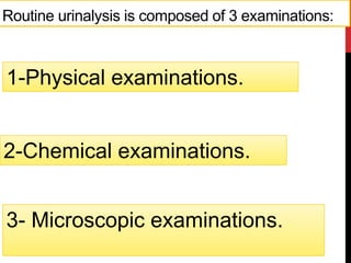
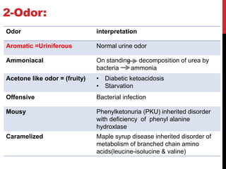
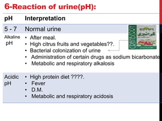
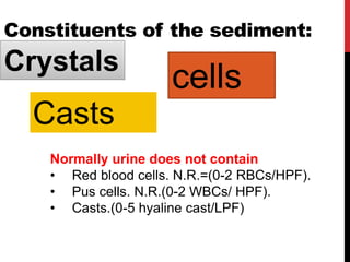
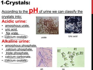
Urine analysis
=Urinalysis.
CUA UA
‫البول‬ ‫تحليل‬
Urine analysis:
1-Urine is a more convenient
sample .
2-Concentrate for many
substances.
3-Detection is easier than in
blood.
Function of the kidney:
Regulation of: water , electrolyte
ADH ALD
ADH
Aldosterone
Acid base balance.
Excretion of the products of :Protein
and nucleic acid metabolism
Urea.
Creatinine
Uric acid.
NPN
ENDOCRINE ORGAN
ADH
Urine formation:
Composition of urine
1-Water :95%.
2- Non protein nitrogenous compound 2.2%:
• Creatinine.
• Urea.
• Uuric acid.2.2%.
3-Dissolved salts and other ions as Na+, K+,H+,
Ca++,Cl-, phospate:2.8%
Urine sample:
It must be analyzed within 1 hour of collection if held at
room temperature or else refrigerated at 2°–8°C for not
more than 8 hours before analysis.
The chemical changes which may occur in urine
specimens stored at room temperature include:
• Breakdown of urea to ammonia by bacteria, leading to
an increase in the pH of the urine. This may cause the
precipitation of calcium and phosphates.
• Destruction of glucose by bacteria.
• Precipitation of urate crystals in acidic urine. These
chemical changes can be slowed down by refrigerating
the urine at 2–8°C or adding preservatives.
3- Microscopic examinations.
Routine urinalysis is composed of 3 examinations:
2-Chemical examinations.
1-Physical examinations.
1. Physical examinations
abnormal characters:
1. Volume.
2. Color.
3. Odor.
4. Specific gravity.
5. Reaction(pH).
6. Aspect.
2. Chemical examinations:
1. Proteins.
2. Glucose.
3. Ketone bodies.
4. Bile salts.
5. Bile pigments.
6. Blood.
3. Microscopic examinations:
For abnormal insoluble constituents:
1. Cells.
2. Crystals.
3. Casts.
Types of urine samples:
Morning
urine
sample:
24 hours
urine
sample
Mid-stream
urine
sample:
Random
urine
sample:
Urine is concentrated
Quantitative
for a culture
Not quantitative
Must stop
antibiotic 48
hrs. before
A) Physical examination of urine:
Volume:
Odor
Color
Specific gravity
Aspect (appearance):
Reaction (PH)
1-Volume:
Normal: 500-2000 ml/day.
Polyuria:
Urinary volume=
>2000 ml/day.
Oliguria:
Urinary volume=
<500 ml/day.
Anuria:
Urinary volume=
<125 ml/day.
A. Polyuria:
Urinary volume= >2000 ml/day.
I- Physiological polyuria
1-Winter.
2-High fluids intake (Tea, coffee, cola, beer
and alcohol Diuretics.
3-High protein diet Urea(osmotic diuresis).
II- Pathological polyuria:
• Diabetes mellitus(D.M) (3-5 L/day).
• Diabetes insipidus (D.I.) (10-15 L/day).
• Diuretics drugs.
3D
Diabetes mellitus(D.M).
Diabetes insipidus(D.I.)
B. Oliguria and anuria:
Oliguria: Urinary volume=<500ml/day.
.
Anuria: Urinary volume=<125ml/day
I-Physiological oliguria:
• Summer (hot weather).
• Fasting.
• Low fluid intake in diet.
• Hyperactivity and physical exercise.
II-Pathological oliguria:
Pre-renal :
• Heart failure(CHF)
• Complete burns.
• Vomiting.
• Diarrhea.
Renal oliguria
• Acute tubular necrosis,
• Acute nephritis.
• Renal failure.
Post-renal oliguria
•Prostate hypertrophy
• Stone.
• Cancer.
2-Odor:
Odor interpretation
Aromatic =Uriniferous Normal urine odor
Ammoniacal On standing decomposition of urea by
bacteria ammonia
Acetone like odor = (fruity) • Diabetic ketoacidosis
• Starvation
Offensive Bacterial infection
Mousy Phenylketonuria (PKU) inherited disorder
with deficiency of phenyl alanine
hydroxlase
Caramelized Maple syrup disease inherited disorder of
metabolism of branched chain amino
acids(leucine-isolucine & valine)
3-Color:
Color Interpretation
Amber yellow Normal urine color
Pale yellow • Very dilute urine
• Infants
• Diabetes mellitus (D.M.)
• Diabetes insipidus (D.I.)
• Increase fluid intake
Dark yellow • Fever
• Hyperthyroidism
• Dehydration
Light brown
(tae like color)
Jaundice
Red • trauma of urinary tract
• Porphyrinuria
Black Alkaptonuria
4- Aspect=Appearance:
Normally, freshly urine transparent &clear.
Turbid urine:
• Pus =Pyuria.
• Red cells =hematuria
• Crystals (calcium phosphate or urate).
• Epithelial cells.
5- Specific gravity:
• It is the density of urine compared with the density of distilled
water that is conveniently fixed as 1 at 20⁰ C.
• It measures the ability of the kidney to concentrate urine.
• It varies directly with the grams of solutes excreted in urine
and inversely with volume.
. Measured using Urinometer
Urinometer:
Specific Gravity Interpretation
1.015 – 1.025 Normal urine
> 1.025 Physiologically:
Summer
First morning specimen.
Pathologically:
• Dehydration.
• Presence of glucose in urine (D.M.)
• Presence of protein in urine (Proteinuria).
• Shock.
• Heart failure.
<1.025 Physiologically:
High fluid intake.
Pathologically:
D.I.
6-Reaction of urine(pH):
pH Interpretation
5 - 7 Normal urine
Alkaline
pH
• After meal.
• High citrus fruits and vegetables??.
• Bacterial colonization of urine
• Administration of certain drugs as sodium bicarbonate
• Metabolic and respiratory alkalosis
Acidic
pH
• High protein diet ????.
• Fever
• D.M.
• Metabolic and respiratory acidosis
B-Microscopic examination of urine:
Preparation of sample:
1- 5.0 ml fresh urine
2-Remove 4.9 ml supernatant fluid.
3-Place a drop of the sediment
4-microscope.
10 min - high speed
Constituents of the sediment:
Crystals
Casts
cells
Normally urine does not contain
• Red blood cells. N.R.=(0-2 RBCs/HPF).
• Pus cells. N.R.(0-2 WBCs/ HPF).
• Casts.(0-5 hyaline cast/LPF)
1-Crystals:
According to the pHof urine we can classify the
crystals into:
Acidic urine:
• amorphous urate.
• uric acid.
• Na urate.
• calcium oxalate.
Alkaline urine:
• amorphous phosphate.
• calcium phosphate.
• triple phosphate.
• calcium carbonate.
• calcium oxalate.
ox
ph
urate Uric acid
2-Cells:
In case of glomerulonephritis the urine
will contain red blood cells; pus cells
and casts (hyaline or granular casts).
3-CASTS:
They are found in the lumen of the distal convoluted tubule
and collecting duct. They are the only elements found in the
urinary sediment that are unique to the kidneys.
C) Chemical examination of urine:
1-Proteins or Albumin: ‫الزالل‬
Most proteins are too large to pass through
the glomeruli.
The glomeruli are negatively charged, so they
repel the negatively charged proteins.
when the glomeruli are damaged, proteins of
various sizes pass through them and appear
in the urine.
Appearance of proteins in urine is referred as
Proteinuria or Albuminuria which is a symptom of
Nephrotic syndrome (damage the
glomeruli capillary walls)
It is characterized by increased glomerular
permeability :
• Glomerulonephritis.
• toxins as gold .
• penicillamine.
Proteinuria results in decrease of serum albumin
concentration generalized edema.
2- GLUCOSE:
Presence of more than the usual amount of glucose
in urine is called glucosuria.
a)A rise in blood glucose concentration
• Untreated diabetes mellitus.
• Glucose infusion.
b) rate of glucose reabsorption:
Tubular damage.
c) rate of glomerular filtration
During pregnancy.
3-ketone bodies:
Acetone.
acetoacetate.
β-hydroxybutyric acids.
Ketonuria :
• diabetic ketoacidosis
• severe starvation.
4-Blood:
Presence of blood in urine is called hematouria.
a)Infections as :
• Schistosoma haematobium.
• urinary tract infections (UTI).
b) Renal causes:
• Glomerulonephritis(Inflammation of the glomeruli) .
• Renal tract stones.
• Kidney tumours.
c) Toxins or drugs as
• Phenols.
• Cyclophosphamide.
5- Bile salts (bile acids):
They are sodium and potassium salts of bile
acids that are metabolic end products of
cholesterol metabolism (e.g.: Taurocholic
acid, Glycocholic acid, Deoxycholic acid).
They act as surfactants and help in the
digestion and absorption of fats.
They are normally excreted in bile to the
intestine so they are normally absent in urine.
The presence of bile salts in urine confirms
the presence of obstructive jaundice.
6-Bile pigments (bilirubin):
Bilirubin is the metabolic end product of
hemoglobin metabolism.
Normal urine is free of bilirubin.
Plasma and hence urinary levels of bilirubin
increase when there is a :
• Biliary obstruction jaundice
• Hepatitis
• Liver cirrhosis.
7) Nitrite:
Normal urine does not contain nitrite.
Presence of nitrite in urine urinary tract infections
(caused by nitrate reducing-bacteria)
URINE ANALYSIS USING DIPSTICK
1) PROTEIN:
This test is based on the color change of the indicator
tetrabromophenol blue. A positive reaction is indicated by
a color change from yellow through green and then to
greenish-blue.
tetrabromophenol blue
2) GLUCOSE:
. First, glucose oxidase catalyzes the formation of gluconic acid
and hydrogen peroxide from the oxidation of glucose.
A second enzyme, peroxidase, catalyzes the reaction of
hydrogen peroxide with potassium iodide chromogen to oxidize
the chromogen to colors ranging from blue through
greenishbrown, and brown to dark-brown.
KI
peroxidase
glucose oxidase
gluconic acid
3) KETONE BODIES:
This test is based on the reaction of acetoacetic acid in the urine with
nitroprusside. The resulting color ranges from tan when no reaction
takes place, to purple for a positive reaction. Normal urine specimens
ordinarily yield negative results with this reagent.
4) BLOOD:
This test is based on the pseudoperoxidase activity of hemoglobin
which catalyzes the reaction of tetramethylbenzidine and buffered
organic peroxide. The resulting color ranges from, greenish-yellow
through bluish-green to dark blue.
5) NITRITE:
This test is based on the reaction of p-arsanilic acid and nitrite in urine
to form a diazonium compound. The diazonium compound in turn
couples with N- (l-naphthyl) ethylenediamine in an acid
medium and the resulting color is pink. Any degree of pink color is
considered positive.
6) UROBILINOGEN:
The test is based on a diazotisation reaction of 4-
Methoxybenzene diazoniurn salt and urinary urobilinogen in a
strong acid medium. The color changes from pink to brown-red.
7) BILIRUBIN:
This test is based on the coupling of bilirubin with 2.4-dichlorobenzene
diazonium salt in a strong acid medium. The color changes from light
tan to pinkish-purple. No bilirubin is detectable in normal urine by even
the most sensitive methods.
Since the bilirubin in samples is sensitive to light, exposure of the urine
samples to light for a long period of time may result in a false negative
test result.
8) LEUCOCYTES:
This test reveals the presence of granulocyte esterases. The esterases
cleave a derivatized pyrazole amino acid ester to liberate derivatized
hydroxy pyrazole. This pyrazole then reacts with a diazonium salt to
produce a purple color.
esterases
diazonium salt
9) REACTION (PH)
This test is based on double indicators (methyl red and bromothymol
blue), which give a broad range of colors covering the entire urinary pH
range. Colors range from orange through greenish yellow and green
to blue. This test indicates the pH values within the range of 5 to 9.
10) SPECIFIC GRAVITY
This test is based on the pka(dissociation constant)
change of certain pretreated polyelectrolytes in relation to the ionic
concentration. In the presence of an indicator, the color changes
from deep blue in urine of low ionic concentration.
1) test for albumin (heat coagulation
test):
Proteins in urine are coagulated (denaturated)
by heat
Procedure:
1) To a clean dry test tube, add 5 ml of urine
sample.
2) To a second tube, add 5 ml distilled water.
3) Heat both tube in boiling water bath for 5
minutes
4) Note the formation of turbidity
2) Test for glucose (Benedict’s test):
Procedure:
1) To a clean dry test tube, add 1 ml of urine
sample.
2) Add 1 ml of Benedict’s reagent.
3) Mix well and heat both tube in boiling water
bath for 5 minutes.
4) Note the formation yellow to orange-red color.
3) test for ketone bodies (rothera test):
Procedure:
1) To a clean dry test tube, add 2 ml of
urine sample.
2) Supersaturate with ammonium sulphate
powder.
3) Add 1 ml of 2% sodium nitroprusside
solution and mix well.
4) Add conc. Ammonia dropwise on the
wall of tube.
5) Note the formation violet ring.
4) test for bile salts (hay’s sulphur test):
Procedure:
1) To a clean dry test tube, add 2 ml of
urine sample.
2) To a second clean dry test tube, add 2
ml water.
3) Sprinkle a little of sulphur powder on
the surface.
4) Sulphur remains on the surface in
normal urine but sinks down in the
presence of bile salts
Urine analysis.pdf

Urine analysis.pdf

  • 1.
  • 3.
    Urine analysis: 1-Urine isa more convenient sample . 2-Concentrate for many substances. 3-Detection is easier than in blood.
  • 6.
    Function of thekidney: Regulation of: water , electrolyte ADH ALD
  • 7.
  • 8.
  • 9.
    Excretion of theproducts of :Protein and nucleic acid metabolism Urea. Creatinine Uric acid. NPN
  • 10.
  • 11.
  • 13.
    Composition of urine 1-Water:95%. 2- Non protein nitrogenous compound 2.2%: • Creatinine. • Urea. • Uuric acid.2.2%. 3-Dissolved salts and other ions as Na+, K+,H+, Ca++,Cl-, phospate:2.8%
  • 15.
    Urine sample: It mustbe analyzed within 1 hour of collection if held at room temperature or else refrigerated at 2°–8°C for not more than 8 hours before analysis. The chemical changes which may occur in urine specimens stored at room temperature include: • Breakdown of urea to ammonia by bacteria, leading to an increase in the pH of the urine. This may cause the precipitation of calcium and phosphates. • Destruction of glucose by bacteria. • Precipitation of urate crystals in acidic urine. These chemical changes can be slowed down by refrigerating the urine at 2–8°C or adding preservatives.
  • 16.
    3- Microscopic examinations. Routineurinalysis is composed of 3 examinations: 2-Chemical examinations. 1-Physical examinations.
  • 17.
    1. Physical examinations abnormalcharacters: 1. Volume. 2. Color. 3. Odor. 4. Specific gravity. 5. Reaction(pH). 6. Aspect.
  • 18.
    2. Chemical examinations: 1.Proteins. 2. Glucose. 3. Ketone bodies. 4. Bile salts. 5. Bile pigments. 6. Blood.
  • 19.
    3. Microscopic examinations: Forabnormal insoluble constituents: 1. Cells. 2. Crystals. 3. Casts.
  • 20.
    Types of urinesamples: Morning urine sample: 24 hours urine sample Mid-stream urine sample: Random urine sample: Urine is concentrated Quantitative for a culture Not quantitative Must stop antibiotic 48 hrs. before
  • 21.
    A) Physical examinationof urine: Volume: Odor Color Specific gravity Aspect (appearance): Reaction (PH)
  • 22.
    1-Volume: Normal: 500-2000 ml/day. Polyuria: Urinaryvolume= >2000 ml/day. Oliguria: Urinary volume= <500 ml/day. Anuria: Urinary volume= <125 ml/day.
  • 23.
    A. Polyuria: Urinary volume=>2000 ml/day. I- Physiological polyuria 1-Winter. 2-High fluids intake (Tea, coffee, cola, beer and alcohol Diuretics. 3-High protein diet Urea(osmotic diuresis).
  • 24.
    II- Pathological polyuria: •Diabetes mellitus(D.M) (3-5 L/day). • Diabetes insipidus (D.I.) (10-15 L/day). • Diuretics drugs. 3D
  • 25.
  • 26.
    B. Oliguria andanuria: Oliguria: Urinary volume=<500ml/day. . Anuria: Urinary volume=<125ml/day
  • 27.
    I-Physiological oliguria: • Summer(hot weather). • Fasting. • Low fluid intake in diet. • Hyperactivity and physical exercise.
  • 28.
    II-Pathological oliguria: Pre-renal : •Heart failure(CHF) • Complete burns. • Vomiting. • Diarrhea.
  • 29.
    Renal oliguria • Acutetubular necrosis, • Acute nephritis. • Renal failure.
  • 30.
  • 31.
    2-Odor: Odor interpretation Aromatic =UriniferousNormal urine odor Ammoniacal On standing decomposition of urea by bacteria ammonia Acetone like odor = (fruity) • Diabetic ketoacidosis • Starvation Offensive Bacterial infection Mousy Phenylketonuria (PKU) inherited disorder with deficiency of phenyl alanine hydroxlase Caramelized Maple syrup disease inherited disorder of metabolism of branched chain amino acids(leucine-isolucine & valine)
  • 32.
    3-Color: Color Interpretation Amber yellowNormal urine color Pale yellow • Very dilute urine • Infants • Diabetes mellitus (D.M.) • Diabetes insipidus (D.I.) • Increase fluid intake Dark yellow • Fever • Hyperthyroidism • Dehydration Light brown (tae like color) Jaundice Red • trauma of urinary tract • Porphyrinuria Black Alkaptonuria
  • 33.
    4- Aspect=Appearance: Normally, freshlyurine transparent &clear. Turbid urine: • Pus =Pyuria. • Red cells =hematuria • Crystals (calcium phosphate or urate). • Epithelial cells.
  • 34.
    5- Specific gravity: •It is the density of urine compared with the density of distilled water that is conveniently fixed as 1 at 20⁰ C. • It measures the ability of the kidney to concentrate urine. • It varies directly with the grams of solutes excreted in urine and inversely with volume. . Measured using Urinometer
  • 35.
  • 36.
    Specific Gravity Interpretation 1.015– 1.025 Normal urine > 1.025 Physiologically: Summer First morning specimen. Pathologically: • Dehydration. • Presence of glucose in urine (D.M.) • Presence of protein in urine (Proteinuria). • Shock. • Heart failure. <1.025 Physiologically: High fluid intake. Pathologically: D.I.
  • 37.
    6-Reaction of urine(pH): pHInterpretation 5 - 7 Normal urine Alkaline pH • After meal. • High citrus fruits and vegetables??. • Bacterial colonization of urine • Administration of certain drugs as sodium bicarbonate • Metabolic and respiratory alkalosis Acidic pH • High protein diet ????. • Fever • D.M. • Metabolic and respiratory acidosis
  • 38.
    B-Microscopic examination ofurine: Preparation of sample: 1- 5.0 ml fresh urine 2-Remove 4.9 ml supernatant fluid. 3-Place a drop of the sediment 4-microscope. 10 min - high speed
  • 39.
    Constituents of thesediment: Crystals Casts cells Normally urine does not contain • Red blood cells. N.R.=(0-2 RBCs/HPF). • Pus cells. N.R.(0-2 WBCs/ HPF). • Casts.(0-5 hyaline cast/LPF)
  • 40.
    1-Crystals: According to thepHof urine we can classify the crystals into: Acidic urine: • amorphous urate. • uric acid. • Na urate. • calcium oxalate. Alkaline urine: • amorphous phosphate. • calcium phosphate. • triple phosphate. • calcium carbonate. • calcium oxalate. ox ph urate Uric acid
  • 41.
    2-Cells: In case ofglomerulonephritis the urine will contain red blood cells; pus cells and casts (hyaline or granular casts).
  • 42.
    3-CASTS: They are foundin the lumen of the distal convoluted tubule and collecting duct. They are the only elements found in the urinary sediment that are unique to the kidneys.
  • 43.
    C) Chemical examinationof urine: 1-Proteins or Albumin: ‫الزالل‬ Most proteins are too large to pass through the glomeruli. The glomeruli are negatively charged, so they repel the negatively charged proteins. when the glomeruli are damaged, proteins of various sizes pass through them and appear in the urine.
  • 44.
    Appearance of proteinsin urine is referred as Proteinuria or Albuminuria which is a symptom of Nephrotic syndrome (damage the glomeruli capillary walls)
  • 45.
    It is characterizedby increased glomerular permeability : • Glomerulonephritis. • toxins as gold . • penicillamine. Proteinuria results in decrease of serum albumin concentration generalized edema.
  • 46.
    2- GLUCOSE: Presence ofmore than the usual amount of glucose in urine is called glucosuria. a)A rise in blood glucose concentration • Untreated diabetes mellitus. • Glucose infusion. b) rate of glucose reabsorption: Tubular damage. c) rate of glomerular filtration During pregnancy.
  • 47.
    3-ketone bodies: Acetone. acetoacetate. β-hydroxybutyric acids. Ketonuria: • diabetic ketoacidosis • severe starvation.
  • 48.
    4-Blood: Presence of bloodin urine is called hematouria. a)Infections as : • Schistosoma haematobium. • urinary tract infections (UTI). b) Renal causes: • Glomerulonephritis(Inflammation of the glomeruli) . • Renal tract stones. • Kidney tumours. c) Toxins or drugs as • Phenols. • Cyclophosphamide.
  • 49.
    5- Bile salts(bile acids): They are sodium and potassium salts of bile acids that are metabolic end products of cholesterol metabolism (e.g.: Taurocholic acid, Glycocholic acid, Deoxycholic acid). They act as surfactants and help in the digestion and absorption of fats. They are normally excreted in bile to the intestine so they are normally absent in urine. The presence of bile salts in urine confirms the presence of obstructive jaundice.
  • 50.
    6-Bile pigments (bilirubin): Bilirubinis the metabolic end product of hemoglobin metabolism. Normal urine is free of bilirubin. Plasma and hence urinary levels of bilirubin increase when there is a : • Biliary obstruction jaundice • Hepatitis • Liver cirrhosis.
  • 51.
    7) Nitrite: Normal urinedoes not contain nitrite. Presence of nitrite in urine urinary tract infections (caused by nitrate reducing-bacteria)
  • 52.
  • 54.
    1) PROTEIN: This testis based on the color change of the indicator tetrabromophenol blue. A positive reaction is indicated by a color change from yellow through green and then to greenish-blue. tetrabromophenol blue
  • 55.
    2) GLUCOSE: . First,glucose oxidase catalyzes the formation of gluconic acid and hydrogen peroxide from the oxidation of glucose. A second enzyme, peroxidase, catalyzes the reaction of hydrogen peroxide with potassium iodide chromogen to oxidize the chromogen to colors ranging from blue through greenishbrown, and brown to dark-brown. KI peroxidase glucose oxidase gluconic acid
  • 56.
    3) KETONE BODIES: Thistest is based on the reaction of acetoacetic acid in the urine with nitroprusside. The resulting color ranges from tan when no reaction takes place, to purple for a positive reaction. Normal urine specimens ordinarily yield negative results with this reagent.
  • 57.
    4) BLOOD: This testis based on the pseudoperoxidase activity of hemoglobin which catalyzes the reaction of tetramethylbenzidine and buffered organic peroxide. The resulting color ranges from, greenish-yellow through bluish-green to dark blue.
  • 58.
    5) NITRITE: This testis based on the reaction of p-arsanilic acid and nitrite in urine to form a diazonium compound. The diazonium compound in turn couples with N- (l-naphthyl) ethylenediamine in an acid medium and the resulting color is pink. Any degree of pink color is considered positive.
  • 59.
    6) UROBILINOGEN: The testis based on a diazotisation reaction of 4- Methoxybenzene diazoniurn salt and urinary urobilinogen in a strong acid medium. The color changes from pink to brown-red.
  • 60.
    7) BILIRUBIN: This testis based on the coupling of bilirubin with 2.4-dichlorobenzene diazonium salt in a strong acid medium. The color changes from light tan to pinkish-purple. No bilirubin is detectable in normal urine by even the most sensitive methods. Since the bilirubin in samples is sensitive to light, exposure of the urine samples to light for a long period of time may result in a false negative test result.
  • 61.
    8) LEUCOCYTES: This testreveals the presence of granulocyte esterases. The esterases cleave a derivatized pyrazole amino acid ester to liberate derivatized hydroxy pyrazole. This pyrazole then reacts with a diazonium salt to produce a purple color. esterases diazonium salt
  • 62.
    9) REACTION (PH) Thistest is based on double indicators (methyl red and bromothymol blue), which give a broad range of colors covering the entire urinary pH range. Colors range from orange through greenish yellow and green to blue. This test indicates the pH values within the range of 5 to 9.
  • 63.
    10) SPECIFIC GRAVITY Thistest is based on the pka(dissociation constant) change of certain pretreated polyelectrolytes in relation to the ionic concentration. In the presence of an indicator, the color changes from deep blue in urine of low ionic concentration.
  • 64.
    1) test foralbumin (heat coagulation test): Proteins in urine are coagulated (denaturated) by heat Procedure: 1) To a clean dry test tube, add 5 ml of urine sample. 2) To a second tube, add 5 ml distilled water. 3) Heat both tube in boiling water bath for 5 minutes 4) Note the formation of turbidity
  • 65.
    2) Test forglucose (Benedict’s test): Procedure: 1) To a clean dry test tube, add 1 ml of urine sample. 2) Add 1 ml of Benedict’s reagent. 3) Mix well and heat both tube in boiling water bath for 5 minutes. 4) Note the formation yellow to orange-red color.
  • 66.
    3) test forketone bodies (rothera test): Procedure: 1) To a clean dry test tube, add 2 ml of urine sample. 2) Supersaturate with ammonium sulphate powder. 3) Add 1 ml of 2% sodium nitroprusside solution and mix well. 4) Add conc. Ammonia dropwise on the wall of tube. 5) Note the formation violet ring.
  • 67.
    4) test forbile salts (hay’s sulphur test): Procedure: 1) To a clean dry test tube, add 2 ml of urine sample. 2) To a second clean dry test tube, add 2 ml water. 3) Sprinkle a little of sulphur powder on the surface. 4) Sulphur remains on the surface in normal urine but sinks down in the presence of bile salts