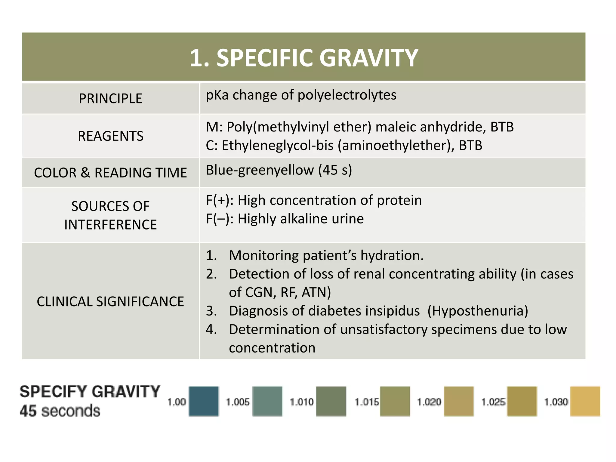
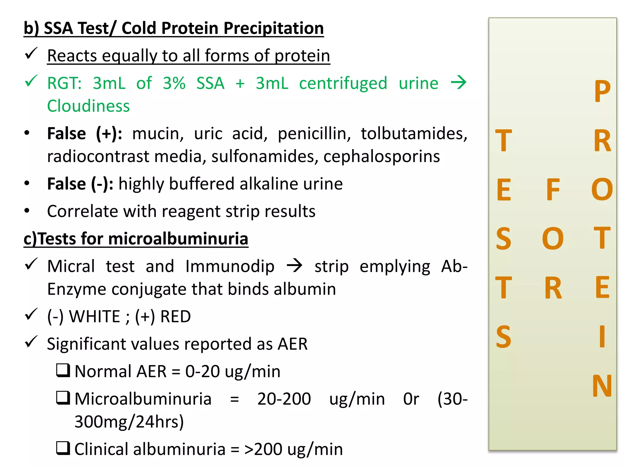
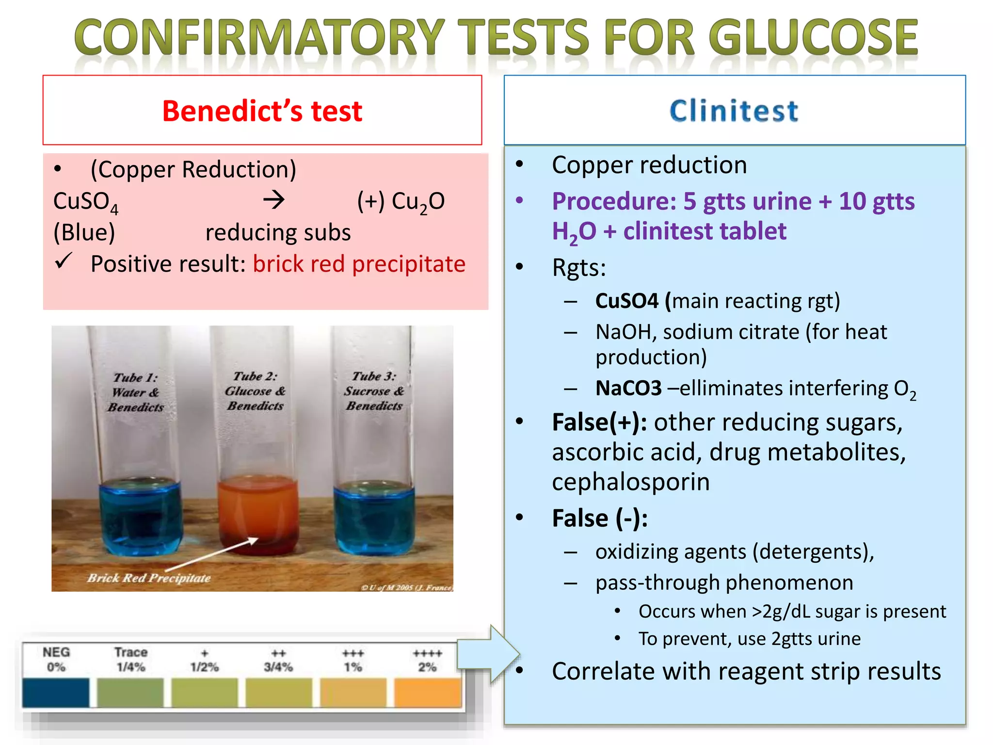
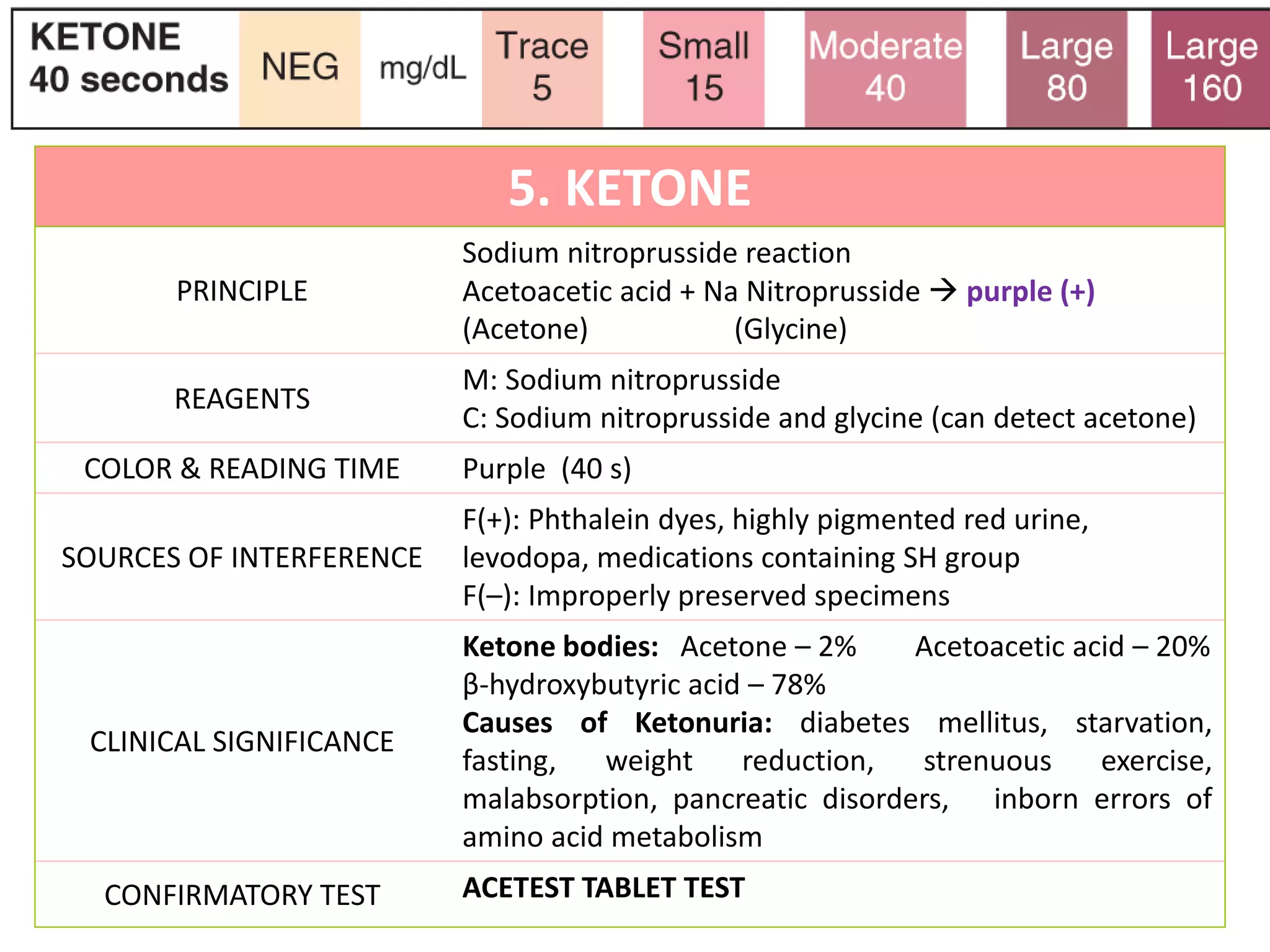
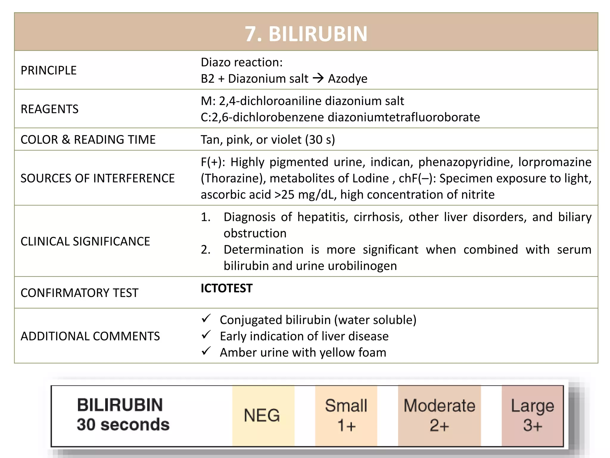
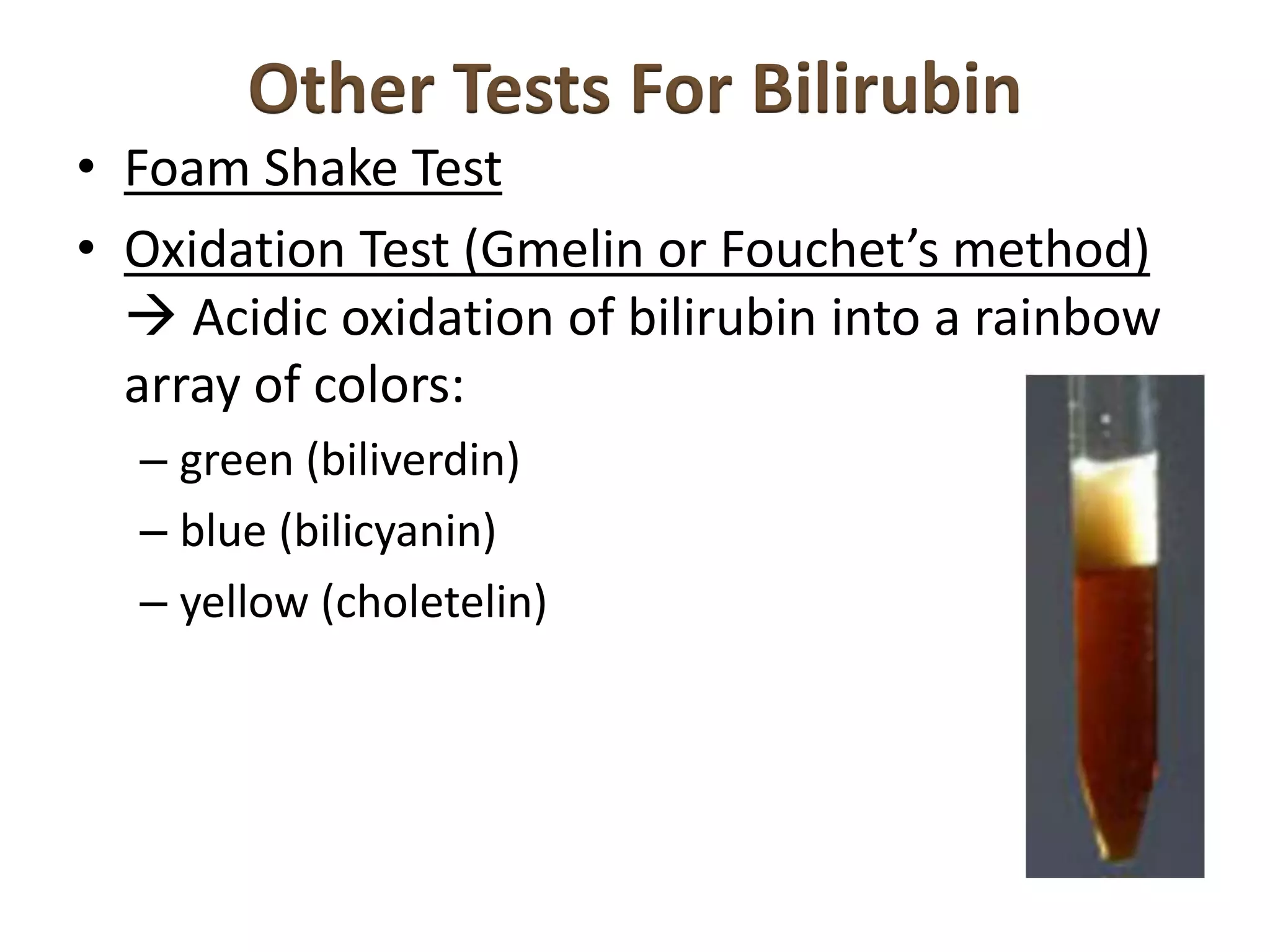
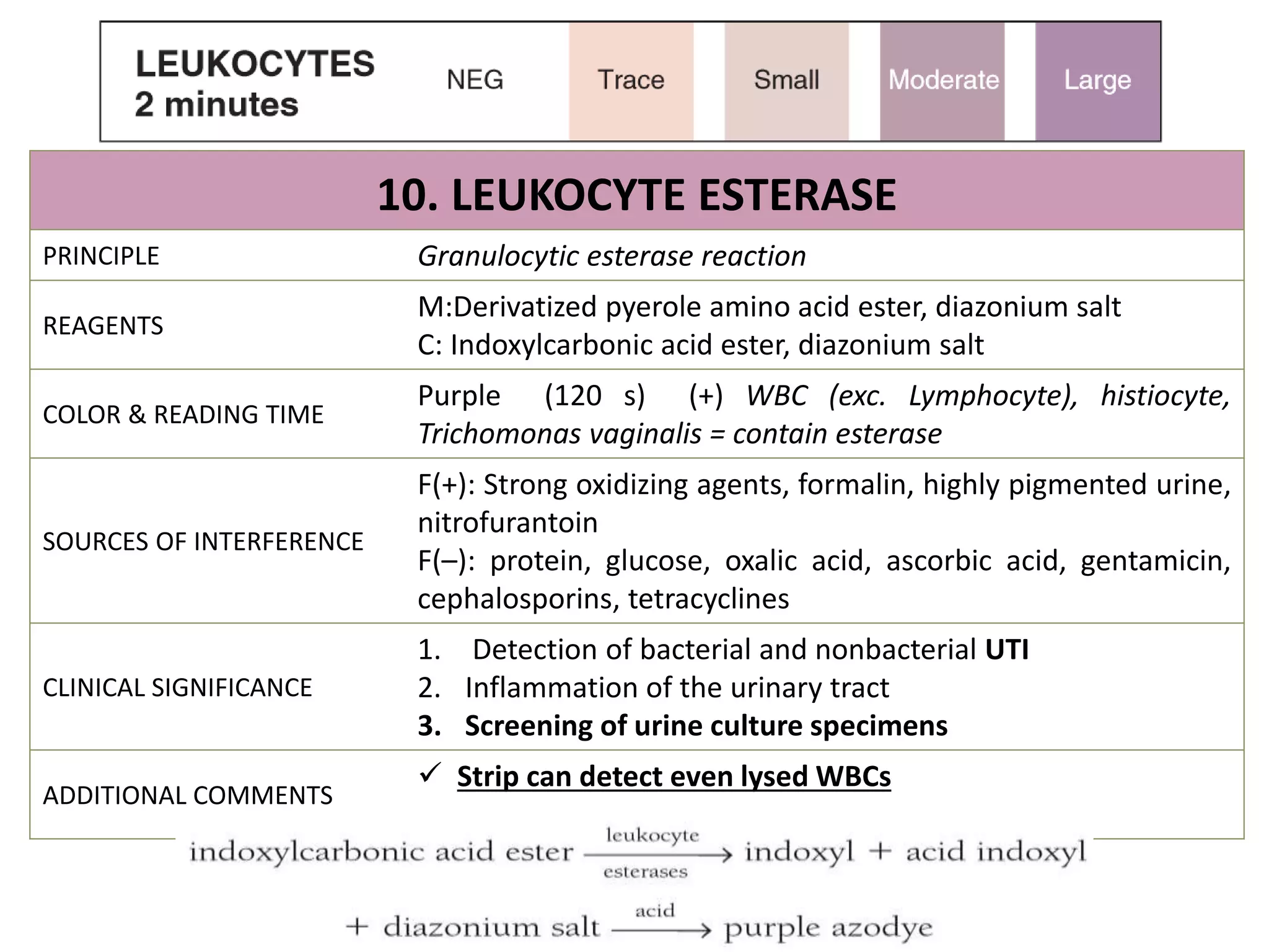
This document provides information on urine analysis tests, including specific gravity, pH, protein, glucose, ketone, blood, and bilirubin. It describes the principle, reagents, expected colors, sources of interference, and clinical significance of each test. The tests can be used to detect and monitor various conditions like diabetes, kidney disease, urinary tract infections, and more. Proper interpretation requires correlating the urine test results with the patient's symptoms and medical history.
An illustrative presentation on Microscopic examination of Urine for Medical, Dental, Pharmacology and Biotechnology students to facilitate easy- learning and self-study..
nephrotic syndrome is characterized by hypoalbuminemia, proteinuria, edema & hyperlipidemia. It is frequently found in children but also not uncommon in adults
Daily bilirubin production - 250-300mg%
85% heme moiety of aged RBC
5% RBC precursors destroyed in bone marrow ( ineffective
erythropoiesis),Catabolism of some heme proteins – myoglobin,
cytochrome, peroxidase
Biomarkers – in Toxicology and Clinical Researchsuruchi71088
A small presentation on growing use of Biomarkers in the field of toxicology and Clinical Research... basically use of various types of bio-markers and its role in drug development process...
It is 2 of the three major genera on which they are responsible for dermatophytosis, hence they are called Dermatophytes. you can see here their basic background and different morphological characteristics.
This presentation was derived from Microbiology 4 books.
Bailley's Scott
Mahon
Alcamo
Jaweitz
Title: Sense of Taste
Presenter: Dr. Faiza, Assistant Professor of Physiology
Qualifications:
MBBS (Best Graduate, AIMC Lahore)
FCPS Physiology
ICMT, CHPE, DHPE (STMU)
MPH (GC University, Faisalabad)
MBA (Virtual University of Pakistan)
Learning Objectives:
Describe the structure and function of taste buds.
Describe the relationship between the taste threshold and taste index of common substances.
Explain the chemical basis and signal transduction of taste perception for each type of primary taste sensation.
Recognize different abnormalities of taste perception and their causes.
Key Topics:
Significance of Taste Sensation:
Differentiation between pleasant and harmful food
Influence on behavior
Selection of food based on metabolic needs
Receptors of Taste:
Taste buds on the tongue
Influence of sense of smell, texture of food, and pain stimulation (e.g., by pepper)
Primary and Secondary Taste Sensations:
Primary taste sensations: Sweet, Sour, Salty, Bitter, Umami
Chemical basis and signal transduction mechanisms for each taste
Taste Threshold and Index:
Taste threshold values for Sweet (sucrose), Salty (NaCl), Sour (HCl), and Bitter (Quinine)
Taste index relationship: Inversely proportional to taste threshold
Taste Blindness:
Inability to taste certain substances, particularly thiourea compounds
Example: Phenylthiocarbamide
Structure and Function of Taste Buds:
Composition: Epithelial cells, Sustentacular/Supporting cells, Taste cells, Basal cells
Features: Taste pores, Taste hairs/microvilli, and Taste nerve fibers
Location of Taste Buds:
Found in papillae of the tongue (Fungiform, Circumvallate, Foliate)
Also present on the palate, tonsillar pillars, epiglottis, and proximal esophagus
Mechanism of Taste Stimulation:
Interaction of taste substances with receptors on microvilli
Signal transduction pathways for Umami, Sweet, Bitter, Sour, and Salty tastes
Taste Sensitivity and Adaptation:
Decrease in sensitivity with age
Rapid adaptation of taste sensation
Role of Saliva in Taste:
Dissolution of tastants to reach receptors
Washing away the stimulus
Taste Preferences and Aversions:
Mechanisms behind taste preference and aversion
Influence of receptors and neural pathways
Impact of Sensory Nerve Damage:
Degeneration of taste buds if the sensory nerve fiber is cut
Abnormalities of Taste Detection:
Conditions: Ageusia, Hypogeusia, Dysgeusia (parageusia)
Causes: Nerve damage, neurological disorders, infections, poor oral hygiene, adverse drug effects, deficiencies, aging, tobacco use, altered neurotransmitter levels
Neurotransmitters and Taste Threshold:
Effects of serotonin (5-HT) and norepinephrine (NE) on taste sensitivity
Supertasters:
25% of the population with heightened sensitivity to taste, especially bitterness
Increased number of fungiform papillae
Ozempic: Preoperative Management of Patients on GLP-1 Receptor Agonists Saeid Safari
Preoperative Management of Patients on GLP-1 Receptor Agonists like Ozempic and Semiglutide
ASA GUIDELINE
NYSORA Guideline
2 Case Reports of Gastric Ultrasound
TEST BANK for Operations Management, 14th Edition by William J. Stevenson, Ve...kevinkariuki227
TEST BANK for Operations Management, 14th Edition by William J. Stevenson, Verified Chapters 1 - 19, Complete Newest Version.pdf
Flu Vaccine Alert in Bangalore Karnatakaaddon Scans
As flu season approaches, health officials in Bangalore, Karnataka, are urging residents to get their flu vaccinations. The seasonal flu, while common, can lead to severe health complications, particularly for vulnerable populations such as young children, the elderly, and those with underlying health conditions.
Dr. Vidisha Kumari, a leading epidemiologist in Bangalore, emphasizes the importance of getting vaccinated. "The flu vaccine is our best defense against the influenza virus. It not only protects individuals but also helps prevent the spread of the virus in our communities," he says.
This year, the flu season is expected to coincide with a potential increase in other respiratory illnesses. The Karnataka Health Department has launched an awareness campaign highlighting the significance of flu vaccinations. They have set up multiple vaccination centers across Bangalore, making it convenient for residents to receive their shots.
To encourage widespread vaccination, the government is also collaborating with local schools, workplaces, and community centers to facilitate vaccination drives. Special attention is being given to ensuring that the vaccine is accessible to all, including marginalized communities who may have limited access to healthcare.
Residents are reminded that the flu vaccine is safe and effective. Common side effects are mild and may include soreness at the injection site, mild fever, or muscle aches. These side effects are generally short-lived and far less severe than the flu itself.
Healthcare providers are also stressing the importance of continuing COVID-19 precautions. Wearing masks, practicing good hand hygiene, and maintaining social distancing are still crucial, especially in crowded places.
Protect yourself and your loved ones by getting vaccinated. Together, we can help keep Bangalore healthy and safe this flu season. For more information on vaccination centers and schedules, residents can visit the Karnataka Health Department’s official website or follow their social media pages.
Stay informed, stay safe, and get your flu shot today!
HOT NEW PRODUCT! BIG SALES FAST SHIPPING NOW FROM CHINA!! EU KU DB BK substit...GL Anaacs
Contact us if you are interested:
Email / Skype : kefaya1771@gmail.com
Threema: PXHY5PDH
New BATCH Ku !!! MUCH IN DEMAND FAST SALE EVERY BATCH HAPPY GOOD EFFECT BIG BATCH !
Contact me on Threema or skype to start big business!!
Hot-sale products:
NEW HOT EUTYLONE WHITE CRYSTAL!!
5cl-adba precursor (semi finished )
5cl-adba raw materials
ADBB precursor (semi finished )
ADBB raw materials
APVP powder
5fadb/4f-adb
Jwh018 / Jwh210
Eutylone crystal
Protonitazene (hydrochloride) CAS: 119276-01-6
Flubrotizolam CAS: 57801-95-3
Metonitazene CAS: 14680-51-4
Payment terms: Western Union,MoneyGram,Bitcoin or USDT.
Deliver Time: Usually 7-15days
Shipping method: FedEx, TNT, DHL,UPS etc.Our deliveries are 100% safe, fast, reliable and discreet.
Samples will be sent for your evaluation!If you are interested in, please contact me, let's talk details.
We specializes in exporting high quality Research chemical, medical intermediate, Pharmaceutical chemicals and so on. Products are exported to USA, Canada, France, Korea, Japan,Russia, Southeast Asia and other countries.
Couples presenting to the infertility clinic- Do they really have infertility...Sujoy Dasgupta
Dr Sujoy Dasgupta presented the study on "Couples presenting to the infertility clinic- Do they really have infertility? – The unexplored stories of non-consummation" in the 13th Congress of the Asia Pacific Initiative on Reproduction (ASPIRE 2024) at Manila on 24 May, 2024.
Pulmonary Thromboembolism - etilogy, types, medical- Surgical and nursing man...VarunMahajani
Disruption of blood supply to lung alveoli due to blockage of one or more pulmonary blood vessels is called as Pulmonary thromboembolism. In this presentation we will discuss its causes, types and its management in depth.
The prostate is an exocrine gland of the male mammalian reproductive system
It is a walnut-sized gland that forms part of the male reproductive system and is located in front of the rectum and just below the urinary bladder
Function is to store and secrete a clear, slightly alkaline fluid that constitutes 10-30% of the volume of the seminal fluid that along with the spermatozoa, constitutes semen
A healthy human prostate measures (4cm-vertical, by 3cm-horizontal, 2cm ant-post ).
It surrounds the urethra just below the urinary bladder. It has anterior, median, posterior and two lateral lobes
It’s work is regulated by androgens which are responsible for male sex characteristics
Generalised disease of the prostate due to hormonal derangement which leads to non malignant enlargement of the gland (increase in the number of epithelial cells and stromal tissue)to cause compression of the urethra leading to symptoms (LUTS
These lecture slides, by Dr Sidra Arshad, offer a quick overview of physiological basis of a normal electrocardiogram.
Learning objectives:
1. Define an electrocardiogram (ECG) and electrocardiography
2. Describe how dipoles generated by the heart produce the waveforms of the ECG
3. Describe the components of a normal electrocardiogram of a typical bipolar leads (limb II)
4. Differentiate between intervals and segments
5. Enlist some common indications for obtaining an ECG
Study Resources:
1. Chapter 11, Guyton and Hall Textbook of Medical Physiology, 14th edition
2. Chapter 9, Human Physiology - From Cells to Systems, Lauralee Sherwood, 9th edition
3. Chapter 29, Ganong’s Review of Medical Physiology, 26th edition
4. Electrocardiogram, StatPearls - https://www.ncbi.nlm.nih.gov/books/NBK549803/
5. ECG in Medical Practice by ABM Abdullah, 4th edition
6. ECG Basics, http://www.nataliescasebook.com/tag/e-c-g-basics
2. 1. SPECIFIC GRAVITY
PRINCIPLE pKa change of polyelectrolytes
REAGENTS
M: Poly(methylvinyl ether) maleic anhydride, BTB
C: Ethyleneglycol-bis (aminoethylether), BTB
COLOR & READING TIME Blue-greenyellow (45 s)
SOURCES OF
INTERFERENCE
F(+): High concentration of protein
F(–): Highly alkaline urine
CLINICAL SIGNIFICANCE
1. Monitoring patient’s hydration.
2. Detection of loss of renal concentrating ability (in cases
of CGN, RF, ATN)
3. Diagnosis of diabetes insipidus (Hyposthenuria)
4. Determination of unsatisfactory specimens due to low
concentration
3.
4. 2. pH
PRINCIPLE Double indicator system
REAGENTS Methyl red; bromthymol blue
COLOR & READING TIME Red-yellow-blue (60 s)
SOURCES OF INTERFERENCE Runover from adjacent pads Old specimens
CLINICAL SIGNIFICANCE
1. Respiratory or metabolic acidosis/ alkalosis 2
2. Renal tubular acidosis
3. Renal calculi formation
4. Treatment of UTI
5. Precipitation/ identification of crystals
6. Determination of unsatisfactory specimen
ADDITIONAL NOTES
Important in the identification of crystals and
determination of unsatisfatory specimens
5. pH
• Normal pH:
– Random 4.5-8.0
– 1st Morning 5.0-6.0
• pH of 9 = unpreserved urine
• Alkaline tide occurs after meals due to withdrawal
of hydrogen ions for the purpose of secretion of HCl
• Causes of acid urine: emphysema, diabetes
mellitus, starvation, diarrhea, dehydration, acid
producing bacteria, high protein diet, medications ,
Cranberry juice ((tx for UTI)
• Causes of alkaline urine: hyperventilation, vomiting,
renal tubular acidosis, urease-producing bacteria,
vegetarian diet, old specimens , after meals
6. 3. PROTEIN
PRINCIPLE
Protein (Sorensen’s) error of indicator ; indicator is
sensitive only to ALBUMIN
REAGENTS
M: Tetrabromphenol blue
C: Tetrachlorophenol tetrabromosulfonphthalein
COLOR & READING TIME Blue-green (60 s)
SOURCES OF INTERFERENCE
F(+): Highly buffered alkaline urine, pigmented
specimens, chlorhexidine, phenazopyridine, QACs
(detergents), antiseptics, loss of buffer, high SG F(–
):Proteins other than albumin, high salt concentration,
microalbuminuria
CLINICAL SIGNIFICANCE
NORMAL: (<30 mg/dL or <150 mg/day; Negative rgt
strip test)
Degrees of proteinuria:
a. Mild – < 1.0 g/day
b. Moderate – 1.0-4.0 g/day
c. Heavy – > 4.0 g/day
ADDITIONAL COMMENT
MOST INDICATIVE OF RENAL DISEASE
WHITE FOAM IN URINE
7. PROTEIN
1. ALBUMIN Major serum protein found in the
urine
Normal values:
<10 mg/dL or 100mg/24 hrs (Strasinger)
<150mg/dL (Henry)
2. OTHER PROTEINS
a. Serum & Tubular microglobulins
b. Tamm-Horsfall protein (aka Uromodulin)
c. Protein derived from prostatic and vaginal
secretion
8. Types of proteinuria:
A. Pre-renal – caused by conditions that affect
plasma prior to its reaching the kidney
intravascular hemolysis
muscle injury
severe infection and inflammation;
Multiple Myeloma- proliferation of Igs-
producing plasma cells (BJP)
Test SPE, Immunofixation electrophoresis
Urine = precipitates at 40-600C (cloudy) and
dissolves at 1000C
9. Types of proteinuria:
B. Renal
A. Glomeruular Proteinuria
1. Diabetic nephropathy- decreased GFR, may lead to renal failure
» Indicator: microalbuminuria
2. Orthostatic Proteinuria/ Cadet/ Postural Proteinuria
Proteinuria when standing due to increased pressure to renal veins
3. Amyloidosis, Glomerulonephritis, Autoimmune Disorders, Toxic
Agents, Hypertension, Strenuous Exercise, Preeclampsia,
Dehydration,
B. Tubular Proteinuria– Reabsorption defective
Fanconi syndrome,
toxic agents
severe viral infections
ORTHOSTATIC PROTEINURIA CLINICAL PROTEINURIA
First Morning NEGATIVE POSITIVE
2 hours after standing POSITIVE POSITIVE
10. Types of proteinuria:
C. Post-renal – after
1. lower UTI;
2. injury or trauma;
3. menstrual contamination;
4. prostatic fluid;
5. spermatozoa;
6. vaginal secretions
11.
12. TESTS
FOR
PROTEIN
a) Heat and Acetic Acid Test
– Grading:
• diffused cloud (+1);
• granular cloud (+2);
• distinct flocculi (+3);
• large flocculi (+4)
b) SSA Test/ Cold Protein Precipitation
13. T
E
S
T
S
F
O
R
P
R
O
T
E
I
N
b) SSA Test/ Cold Protein Precipitation
Reacts equally to all forms of protein
RGT: 3mL of 3% SSA + 3mL centrifuged urine
Cloudiness
• False (+): mucin, uric acid, penicillin, tolbutamides,
radiocontrast media, sulfonamides, cephalosporins
• False (-): highly buffered alkaline urine
• Correlate with reagent strip results
c)Tests for microalbuminuria
Micral test and Immunodip strip emplying Ab-
Enzyme conjugate that binds albumin
(-) WHITE ; (+) RED
Significant values reported as AER
Normal AER = 0-20 ug/min
Microalbuminuria = 20-200 ug/min 0r (30-
300mg/24hrs)
Clinical albuminuria = >200 ug/min
14. 4. GLUCOSE
PRINCIPLE Glucose oxidase reaction / Double sequential enzyme rxn
REAGENTS
M: Glucose oxidase, peroxidase, KI
C: Glucose oxidase, peroxidase, TMB
COLOR & READING TIME M: greenbrown C: yellowgreen (30 s)
SOURCES OF INTERFERENCE
F(+): Oxidizing agents, detergents
F(–): Ascorbic acid, ketones, high SG, low temperatures,
improperly preserved specimens
CLINICAL SIGNIFICANCE
Types of Glucosuria:
a. Hyperglycemia-associated – diabetes mellitus,
endocrine disorders,( Cushing’s, Pheochromocytoma,
Acromegaly, Hyperthyroidism) pancreatic disorders,
CNS disorders, disturbance in metabolism, liver
disease, drugs, gestational diabetes mellitus
b. Renal-associated – renal tubular dysfunction, tubular
necrosis, Fanconi syndrome, osteomalacia, pregnancy
ADDITIONAL COMMENT Most frequently tested in urine; threshold substance
16. Non-specific test for reducing sugars
• Fructose (Levulose)
• Galactose
• Lactose (Glu-Gal)
• Pentose (Xylulose, Arabinose)
• Sucrose (Glu-Fru) – non reducing sugar,
thus negative
NOTE:
17. 5. KETONE
PRINCIPLE
Sodium nitroprusside reaction
Acetoacetic acid + Na Nitroprusside purple (+)
(Acetone) (Glycine)
REAGENTS
M: Sodium nitroprusside
C: Sodium nitroprusside and glycine (can detect acetone)
COLOR & READING TIME Purple (40 s)
SOURCES OF INTERFERENCE
F(+): Phthalein dyes, highly pigmented red urine,
levodopa, medications containing SH group
F(–): Improperly preserved specimens
CLINICAL SIGNIFICANCE
Ketone bodies: Acetone – 2% Acetoacetic acid – 20%
β-hydroxybutyric acid – 78%
Causes of Ketonuria: diabetes mellitus, starvation,
fasting, weight reduction, strenuous exercise,
malabsorption, pancreatic disorders, inborn errors of
amino acid metabolism
CONFIRMATORY TEST ACETEST TABLET TEST
18. • CONTAINS:
– Sodium nitroprusside
– Disodium phosphate (strong alkaline buffer)
– Lactose (helps to enhance the color)
• 10 times sensitive to diacetic acid than acetone
• Can detect in urine:
– 5–10 mg/dL of diacetic acid and 20–25 mg/dL of
acetone
19. 1. Place the tablet on a piece of clean, dry white paper.
2. Put one drop of urine, serum, plasma, or whole blood
directly on top of the tablet.
3. For urine, compare the color of the tablet with the color
chart at 30 seconds. For serum or plasma, compare the color
after 2 minutes. For whole blood, remove the clotted blood
from the tablet after 10 minutes and compare the color of
the tablet with the chart.
Note: for serum, plasma and whole blood, the loest limit of
detection s 10mg of diacetic acid per 100mL
5-10mg/dL
Diacetic acid
30-40mg/dL
Diacetic acid
80-100mg/dL
Diacetic acid
21. 6. BLOOD
PRINCIPLE
Pseudoperoxidase activity of heme
Hgb
H2O2 + Chromogen oxidized chromogen + H2O
Pseudoperoxidase
REAGENTS
M:Diisopropylbenzene dihydroperoxide TMB
C: 2,5-dimethyl 2,5dihydroperoxyhexane TMB
COLOR & READING TIME
(-) yellow ; (+)Blue-green (60 s)
Uniformed green/blue color= Hgb/Mgb
Speckled/Spotted = Hematuria (intact RBCs)
SOURCES OF INTERFERENCE
F(+): Strong oxidizing agents, bacterial peroxidases, menstrual
contamination
F(–): High SG, crenated cells, formalin, captopril, nitrite, ascorbic
acid, unmixed specimen
CLINICAL SIGNIFICANCE
a. Hematuria
b. Hemoglobinuria
c. Myoglobinuria
CONFIRMATORY TEST MICROSCOPIC ANALYSIS
22. HEMATURIA HEMOGLOBINURIA MYOGLOBINURIA
Cloudy red urine Clear red urine Clear red urine/ reddish
brown/tea-colored
Seen in:
Glomerulonephritis
Renal calculi
Microscopic: INTACT RBCS
Seen in:
Intravascular hemolysis
Transfusion rxns
Hemolytic Anemia
Seen in: Rhabdomyolysis
Heme portion of the
myoglobin is toxic to the
renal tubules
HEMOGLOBIN vs MYOGLOBIN
TEST HEMOGLOBIN MYOGLOBIN
1. Plasma Examination Red/Pink plasma
Decrease Haptoglobin
CK Slightly increased
LD 1 & 2 increased
Pale yellow plasma
CK Markedly increased
and Aldolase increased
LD 4 & 5 increased
2. Blondheim’s test
(Ammonium sulfate)
Proc: Urine + 2.8 NH4Sulfate (80%
satd) filter centri test
supernatant for blood with rgt strip
Precipitated at the bottom
Supernatant test (-) for blood
Not precipitated
Supernatant test (+) blood
23. OTHER TESTS FOR URINE BLOOD
• Enzyme assays
• Absorption Spectrophotometry
• Immunodiffusion Technique
• Electrophoresis
24. 7. BILIRUBIN
PRINCIPLE
Diazo reaction:
B2 + Diazonium salt Azodye
REAGENTS
M: 2,4-dichloroaniline diazonium salt
C:2,6-dichlorobenzene diazoniumtetrafluoroborate
COLOR & READING TIME Tan, pink, or violet (30 s)
SOURCES OF INTERFERENCE
F(+): Highly pigmented urine, indican, phenazopyridine, lorpromazine
(Thorazine), metabolites of Lodine , chF(–): Specimen exposure to light,
ascorbic acid >25 mg/dL, high concentration of nitrite
CLINICAL SIGNIFICANCE
1. Diagnosis of hepatitis, cirrhosis, other liver disorders, and biliary
obstruction
2. Determination is more significant when combined with serum
bilirubin and urine urobilinogen
CONFIRMATORY TEST ICTOTEST
ADDITIONAL COMMENTS
Conjugated bilirubin (water soluble)
Early indication of liver disease
Amber urine with yellow foam
25. Procedure:
1. Place five drops of urine on
one square of the special test
mat supplied with Ictotest.
2. Place a tablet in the center of
the moistened area.
3. Flow two drops of water onto
the tablet so that the water
runs off of the tablet and onto
the mat.
4. Observe the color of the mat
around the tablet at the end
of 30 seconds. If a blue or
purple color develops, the test
is positive.
able to detect as little as
0.05 mg/dL bilirubin
Contains:
P-nitrobenzene-diazonium
p-toluenesulfonate
SSA (provide acidity and act
with Sodium carbonate to
provide effervescence
Sodium carbonate
Boric acid
26. Other Tests For Bilirubin
• Foam Shake Test
• Oxidation Test (Gmelin or Fouchet’s method)
Acidic oxidation of bilirubin into a rainbow
array of colors:
– green (biliverdin)
– blue (bilicyanin)
– yellow (choletelin)
27. 8. UROBILINOGEN
PRINCIPLE Ehrlich reaction : UBG +PDAB (+) RED
REAGENTS
M: paradiethylaminobenzaldehyde
C: 4-methoxybenzene diazonium tetrafluoroborate
COLOR & READING TIME Red (60 s)
SOURCES OF INTERFERENCE
F(+): PBG, indican, procaine, p-aminosalicylic acid,
sulfonamides, methyldopa, chlorpromazine, pigmented urine
F(–): Old specimens, formalin, high concentration of nitrite
CLINICAL SIGNIFICANCE
1. Only a small amount is normally found in urine.
2. Useful for the early detection of liver disease and for the
diagnosis of hemolytic disorders, hepatitis, cirrhosis, and
carcinoma
3. Absence in urine may indicate biliary obstruction
TEST USED TO DIFFERENTIATE
UROBILINOGEN TO
PORPHOBILINOGEN
WATSON-SCWARTZ DIFFERENTIATION TEST
ADDITIONAL COMMENTS
(< 1.0 mg/dL or <1.0 Ehrlich unit)
Specimen: Afternoon urine (2-4PM due to alkaline tide)
28. 2. WATSON-SCHWARTZ TEST
• Uses extraction with organic solvents Chloroform and Butanol
• Sodium acetate enhances reaction
1. Ehrlich’s Tube Test
• Reagent: p-dimethylaminobenzaldehyde
• (+) Result: cherry red color
29. • INVERSE EHRLICH REACTION
• Rapid screening test for Porphobilinogen
(>2 mg/dL)
Procedure:
2 gtts urine + 2mL Hoesch rgt
( Ehrlich’s rgt in 6M or 6N HCl)
• Immediate development of a cherry-red color at
the top of the mixture indicates a positive result
30.
31. 9. NITRITE
PRINCIPLE Greiss reaction
REAGENTS
M: p-arsanilic acid, tetrahydrobenzoquinolinol
C: sulfanilamide, 3hydroxy- 1,2,3,4- tetrahydro- 7,8-
benzoquinoline
COLOR & READING TIME
Uniform Pink (+) = 100,000 org/mL
not pink spots/edge (-) (60 s)
SOURCES OF INTERFERENCE
F(+): Old specimen, highly pigmented urine
F(–):Non-reductase-containing bacteria, lack of urinary nitrate,
insufficient contact time between bacteria and nitrate,
bacteria converting nitrite to nitrogen, antibiotics, ascorbic
acid, high SG
CLINICAL SIGNIFICANCE
1. Diagnosis of cystitis and pyelonephritis
2. evaluation of antibiotic therapy
3. Monitoring of patients at high risk for UTI
4. Screening of urine culture specimens
ADDITIONAL COMMENTS
Rapid screening test for UTI/bacteriuria
Specimen: 1st morning or 4hr urine
32. 10. LEUKOCYTE ESTERASE
PRINCIPLE Granulocytic esterase reaction
REAGENTS
M:Derivatized pyerole amino acid ester, diazonium salt
C: Indoxylcarbonic acid ester, diazonium salt
COLOR & READING TIME
Purple (120 s) (+) WBC (exc. Lymphocyte), histiocyte,
Trichomonas vaginalis = contain esterase
SOURCES OF INTERFERENCE
F(+): Strong oxidizing agents, formalin, highly pigmented urine,
nitrofurantoin
F(–): protein, glucose, oxalic acid, ascorbic acid, gentamicin,
cephalosporins, tetracyclines
CLINICAL SIGNIFICANCE
1. Detection of bacterial and nonbacterial UTI
2. Inflammation of the urinary tract
3. Screening of urine culture specimens
ADDITIONAL COMMENTS
Strip can detect even lysed WBCs