SlideShare a Scribd company logo
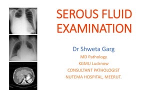
SEROUS FLUID
EXAMINATION
Dr Shweta Garg
MD Pathology
KGMU Lucknow
CONSULTANT PATHOLOGIST
NUTEMA HOSPITAL, MEERUT.
LEARNING OBJECTIVE
• NORMAL FORMATION OF SEROUS FLUID
• DIFFERENTIATE BETWEEN TRANSUDATE AND
EXUDATE
• VARIOUS OTHER ROUTINE & SPECIAL TEST &
THEIR CLINICAL & DIAGNOSTIC SIGNIFICANCE
GENERAL CONSIDERATION
• Serous fluids – secreted by serous membrane (Parietal and
visceral), lining the close cavity of body
• These are named as per location
• Pleural
• Pericardial
• Peritoneal
• Their function is to provide lubrication between two
membranes
FLUID FORMATION
- These are extravascular fluid
collected in intercellular spaces
(body cavity) and come from
vascular space.
- It is produced by exertion of
hydrostatic pressure and oncotic
pressure and small amount is
absorbed by lypmphatics
HOW DOES EFFUSION OCCUR ?
• Increase venous pressure i.e Hydrostatic pressure
• Greater exit of fluid from the vascular system than it is
absorbed
• Capillary permeability does not change
• Fluid resembles like normal tissue fluid – few cells & very low
protein
• Congestive heart failure, salt & fluid retention
• Increase capillary permeability
• Due to inflammation or toxic damage to capillaries –
microbial infection
• Contain high concentration of protein
HOW DOES EFFUSION OCCUR ?
• Decrease in plasma colloidal pressure
• In cases of Hypoproteinaemia – like nephrotic Syndrome
• Hepatic cirrhosis
• Malnutrition, Protein losing enteropathy
• Interference with lymphatic flow
• Due to obstruction of lymphatic flow – Filaria, Cancer,
Scar tissue, thoracic duct injury
• Contain high concentration of protein & lipids
TRANSUDATE & EXUDATE
• Transudate is fluid buildup caused by systemic conditions
that alter the pressure in blood vessels causing fluid to leave
the vascular system.
• Exudate is fluid build up caused by tissue leakage due to
inflammation or local cellular damage.
LIGHT’S CRITERIA
The fluid is exudate if one of the following Light’s criteria is
present:
1. Effusion protein/ serum protein ratio > 0.5
2. Effusion lactate dehydrogenase( LDH) / serum LDH ratio
>0.6
3. Effusion LDH level greater than two thirds the upper limit
of reference range of LDH
GENERAL LAB TESTING
• Specimen collection and handling
• Examination of Fluid
• Physical examination
• Microscopic examination
• Chemical examination
• Microbiological and serological test
• Ancillary test
SPECIMEN COLLECTION AND HANDLING
• Pleural fluid: Thoracocentesis is indicated for therapeutic
purposes in patients with massive symptomatic effusion.
• Peritoneal fluid: Diagnostic Paracentesis is performed in most
pts of ascites. Min of 30 ml is required.
• Pericardial fluid:
• Sample is obtained by Pericardiocentesis in a wide mouth
universal container.
• Post test the samples are stored at 4-8 ֯C for 48 hours.
COLLECTION OF SAMPLE
• To be collected in three tubes
• EDTA for Haematolgy
• Plain for Biochemistry
• Sterile heparinised / Plain for microbiology & Cytology
• It should be examined as early as possible to avoid chemical
change, bacterial growth & cellular disintegration
• Remaining sample can be stored at 2 – 4 C for further
ancillary testing
PROCEDURAL STEPS
Physical examination:
• Volume
• Color
• Appearance
• Presence/absence of coagulum
Coagulum formation occurs due to substantial
inflammatory reaction and presence of fibrinogen due to
capillary wall damage
MICROSCOPIC EXAMINATION
• Done for routine TLC & DLC and cytological purpose
• For TLC, mix the specimen carefully, if clear, use undiluted.
• If blood- tinged prepare 1:2 dilutions with diluting fluid
• If cloudy, prepare 1:20 dilution with diluting fluid or normal saline
(composition of Turk solution: Glacial acetic acid 4 ml, methylene
blue solution 10 drops, & distilled water to make 200 ml).
• Count the cells in the corner 4 squares of neubauer chamber.
• Calculation: TLC/cumm= n ×D/V
where “n”= no. of cells in four corner squares
“D”= dilution factor
“V”= volume (vol. of 4 wbc chamber)
NEUBAUER CHAMBER
• Now the fluid is centrifuged at 1500 rpm for 5 minutes &
sediment is used to prepare smears for DLC, malignant cell
& cytology.
• Smears are stained with Leishman, Giemsa & PAP stain.
• Limitations: Counts should be performed as soon as
possible.
• Specimen should be stored at 2-8֯ C for 48 hrs
FLUID CYTOLOGY
Chemical Examination
• Following chemical test are preformed
• Protein & Albumin in some
• Glucose
• LDH
• Others –
• Triglyceride to rule out chylous effusion
• Amylase – to rule out pancreatitis or esophageal rupture
• ADA – helps in making diagnosis of tuberculosis ( Usually >
40 IU/L in tubercular effusion & ascites)
APPROACH TO PLEURAL
FLUID EXAMINATION
HAEMATOLOGY TEST
• Increase Neutrophil
• Bacterial infection, Pancreatitis, Pulmonary infarction
• Increase Lymphocytes
• Tuberculosis, Viral infection, Autoimmune disorders,
Malignancy
• Increase Eosinophils > 10 %
• Trauma introducing air and blood, allergic reaction,
parasite / fungal infection, pulmonary infarction, Drugs
Eosinophilic effusion
PERICADIAL FLUID
EXAMINATION
PERICADIAL FLUID EXAMINATION
• Normally 10 -50 ml of pericardial fluid
• Straw coloured
• Transudate (Pale yellow, clear) – Hypothyroidism, Uraemia,
Autoimmune disorder
• Exuduative (Turbid ) – Infection , Malignancy
• Haemorrhagic – Trauma, Uraemia, Tubercular
• Milky – Chylous and Pseudochylous
PERICADIAL FLUID EXAMINATION
• CHEMICAL EXAMINATION
• Protein – little use
• Glucose < 40 mg/dl in Tuberculosis, bacterial and
Rheumatic diseases and malignancy
• ADA – increase in Tuberculosis
• MICROSCOPY
• WBC > 1000 cells/cu mm in Bacterial infection and TB
• Malignant cell – Metastatic from lung and breast
PERICADIAL FLUID
• MICROBIOLOGICAL EXAMINATION
• Gram Stain – positive in 50% bacterial infections
• Culture – Positive in 80% of bacterial infections
• AFB – Positive in 50% of Tubercular cases
ASCITIC FLUID
EXAMINATION
PERITONEAL FLUID
• Peritoneal cavity normally contain upto 50ml of clear straw
colored fluid
• Patient with peritoneal effusion is said to have ascitis and it
is called ascitic fluid
• The procedure of collecting the ascitic fluid is called
abdominal paracentesis
• Indications: ascites of unknown etiology, acute abdominal
pain, post operative hypotension, intra abdominal
hemorrhage etc
• Specimen collected into tubes same as for other fluid
CAUSES OF ASCITES
PHYSICAL EXAMINATION
• Colour and appearance: normally clear and pale yellow
• Turbid: appendicitis, pancreatitis etc.
• Green: intestinal perforation, cholecystitis
• Milky: nephrotic syndrome, carcinoma, parasitic infection
• Bloody: hemorrhagic pacreatitis, reptured spleen or liver
• Examine for clot formation
MICROSCPIC EXAMINATION
• Total leukocyte useful in spontaneous bacterial peritonitis
(SBP)
• Approximately 90% of (SBP) have leukocyte count
>500/cumm and over 50% neutrophiles
• Increase lymphocyte – Favours TB
• Eosinophilia >10% most commonly associates with CHF,
vasculitis, lymphoma and ruptured hydatid cyst
CHEMICAL EXAMINATION
• Estimation of glucose has little value
• Decreased in peritonitis ,malignancy
• Estimation of amylase - Increased in acute pancreatitis,
(more than 3 times of serum values) , Gi perforation
• Estimation of ALP - Elevated in intestinal perforation
• Estimation of LDH – Increase in Malignancy
CHEMICAL EXAMINATION
• Estimation Urea and Creatinine – Traumatic rupture of
urinary tract or in renal transplant surgery (ureteric
dehiscence)
• ADA – increase more than 40 unit/liter in TB peritonitis
• Tumour marker
• Presence of CA 125 antigen with a negative CEA suggests
the source is from ovaries, fallopian tube, or
endometrium
• Presence of CEA suggests source is gastrointestinal
MICROBIOLOGY TESTS
• Gram stains and aerobic and anaerobic cultures
• Aerobic cultures : inoculate blood culture in blood culture
bottles , sensitivity increases from 50 % to 80% if done with
BACTEC vis a vis conventional
• Acid fast smear , adenosine deaminase and culture for TB
THANK YOU
Examination of pleural fluid
• Ascites • Diagnosis: • established with a combination of a physical
examination & an imaging test (USG). • Approx 1500 mL of fluid had
to be present for flank dullness to be detected • lesser degrees of
ascites can be missed. • Ultrasonography can be helpful when the
physical examination is not definitive
• Causes of Ascites • Ascites can be classified based on the underlying
pathophysiology: • Portal hypertension – Cirrhosis – Alcoholic
hepatitis – Acute liver – Hepatic veno-occlusive disease (eg, Budd-
Chiari syndrome) – Heart failure – Constrictive pericarditis –
Hemodialysis-associated ascites (nephrogenic ascites)
• Causes of Ascites • Hypoalbuminemia – Nephrotic syndrome –
Protein-losing enteropathy – Severe malnutrition • Peritoneal disease
– Malignant ascites (eg, ovarian cancer, mesothelioma) – Infectious
peritonitis (eg, tuberculosis or fungal infection) – Eosinophilic
gastroenteritis – Starch granulomatous peritonitis – Peritoneal dialysis
• Causes of Ascites • Other etiologies – Chylous ascites – Pancreatic
ascites (disrupted pancreatic duct) – Myxedema – Hemoperitoneum
• International Ascites Club Grading system • Grade 1 – Mild ascites
detectable only by ultrasound examination • Grade 2 – Moderate
ascites manifested by moderate symmetrical distension of the
abdomen • Grade 3 – Large or gross ascites with marked abdominal
distension
• Abdominal Paracentesis • Most efficient way to confirm the presence
of ascites, diagnose its cause, and determine if the fluid is infected. •
Safe procedure, with an extremely low incidence of serious
complications despite the coagulopathy that is usually present in
patients with cirrhosis. • Coagulation parameters beyond which
paracentesis should be avoided. • There are no data-supported
however, patients with clinically evident fibrinolysis or disseminated
intravascular coagulation should not undergo paracentesis.
TESTS PERFORMED ON ASCITIC FLUID
• Routine tests
• Cell count and differential
• Albumin concentration
• Total protein concentration
• Culture in blood culture bottles
• Optional tests
• Glucose concentration
• LDH concentration
• Gram stain
• Amylase concentration
• Other tests •
• Tuberculosis smear and culture •
• Cytology •
• Triglyceride concentration •
• Bilirubin concentration
• Cell count and differential • The cell count with differential is the
single most helpful test performed on ascitic fluid to evaluate for
infection. • Polymorphonuclear count ≥ 250/mm3 – spontaneous
bacterial peritonitis. • In bloody ascites: – one neutrophil should be
subtracted from the absolute neutrophil count for every 250 red cells
to yield the "corrected neutrophil count“
SERUM-TO-ASCITES ALBUMIN GRADIENT
• • The serum-to-ascites albumin gradient (SAAG) accurately identifies
the presence of portal hypertension and is more useful than the
proteinbased exudate/transudate concept. • SAAG – serum albumin
value - ascitic fluid albumin – (obtained on the same day). • SAAG ≥
1.1 g/dL (11 g/L) – Indicates portal hypertension – (Budd-Chiari
syndrome, heart failure, or liver cirrhosis) • SAAG
• Sending Cultures • Bacterial cultures of ascitic fluid should be sent
from patients with – new onset ascites • admitted with ascites • Who
deteriorate with – Fever, – Abdominal pain – Azotemia, – Acidosis –
confusion
• Protein, Glucose, LDH • Protein — Ascitic fluid had been classified as
an exudate if the total protein concentration is ≥2.5 or 3 g/dL and a
transudate if it is below this cut-off. However, the exudate/transudate
system of ascitic fluid classification has been replaced by the SAAG. •
Measurement of total protein, glucose, and lactate dehydrogenase
(LDH) in ascites may also be of value in distinguishing SBP from gut
perforation into ascites • Patients with ascitic fluid that has a
neutrophil count ≥250 cells/mm3 and meets two out of the following
three criteria are unlikely to have SBP and warrant immediate
evaluation to determine if gut perforation into ascites has occurred. –
Total protein >1 g/dL – Glucose
TESTS FOR TUBERCULOUS PERITONITIS
• Direct smear - 0 to 2% sensitivity in detecting Mycobacteria
• Culture - When one litre of fluid is cultured, sensitivity for
Mycobacteria 62 to 83%
• Fluid for PCR for tuberculosis
• Cell count - Tuberculous peritonitis can mimic the culture-negative
variant of SBP, but lymphocyte cells usually predominate in
tuberculosis
• Adenosine deaminase
• Adenosine deaminase activity of ascitic fluid has been proposed as a
useful non-culture method of detecting tuberculous peritonitis;
however, patients with cirrhosis and tuberculous peritonitis usually
have falsely low values .
serous fluid Dr shweta [Autosaved].pptx

More Related Content

What's hot

Quality Assurance in Hematology laboratory
Quality Assurance in Hematology laboratoryQuality Assurance in Hematology laboratory
Quality Assurance in Hematology laboratory
Mohammed Abdalla Elhassan
 
special and routine stains in haematology 1
special and routine stains in haematology 1special and routine stains in haematology 1
special and routine stains in haematology 1
Dr.SHAHID Raza
 
Buffy coat
Buffy coatBuffy coat
Buffy coat
kabitachatterjee
 
Romanowsky stains
Romanowsky stainsRomanowsky stains
Preparation and staining of peripheral blood smear
Preparation and staining of peripheral blood smearPreparation and staining of peripheral blood smear
Preparation and staining of peripheral blood smear
MLT LECTURES BY TANVEER TARA
 
Pas staining
Pas staining Pas staining
Blood component preparation blood banking
Blood component preparation blood bankingBlood component preparation blood banking
Blood component preparation blood banking
Appy Akshay Agarwal
 
Compatability testing
Compatability testingCompatability testing
Compatability testing
SUNIL KUMAR PEDDANA
 
Automated cell counters: principle and types
Automated cell counters: principle and typesAutomated cell counters: principle and types
Automated cell counters: principle and types
Sivaranjini N
 
Automation in hematology part 1
Automation in hematology part 1Automation in hematology part 1
Automation in hematology part 1
Dr. Varughese George
 
Frozen Section Basics
Frozen Section BasicsFrozen Section Basics
Frozen Section Basics
FARUQ BSMMU R9
 
Synovial fluid
Synovial fluidSynovial fluid
Synovial fluid
dattasrisaila
 
Final ppt sickle cell
Final ppt sickle cellFinal ppt sickle cell
Final ppt sickle cell
Darlasrinivasarao Srinu
 
Ascitic fluid examination
Ascitic fluid examinationAscitic fluid examination
AUTOMATION IN HEMATOLOGY
AUTOMATION IN HEMATOLOGYAUTOMATION IN HEMATOLOGY
AUTOMATION IN HEMATOLOGY
Dr. Ajit Surya Singh
 
General criteria of malignancy
General criteria of malignancyGeneral criteria of malignancy
General criteria of malignancy
san yu maung
 
Automation in hematology
Automation in hematologyAutomation in hematology
Automation in hematology
Dr Siddartha
 
Cross matching
Cross matchingCross matching
Cross matching
Mitalisingh30
 
CSF - Cerebrospinal fluid examination - from tapping to pathological diagnosis
CSF - Cerebrospinal fluid examination - from tapping to pathological diagnosisCSF - Cerebrospinal fluid examination - from tapping to pathological diagnosis
CSF - Cerebrospinal fluid examination - from tapping to pathological diagnosis
Ashish Jawarkar
 
HistoPATHOLOGY
HistoPATHOLOGYHistoPATHOLOGY
HistoPATHOLOGY
Musa Khan
 

What's hot (20)

Quality Assurance in Hematology laboratory
Quality Assurance in Hematology laboratoryQuality Assurance in Hematology laboratory
Quality Assurance in Hematology laboratory
 
special and routine stains in haematology 1
special and routine stains in haematology 1special and routine stains in haematology 1
special and routine stains in haematology 1
 
Buffy coat
Buffy coatBuffy coat
Buffy coat
 
Romanowsky stains
Romanowsky stainsRomanowsky stains
Romanowsky stains
 
Preparation and staining of peripheral blood smear
Preparation and staining of peripheral blood smearPreparation and staining of peripheral blood smear
Preparation and staining of peripheral blood smear
 
Pas staining
Pas staining Pas staining
Pas staining
 
Blood component preparation blood banking
Blood component preparation blood bankingBlood component preparation blood banking
Blood component preparation blood banking
 
Compatability testing
Compatability testingCompatability testing
Compatability testing
 
Automated cell counters: principle and types
Automated cell counters: principle and typesAutomated cell counters: principle and types
Automated cell counters: principle and types
 
Automation in hematology part 1
Automation in hematology part 1Automation in hematology part 1
Automation in hematology part 1
 
Frozen Section Basics
Frozen Section BasicsFrozen Section Basics
Frozen Section Basics
 
Synovial fluid
Synovial fluidSynovial fluid
Synovial fluid
 
Final ppt sickle cell
Final ppt sickle cellFinal ppt sickle cell
Final ppt sickle cell
 
Ascitic fluid examination
Ascitic fluid examinationAscitic fluid examination
Ascitic fluid examination
 
AUTOMATION IN HEMATOLOGY
AUTOMATION IN HEMATOLOGYAUTOMATION IN HEMATOLOGY
AUTOMATION IN HEMATOLOGY
 
General criteria of malignancy
General criteria of malignancyGeneral criteria of malignancy
General criteria of malignancy
 
Automation in hematology
Automation in hematologyAutomation in hematology
Automation in hematology
 
Cross matching
Cross matchingCross matching
Cross matching
 
CSF - Cerebrospinal fluid examination - from tapping to pathological diagnosis
CSF - Cerebrospinal fluid examination - from tapping to pathological diagnosisCSF - Cerebrospinal fluid examination - from tapping to pathological diagnosis
CSF - Cerebrospinal fluid examination - from tapping to pathological diagnosis
 
HistoPATHOLOGY
HistoPATHOLOGYHistoPATHOLOGY
HistoPATHOLOGY
 

Similar to serous fluid Dr shweta [Autosaved].pptx

body fluids analysis - corrected - Copy.pptx
body fluids analysis - corrected - Copy.pptxbody fluids analysis - corrected - Copy.pptx
body fluids analysis - corrected - Copy.pptx
SURAJ PANCHAL
 
Fluid cytology in serous cavity effusions
Fluid cytology in serous cavity effusionsFluid cytology in serous cavity effusions
Fluid cytology in serous cavity effusions
tashagarwal
 
Approach to patients with pleural effusion (1).pptx
Approach to patients with pleural effusion (1).pptxApproach to patients with pleural effusion (1).pptx
Approach to patients with pleural effusion (1).pptx
aashishkoirala6
 
Pleural effusion
Pleural effusionPleural effusion
Pleural effusion
Greeshma Mandali
 
asciticfluidexamination-200420102558 (2).pdf
asciticfluidexamination-200420102558 (2).pdfasciticfluidexamination-200420102558 (2).pdf
asciticfluidexamination-200420102558 (2).pdf
Rubab161509
 
Pleural Effusion By Akhtar Totakhail.pptx
Pleural Effusion By Akhtar Totakhail.pptxPleural Effusion By Akhtar Totakhail.pptx
Pleural Effusion By Akhtar Totakhail.pptx
DrAkhtarMohammadTota
 
Hd for residents (2) copy
Hd for residents (2)   copyHd for residents (2)   copy
Hd for residents (2) copy
FarragBahbah
 
Approach To a Patient with Ascitis
Approach To a Patient with AscitisApproach To a Patient with Ascitis
Approach To a Patient with Ascitis
Dr. Ahammad Shamir Shawrov
 
Pleural Effusion for Undergraduates
Pleural Effusion for UndergraduatesPleural Effusion for Undergraduates
Pleural Effusion for Undergraduates
Sesha Sai
 
Ascitic fluid analysis
Ascitic fluid analysis Ascitic fluid analysis
Ascitic fluid analysis
Muhammad Asim Rana
 
1a hematology slides
1a hematology slides1a hematology slides
1a hematology slides
Samantha Watson
 
Pleural effusion
Pleural effusionPleural effusion
Pleural effusion
ahmad tanweer
 
PLEURAL EFFUSION BY Mr. AKRAM KHAN
PLEURAL EFFUSION BY Mr. AKRAM KHANPLEURAL EFFUSION BY Mr. AKRAM KHAN
PLEURAL EFFUSION BY Mr. AKRAM KHAN
Akram Khan
 
CLD Diagnosis and management………………..pptx
CLD Diagnosis and management………………..pptxCLD Diagnosis and management………………..pptx
CLD Diagnosis and management………………..pptx
hamid15abass
 
URINE & STOOL EXAMINATION.pptx
URINE & STOOL EXAMINATION.pptxURINE & STOOL EXAMINATION.pptx
URINE & STOOL EXAMINATION.pptx
amanjotkaursidhu
 
Upper gi bleeding
Upper gi bleedingUpper gi bleeding
Upper gi bleeding
niteshpansari
 
uppergibleeding-150402032909-conversion-gate01.pdf
uppergibleeding-150402032909-conversion-gate01.pdfuppergibleeding-150402032909-conversion-gate01.pdf
uppergibleeding-150402032909-conversion-gate01.pdf
DakaneMaalim
 
Management of ascites(3).pptx
Management of ascites(3).pptxManagement of ascites(3).pptx
Management of ascites(3).pptx
Kemi Adaramola
 
Uppergibleeding 150402032909-conversion-gate01-converted
Uppergibleeding 150402032909-conversion-gate01-convertedUppergibleeding 150402032909-conversion-gate01-converted
Uppergibleeding 150402032909-conversion-gate01-converted
Mohammad Rehan
 
Upper GI bleeding (UGIB) Lecture Ppt.pptx
Upper GI bleeding (UGIB) Lecture Ppt.pptxUpper GI bleeding (UGIB) Lecture Ppt.pptx
Upper GI bleeding (UGIB) Lecture Ppt.pptx
Shashi Prakash
 

Similar to serous fluid Dr shweta [Autosaved].pptx (20)

body fluids analysis - corrected - Copy.pptx
body fluids analysis - corrected - Copy.pptxbody fluids analysis - corrected - Copy.pptx
body fluids analysis - corrected - Copy.pptx
 
Fluid cytology in serous cavity effusions
Fluid cytology in serous cavity effusionsFluid cytology in serous cavity effusions
Fluid cytology in serous cavity effusions
 
Approach to patients with pleural effusion (1).pptx
Approach to patients with pleural effusion (1).pptxApproach to patients with pleural effusion (1).pptx
Approach to patients with pleural effusion (1).pptx
 
Pleural effusion
Pleural effusionPleural effusion
Pleural effusion
 
asciticfluidexamination-200420102558 (2).pdf
asciticfluidexamination-200420102558 (2).pdfasciticfluidexamination-200420102558 (2).pdf
asciticfluidexamination-200420102558 (2).pdf
 
Pleural Effusion By Akhtar Totakhail.pptx
Pleural Effusion By Akhtar Totakhail.pptxPleural Effusion By Akhtar Totakhail.pptx
Pleural Effusion By Akhtar Totakhail.pptx
 
Hd for residents (2) copy
Hd for residents (2)   copyHd for residents (2)   copy
Hd for residents (2) copy
 
Approach To a Patient with Ascitis
Approach To a Patient with AscitisApproach To a Patient with Ascitis
Approach To a Patient with Ascitis
 
Pleural Effusion for Undergraduates
Pleural Effusion for UndergraduatesPleural Effusion for Undergraduates
Pleural Effusion for Undergraduates
 
Ascitic fluid analysis
Ascitic fluid analysis Ascitic fluid analysis
Ascitic fluid analysis
 
1a hematology slides
1a hematology slides1a hematology slides
1a hematology slides
 
Pleural effusion
Pleural effusionPleural effusion
Pleural effusion
 
PLEURAL EFFUSION BY Mr. AKRAM KHAN
PLEURAL EFFUSION BY Mr. AKRAM KHANPLEURAL EFFUSION BY Mr. AKRAM KHAN
PLEURAL EFFUSION BY Mr. AKRAM KHAN
 
CLD Diagnosis and management………………..pptx
CLD Diagnosis and management………………..pptxCLD Diagnosis and management………………..pptx
CLD Diagnosis and management………………..pptx
 
URINE & STOOL EXAMINATION.pptx
URINE & STOOL EXAMINATION.pptxURINE & STOOL EXAMINATION.pptx
URINE & STOOL EXAMINATION.pptx
 
Upper gi bleeding
Upper gi bleedingUpper gi bleeding
Upper gi bleeding
 
uppergibleeding-150402032909-conversion-gate01.pdf
uppergibleeding-150402032909-conversion-gate01.pdfuppergibleeding-150402032909-conversion-gate01.pdf
uppergibleeding-150402032909-conversion-gate01.pdf
 
Management of ascites(3).pptx
Management of ascites(3).pptxManagement of ascites(3).pptx
Management of ascites(3).pptx
 
Uppergibleeding 150402032909-conversion-gate01-converted
Uppergibleeding 150402032909-conversion-gate01-convertedUppergibleeding 150402032909-conversion-gate01-converted
Uppergibleeding 150402032909-conversion-gate01-converted
 
Upper GI bleeding (UGIB) Lecture Ppt.pptx
Upper GI bleeding (UGIB) Lecture Ppt.pptxUpper GI bleeding (UGIB) Lecture Ppt.pptx
Upper GI bleeding (UGIB) Lecture Ppt.pptx
 

Recently uploaded

Pediatric Emergency Care for Children | Apollo Hospital
Pediatric Emergency Care for Children | Apollo HospitalPediatric Emergency Care for Children | Apollo Hospital
Pediatric Emergency Care for Children | Apollo Hospital
Apollo 24/7 Adult & Paediatric Emergency Services
 
The Power of Superfoods and Exercise.pdf
The Power of Superfoods and Exercise.pdfThe Power of Superfoods and Exercise.pdf
The Power of Superfoods and Exercise.pdf
Dr Rachana Gujar
 
Pneumothorax and role of Physiotherapy in it.
Pneumothorax and role of Physiotherapy in it.Pneumothorax and role of Physiotherapy in it.
Pneumothorax and role of Physiotherapy in it.
Vishal kr Thakur
 
Bringing AI into a Mid-Sized Company: A structured Approach
Bringing AI into a Mid-Sized Company: A structured ApproachBringing AI into a Mid-Sized Company: A structured Approach
Bringing AI into a Mid-Sized Company: A structured Approach
Brian Frerichs
 
Bath patient Fundamental of Nursing.pptx
Bath patient Fundamental of Nursing.pptxBath patient Fundamental of Nursing.pptx
Bath patient Fundamental of Nursing.pptx
MianProductions
 
2024 HIPAA Compliance Training Guide to the Compliance Officers
2024 HIPAA Compliance Training Guide to the Compliance Officers2024 HIPAA Compliance Training Guide to the Compliance Officers
2024 HIPAA Compliance Training Guide to the Compliance Officers
Conference Panel
 
Professional Secrecy: Forensic Medicine Lecture
Professional Secrecy: Forensic Medicine LectureProfessional Secrecy: Forensic Medicine Lecture
Professional Secrecy: Forensic Medicine Lecture
DIVYANSHU740006
 
Can coffee help me lose weight? Yes, 25,422 users in the USA use it for that ...
Can coffee help me lose weight? Yes, 25,422 users in the USA use it for that ...Can coffee help me lose weight? Yes, 25,422 users in the USA use it for that ...
Can coffee help me lose weight? Yes, 25,422 users in the USA use it for that ...
nirahealhty
 
Time line.ppQAWSDRFTGYUIOPÑLKIUYTREWASDFTGY
Time line.ppQAWSDRFTGYUIOPÑLKIUYTREWASDFTGYTime line.ppQAWSDRFTGYUIOPÑLKIUYTREWASDFTGY
Time line.ppQAWSDRFTGYUIOPÑLKIUYTREWASDFTGY
DianaRodriguez639773
 
NKTI Annual Report - Annual Report FY 2022
NKTI Annual Report - Annual Report FY 2022NKTI Annual Report - Annual Report FY 2022
NKTI Annual Report - Annual Report FY 2022
nktiacc3
 
Exploring the Benefits of Binaural Hearing: Why Two Hearing Aids Are Better T...
Exploring the Benefits of Binaural Hearing: Why Two Hearing Aids Are Better T...Exploring the Benefits of Binaural Hearing: Why Two Hearing Aids Are Better T...
Exploring the Benefits of Binaural Hearing: Why Two Hearing Aids Are Better T...
Ear Solutions (ESPL)
 
Vicarious movements or trick movements_AB.pdf
Vicarious movements or trick movements_AB.pdfVicarious movements or trick movements_AB.pdf
Vicarious movements or trick movements_AB.pdf
Arunima620542
 
Tips for Pet Care in winters How to take care of pets.
Tips for Pet Care in winters How to take care of pets.Tips for Pet Care in winters How to take care of pets.
Tips for Pet Care in winters How to take care of pets.
Dinesh Chauhan
 
Unlocking the Secrets to Safe Patient Handling.pdf
Unlocking the Secrets to Safe Patient Handling.pdfUnlocking the Secrets to Safe Patient Handling.pdf
Unlocking the Secrets to Safe Patient Handling.pdf
Lift Ability
 
一比一原版(EUR毕业证)鹿特丹伊拉斯姆斯大学毕业证如何办理
一比一原版(EUR毕业证)鹿特丹伊拉斯姆斯大学毕业证如何办理一比一原版(EUR毕业证)鹿特丹伊拉斯姆斯大学毕业证如何办理
一比一原版(EUR毕业证)鹿特丹伊拉斯姆斯大学毕业证如何办理
gjsma0ep
 
Innovative Minds France's Most Impactful Healthcare Leaders.pdf
Innovative Minds France's Most Impactful Healthcare Leaders.pdfInnovative Minds France's Most Impactful Healthcare Leaders.pdf
Innovative Minds France's Most Impactful Healthcare Leaders.pdf
eurohealthleaders
 
LEAD Innovation Launch_WHO Innovation Initiative.pptx
LEAD Innovation Launch_WHO Innovation Initiative.pptxLEAD Innovation Launch_WHO Innovation Initiative.pptx
LEAD Innovation Launch_WHO Innovation Initiative.pptx
ChetanSharma78255
 
Top massage center in ajman chandrima Spa
Top massage center in ajman chandrima  SpaTop massage center in ajman chandrima  Spa
Top massage center in ajman chandrima Spa
Chandrima Spa Ajman
 
MBC Support Group for Black Women – Insights in Genetic Testing.pdf
MBC Support Group for Black Women – Insights in Genetic Testing.pdfMBC Support Group for Black Women – Insights in Genetic Testing.pdf
MBC Support Group for Black Women – Insights in Genetic Testing.pdf
bkling
 
Gemma Wean- Nutritional solution for Artemia
Gemma Wean- Nutritional solution for ArtemiaGemma Wean- Nutritional solution for Artemia
Gemma Wean- Nutritional solution for Artemia
smuskaan0008
 

Recently uploaded (20)

Pediatric Emergency Care for Children | Apollo Hospital
Pediatric Emergency Care for Children | Apollo HospitalPediatric Emergency Care for Children | Apollo Hospital
Pediatric Emergency Care for Children | Apollo Hospital
 
The Power of Superfoods and Exercise.pdf
The Power of Superfoods and Exercise.pdfThe Power of Superfoods and Exercise.pdf
The Power of Superfoods and Exercise.pdf
 
Pneumothorax and role of Physiotherapy in it.
Pneumothorax and role of Physiotherapy in it.Pneumothorax and role of Physiotherapy in it.
Pneumothorax and role of Physiotherapy in it.
 
Bringing AI into a Mid-Sized Company: A structured Approach
Bringing AI into a Mid-Sized Company: A structured ApproachBringing AI into a Mid-Sized Company: A structured Approach
Bringing AI into a Mid-Sized Company: A structured Approach
 
Bath patient Fundamental of Nursing.pptx
Bath patient Fundamental of Nursing.pptxBath patient Fundamental of Nursing.pptx
Bath patient Fundamental of Nursing.pptx
 
2024 HIPAA Compliance Training Guide to the Compliance Officers
2024 HIPAA Compliance Training Guide to the Compliance Officers2024 HIPAA Compliance Training Guide to the Compliance Officers
2024 HIPAA Compliance Training Guide to the Compliance Officers
 
Professional Secrecy: Forensic Medicine Lecture
Professional Secrecy: Forensic Medicine LectureProfessional Secrecy: Forensic Medicine Lecture
Professional Secrecy: Forensic Medicine Lecture
 
Can coffee help me lose weight? Yes, 25,422 users in the USA use it for that ...
Can coffee help me lose weight? Yes, 25,422 users in the USA use it for that ...Can coffee help me lose weight? Yes, 25,422 users in the USA use it for that ...
Can coffee help me lose weight? Yes, 25,422 users in the USA use it for that ...
 
Time line.ppQAWSDRFTGYUIOPÑLKIUYTREWASDFTGY
Time line.ppQAWSDRFTGYUIOPÑLKIUYTREWASDFTGYTime line.ppQAWSDRFTGYUIOPÑLKIUYTREWASDFTGY
Time line.ppQAWSDRFTGYUIOPÑLKIUYTREWASDFTGY
 
NKTI Annual Report - Annual Report FY 2022
NKTI Annual Report - Annual Report FY 2022NKTI Annual Report - Annual Report FY 2022
NKTI Annual Report - Annual Report FY 2022
 
Exploring the Benefits of Binaural Hearing: Why Two Hearing Aids Are Better T...
Exploring the Benefits of Binaural Hearing: Why Two Hearing Aids Are Better T...Exploring the Benefits of Binaural Hearing: Why Two Hearing Aids Are Better T...
Exploring the Benefits of Binaural Hearing: Why Two Hearing Aids Are Better T...
 
Vicarious movements or trick movements_AB.pdf
Vicarious movements or trick movements_AB.pdfVicarious movements or trick movements_AB.pdf
Vicarious movements or trick movements_AB.pdf
 
Tips for Pet Care in winters How to take care of pets.
Tips for Pet Care in winters How to take care of pets.Tips for Pet Care in winters How to take care of pets.
Tips for Pet Care in winters How to take care of pets.
 
Unlocking the Secrets to Safe Patient Handling.pdf
Unlocking the Secrets to Safe Patient Handling.pdfUnlocking the Secrets to Safe Patient Handling.pdf
Unlocking the Secrets to Safe Patient Handling.pdf
 
一比一原版(EUR毕业证)鹿特丹伊拉斯姆斯大学毕业证如何办理
一比一原版(EUR毕业证)鹿特丹伊拉斯姆斯大学毕业证如何办理一比一原版(EUR毕业证)鹿特丹伊拉斯姆斯大学毕业证如何办理
一比一原版(EUR毕业证)鹿特丹伊拉斯姆斯大学毕业证如何办理
 
Innovative Minds France's Most Impactful Healthcare Leaders.pdf
Innovative Minds France's Most Impactful Healthcare Leaders.pdfInnovative Minds France's Most Impactful Healthcare Leaders.pdf
Innovative Minds France's Most Impactful Healthcare Leaders.pdf
 
LEAD Innovation Launch_WHO Innovation Initiative.pptx
LEAD Innovation Launch_WHO Innovation Initiative.pptxLEAD Innovation Launch_WHO Innovation Initiative.pptx
LEAD Innovation Launch_WHO Innovation Initiative.pptx
 
Top massage center in ajman chandrima Spa
Top massage center in ajman chandrima  SpaTop massage center in ajman chandrima  Spa
Top massage center in ajman chandrima Spa
 
MBC Support Group for Black Women – Insights in Genetic Testing.pdf
MBC Support Group for Black Women – Insights in Genetic Testing.pdfMBC Support Group for Black Women – Insights in Genetic Testing.pdf
MBC Support Group for Black Women – Insights in Genetic Testing.pdf
 
Gemma Wean- Nutritional solution for Artemia
Gemma Wean- Nutritional solution for ArtemiaGemma Wean- Nutritional solution for Artemia
Gemma Wean- Nutritional solution for Artemia
 

serous fluid Dr shweta [Autosaved].pptx

  • 1. SEROUS FLUID EXAMINATION Dr Shweta Garg MD Pathology KGMU Lucknow CONSULTANT PATHOLOGIST NUTEMA HOSPITAL, MEERUT.
  • 2. LEARNING OBJECTIVE • NORMAL FORMATION OF SEROUS FLUID • DIFFERENTIATE BETWEEN TRANSUDATE AND EXUDATE • VARIOUS OTHER ROUTINE & SPECIAL TEST & THEIR CLINICAL & DIAGNOSTIC SIGNIFICANCE
  • 3. GENERAL CONSIDERATION • Serous fluids – secreted by serous membrane (Parietal and visceral), lining the close cavity of body • These are named as per location • Pleural • Pericardial • Peritoneal • Their function is to provide lubrication between two membranes
  • 4. FLUID FORMATION - These are extravascular fluid collected in intercellular spaces (body cavity) and come from vascular space. - It is produced by exertion of hydrostatic pressure and oncotic pressure and small amount is absorbed by lypmphatics
  • 5. HOW DOES EFFUSION OCCUR ? • Increase venous pressure i.e Hydrostatic pressure • Greater exit of fluid from the vascular system than it is absorbed • Capillary permeability does not change • Fluid resembles like normal tissue fluid – few cells & very low protein • Congestive heart failure, salt & fluid retention • Increase capillary permeability • Due to inflammation or toxic damage to capillaries – microbial infection • Contain high concentration of protein
  • 6. HOW DOES EFFUSION OCCUR ? • Decrease in plasma colloidal pressure • In cases of Hypoproteinaemia – like nephrotic Syndrome • Hepatic cirrhosis • Malnutrition, Protein losing enteropathy • Interference with lymphatic flow • Due to obstruction of lymphatic flow – Filaria, Cancer, Scar tissue, thoracic duct injury • Contain high concentration of protein & lipids
  • 7. TRANSUDATE & EXUDATE • Transudate is fluid buildup caused by systemic conditions that alter the pressure in blood vessels causing fluid to leave the vascular system. • Exudate is fluid build up caused by tissue leakage due to inflammation or local cellular damage.
  • 8.
  • 9. LIGHT’S CRITERIA The fluid is exudate if one of the following Light’s criteria is present: 1. Effusion protein/ serum protein ratio > 0.5 2. Effusion lactate dehydrogenase( LDH) / serum LDH ratio >0.6 3. Effusion LDH level greater than two thirds the upper limit of reference range of LDH
  • 10. GENERAL LAB TESTING • Specimen collection and handling • Examination of Fluid • Physical examination • Microscopic examination • Chemical examination • Microbiological and serological test • Ancillary test
  • 11. SPECIMEN COLLECTION AND HANDLING • Pleural fluid: Thoracocentesis is indicated for therapeutic purposes in patients with massive symptomatic effusion. • Peritoneal fluid: Diagnostic Paracentesis is performed in most pts of ascites. Min of 30 ml is required. • Pericardial fluid: • Sample is obtained by Pericardiocentesis in a wide mouth universal container. • Post test the samples are stored at 4-8 ֯C for 48 hours.
  • 12.
  • 13. COLLECTION OF SAMPLE • To be collected in three tubes • EDTA for Haematolgy • Plain for Biochemistry • Sterile heparinised / Plain for microbiology & Cytology • It should be examined as early as possible to avoid chemical change, bacterial growth & cellular disintegration • Remaining sample can be stored at 2 – 4 C for further ancillary testing
  • 14. PROCEDURAL STEPS Physical examination: • Volume • Color • Appearance • Presence/absence of coagulum Coagulum formation occurs due to substantial inflammatory reaction and presence of fibrinogen due to capillary wall damage
  • 15. MICROSCOPIC EXAMINATION • Done for routine TLC & DLC and cytological purpose • For TLC, mix the specimen carefully, if clear, use undiluted. • If blood- tinged prepare 1:2 dilutions with diluting fluid • If cloudy, prepare 1:20 dilution with diluting fluid or normal saline (composition of Turk solution: Glacial acetic acid 4 ml, methylene blue solution 10 drops, & distilled water to make 200 ml). • Count the cells in the corner 4 squares of neubauer chamber.
  • 16. • Calculation: TLC/cumm= n ×D/V where “n”= no. of cells in four corner squares “D”= dilution factor “V”= volume (vol. of 4 wbc chamber)
  • 18. • Now the fluid is centrifuged at 1500 rpm for 5 minutes & sediment is used to prepare smears for DLC, malignant cell & cytology. • Smears are stained with Leishman, Giemsa & PAP stain. • Limitations: Counts should be performed as soon as possible. • Specimen should be stored at 2-8֯ C for 48 hrs
  • 20. Chemical Examination • Following chemical test are preformed • Protein & Albumin in some • Glucose • LDH • Others – • Triglyceride to rule out chylous effusion • Amylase – to rule out pancreatitis or esophageal rupture • ADA – helps in making diagnosis of tuberculosis ( Usually > 40 IU/L in tubercular effusion & ascites)
  • 22.
  • 23.
  • 24.
  • 25.
  • 26. HAEMATOLOGY TEST • Increase Neutrophil • Bacterial infection, Pancreatitis, Pulmonary infarction • Increase Lymphocytes • Tuberculosis, Viral infection, Autoimmune disorders, Malignancy • Increase Eosinophils > 10 % • Trauma introducing air and blood, allergic reaction, parasite / fungal infection, pulmonary infarction, Drugs
  • 28.
  • 29.
  • 31. PERICADIAL FLUID EXAMINATION • Normally 10 -50 ml of pericardial fluid • Straw coloured • Transudate (Pale yellow, clear) – Hypothyroidism, Uraemia, Autoimmune disorder • Exuduative (Turbid ) – Infection , Malignancy • Haemorrhagic – Trauma, Uraemia, Tubercular • Milky – Chylous and Pseudochylous
  • 32. PERICADIAL FLUID EXAMINATION • CHEMICAL EXAMINATION • Protein – little use • Glucose < 40 mg/dl in Tuberculosis, bacterial and Rheumatic diseases and malignancy • ADA – increase in Tuberculosis • MICROSCOPY • WBC > 1000 cells/cu mm in Bacterial infection and TB • Malignant cell – Metastatic from lung and breast
  • 33. PERICADIAL FLUID • MICROBIOLOGICAL EXAMINATION • Gram Stain – positive in 50% bacterial infections • Culture – Positive in 80% of bacterial infections • AFB – Positive in 50% of Tubercular cases
  • 34.
  • 36. PERITONEAL FLUID • Peritoneal cavity normally contain upto 50ml of clear straw colored fluid • Patient with peritoneal effusion is said to have ascitis and it is called ascitic fluid • The procedure of collecting the ascitic fluid is called abdominal paracentesis • Indications: ascites of unknown etiology, acute abdominal pain, post operative hypotension, intra abdominal hemorrhage etc • Specimen collected into tubes same as for other fluid
  • 38.
  • 39. PHYSICAL EXAMINATION • Colour and appearance: normally clear and pale yellow • Turbid: appendicitis, pancreatitis etc. • Green: intestinal perforation, cholecystitis • Milky: nephrotic syndrome, carcinoma, parasitic infection • Bloody: hemorrhagic pacreatitis, reptured spleen or liver • Examine for clot formation
  • 40. MICROSCPIC EXAMINATION • Total leukocyte useful in spontaneous bacterial peritonitis (SBP) • Approximately 90% of (SBP) have leukocyte count >500/cumm and over 50% neutrophiles • Increase lymphocyte – Favours TB • Eosinophilia >10% most commonly associates with CHF, vasculitis, lymphoma and ruptured hydatid cyst
  • 41.
  • 42.
  • 43.
  • 44. CHEMICAL EXAMINATION • Estimation of glucose has little value • Decreased in peritonitis ,malignancy • Estimation of amylase - Increased in acute pancreatitis, (more than 3 times of serum values) , Gi perforation • Estimation of ALP - Elevated in intestinal perforation • Estimation of LDH – Increase in Malignancy
  • 45. CHEMICAL EXAMINATION • Estimation Urea and Creatinine – Traumatic rupture of urinary tract or in renal transplant surgery (ureteric dehiscence) • ADA – increase more than 40 unit/liter in TB peritonitis • Tumour marker • Presence of CA 125 antigen with a negative CEA suggests the source is from ovaries, fallopian tube, or endometrium • Presence of CEA suggests source is gastrointestinal
  • 46. MICROBIOLOGY TESTS • Gram stains and aerobic and anaerobic cultures • Aerobic cultures : inoculate blood culture in blood culture bottles , sensitivity increases from 50 % to 80% if done with BACTEC vis a vis conventional • Acid fast smear , adenosine deaminase and culture for TB
  • 47.
  • 48.
  • 50.
  • 52. • Ascites • Diagnosis: • established with a combination of a physical examination & an imaging test (USG). • Approx 1500 mL of fluid had to be present for flank dullness to be detected • lesser degrees of ascites can be missed. • Ultrasonography can be helpful when the physical examination is not definitive
  • 53. • Causes of Ascites • Ascites can be classified based on the underlying pathophysiology: • Portal hypertension – Cirrhosis – Alcoholic hepatitis – Acute liver – Hepatic veno-occlusive disease (eg, Budd- Chiari syndrome) – Heart failure – Constrictive pericarditis – Hemodialysis-associated ascites (nephrogenic ascites)
  • 54. • Causes of Ascites • Hypoalbuminemia – Nephrotic syndrome – Protein-losing enteropathy – Severe malnutrition • Peritoneal disease – Malignant ascites (eg, ovarian cancer, mesothelioma) – Infectious peritonitis (eg, tuberculosis or fungal infection) – Eosinophilic gastroenteritis – Starch granulomatous peritonitis – Peritoneal dialysis
  • 55. • Causes of Ascites • Other etiologies – Chylous ascites – Pancreatic ascites (disrupted pancreatic duct) – Myxedema – Hemoperitoneum
  • 56. • International Ascites Club Grading system • Grade 1 – Mild ascites detectable only by ultrasound examination • Grade 2 – Moderate ascites manifested by moderate symmetrical distension of the abdomen • Grade 3 – Large or gross ascites with marked abdominal distension
  • 57. • Abdominal Paracentesis • Most efficient way to confirm the presence of ascites, diagnose its cause, and determine if the fluid is infected. • Safe procedure, with an extremely low incidence of serious complications despite the coagulopathy that is usually present in patients with cirrhosis. • Coagulation parameters beyond which paracentesis should be avoided. • There are no data-supported however, patients with clinically evident fibrinolysis or disseminated intravascular coagulation should not undergo paracentesis.
  • 58. TESTS PERFORMED ON ASCITIC FLUID • Routine tests • Cell count and differential • Albumin concentration • Total protein concentration • Culture in blood culture bottles • Optional tests • Glucose concentration • LDH concentration • Gram stain • Amylase concentration • Other tests • • Tuberculosis smear and culture • • Cytology • • Triglyceride concentration • • Bilirubin concentration
  • 59.
  • 60. • Cell count and differential • The cell count with differential is the single most helpful test performed on ascitic fluid to evaluate for infection. • Polymorphonuclear count ≥ 250/mm3 – spontaneous bacterial peritonitis. • In bloody ascites: – one neutrophil should be subtracted from the absolute neutrophil count for every 250 red cells to yield the "corrected neutrophil count“
  • 61. SERUM-TO-ASCITES ALBUMIN GRADIENT • • The serum-to-ascites albumin gradient (SAAG) accurately identifies the presence of portal hypertension and is more useful than the proteinbased exudate/transudate concept. • SAAG – serum albumin value - ascitic fluid albumin – (obtained on the same day). • SAAG ≥ 1.1 g/dL (11 g/L) – Indicates portal hypertension – (Budd-Chiari syndrome, heart failure, or liver cirrhosis) • SAAG
  • 62. • Sending Cultures • Bacterial cultures of ascitic fluid should be sent from patients with – new onset ascites • admitted with ascites • Who deteriorate with – Fever, – Abdominal pain – Azotemia, – Acidosis – confusion
  • 63. • Protein, Glucose, LDH • Protein — Ascitic fluid had been classified as an exudate if the total protein concentration is ≥2.5 or 3 g/dL and a transudate if it is below this cut-off. However, the exudate/transudate system of ascitic fluid classification has been replaced by the SAAG. • Measurement of total protein, glucose, and lactate dehydrogenase (LDH) in ascites may also be of value in distinguishing SBP from gut perforation into ascites • Patients with ascitic fluid that has a neutrophil count ≥250 cells/mm3 and meets two out of the following three criteria are unlikely to have SBP and warrant immediate evaluation to determine if gut perforation into ascites has occurred. – Total protein >1 g/dL – Glucose
  • 64. TESTS FOR TUBERCULOUS PERITONITIS • Direct smear - 0 to 2% sensitivity in detecting Mycobacteria • Culture - When one litre of fluid is cultured, sensitivity for Mycobacteria 62 to 83% • Fluid for PCR for tuberculosis • Cell count - Tuberculous peritonitis can mimic the culture-negative variant of SBP, but lymphocyte cells usually predominate in tuberculosis • Adenosine deaminase • Adenosine deaminase activity of ascitic fluid has been proposed as a useful non-culture method of detecting tuberculous peritonitis; however, patients with cirrhosis and tuberculous peritonitis usually have falsely low values .