SlideShare a Scribd company logo
PREPARED BY:
PRINCESS ALEN I. AGUILAR,RMT
SOURCE: HO BY CUA
HEMATOLOGY BOOKS BY WILLIAM, HOFF’S, TURGEON
1828The disorder was first described
in a thesis published by Hoff
1842 Platelets were described
1905 Theory of Blood Coagulation by
Paul Morawitz was accepted
1913  Lee and White WB CT was
performed
1930 PT Time was introduced by Quick
1940 Other tests for evaluating
hemostatic mechanisms, like platelet
count and bleeding time were introduced
1964 Cascade and Waterfall Theory of
coagulation was introduced
2ND Century A.D.  Hemophilia was first
recognized
12th Century A.D. Moises Maimonides
described 2 male siblings who died because
of excessive bleeding after circumcision
1803  Clinical description of families
with haemophilia was first published. The
disorder was given the name haemophilia
which means “love of haemorrhage” by
Schonlein
2ND Century A.D. 
Hemophilia was first
recognized
• Size: 20-50 µm in diameter.
• Cytoplasm: varying shades of blue
• Usually darker than the myeloblast.
• May have small, blunt pseudopods.
• Small to moderate amount. Usually a narrow
band around the nucleus. As the cell matures,
the amount of cytoplasm increases
• Usually nongranular.
• Nucleus:
• Round,oval or may be kidney shaped.
• Fine chromatin pattern
• Multiple nucleoli that generally stain blue
• N/C Ratio is about 10:1
• Size: 20-60 µm in diameter
• Cytoplasm: More abundant than
previous stage.
• Less basophilic than in the blast.
• Granules begin to form in the golgi region.
• Nucleus: Chromatin becomes more
coarse.
• Multiple nucleoli are visible.
• Irregular in shape; may even show slight
lobulation.
• N/C Ratio 4:1 to 7:1 depending on the
ploidy
• Size:30-90 µm in diameter
• Cytoplasm: abundant
• Pinkish blue in color
• Very fine and diffusely granular
• Usually has an irregular peripheral border
• Nucleus: small in comparison to cell size
• Multiple nuclei may be visible or the nucleus
may show multi-lobulation.
• Chromatin is coarser than in the previous
stage.
• No nuclei are visible.
• N/C ratio is 2:1 to 1:1
• Size: 40-120 µm in diameter
• Cytoplasm:
• contains coarse clumps of granules
aggregating into little bundles, which
bud off from the periphery to
become platelets
• Nucleus:
• multiple nuclei are present, or the
nucleus is multilobulated
• No nucleoli are visible.
• N/C Ratio is less than 1:1
• Size:1-4 µm in diameter
• Cytoplasm: light blue to purple.
• Very granular.
• Consists of two parts:
(1) the chromomere, which is
granular and located generally.
(2) the hyalomere, which surrounds
the chromomere and is non
granular and clear to light blue.
Stoppage of blood flow
Involves the interaction of blood vessels, platelets, the
coagulation mechanism, fibrinolysis and tissue repair
A complex process that:
Produces a clot to stop the bleeding
Keeps the clot confined
Dissolves the clot as the wound heals
CELLULAR ELEMENTS OF HEMOSTASIS:
Extravascular Tissue Factor  tissue surrounding the vessel
Vascular Intima  blood vessel through which the blood flows
Intravascular component  plasma proteins and platelets
A. PRIMARY HEMOSTASIS
Refers to the role of Blood
vessels (vascular system which
includes the arteries,veins,and
capillaries) and platelets in the
primary formation og platelet
plug in response to vascular
injury
TEST: BLEEDING TIME
1. PLATELET ADHESION  plt-
non plt substance;
Contact to plts with the
subendothelium and their
adhesion to it
Plts adheres to Collagen
Occurs in presence of vWF
vWF disease
Bernard-Soulier disease
A. PRIMARY HEMOSTASIS
3. PLATELET SECRETION
Release of granules
4. PLATELET AGGREGATION
Platelet to another platelet
Aggregate plt for oxygen
exchange
Requires fibrinogen (Bridge)
*Glanzmann’s thrombasthenia
2. PLATELET ACTIVATION
Morphologic and functional
changes in platelets (platelet
change shepe)
Form discoid to spherical with
pseudopod formation
Ca2+,actin and
myosin/thrombosthenin
B. SECONDARY HEMOSTASIS
Involves the enzymatic activation of series of
plasma proteins in the coagulation system
(coag factors) to form a fibrin meshwork (fibrin
clot)
TEST: CLOTTING TIME
Factor Trivial Name(s) Functions
Prekallikrein (PK) Fletcher factor
High molecular weight
kininogen (HMWK)
contact activation cofactor; Fitzgerald,
Flaujeac Williams factor
I Fibrinogen Polymerizes fibrin
II Prothrombin Serine proteases
III Tissue Factor Co-Factor
IV Calcium Mineral
V
Proaccelerin, labile factor, accelerator (Ac-)
globulin
Co-Factor
VI (same as Va) Accelerin
VII
Proconvertin, serum prothrombin conversion
accelerator (SPCA), cothromboplastin
Serine proteases
VIII
Antihemophiliac factor A, antihemophilic
globulin (AHG)
Co-Factor
IX
Christmas Factor, antihemophilic factor
B,plasma thromboplastin component (PTC)
Serine proteases
X Stuart-Prower Factor Serine proteases
XI Plasma thromboplastin antecedent (PTA) Serine proteases
XII Hageman Factor Serine proteases
XIII
Protransglutaminase, fibrin stabilizing factor
(FSF), fibrinoligase
Stabilizing Factor
Petechiae- purplish red pinpoint hemorrhagic spots in the skin caused by loss of capillary
ability to withstand normal blood pressure and trauma
Purpura – haemorrhage of blood into small areas of skin, mucous membranes, and other
tissues
Ecchymosis- form of purpura in which blood escapes into large areas of skin and mucous
membranes, but not into deep tissues
Epistaxis – nosebleed
Hemarthrosis – leakage of blood into joint activities
Hematemesis – vomiting of blood
Hemoptysis-Bloody sputum
Hematochezia- “red stool”; asstd with lower GIT bleeding
Hematoma –
Hemoglobinuria – hgb in urine
Hemorrhage- is a severe form of bleeding tha requires immediate intervention and transfusion
Melena- stool containing dark red or black blood; “ black tarry stool
Menorrhagia – excessive menstrual bleeding
INHIBITOR!
SPECIMEN COLLECTION: is a
prenalytic that may have serious
implications in laboratory testing.
According to studies done in recent
years, 32-75% of testing errors
occure during the pre-analytic
phase.
SPECIMEN : EDTA anticoagulated
WB- most commonly used
A. QUANTITATIVE PLATELET EVALUATION
PLATELET SATELLITOSIS
A form of pseudo-thrombocytopenia
Antibodies directed against GPIIb-IIIa react with
the leukocyte Fc gamma receptor III and attach
the platelet to neutrophils are the most
frequently involved; occasionally monocytes
Naturally occurring, ut exposure of antigen on
EDTA-treated platelets and leukocytes may
trigger the phenomenon.
Using sodium citrate as an anticoagulant should
correct this problem. Because of the dilution in
the citrate tubes, it is necessary to multiply the
obtained platelet count by 1.1
Platelets are counted in a
hemocytometer as in erythrocytes and
leukocytes
A. REESE-ECKER
DILUENT: isotonic
Sodium Citrate – prevent platelet aggregation
Formaldehyde- Preservative
BCB- Stain/dye
Microscopy: Light Microscopy
Appearance of platelets: BLUISH
B. BRECKER-CRONKITE- reference method
Diluent: Hypotonic= 1% ammonium oxalate
Microscopy: Phase-Contrast Microscopy (best
microscope for platelet morph evaluation)
I. DIRECT PLATELET COUNT
A. QUANTITATIVE PLATELET EVALUATION
C. GUY AND LEAKE
Diluent: Isotonic
 Sodium oxalate- prevent platelet
aggregation
Formaldehyde- preservative
CV- stain/dye
Mircoscopy: Light Microscopy
Appearance of platelets: LILAC
refractile object
D. UNOPETTE
Diluent: 1% ammonium oxalate
Dilution: 1:100
Platelets are counted in their relationship
to red cells on a fixed-stained smear. This
method is not reliable because the results
depend upon the distribution of platelets
and on the red cell count.
Stained by Giemsa (10 fields)
A. FONIO’S
14% Magnesium sulphate
Wright’s stain
B. DAMESHECK
BCB
Wright’s stain
C. OLEF’S
II. INDIRECT PLATELET COUNT
A. QUANTITATIVE PLATELET EVALUATION
Red cells must first be removed
from whole blood, either by
sedimentation or by controlled
centrifugation.
PRINCIPLE:
VOLTAGE PULSE COUNTING
ELECTRO-OPTICAL COUNTING
III. AUTOMATED PLATELET COUNT
Less than 100,000/uL  abnormally low
30-50,000/uL  bleeding possible with trauma
Less than 30,000/uL  spontaneous bleeding possible
Less than 5,000/uL  severe spontaneous bleeding
Note: normal platelet count + prolonged BT= QUALITATIVE PLATELET
ABNORMALITY, primary vascular abnormality and von Willebrand’s
syndrome
Low platelet count + normal BT: autoimmune thrombocytopenia
Low platelet count + very prolonged BT: Simultaneous quantitative and
qualitative platelet deficiency
Assess the ability of platelets to
aggregate after the addition of
aggregating agents
Sample: PRP (Platelet Rich Plasma)
Aggregating agents:
Collagen
ADP
Ristocetin
Epinephrine
Also: thrombin, arachidonic acid, serotonin
B. PLATELET AGGREGATION TEST
TEST CONSIDERATIONS:
No hemolysis
Fasting: 8 hours
pH: 6.5-8.5
Within 3 hours
The adhesiveness of platelets may be
measured in vitro by their ability to
adhere on glass surfaces with beads
Salzman method: test of the retention
of platelets within glass bead column
C. PLATELET RETENTION TEST (ADHESIVENESS)
Platelet adhesion may be tested in vitro by
their ability to adhere to glass surfaces. The
number of platelets in the blood, collected
through the glass bead collecting system, will
be lower than the number obtained by routine
venipuncture. (Brown)
C. PLATELET RETENTION TEST (ADHESIVENESS)
When blood is passed
through a glass bead
column, normal
platelets that have
access to normal vWF
will adhere and
aggregate to the beads
such that the effluent
from the column will
have a much lower
platelet count than the
starting sample
(Steininger).
DECREASED PLATELET
RETENTION
INCREASED PLATELET
RETENTION
Bernard Soulier Thrombotic disorder
Glanzmann Thrombasthenia Hyperlipidemia
vWD Carcinoma
Chediak-Higashi Oral Contraceptives
Myeloproliferative disorders Pregnancy
Uremia
Aspirin Administration
ABNORMAL PLATELET RETENTION TEST
REFERENCE INTERVALS:
• 26-60% (Brown)
• 70% or greater (Steininger)
• Each laboratory should establish its own reference range with the
equipment used.
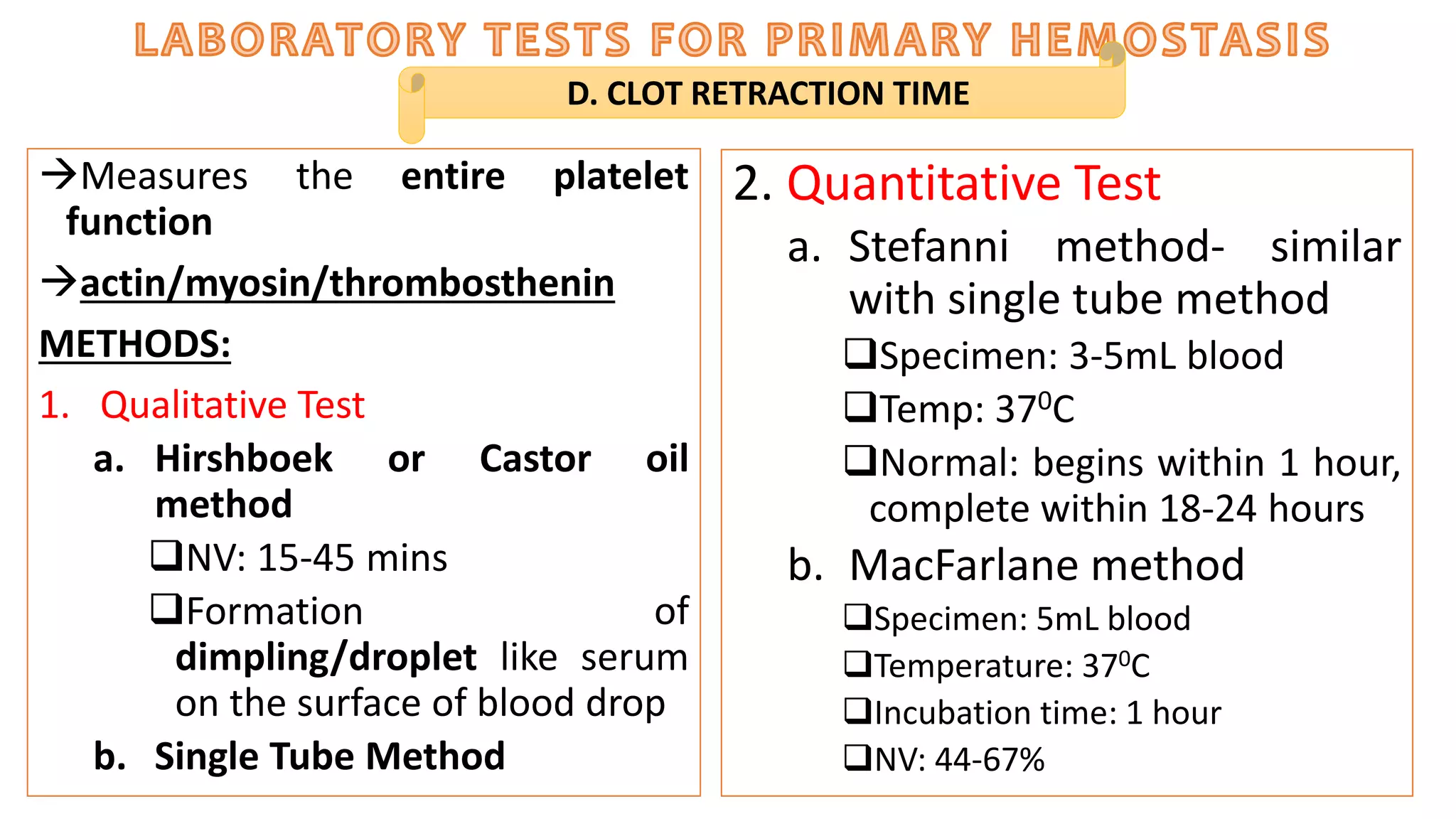
Measures the entire platelet
function
actin/myosin/thrombosthenin
METHODS:
1. Qualitative Test
a. Hirshboek or Castor oil
method
NV: 15-45 mins
Formation of
dimpling/droplet like serum
on the surface of blood drop
b. Single Tube Method
D. CLOT RETRACTION TIME
2. Quantitative Test
a. Stefanni method- similar
with single tube method
Specimen: 3-5mL blood
Temp: 370C
Normal: begins within 1 hour,
complete within 18-24 hours
b. MacFarlane method
Specimen: 5mL blood
Temperature: 370C
Incubation time: 1 hour
NV: 44-67%
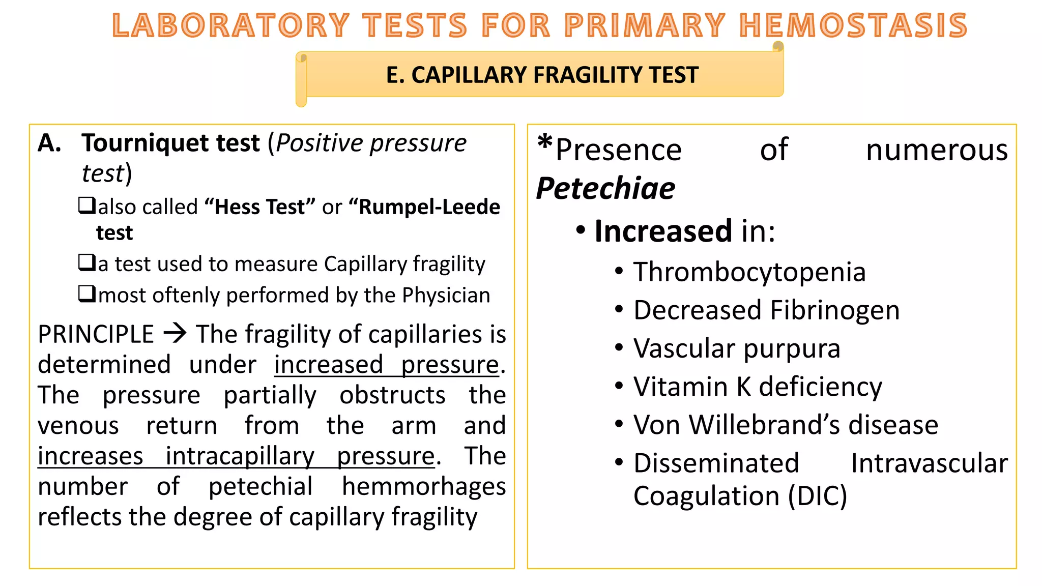
A. Tourniquet test (Positive pressure
test)
also called “Hess Test” or “Rumpel-Leede
test
a test used to measure Capillary fragility
most oftenly performed by the Physician
PRINCIPLE  The fragility of capillaries is
determined under increased pressure.
The pressure partially obstructs the
venous return from the arm and
increases intracapillary pressure. The
number of petechial hemmorhages
reflects the degree of capillary fragility
*Presence of numerous
Petechiae
• Increased in:
• Thrombocytopenia
• Decreased Fibrinogen
• Vascular purpura
• Vitamin K deficiency
• Von Willebrand’s disease
• Disseminated Intravascular
Coagulation (DIC)
E. CAPILLARY FRAGILITY TEST
• Equipments
Stethoscope
Sphygmomanometer
The test result may be graded roughly
as follows:
(0-10) 1+ = a Few petechiae on the
anterior part of the forearm
(10-20) 2+ = Many petechiae on the
anterior part of the forearm
(20-50) 3+ = Multiple petechiae over the
whole arm and back of the hand
(over 50) 4+ = Confluent petechiae on
the arm and back of the hand
B. Suction cup (Negative pressure
test) Second way to test Capillary
fragility
PROCEDURE:
1) Lubricate suction cup.
2) Apply suction cup to upper arm.
Keep negative pressure for 5 minute.
3) After 5 minutes, inspect skin under
the suction cup for Petechiae.
E. CAPILLARY FRAGILITY TEST
•Used to measure the
duration of bleeding after
measured skin incision.
•It depends on the elasticity
of blood vessel wall and on
the number and functional
capacity of platelets.
PRINCIPLE:
 It is performed by using a
sterile blood lancet to make a
measured skin puncture in the
earlobe or forearm. The time is
started immediately after the
puncture. Touching a piece of
filter paper to cut every 30
seconds until bleeding ceases
and record time and reported as
bleeding time.
F. BLEEDING TIME
The ff. are the drugs that
causes bleeding time prolonged:
Sulphonamides
Thiazide diuretics
Antineoplastic
anticoagulants,
Non-steroidal anti-inflammatory
drugs
Aspirin and aspirin compounds
Some non-narcotic analgesics
METHODS:
• Duke’s method (Template Bleeding
Time)
• Modified Ivy’s method best method to
assess platelet adhesiveness
• Coply Lalitch Method
• Adelson-Crosby Method
• MacFarlane’s Method  Same principle
with Adelson-Crosby Method but it only
uses earlobe as site of puncture
• Aspirin Tolerance test  Assesses the
effect of a standard dose of aspirin on the
duke’s bleeding
F. BLEEDING TIME
Hemostasis (COAGULATION)

More Related Content

What's hot

special and routine stains in haematology 1
special and routine stains in haematology 1special and routine stains in haematology 1
special and routine stains in haematology 1
Dr.SHAHID Raza
 
Reticulocyte count
Reticulocyte countReticulocyte count
Reticulocyte count
Prbn Shah
 

What's hot (20)

Making a 3 5% rbc suspension
Making a 3 5% rbc suspensionMaking a 3 5% rbc suspension
Making a 3 5% rbc suspension
 
Coombs test
Coombs testCoombs test
Coombs test
 
special and routine stains in haematology 1
special and routine stains in haematology 1special and routine stains in haematology 1
special and routine stains in haematology 1
 
Clotting time - Coagulation of whole blood
Clotting time - Coagulation of whole bloodClotting time - Coagulation of whole blood
Clotting time - Coagulation of whole blood
 
Gel card technology ppt nc
Gel card technology ppt ncGel card technology ppt nc
Gel card technology ppt nc
 
Automation in blood banking
Automation in blood bankingAutomation in blood banking
Automation in blood banking
 
Erythrocyte Sedimentation Rate
Erythrocyte Sedimentation RateErythrocyte Sedimentation Rate
Erythrocyte Sedimentation Rate
 
Coagulation profile
Coagulation profileCoagulation profile
Coagulation profile
 
Demonstration of le cells
Demonstration of le cellsDemonstration of le cells
Demonstration of le cells
 
Blood Bank
Blood BankBlood Bank
Blood Bank
 
Gel tech
Gel techGel tech
Gel tech
 
‫Pretransfusion testing final- ab screening - NAGLAA MAKRAM
‫Pretransfusion testing  final- ab screening - NAGLAA MAKRAM ‫Pretransfusion testing  final- ab screening - NAGLAA MAKRAM
‫Pretransfusion testing final- ab screening - NAGLAA MAKRAM
 
DECALCIFICATION
DECALCIFICATIONDECALCIFICATION
DECALCIFICATION
 
Le cell
Le cellLe cell
Le cell
 
coombs test
coombs test coombs test
coombs test
 
Immunohema
ImmunohemaImmunohema
Immunohema
 
Romanowsky stains
Romanowsky stainsRomanowsky stains
Romanowsky stains
 
Investigation of transfusion reaction
Investigation of transfusion reactionInvestigation of transfusion reaction
Investigation of transfusion reaction
 
Reticulocyte count
Reticulocyte countReticulocyte count
Reticulocyte count
 
Special stains in hematology
Special stains in hematologySpecial stains in hematology
Special stains in hematology
 

Similar to Hemostasis (COAGULATION)

GIẢM TIỂU CẦU - HỘI CHỨNG HELLP
GIẢM TIỂU CẦU - HỘI CHỨNG HELLPGIẢM TIỂU CẦU - HỘI CHỨNG HELLP
GIẢM TIỂU CẦU - HỘI CHỨNG HELLP
SoM
 
Approach to diagnosis of bleeding disorders
Approach to diagnosis of bleeding disordersApproach to diagnosis of bleeding disorders
Approach to diagnosis of bleeding disorders
Biswajeeta Saha
 

Similar to Hemostasis (COAGULATION) (20)

ix of hemorrhagic disease.pptx
ix of hemorrhagic disease.pptxix of hemorrhagic disease.pptx
ix of hemorrhagic disease.pptx
 
Thrombocytopenia
ThrombocytopeniaThrombocytopenia
Thrombocytopenia
 
GIẢM TIỂU CẦU - HỘI CHỨNG HELLP
GIẢM TIỂU CẦU - HỘI CHỨNG HELLPGIẢM TIỂU CẦU - HỘI CHỨNG HELLP
GIẢM TIỂU CẦU - HỘI CHỨNG HELLP
 
Blood count
Blood countBlood count
Blood count
 
Anti thrombiotic therapy (1)
Anti thrombiotic therapy (1)Anti thrombiotic therapy (1)
Anti thrombiotic therapy (1)
 
Anti thrombiotic therapy (1)
Anti thrombiotic therapy (1)Anti thrombiotic therapy (1)
Anti thrombiotic therapy (1)
 
Anti thrombiotic therapy
Anti thrombiotic therapy Anti thrombiotic therapy
Anti thrombiotic therapy
 
Thrombosis & embolism
Thrombosis & embolismThrombosis & embolism
Thrombosis & embolism
 
Platelets .pptx
Platelets .pptxPlatelets .pptx
Platelets .pptx
 
Bleeding vs coagulation disorders
Bleeding vs coagulation disordersBleeding vs coagulation disorders
Bleeding vs coagulation disorders
 
Hemostasis disorders
Hemostasis disordersHemostasis disorders
Hemostasis disorders
 
Pathology-bleeding disorders.ppt
Pathology-bleeding disorders.pptPathology-bleeding disorders.ppt
Pathology-bleeding disorders.ppt
 
HEMATOLOGY METHODOLOGY .pptx
HEMATOLOGY METHODOLOGY             .pptxHEMATOLOGY METHODOLOGY             .pptx
HEMATOLOGY METHODOLOGY .pptx
 
Approach to diagnosis of bleeding disorders
Approach to diagnosis of bleeding disordersApproach to diagnosis of bleeding disorders
Approach to diagnosis of bleeding disorders
 
Hemostatsis & blood transfusion
Hemostatsis & blood transfusionHemostatsis & blood transfusion
Hemostatsis & blood transfusion
 
1600_approach_to_coagulopath.pdf
1600_approach_to_coagulopath.pdf1600_approach_to_coagulopath.pdf
1600_approach_to_coagulopath.pdf
 
05 blood liquid connective tissue
05 blood liquid connective tissue05 blood liquid connective tissue
05 blood liquid connective tissue
 
Hemostasis and coagulation of blood For M.Sc & Basic Medical Students by Pand...
Hemostasis and coagulation of blood For M.Sc & Basic Medical Students by Pand...Hemostasis and coagulation of blood For M.Sc & Basic Medical Students by Pand...
Hemostasis and coagulation of blood For M.Sc & Basic Medical Students by Pand...
 
Approach to bleeding disorders part II
Approach to bleeding disorders part IIApproach to bleeding disorders part II
Approach to bleeding disorders part II
 
NORMAL HEMOSTASIS, PLATELET AND VASCULAR DISORDERS
NORMAL HEMOSTASIS, PLATELET AND VASCULAR DISORDERSNORMAL HEMOSTASIS, PLATELET AND VASCULAR DISORDERS
NORMAL HEMOSTASIS, PLATELET AND VASCULAR DISORDERS
 

More from Princess Alen Aguilar-Cabunoc

More from Princess Alen Aguilar-Cabunoc (20)

Amniotic fluid,hcg, sputum, bal & sweat
Amniotic fluid,hcg, sputum, bal & sweatAmniotic fluid,hcg, sputum, bal & sweat
Amniotic fluid,hcg, sputum, bal & sweat
 
Serous fluid & gastric fluid
Serous fluid & gastric fluidSerous fluid & gastric fluid
Serous fluid & gastric fluid
 
Vaginal secretions
Vaginal secretionsVaginal secretions
Vaginal secretions
 
Urine screening for metabolic disorders
Urine screening for metabolic disordersUrine screening for metabolic disorders
Urine screening for metabolic disorders
 
Renal diseases
Renal diseasesRenal diseases
Renal diseases
 
Chemical examination of urine (rgt strip)
Chemical examination of urine (rgt strip)Chemical examination of urine (rgt strip)
Chemical examination of urine (rgt strip)
 
Week3 physical examination of urine
Week3 physical examination of urineWeek3 physical examination of urine
Week3 physical examination of urine
 
Renal function tests and intro to urinalysis
Renal function tests and intro to urinalysisRenal function tests and intro to urinalysis
Renal function tests and intro to urinalysis
 
Adrenal, pacreas, reproductive glands
Adrenal, pacreas, reproductive glandsAdrenal, pacreas, reproductive glands
Adrenal, pacreas, reproductive glands
 
Hypothalamus, Pituitary, Para/Thyroid
Hypothalamus, Pituitary, Para/ThyroidHypothalamus, Pituitary, Para/Thyroid
Hypothalamus, Pituitary, Para/Thyroid
 
Platelet disorders
Platelet disordersPlatelet disorders
Platelet disorders
 
Abo ag, ab, & c
Abo ag, ab, & cAbo ag, ab, & c
Abo ag, ab, & c
 
Chlorides
ChloridesChlorides
Chlorides
 
Beta & gamma herpesvirinae
Beta & gamma herpesvirinaeBeta & gamma herpesvirinae
Beta & gamma herpesvirinae
 
Enveloped dna viruses alpha and hhv6
Enveloped dna viruses alpha and hhv6Enveloped dna viruses alpha and hhv6
Enveloped dna viruses alpha and hhv6
 
Hepatitis B/ Chronic Hepatitis/Serum Hepatitis
Hepatitis B/ Chronic Hepatitis/Serum HepatitisHepatitis B/ Chronic Hepatitis/Serum Hepatitis
Hepatitis B/ Chronic Hepatitis/Serum Hepatitis
 
Hepatitis type D, E, F G
Hepatitis type D, E, F GHepatitis type D, E, F G
Hepatitis type D, E, F G
 
Immunology and Serology (transplantation reactions)
Immunology and Serology  (transplantation reactions)Immunology and Serology  (transplantation reactions)
Immunology and Serology (transplantation reactions)
 
Wbc & platelets anomalies
Wbc & platelets anomaliesWbc & platelets anomalies
Wbc & platelets anomalies
 
Opportunistic & systemic mycoses tabulated summary
Opportunistic & systemic mycoses tabulated summaryOpportunistic & systemic mycoses tabulated summary
Opportunistic & systemic mycoses tabulated summary
 

Recently uploaded

New Directions in Targeted Therapeutic Approaches for Older Adults With Mantl...
New Directions in Targeted Therapeutic Approaches for Older Adults With Mantl...New Directions in Targeted Therapeutic Approaches for Older Adults With Mantl...
New Directions in Targeted Therapeutic Approaches for Older Adults With Mantl...
i3 Health
 

Recently uploaded (20)

Prix Galien International 2024 Forum Program
Prix Galien International 2024 Forum ProgramPrix Galien International 2024 Forum Program
Prix Galien International 2024 Forum Program
 
PT MANAGEMENT OF URINARY INCONTINENCE.pptx
PT MANAGEMENT OF URINARY INCONTINENCE.pptxPT MANAGEMENT OF URINARY INCONTINENCE.pptx
PT MANAGEMENT OF URINARY INCONTINENCE.pptx
 
Compare home pulse pressure components collected directly from home
Compare home pulse pressure components collected directly from homeCompare home pulse pressure components collected directly from home
Compare home pulse pressure components collected directly from home
 
New Directions in Targeted Therapeutic Approaches for Older Adults With Mantl...
New Directions in Targeted Therapeutic Approaches for Older Adults With Mantl...New Directions in Targeted Therapeutic Approaches for Older Adults With Mantl...
New Directions in Targeted Therapeutic Approaches for Older Adults With Mantl...
 
PT MANAGEMENT OF URINARY INCONTINENCE.pptx
PT MANAGEMENT OF URINARY INCONTINENCE.pptxPT MANAGEMENT OF URINARY INCONTINENCE.pptx
PT MANAGEMENT OF URINARY INCONTINENCE.pptx
 
linearity concept of significance, standard deviation, chi square test, stude...
linearity concept of significance, standard deviation, chi square test, stude...linearity concept of significance, standard deviation, chi square test, stude...
linearity concept of significance, standard deviation, chi square test, stude...
 
Presentació "Advancing Emergency Medicine Education through Virtual Reality"
Presentació "Advancing Emergency Medicine Education through Virtual Reality"Presentació "Advancing Emergency Medicine Education through Virtual Reality"
Presentació "Advancing Emergency Medicine Education through Virtual Reality"
 
Multiple sclerosis diet.230524.ppt3.pptx
Multiple sclerosis diet.230524.ppt3.pptxMultiple sclerosis diet.230524.ppt3.pptx
Multiple sclerosis diet.230524.ppt3.pptx
 
1130525--家醫計畫2.0糖尿病照護研討會-社團法人高雄市醫師公會.pdf
1130525--家醫計畫2.0糖尿病照護研討會-社團法人高雄市醫師公會.pdf1130525--家醫計畫2.0糖尿病照護研討會-社團法人高雄市醫師公會.pdf
1130525--家醫計畫2.0糖尿病照護研討會-社團法人高雄市醫師公會.pdf
 
Arterial health throughout cancer treatment and exercise rehabilitation in wo...
Arterial health throughout cancer treatment and exercise rehabilitation in wo...Arterial health throughout cancer treatment and exercise rehabilitation in wo...
Arterial health throughout cancer treatment and exercise rehabilitation in wo...
 
Retinal consideration in cataract surgery
Retinal consideration in cataract surgeryRetinal consideration in cataract surgery
Retinal consideration in cataract surgery
 
US E-cigarette Summit: Taming the nicotine industrial complex
US E-cigarette Summit: Taming the nicotine industrial complexUS E-cigarette Summit: Taming the nicotine industrial complex
US E-cigarette Summit: Taming the nicotine industrial complex
 
"Central Hypertension"‚ in China: Towards the nation-wide use of SphygmoCor t...
"Central Hypertension"‚ in China: Towards the nation-wide use of SphygmoCor t..."Central Hypertension"‚ in China: Towards the nation-wide use of SphygmoCor t...
"Central Hypertension"‚ in China: Towards the nation-wide use of SphygmoCor t...
 
Effects of vaping e-cigarettes on arterial health
Effects of vaping e-cigarettes on arterial healthEffects of vaping e-cigarettes on arterial health
Effects of vaping e-cigarettes on arterial health
 
Is preeclampsia and spontaneous preterm delivery associate with vascular and ...
Is preeclampsia and spontaneous preterm delivery associate with vascular and ...Is preeclampsia and spontaneous preterm delivery associate with vascular and ...
Is preeclampsia and spontaneous preterm delivery associate with vascular and ...
 
Blue Printing in medical education by Dr.Mumtaz Ali.pptx
Blue Printing in medical education by Dr.Mumtaz Ali.pptxBlue Printing in medical education by Dr.Mumtaz Ali.pptx
Blue Printing in medical education by Dr.Mumtaz Ali.pptx
 
Aptopadesha Pramana / Pariksha: The Verbal Testimony
Aptopadesha Pramana / Pariksha: The Verbal TestimonyAptopadesha Pramana / Pariksha: The Verbal Testimony
Aptopadesha Pramana / Pariksha: The Verbal Testimony
 
Young at heart: Cardiovascular health stations to empower healthy lifestyle b...
Young at heart: Cardiovascular health stations to empower healthy lifestyle b...Young at heart: Cardiovascular health stations to empower healthy lifestyle b...
Young at heart: Cardiovascular health stations to empower healthy lifestyle b...
 
TEST BANK For Advanced Practice Nursing in the Care of Older Adults, 2nd Edit...
TEST BANK For Advanced Practice Nursing in the Care of Older Adults, 2nd Edit...TEST BANK For Advanced Practice Nursing in the Care of Older Adults, 2nd Edit...
TEST BANK For Advanced Practice Nursing in the Care of Older Adults, 2nd Edit...
 
How to Give Better Lectures: Some Tips for Doctors
How to Give Better Lectures: Some Tips for DoctorsHow to Give Better Lectures: Some Tips for Doctors
How to Give Better Lectures: Some Tips for Doctors
 

Hemostasis (COAGULATION)

  • 1. PREPARED BY: PRINCESS ALEN I. AGUILAR,RMT SOURCE: HO BY CUA HEMATOLOGY BOOKS BY WILLIAM, HOFF’S, TURGEON
  • 2. 1828The disorder was first described in a thesis published by Hoff 1842 Platelets were described 1905 Theory of Blood Coagulation by Paul Morawitz was accepted 1913  Lee and White WB CT was performed 1930 PT Time was introduced by Quick 1940 Other tests for evaluating hemostatic mechanisms, like platelet count and bleeding time were introduced 1964 Cascade and Waterfall Theory of coagulation was introduced 2ND Century A.D.  Hemophilia was first recognized 12th Century A.D. Moises Maimonides described 2 male siblings who died because of excessive bleeding after circumcision 1803  Clinical description of families with haemophilia was first published. The disorder was given the name haemophilia which means “love of haemorrhage” by Schonlein 2ND Century A.D.  Hemophilia was first recognized
  • 3.
  • 4.
  • 5. • Size: 20-50 µm in diameter. • Cytoplasm: varying shades of blue • Usually darker than the myeloblast. • May have small, blunt pseudopods. • Small to moderate amount. Usually a narrow band around the nucleus. As the cell matures, the amount of cytoplasm increases • Usually nongranular. • Nucleus: • Round,oval or may be kidney shaped. • Fine chromatin pattern • Multiple nucleoli that generally stain blue • N/C Ratio is about 10:1
  • 6. • Size: 20-60 µm in diameter • Cytoplasm: More abundant than previous stage. • Less basophilic than in the blast. • Granules begin to form in the golgi region. • Nucleus: Chromatin becomes more coarse. • Multiple nucleoli are visible. • Irregular in shape; may even show slight lobulation. • N/C Ratio 4:1 to 7:1 depending on the ploidy
  • 7. • Size:30-90 µm in diameter • Cytoplasm: abundant • Pinkish blue in color • Very fine and diffusely granular • Usually has an irregular peripheral border • Nucleus: small in comparison to cell size • Multiple nuclei may be visible or the nucleus may show multi-lobulation. • Chromatin is coarser than in the previous stage. • No nuclei are visible. • N/C ratio is 2:1 to 1:1
  • 8. • Size: 40-120 µm in diameter • Cytoplasm: • contains coarse clumps of granules aggregating into little bundles, which bud off from the periphery to become platelets • Nucleus: • multiple nuclei are present, or the nucleus is multilobulated • No nucleoli are visible. • N/C Ratio is less than 1:1
  • 9. • Size:1-4 µm in diameter • Cytoplasm: light blue to purple. • Very granular. • Consists of two parts: (1) the chromomere, which is granular and located generally. (2) the hyalomere, which surrounds the chromomere and is non granular and clear to light blue.
  • 10. Stoppage of blood flow Involves the interaction of blood vessels, platelets, the coagulation mechanism, fibrinolysis and tissue repair A complex process that: Produces a clot to stop the bleeding Keeps the clot confined Dissolves the clot as the wound heals CELLULAR ELEMENTS OF HEMOSTASIS: Extravascular Tissue Factor  tissue surrounding the vessel Vascular Intima  blood vessel through which the blood flows Intravascular component  plasma proteins and platelets
  • 11.
  • 12.
  • 13.
  • 14. A. PRIMARY HEMOSTASIS Refers to the role of Blood vessels (vascular system which includes the arteries,veins,and capillaries) and platelets in the primary formation og platelet plug in response to vascular injury TEST: BLEEDING TIME 1. PLATELET ADHESION  plt- non plt substance; Contact to plts with the subendothelium and their adhesion to it Plts adheres to Collagen Occurs in presence of vWF vWF disease Bernard-Soulier disease
  • 15. A. PRIMARY HEMOSTASIS 3. PLATELET SECRETION Release of granules 4. PLATELET AGGREGATION Platelet to another platelet Aggregate plt for oxygen exchange Requires fibrinogen (Bridge) *Glanzmann’s thrombasthenia 2. PLATELET ACTIVATION Morphologic and functional changes in platelets (platelet change shepe) Form discoid to spherical with pseudopod formation Ca2+,actin and myosin/thrombosthenin
  • 16.
  • 17.
  • 18.
  • 19.
  • 20. B. SECONDARY HEMOSTASIS Involves the enzymatic activation of series of plasma proteins in the coagulation system (coag factors) to form a fibrin meshwork (fibrin clot) TEST: CLOTTING TIME
  • 21. Factor Trivial Name(s) Functions Prekallikrein (PK) Fletcher factor High molecular weight kininogen (HMWK) contact activation cofactor; Fitzgerald, Flaujeac Williams factor I Fibrinogen Polymerizes fibrin II Prothrombin Serine proteases III Tissue Factor Co-Factor IV Calcium Mineral V Proaccelerin, labile factor, accelerator (Ac-) globulin Co-Factor VI (same as Va) Accelerin VII Proconvertin, serum prothrombin conversion accelerator (SPCA), cothromboplastin Serine proteases VIII Antihemophiliac factor A, antihemophilic globulin (AHG) Co-Factor IX Christmas Factor, antihemophilic factor B,plasma thromboplastin component (PTC) Serine proteases X Stuart-Prower Factor Serine proteases XI Plasma thromboplastin antecedent (PTA) Serine proteases XII Hageman Factor Serine proteases XIII Protransglutaminase, fibrin stabilizing factor (FSF), fibrinoligase Stabilizing Factor
  • 22. Petechiae- purplish red pinpoint hemorrhagic spots in the skin caused by loss of capillary ability to withstand normal blood pressure and trauma Purpura – haemorrhage of blood into small areas of skin, mucous membranes, and other tissues Ecchymosis- form of purpura in which blood escapes into large areas of skin and mucous membranes, but not into deep tissues Epistaxis – nosebleed Hemarthrosis – leakage of blood into joint activities Hematemesis – vomiting of blood Hemoptysis-Bloody sputum Hematochezia- “red stool”; asstd with lower GIT bleeding Hematoma – Hemoglobinuria – hgb in urine Hemorrhage- is a severe form of bleeding tha requires immediate intervention and transfusion Melena- stool containing dark red or black blood; “ black tarry stool Menorrhagia – excessive menstrual bleeding
  • 23.
  • 24.
  • 25.
  • 26.
  • 28.
  • 29.
  • 30.
  • 31. SPECIMEN COLLECTION: is a prenalytic that may have serious implications in laboratory testing. According to studies done in recent years, 32-75% of testing errors occure during the pre-analytic phase. SPECIMEN : EDTA anticoagulated WB- most commonly used A. QUANTITATIVE PLATELET EVALUATION PLATELET SATELLITOSIS A form of pseudo-thrombocytopenia Antibodies directed against GPIIb-IIIa react with the leukocyte Fc gamma receptor III and attach the platelet to neutrophils are the most frequently involved; occasionally monocytes Naturally occurring, ut exposure of antigen on EDTA-treated platelets and leukocytes may trigger the phenomenon. Using sodium citrate as an anticoagulant should correct this problem. Because of the dilution in the citrate tubes, it is necessary to multiply the obtained platelet count by 1.1
  • 32. Platelets are counted in a hemocytometer as in erythrocytes and leukocytes A. REESE-ECKER DILUENT: isotonic Sodium Citrate – prevent platelet aggregation Formaldehyde- Preservative BCB- Stain/dye Microscopy: Light Microscopy Appearance of platelets: BLUISH B. BRECKER-CRONKITE- reference method Diluent: Hypotonic= 1% ammonium oxalate Microscopy: Phase-Contrast Microscopy (best microscope for platelet morph evaluation) I. DIRECT PLATELET COUNT A. QUANTITATIVE PLATELET EVALUATION C. GUY AND LEAKE Diluent: Isotonic  Sodium oxalate- prevent platelet aggregation Formaldehyde- preservative CV- stain/dye Mircoscopy: Light Microscopy Appearance of platelets: LILAC refractile object D. UNOPETTE Diluent: 1% ammonium oxalate Dilution: 1:100
  • 33. Platelets are counted in their relationship to red cells on a fixed-stained smear. This method is not reliable because the results depend upon the distribution of platelets and on the red cell count. Stained by Giemsa (10 fields) A. FONIO’S 14% Magnesium sulphate Wright’s stain B. DAMESHECK BCB Wright’s stain C. OLEF’S II. INDIRECT PLATELET COUNT A. QUANTITATIVE PLATELET EVALUATION Red cells must first be removed from whole blood, either by sedimentation or by controlled centrifugation. PRINCIPLE: VOLTAGE PULSE COUNTING ELECTRO-OPTICAL COUNTING III. AUTOMATED PLATELET COUNT
  • 34. Less than 100,000/uL  abnormally low 30-50,000/uL  bleeding possible with trauma Less than 30,000/uL  spontaneous bleeding possible Less than 5,000/uL  severe spontaneous bleeding Note: normal platelet count + prolonged BT= QUALITATIVE PLATELET ABNORMALITY, primary vascular abnormality and von Willebrand’s syndrome Low platelet count + normal BT: autoimmune thrombocytopenia Low platelet count + very prolonged BT: Simultaneous quantitative and qualitative platelet deficiency
  • 35. Assess the ability of platelets to aggregate after the addition of aggregating agents Sample: PRP (Platelet Rich Plasma) Aggregating agents: Collagen ADP Ristocetin Epinephrine Also: thrombin, arachidonic acid, serotonin B. PLATELET AGGREGATION TEST TEST CONSIDERATIONS: No hemolysis Fasting: 8 hours pH: 6.5-8.5 Within 3 hours
  • 36. The adhesiveness of platelets may be measured in vitro by their ability to adhere on glass surfaces with beads Salzman method: test of the retention of platelets within glass bead column C. PLATELET RETENTION TEST (ADHESIVENESS) Platelet adhesion may be tested in vitro by their ability to adhere to glass surfaces. The number of platelets in the blood, collected through the glass bead collecting system, will be lower than the number obtained by routine venipuncture. (Brown)
  • 37. C. PLATELET RETENTION TEST (ADHESIVENESS) When blood is passed through a glass bead column, normal platelets that have access to normal vWF will adhere and aggregate to the beads such that the effluent from the column will have a much lower platelet count than the starting sample (Steininger). DECREASED PLATELET RETENTION INCREASED PLATELET RETENTION Bernard Soulier Thrombotic disorder Glanzmann Thrombasthenia Hyperlipidemia vWD Carcinoma Chediak-Higashi Oral Contraceptives Myeloproliferative disorders Pregnancy Uremia Aspirin Administration ABNORMAL PLATELET RETENTION TEST REFERENCE INTERVALS: • 26-60% (Brown) • 70% or greater (Steininger) • Each laboratory should establish its own reference range with the equipment used.
  • 38. Measures the entire platelet function actin/myosin/thrombosthenin METHODS: 1. Qualitative Test a. Hirshboek or Castor oil method NV: 15-45 mins Formation of dimpling/droplet like serum on the surface of blood drop b. Single Tube Method D. CLOT RETRACTION TIME 2. Quantitative Test a. Stefanni method- similar with single tube method Specimen: 3-5mL blood Temp: 370C Normal: begins within 1 hour, complete within 18-24 hours b. MacFarlane method Specimen: 5mL blood Temperature: 370C Incubation time: 1 hour NV: 44-67%
  • 39. A. Tourniquet test (Positive pressure test) also called “Hess Test” or “Rumpel-Leede test a test used to measure Capillary fragility most oftenly performed by the Physician PRINCIPLE  The fragility of capillaries is determined under increased pressure. The pressure partially obstructs the venous return from the arm and increases intracapillary pressure. The number of petechial hemmorhages reflects the degree of capillary fragility *Presence of numerous Petechiae • Increased in: • Thrombocytopenia • Decreased Fibrinogen • Vascular purpura • Vitamin K deficiency • Von Willebrand’s disease • Disseminated Intravascular Coagulation (DIC) E. CAPILLARY FRAGILITY TEST
  • 40. • Equipments Stethoscope Sphygmomanometer The test result may be graded roughly as follows: (0-10) 1+ = a Few petechiae on the anterior part of the forearm (10-20) 2+ = Many petechiae on the anterior part of the forearm (20-50) 3+ = Multiple petechiae over the whole arm and back of the hand (over 50) 4+ = Confluent petechiae on the arm and back of the hand B. Suction cup (Negative pressure test) Second way to test Capillary fragility PROCEDURE: 1) Lubricate suction cup. 2) Apply suction cup to upper arm. Keep negative pressure for 5 minute. 3) After 5 minutes, inspect skin under the suction cup for Petechiae. E. CAPILLARY FRAGILITY TEST
  • 41. •Used to measure the duration of bleeding after measured skin incision. •It depends on the elasticity of blood vessel wall and on the number and functional capacity of platelets. PRINCIPLE:  It is performed by using a sterile blood lancet to make a measured skin puncture in the earlobe or forearm. The time is started immediately after the puncture. Touching a piece of filter paper to cut every 30 seconds until bleeding ceases and record time and reported as bleeding time. F. BLEEDING TIME
  • 42. The ff. are the drugs that causes bleeding time prolonged: Sulphonamides Thiazide diuretics Antineoplastic anticoagulants, Non-steroidal anti-inflammatory drugs Aspirin and aspirin compounds Some non-narcotic analgesics METHODS: • Duke’s method (Template Bleeding Time) • Modified Ivy’s method best method to assess platelet adhesiveness • Coply Lalitch Method • Adelson-Crosby Method • MacFarlane’s Method  Same principle with Adelson-Crosby Method but it only uses earlobe as site of puncture • Aspirin Tolerance test  Assesses the effect of a standard dose of aspirin on the duke’s bleeding F. BLEEDING TIME

Editor's Notes

  1. AASA vwF-needed for normal plt adhesion
  2. pseudopod formation- to facilitate the release of agonist / calcium and TXA2 Thrombosthenin AKA microfilaments Agonist-substances that initiates activation
  3. AASA vwF-needed for normal plt adhesion
  4. Thus deficiency in contact factors does not exhibit bleeding disorder rather thrombotic disorder
  5. Factor in platelet counting  20,000
  6. CARE
  7. This test is difficult to standardize and the results are affected by the rate at which the blood flows through the glass bead column, the length of this column, the size of the glass beads, the hematocrit, and the fragility of the blood cells. In vivo testing of platelet retention/platelet adhesiveness
  8. Factor in platelet counting  20,000
  9. Dukes; NV (6-10mins)