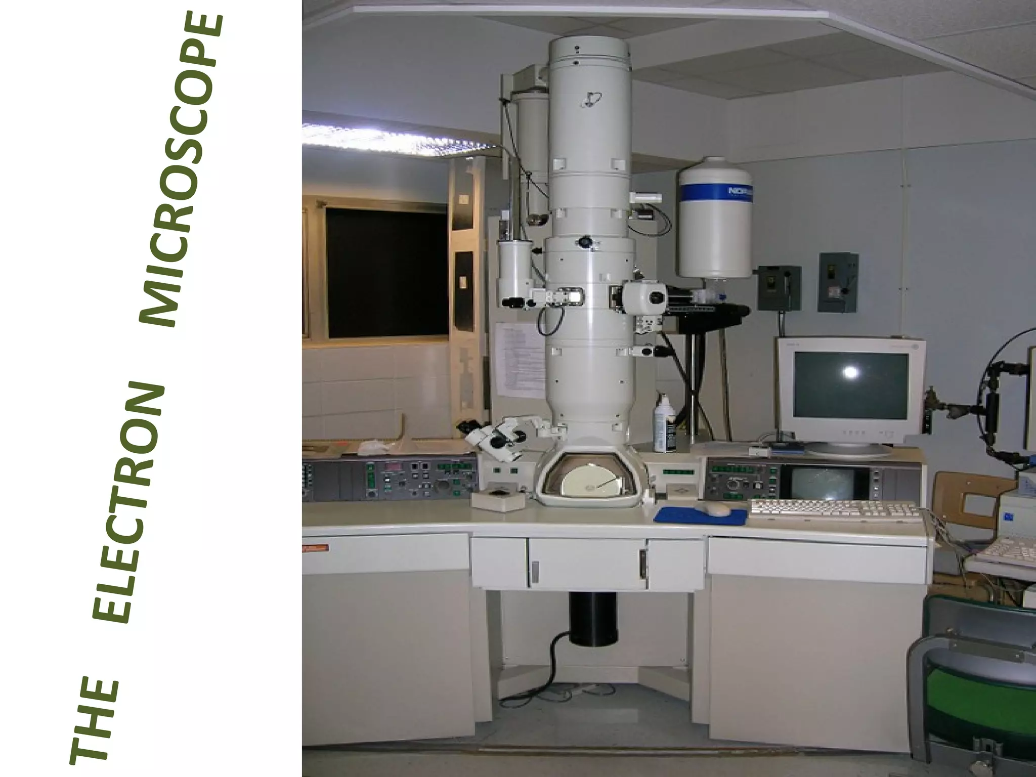
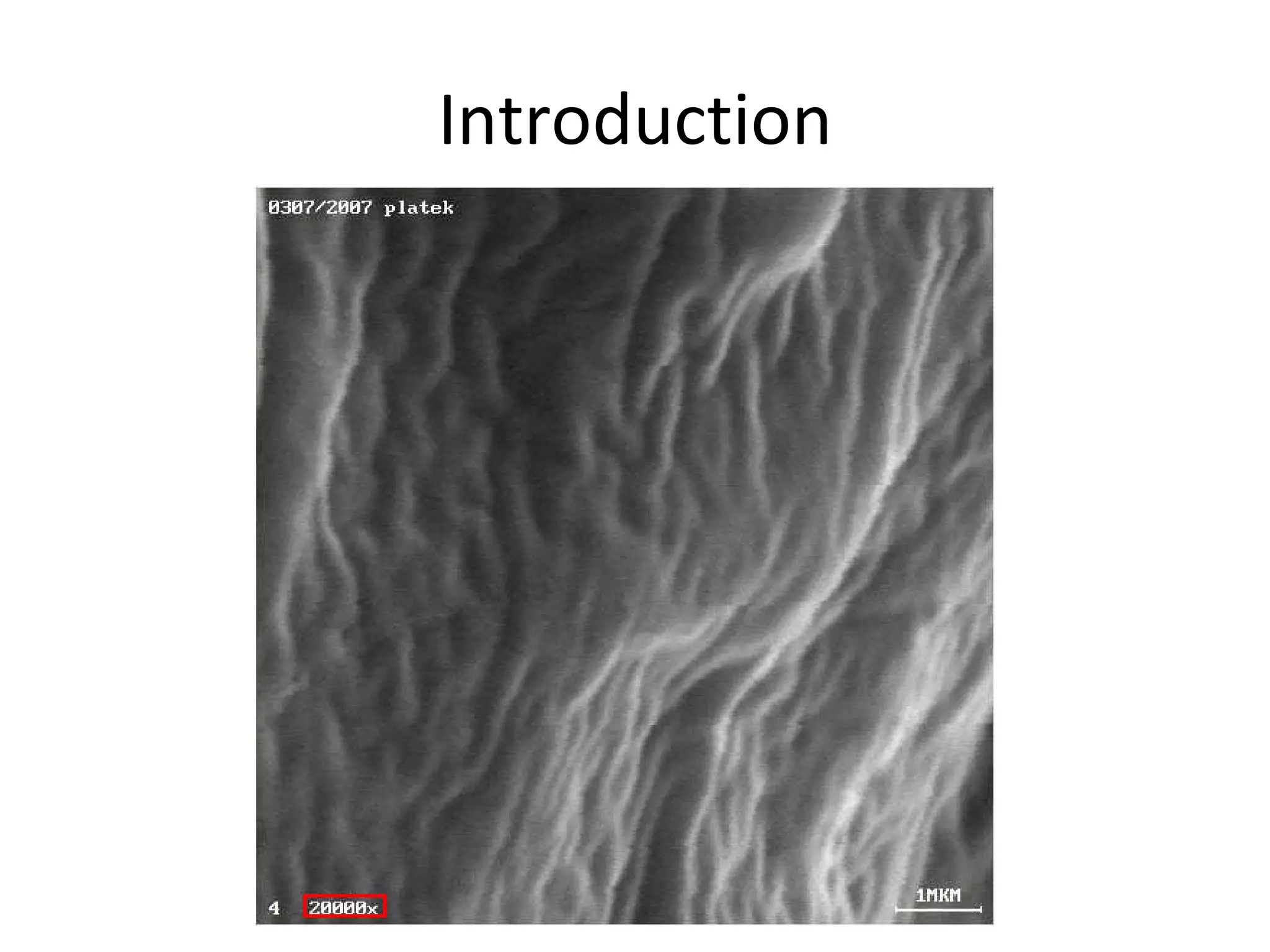
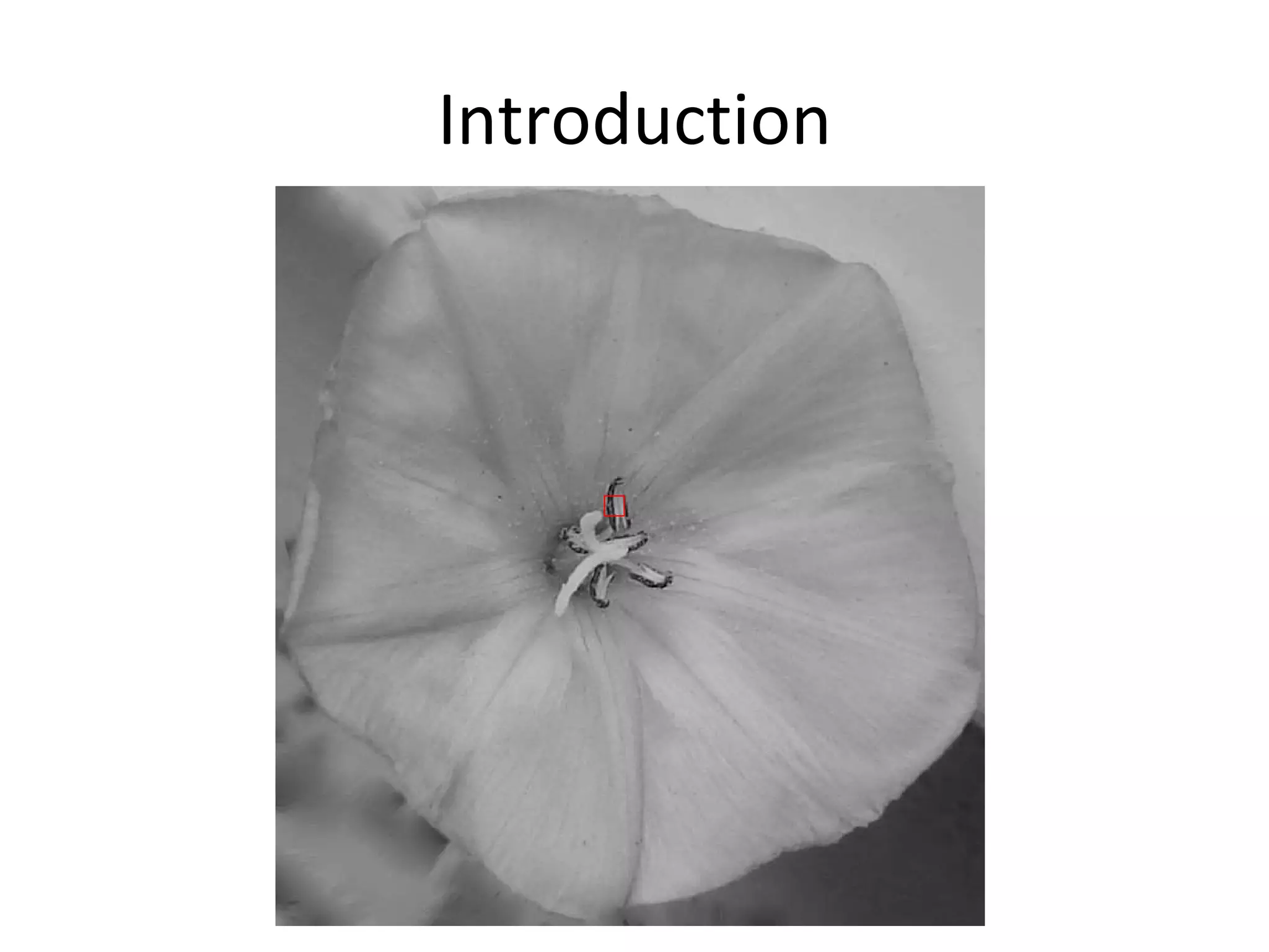
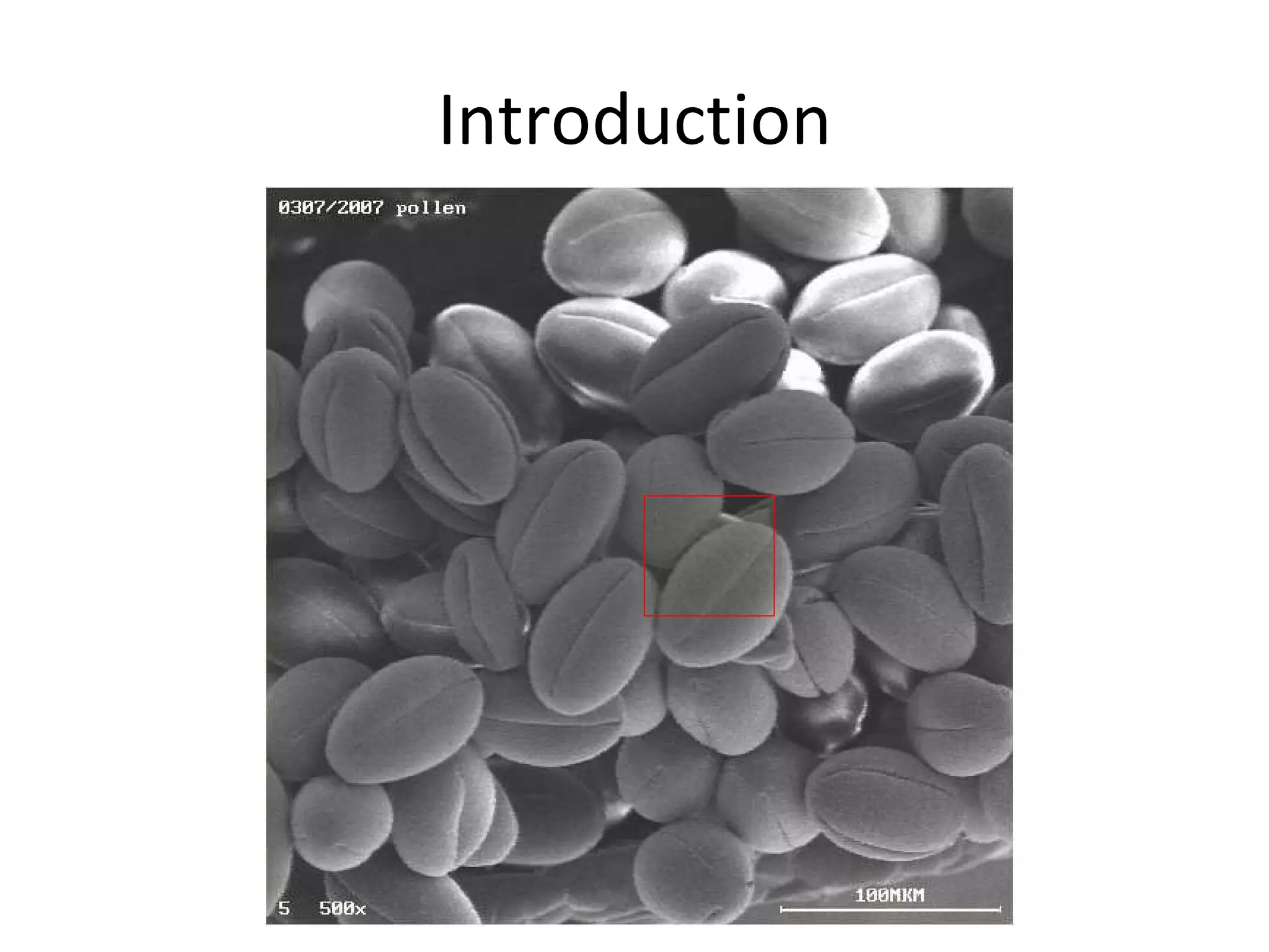
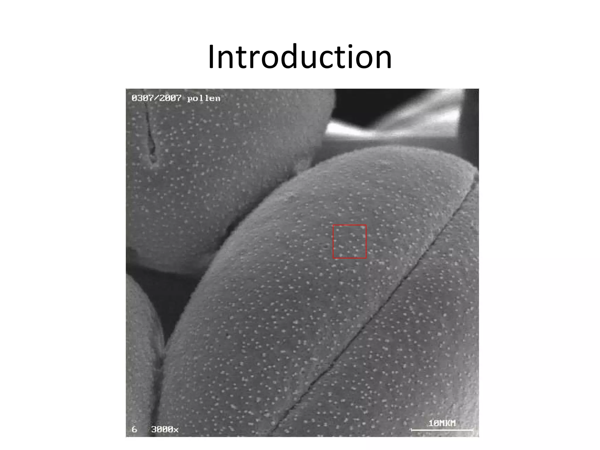
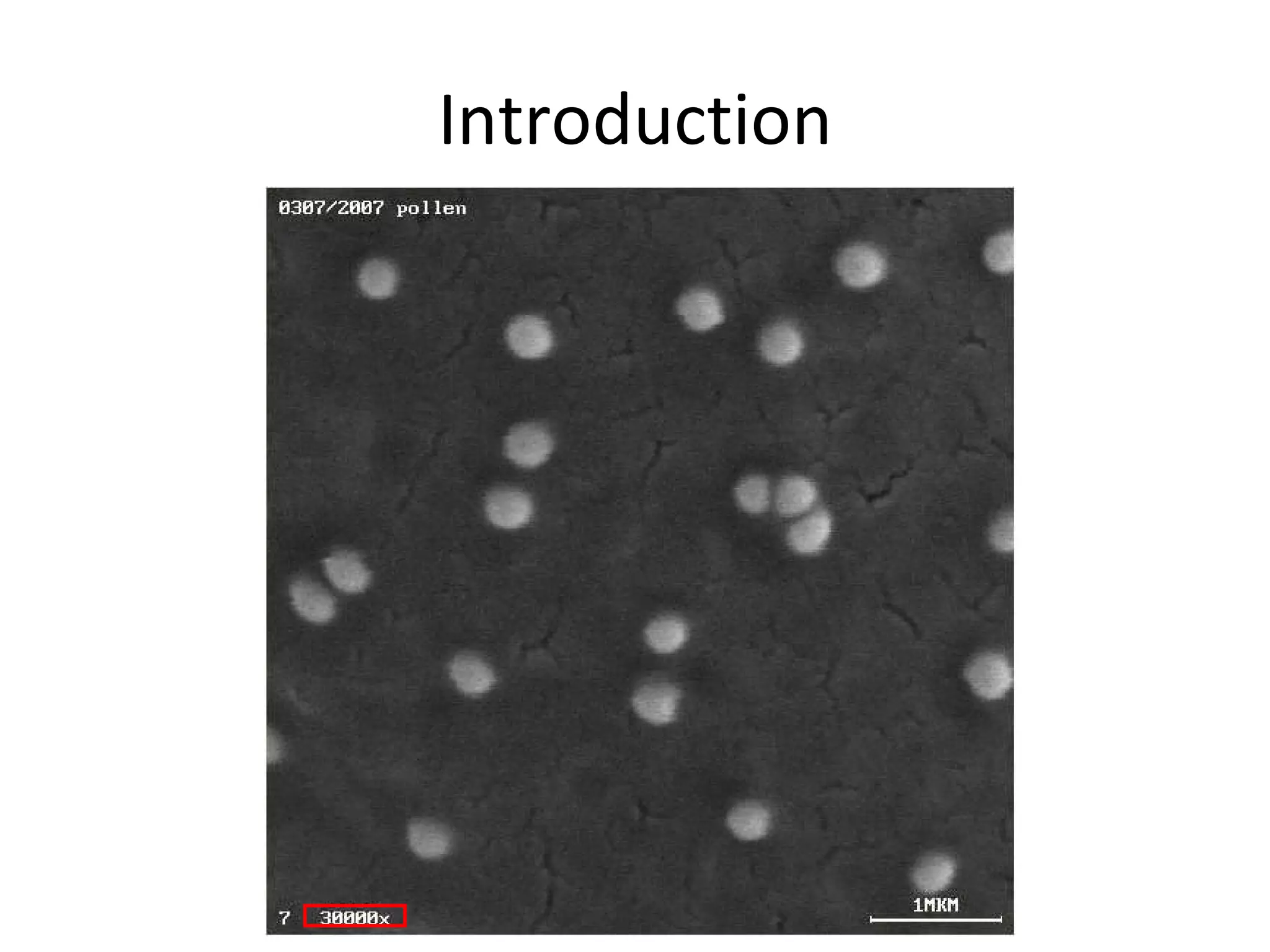
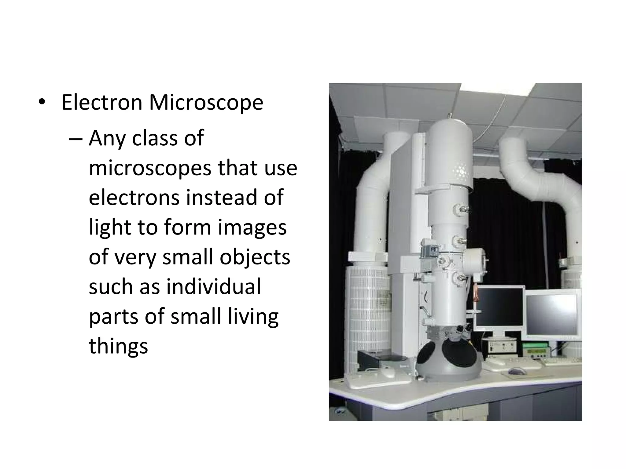
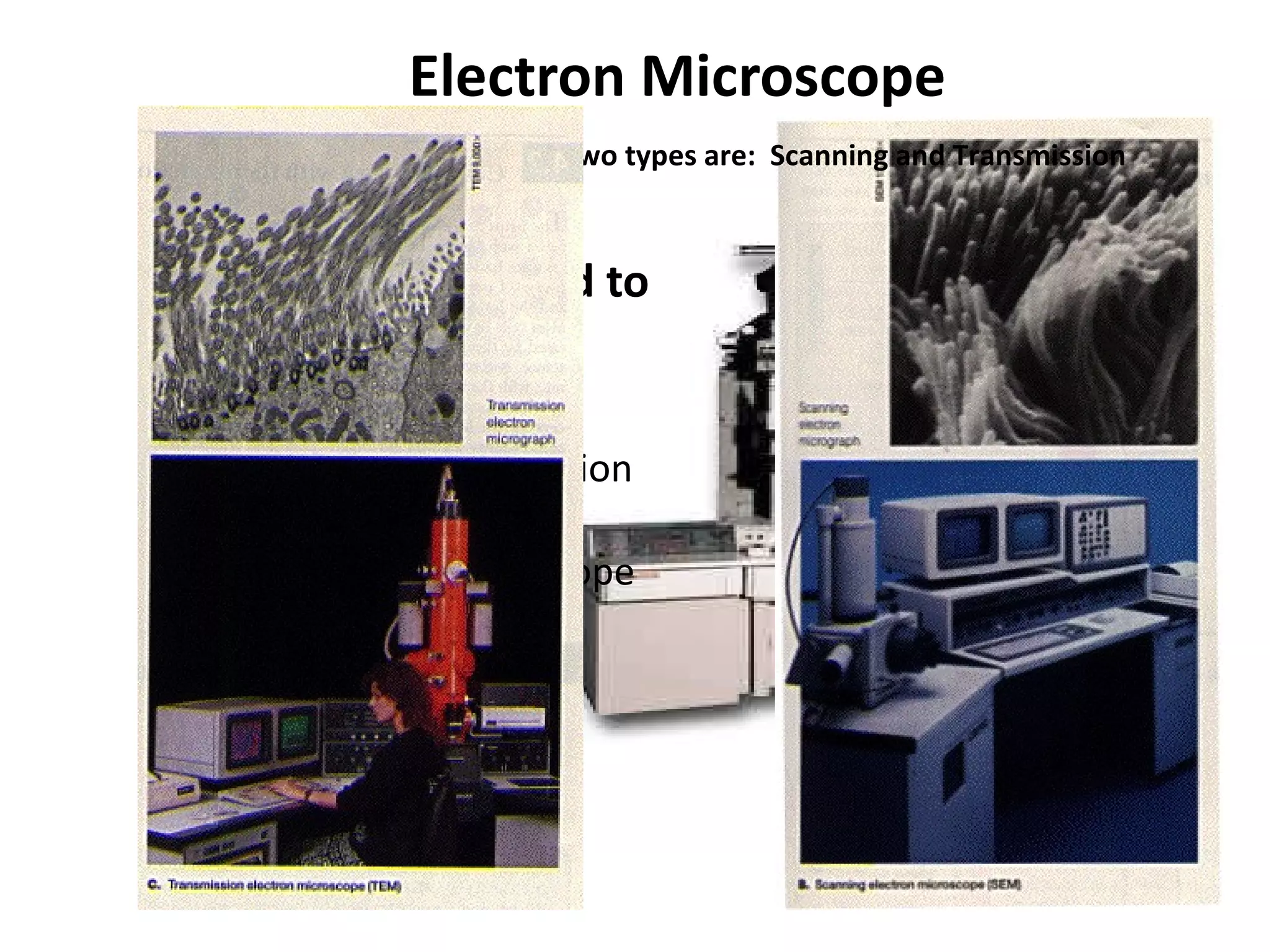
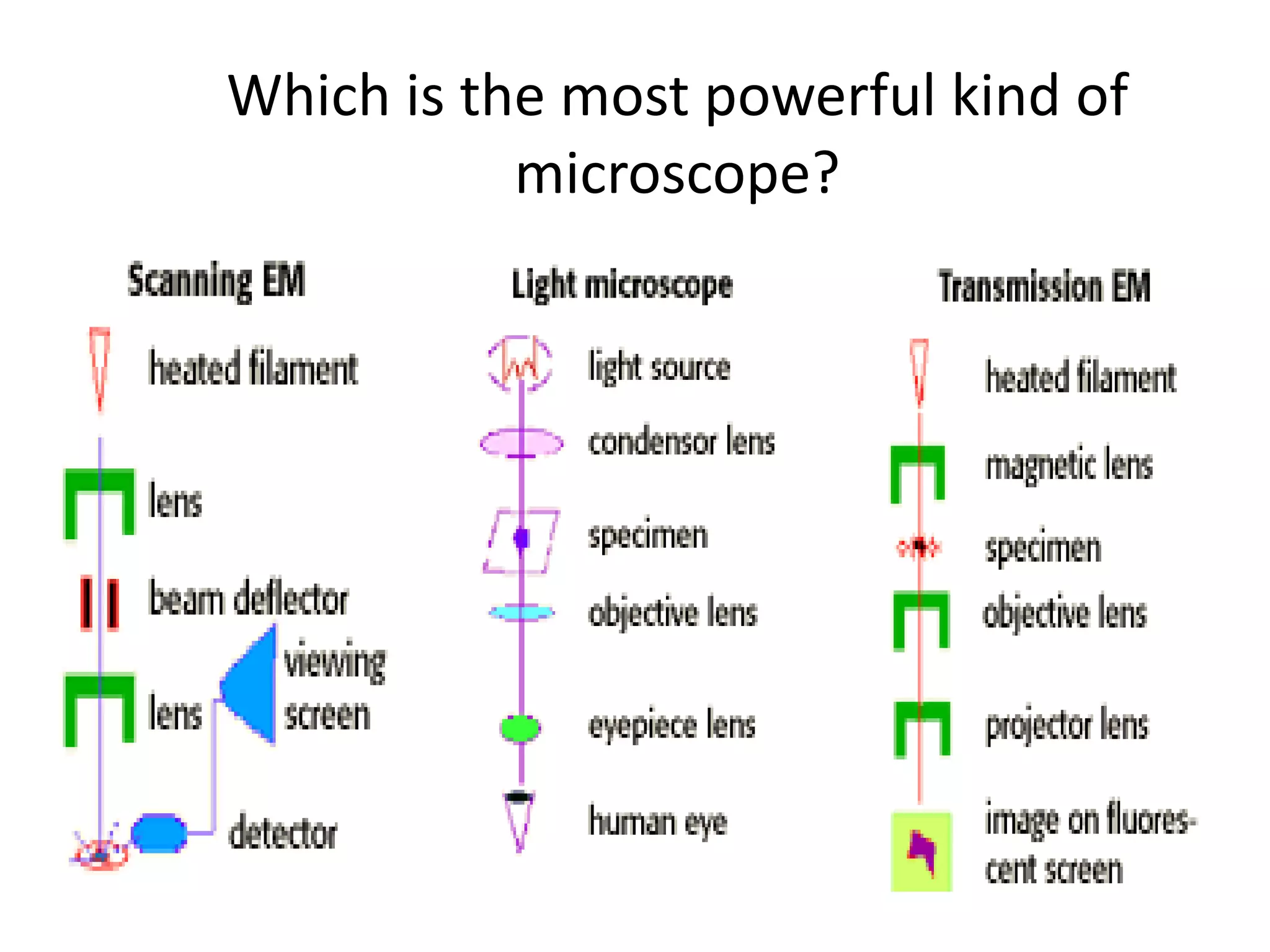
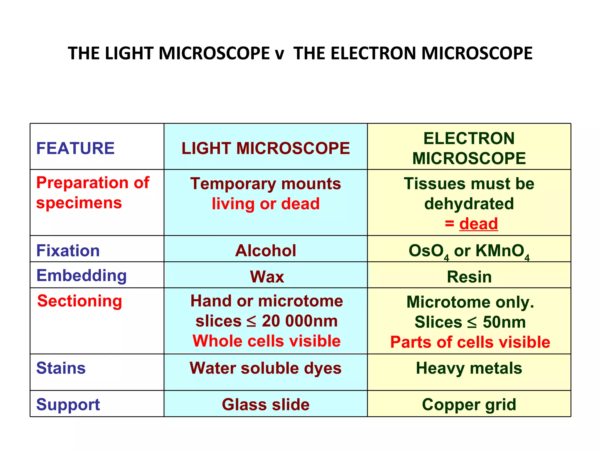
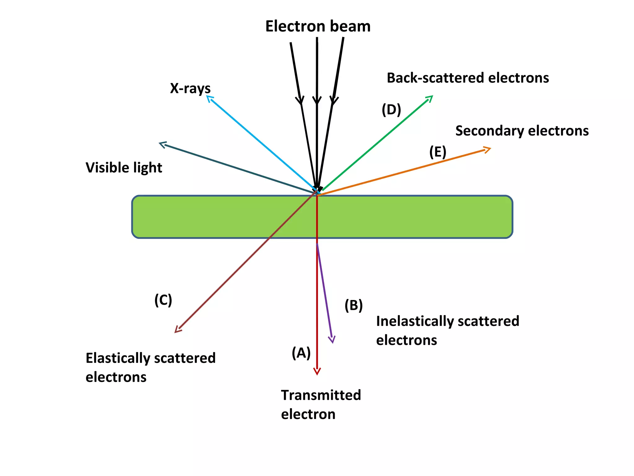
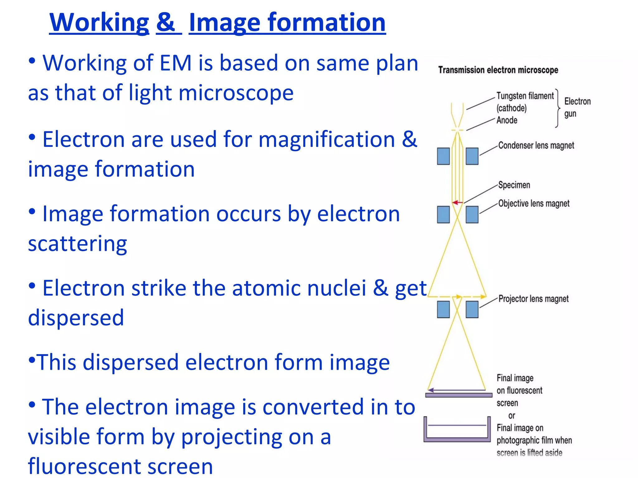
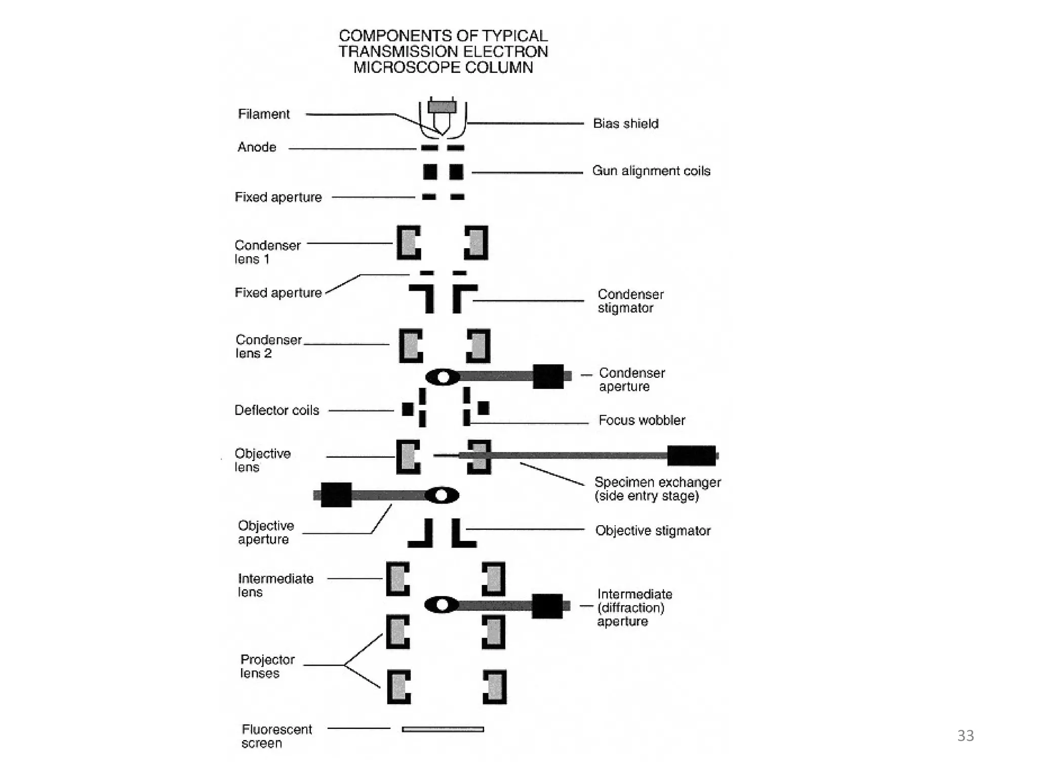
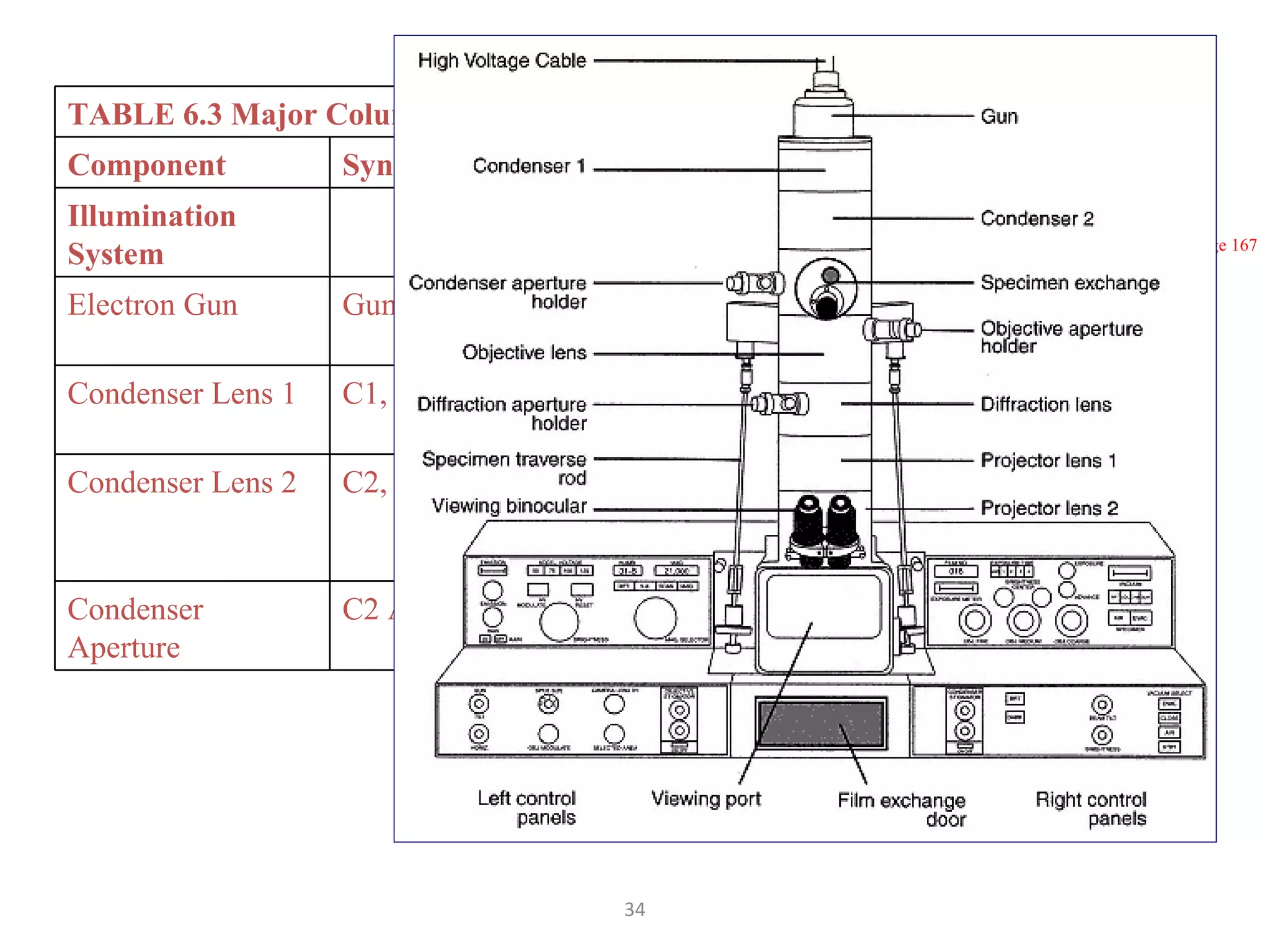
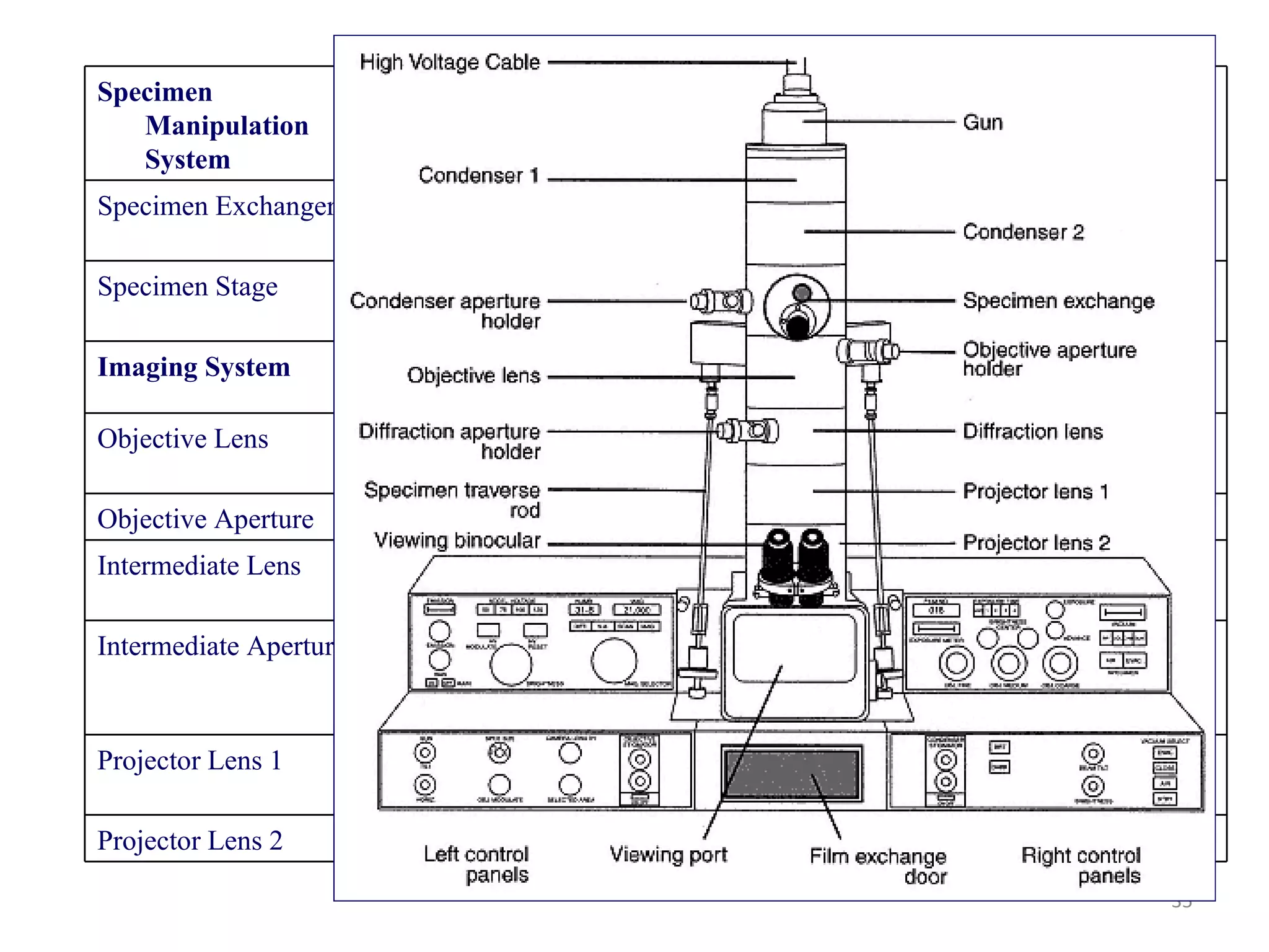
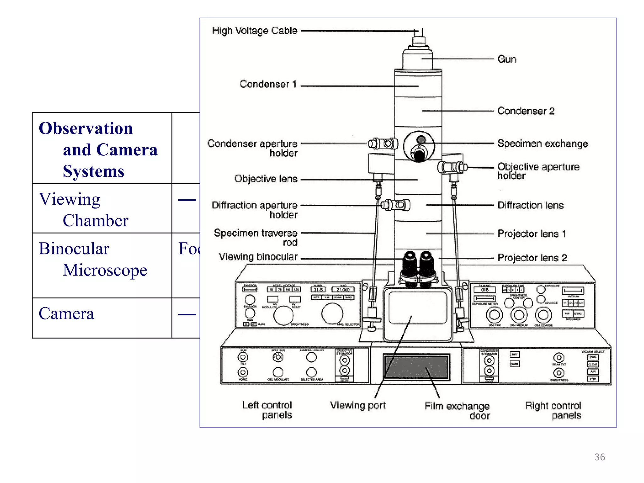
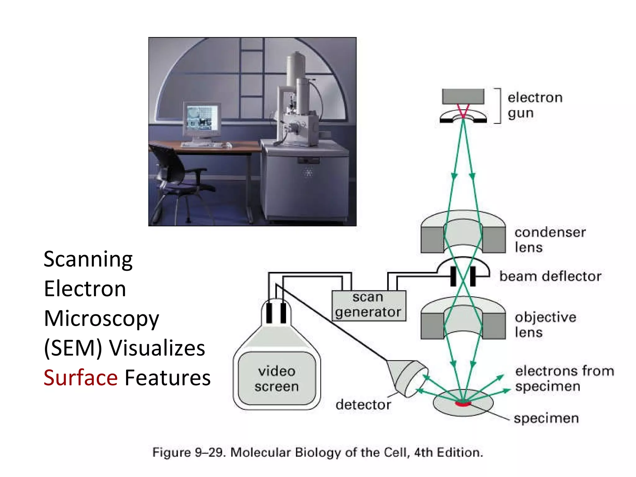
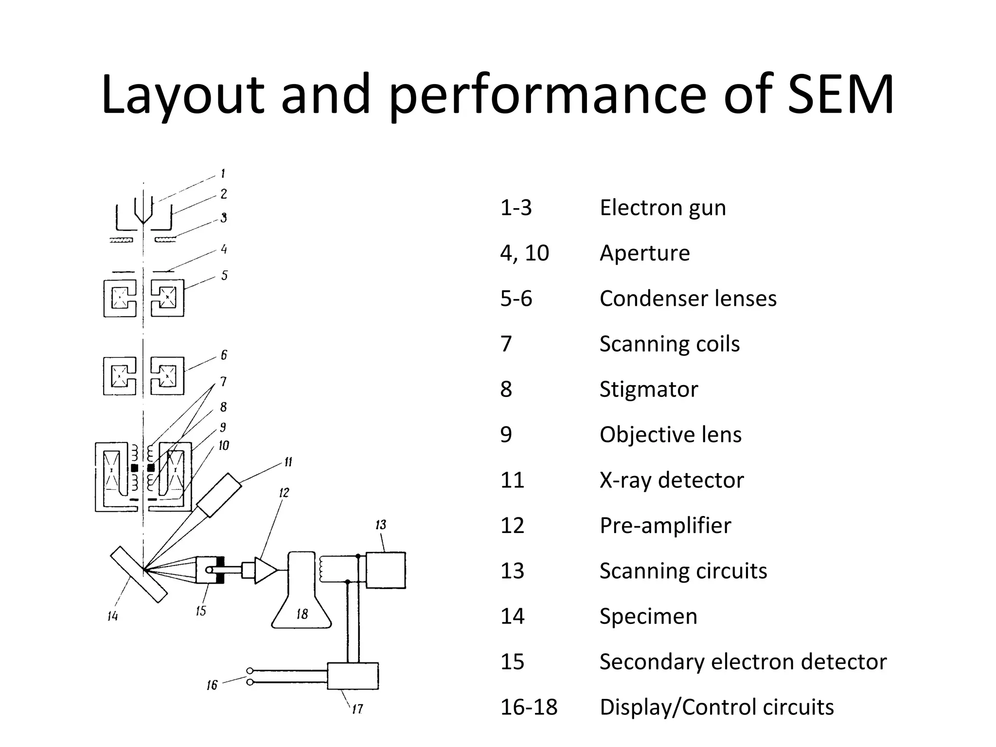
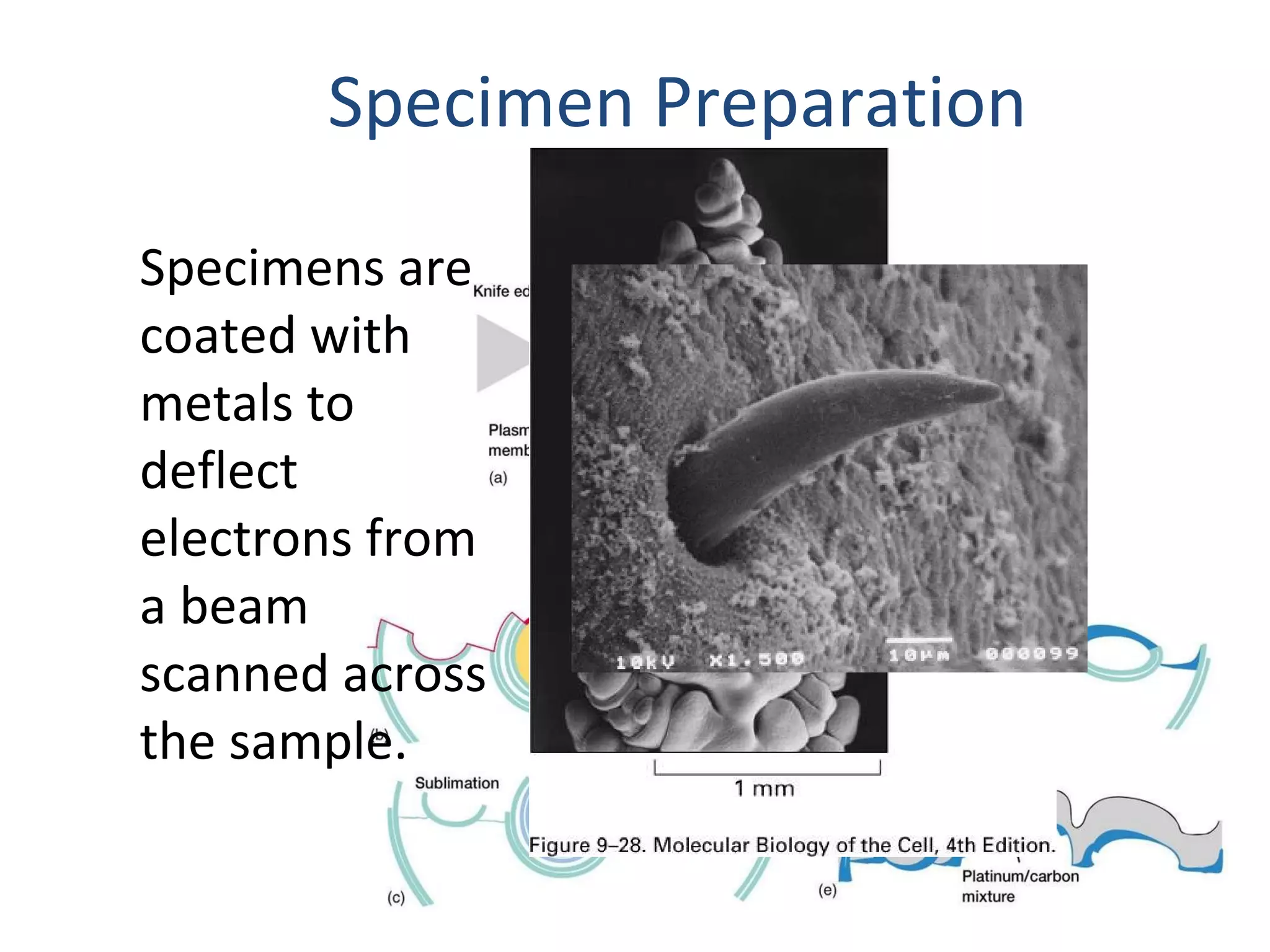
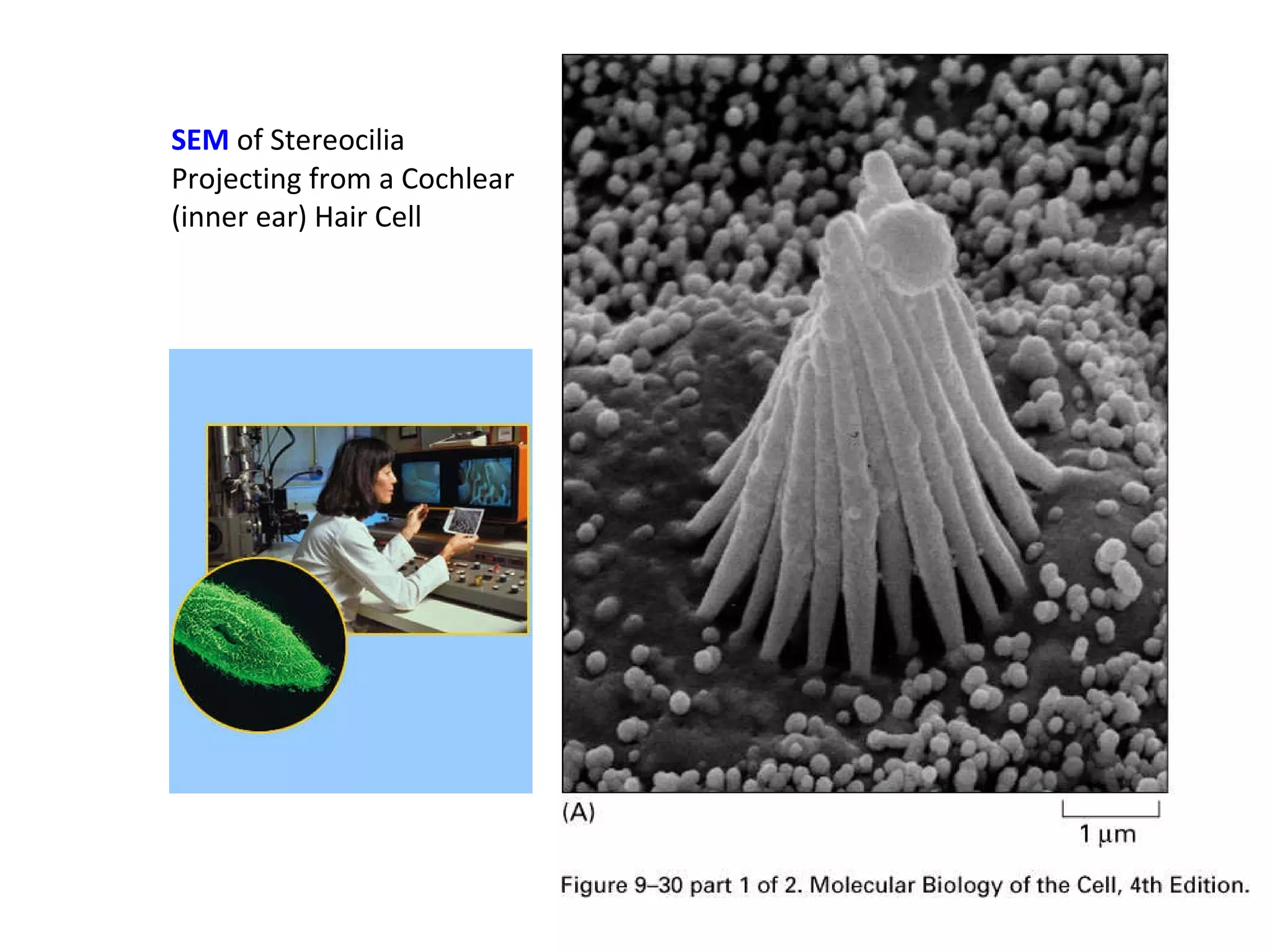
15. THE LIGHT MICROSCOPE v THE ELECTRON MICROSCOPE fluorescent (TV) screen, photographic film Human eye (retina), photographic film Focussing screen Vacuum Air-filled Interior Magnets Glass Lenses High voltage (50kV) tungsten lamp Tungsten or quartz halogen lamp Radiation source x500 000 x1000 – x1500 Maximum magnification 0.2nm Fine detail app. 200nm Maximum resolving power Electrons app. 4nm Monochrome Visible light 760nm (red) – 390nm Colours visible Electromagnetic spectrum used ELECTRON MICROSCOPE LIGHT MICROSCOPE FEATURE
16. THE LIGHT MICROSCOPE v THE ELECTRON MICROSCOPE Copper grid Glass slide Support Heavy metals Water soluble dyes Stains Microtome only. Slices 50nm Parts of cells visible Hand or microtome slices 20 000nm Whole cells visible Sectioning Resin Wax Embedding OsO 4 or KMnO 4 Alcohol Fixation Tissues must be dehydrated = dead Temporary mounts living or dead Preparation of specimens ELECTRON MICROSCOPE LIGHT MICROSCOPE FEATURE
34. Page 167 TABLE 6.3 Major Column Components of the TEM* Component Synonyms Function of Components Illumination System Electron Gun Gun, Source Generates electrons and provides first coherent crossover of electron beam Condenser Lens 1 C1, Spot Size Determines smallest illumination spot size on specimen (see Spot Size in Table 6.4) Condenser Lens 2 C2, Brightness Varies amount of illumination on specimen — in combination with C1 (see Brightness in Table 6.4) Condenser Aperture C2 Aperture Reduces spherical aberration, helps control amount of illumination striking specimen
35. Specimen Manipulation System Specimen Exchanger Specimen Air Lock Chamber and mechanism for inserting specimen holder Specimen Stage Stage Mechanism for moving specimen inside column of microscope Imaging System Objective Lens — Forms, magnifies, and focuses first image (see Focus in Table 6.4) Objective Aperture — Controls contrast and spherical aberration Intermediate Lens Diffraction Lens Normally used to help magnify image from objective lens and to focus diffraction pattern Intermediate Aperture Diffraction Aperture, Field Limiting Aperture Selects area to be diffracted Projector Lens 1 P1 Helps magnify image, possibly used in some diffraction work Projector Lens 2 P2 Same as P1
36. Observation and Camera Systems Viewing Chamber — Contains viewing screen for final image Binocular Microscope Focusing Scope Magnifies image on viewing screen for accurate focusing Camera — Contains film for recording