The document summarizes the key differences between light microscopes and electron microscopes. Light microscopes have a resolving power of about half the wavelength of visible light, limiting magnification to around 1500x. Electron microscopes use short wavelength electrons, allowing much higher resolving power and magnification over 500,000x. There are two main types: transmission electron microscopes (TEM) that pass electrons through thin samples, and scanning electron microscopes (SEM) that detect electrons reflected from surfaces to generate 3D images. TEMs require harsh chemical fixation of cells that can introduce artifacts.
Introduction to AS Biology and the importance of microscopy; differences between light and electron microscopy.
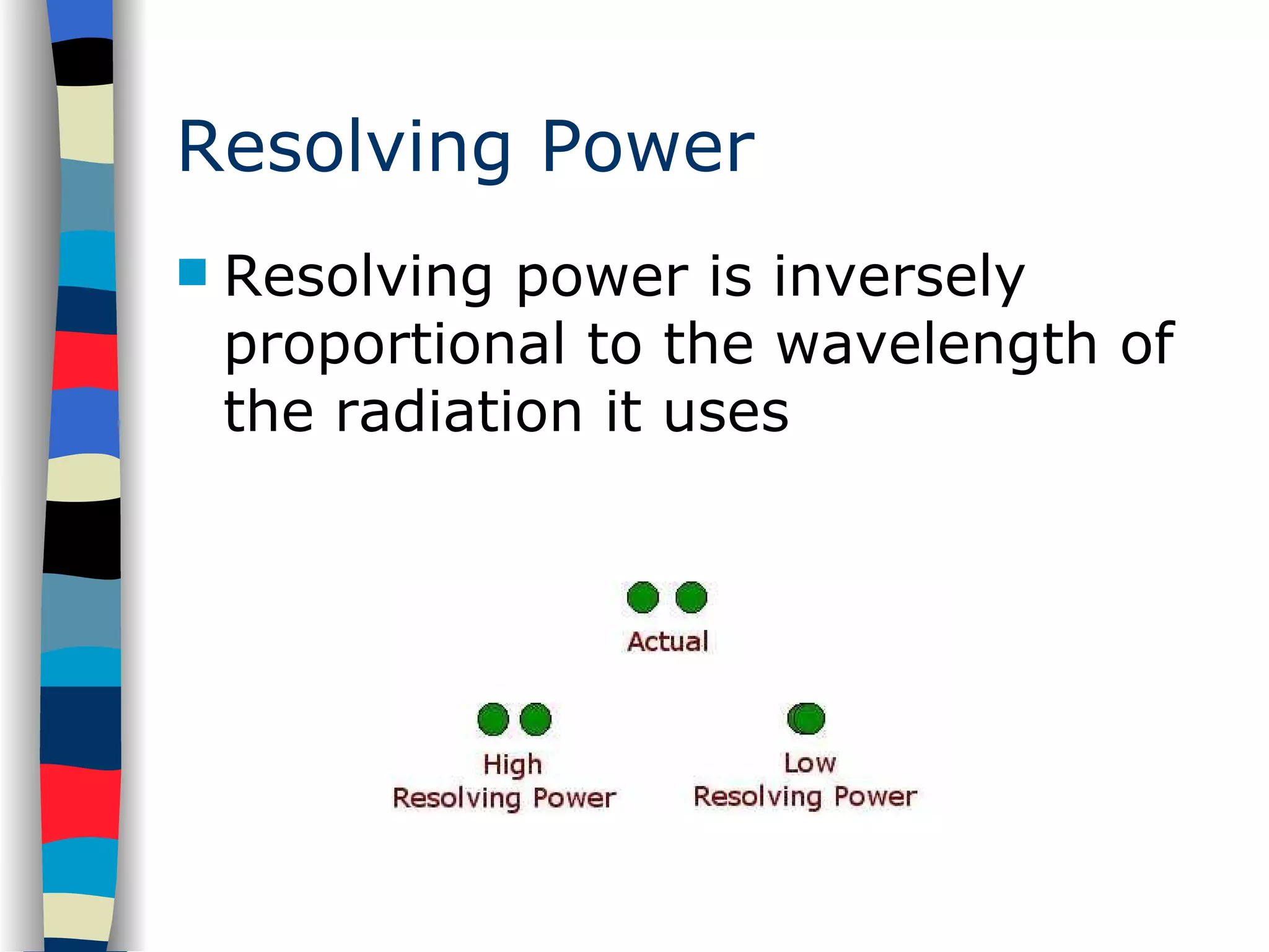
Defining resolving power and its significance in microscopy; affected by wavelength of radiation.
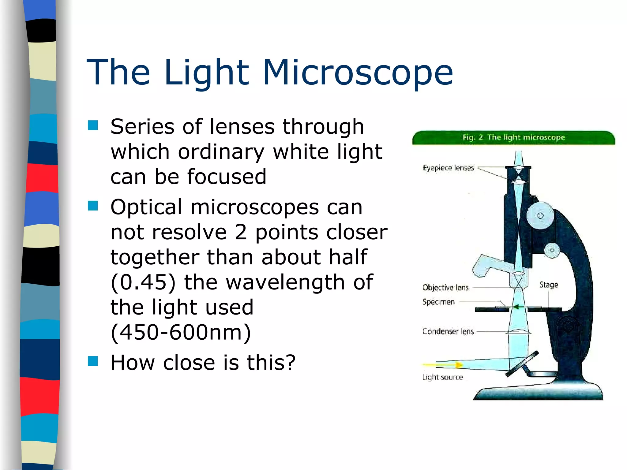
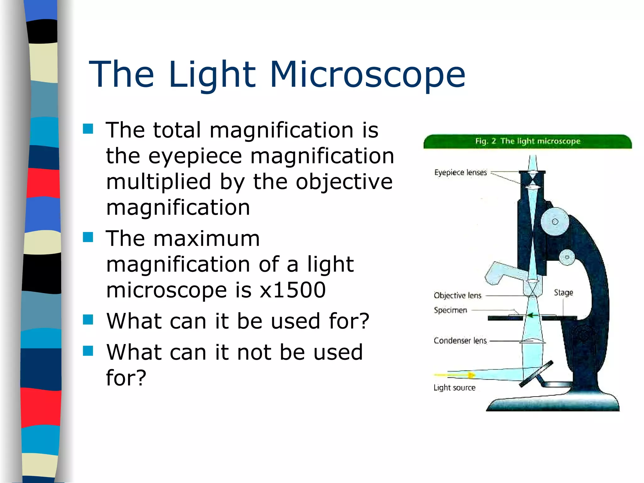
Description of light microscopes, their limitations, total magnification, and practical uses.
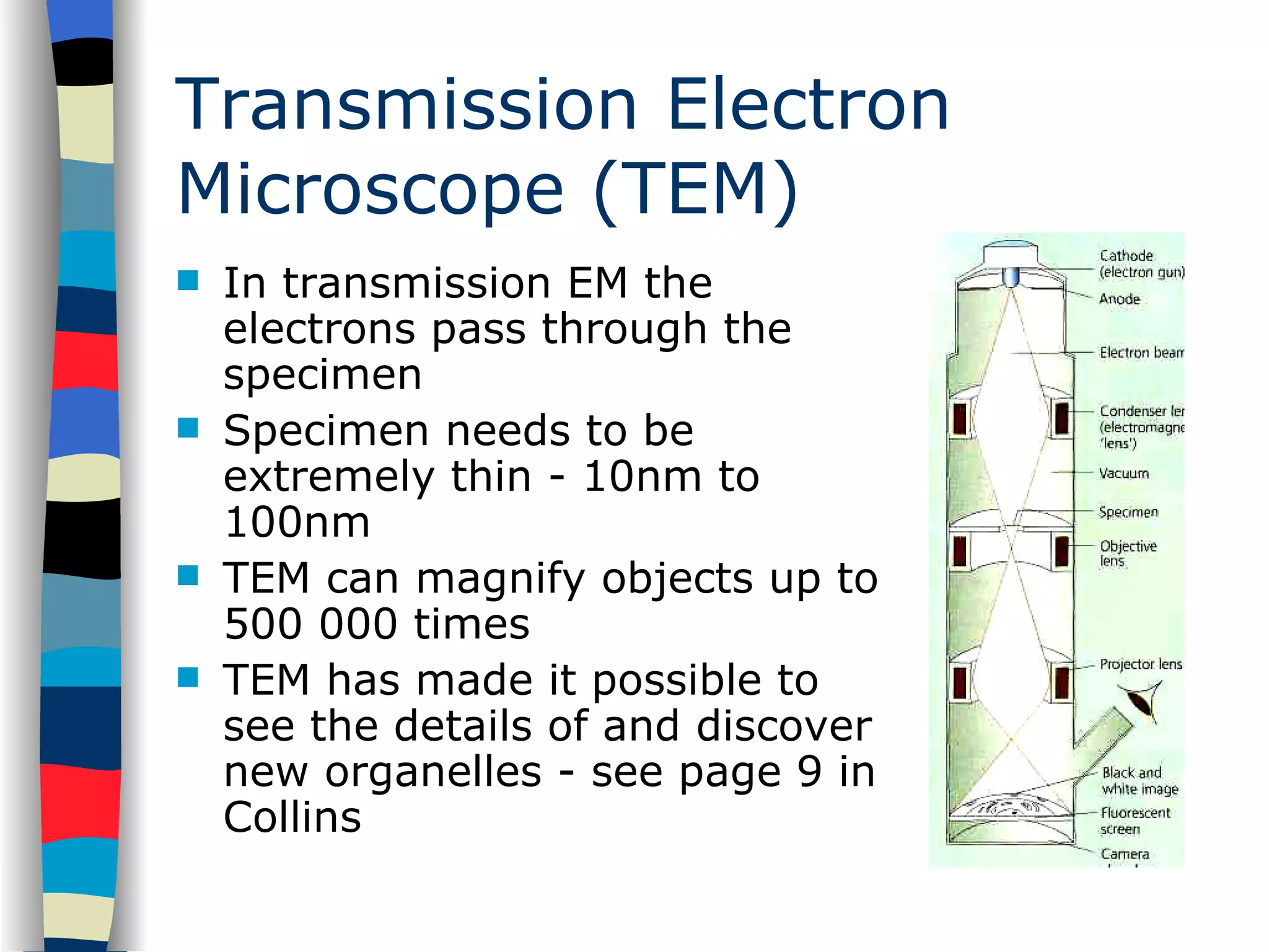
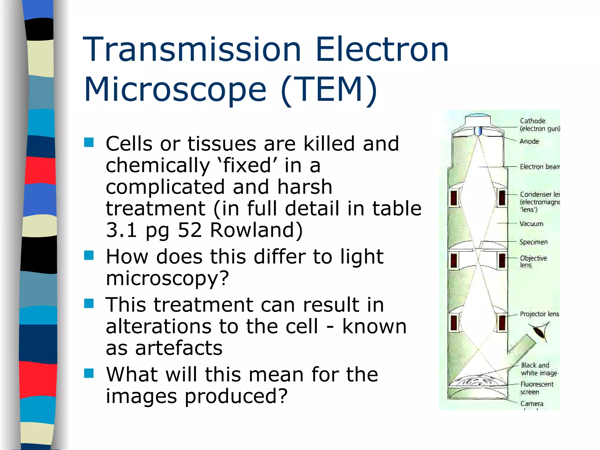
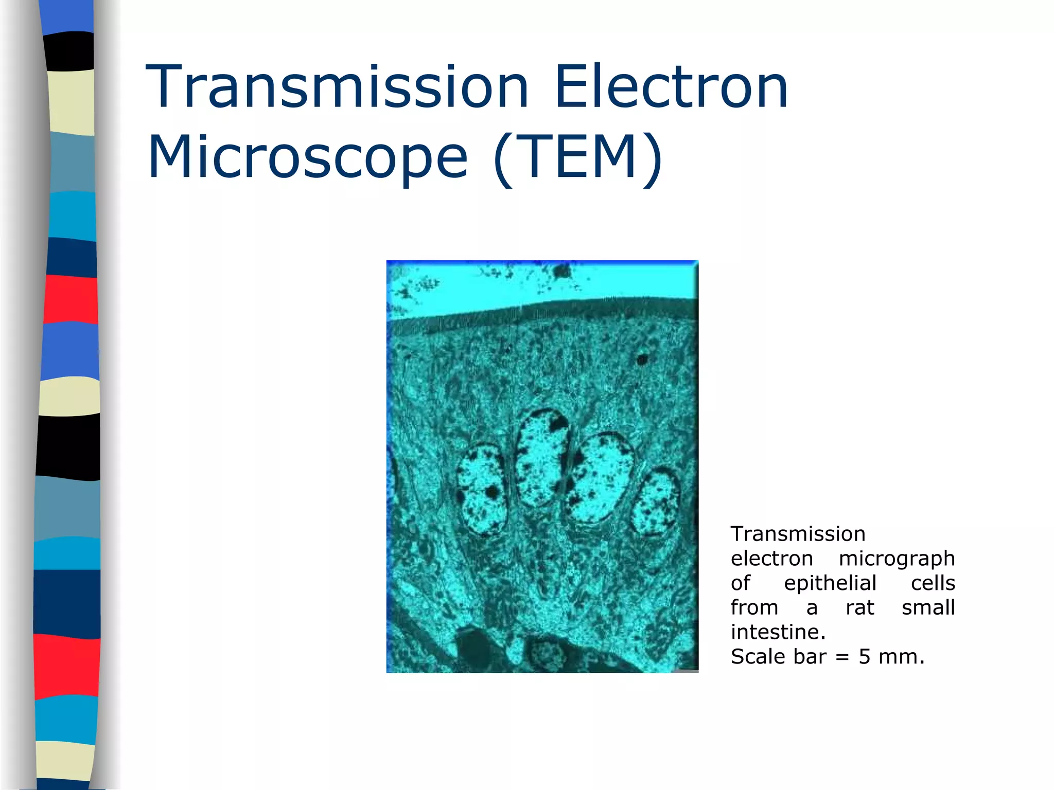
Explains electron microscopes; types: transmission and scanning, and their applications and requirements. TEM specifics: capabilities, specimen requirements, and impact on cell imaging with examples.
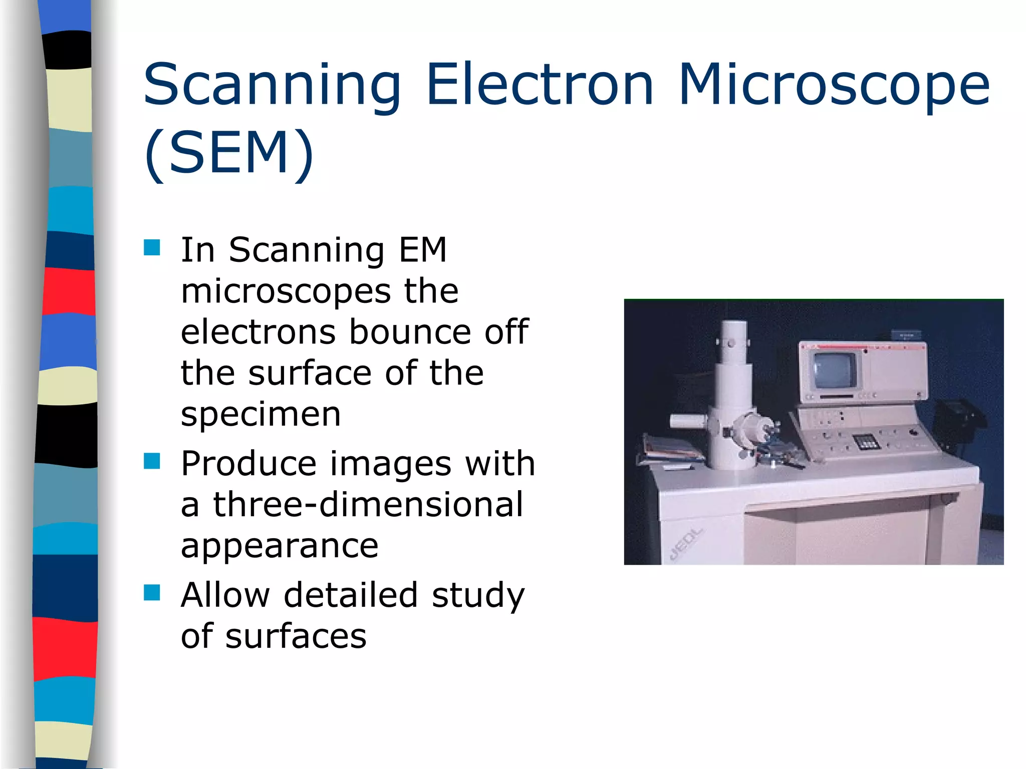
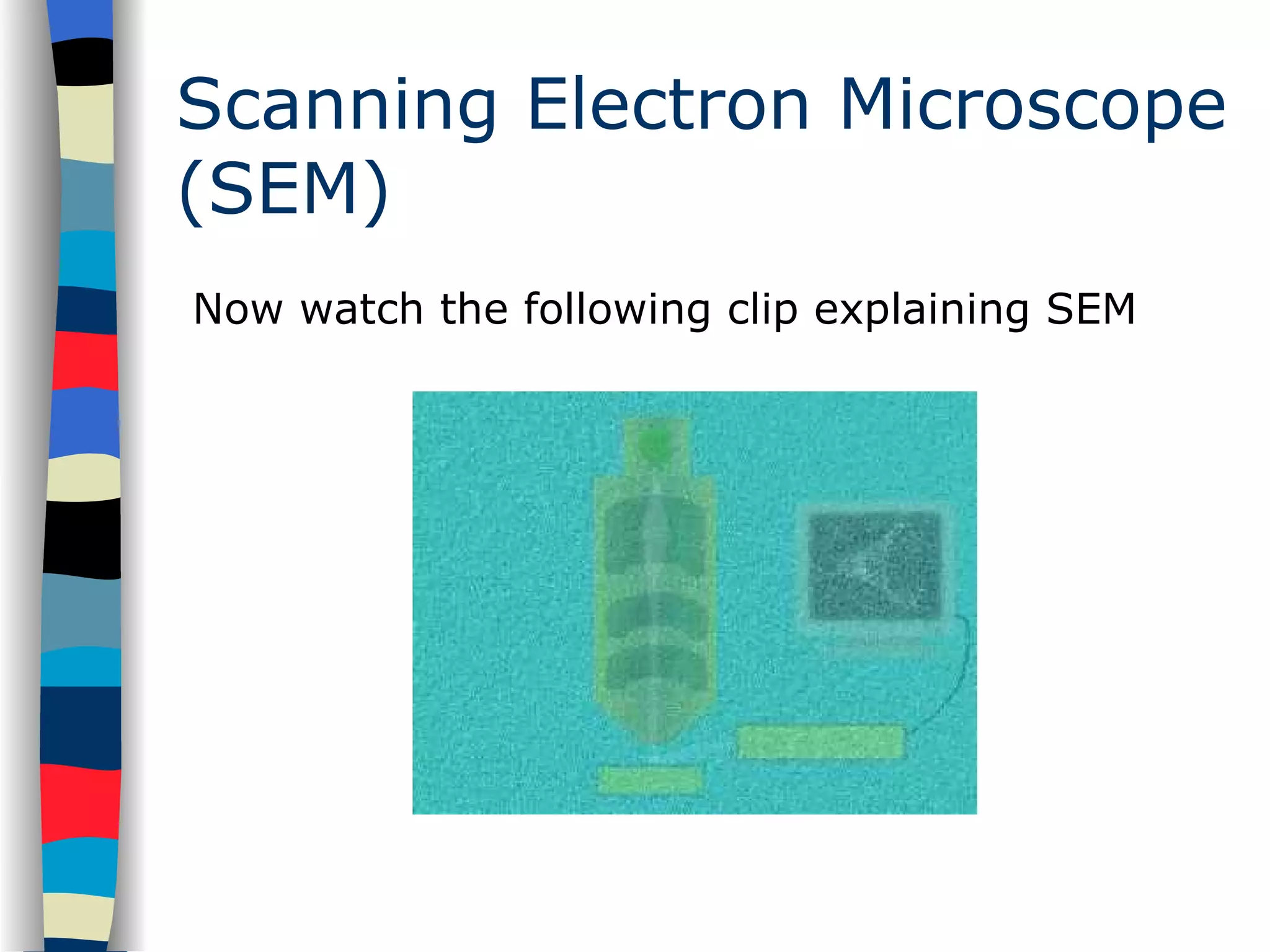
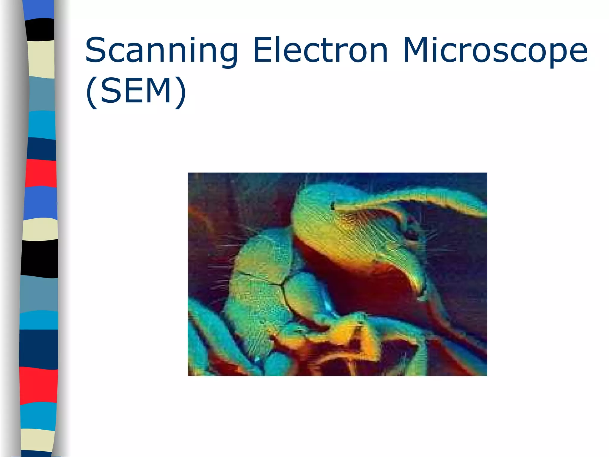
Examining the SEM's function, output, and 3D imaging capabilities with a recommendation for further viewing.
Further resources for learning about electron microscopy and related techniques.
AS Biology Core Principles The Electron Microscope dr shabeel p n
2.
Aims Resolving powerThe resolving power of light & electron microscopes The difference between the light & electron microscope Transmission & scanning electron microscopy
3.
Introduction Microscopes magnify& resolve images Microscopy began in 1665 when Robert Hooke coined the word ‘cells’ to describe the structure of cork You need to know about 2 types of microscope - light & electron You need to know how they work and the differences between them ‘ Its not how much they magnify that is key - but how well they resolve…’
4.
Resolving Power Thelimit of resolution of a microscope is the smallest distance between 2 points that can be seen using a microscope This is a measure of the clarity of the image A microscope with a high resolving power will allow 2 small objects which are close together to be seen as 2 distinct objects
The Light MicroscopeSeries of lenses through which ordinary white light can be focused Optical microscopes can not resolve 2 points closer together than about half (0.45) the wavelength of the light used (450-600nm) How close is this?
7.
The Light MicroscopeThe total magnification is the eyepiece magnification multiplied by the objective magnification The maximum magnification of a light microscope is x1500 What can it be used for? What can it not be used for?
8.
The Electron MicroscopeElectrons (negatively charged, very small particles) can behave as waves The wavelength of electrons is about 0.005nm What will this mean for the limit of resolution? Electrons are ‘fired’ from an electron gun at the specimen and onto a fluorescent screen or photographic plate Where is this technique commonly used? There are 2 types of electron microscopy - transmission and scanning Both focus an electron beam onto the specimen using electromagnets
9.
Transmission Electron Microscope(TEM) In transmission EM the electrons pass through the specimen Specimen needs to be extremely thin - 10nm to 100nm TEM can magnify objects up to 500 000 times TEM has made it possible to see the details of and discover new organelles - see page 9 in Collins
10.
Transmission Electron Microscope(TEM) Cells or tissues are killed and chemically ‘fixed’ in a complicated and harsh treatment (in full detail in table 3.1 pg 52 Rowland) How does this differ to light microscopy? This treatment can result in alterations to the cell - known as artefacts What will this mean for the images produced?
11.
Transmission Electron Microscope(TEM) Transmission electron micrograph of epithelial cells from a rat small intestine. Scale bar = 5 mm.
12.
Scanning Electron Microscope(SEM) In Scanning EM microscopes the electrons bounce off the surface of the specimen Produce images with a three-dimensional appearance Allow detailed study of surfaces