Downloaded 450 times
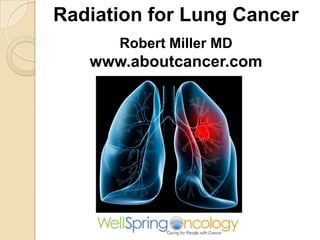
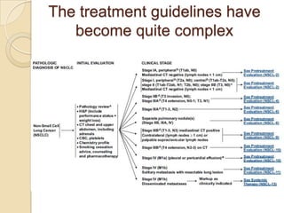
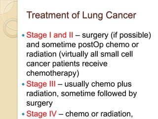
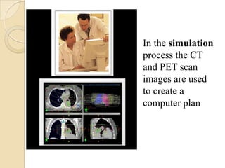
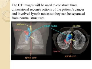
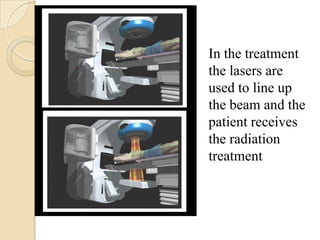
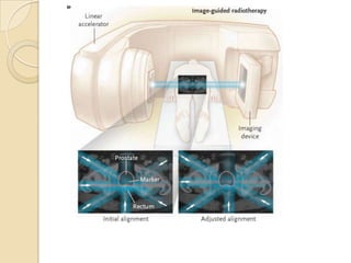
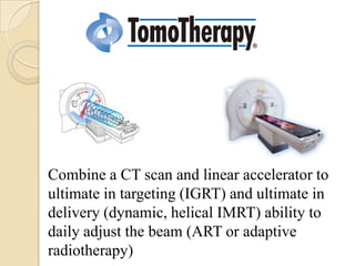
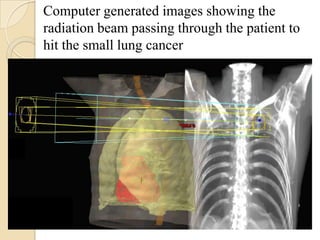
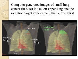
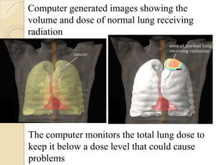
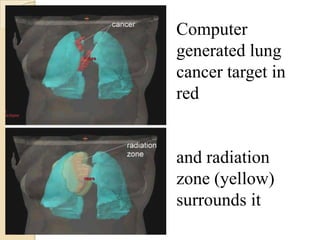
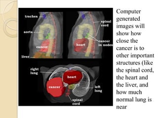
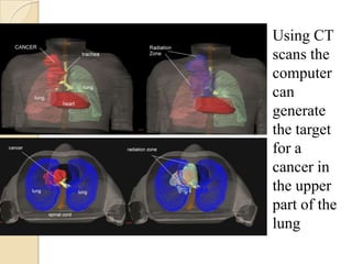
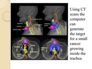
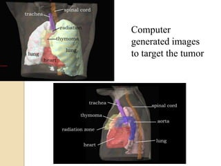
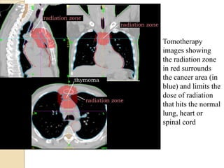
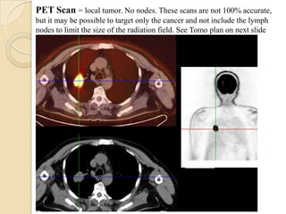
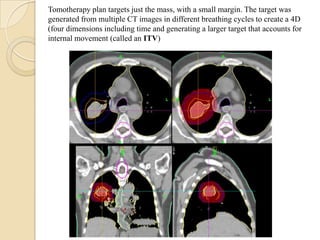
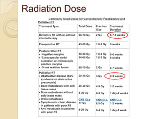
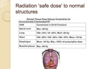
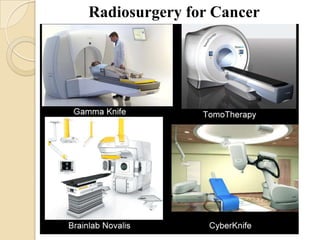
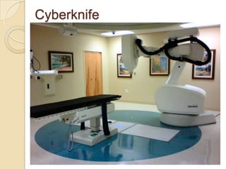
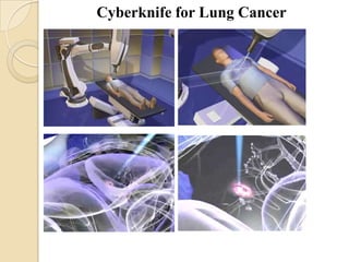
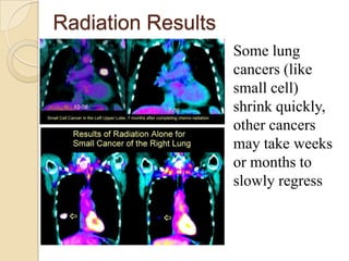
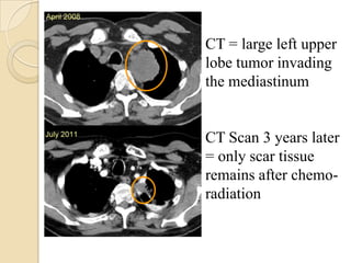
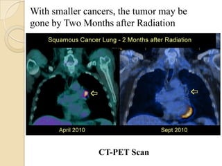
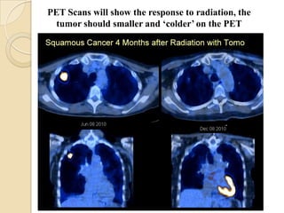
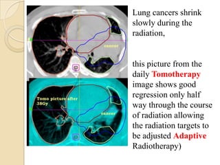
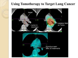
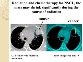
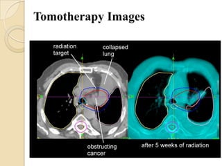
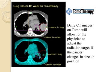
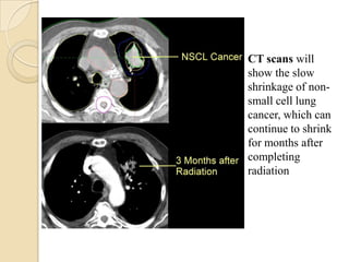
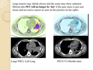
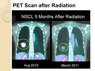
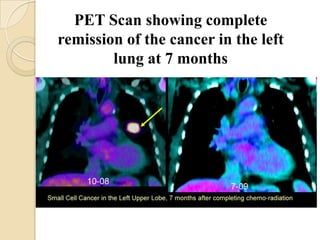
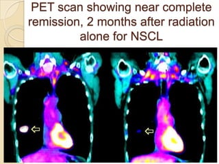
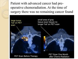
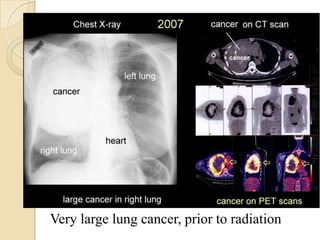
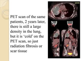
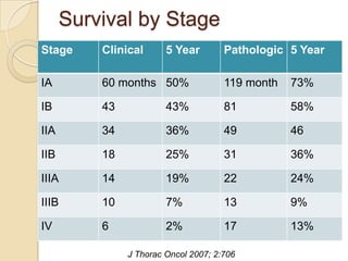
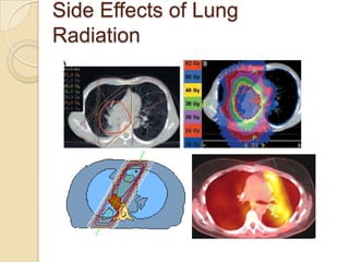
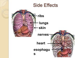
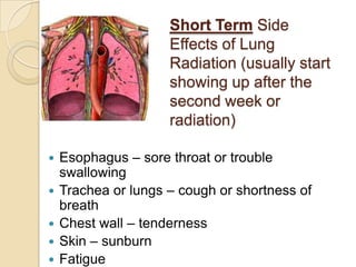
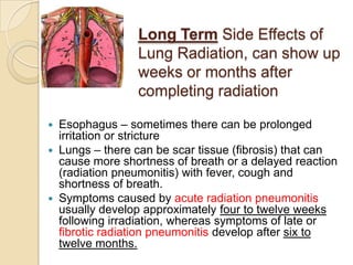
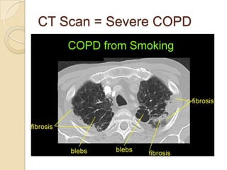
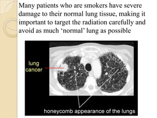
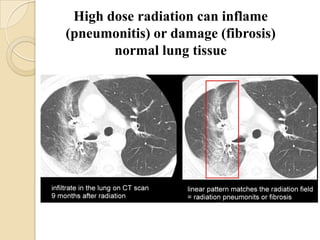
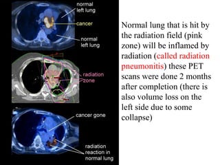
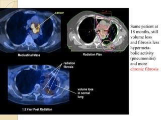
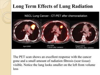
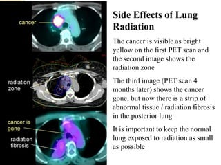
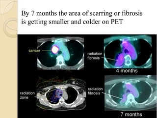
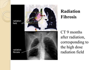
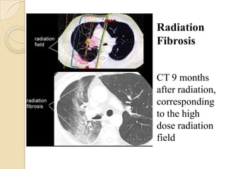
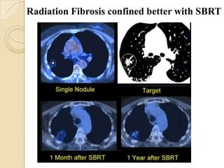

This document discusses the treatment of lung cancer with radiation. Stage I-II lung cancers are typically treated with surgery and sometimes post-operative chemotherapy or radiation. Stage III cancers usually receive chemotherapy and radiation, sometimes followed by surgery. Stage IV cancers are treated with chemotherapy or radiation. Advanced techniques like CT-guided planning, adaptive radiotherapy using daily CT images, and stereotactic body radiation therapy can help target radiation doses precisely to tumors while minimizing exposure to healthy lung tissue. Radiation is generally well-tolerated but can cause short-term effects like cough and long-term effects like fibrosis. Careful treatment planning aims to limit radiation doses to normal lungs.


![CASE_PRESENTATION_ON_subdural_hematoma(SDH)[1 FINAL PPT]-1.pptx](https://cdn.slidesharecdn.com/ss_thumbnails/casepresentationonsubduralhematomasdh1finalppt-1-260129172522-d405d375-thumbnail.jpg?width=640&height=640&fit=bounds)


