


![C 2008 by Blackwell Publishing
Blackwell Publishing, Inc., 350 Main Street, Malden, Massachusetts 02148-5020, USA
Blackwell Publishing Ltd, 9600 Garsington Road, Oxford OX4 2DQ, UK
Blackwell Publishing Asia Pty Ltd, 550 Swanston Street, Carlton, Victoria 3053, Australia
The right of the Author to be identified as the Author of this Work has been asserted in accordance with
the Copyright, Designs and Patents Act 1988.
All rights reserved. No part of this publication may be reproduced, stored in a retrieval system, or
transmitted, in any form or by any means, electronic, mechanical, photocopying, recording or
otherwise, except as permitted by the UK Copyright, Designs and Patents Act 1988, without the prior
permission of the publisher.
First published 1998
Third edition 2008
1 2008
Library of Congress Cataloging-in-Publication Data
Lung cancer / edited by Jack A. Roth, James D. Cox, Waun Ki Hong. – 3rd ed.
p. ; cm.
Includes bibliographical references and index.
ISBN 978-1-4051-5112-2 (alk. paper)
1. Lungs–Cancer. I. Roth, Jack A. II. Cox, James D. (James Daniel), 1938–
III. Hong, Waun Ki.
[DNLM: 1. Lung Neoplasms–therapy. 2. Lung Neoplasms–diagnosis. 3. Lung Neoplasms–genetics.
WF 658 L9604 2008]
RC280.L8L765 2008
616.99 424–dc22
ISBN: 978-1-4051-5112-2
A catalogue record for this title is available from the British Library
Set in 9.5/12pt Meridien by Aptara Inc., New Delhi, India
Printed and bound in Singapore by Fabulous Printers Pte Ltd
For further information on Blackwell Publishing, visit our website:
http://www.blackwellpublishing.com
The publisher’s policy is to use permanent paper from mills that operate a sustainable forestry policy,
and which has been manufactured from pulp processed using acid-free and elementary chlorine-free
practices. Furthermore, the publisher ensures that the text paper and cover board used have met
acceptable environmental accreditation standards.
Designations used by companies to distinguish their products are often claimed as trademarks. All brand
names and product names used in this book are trade names, service marks, trademarks or registered
trademarks of their respective owners. The Publisher is not associated with any product or vendor
mentioned in this book.
The contents of this work are intended to further general scientific research, understanding, and
discussion only and are not intended and should not be relied upon as recommending or promoting a
specific method, diagnosis, or treatment by physicians for any particular patient. The publisher and the
author make no representations or warranties with respect to the accuracy or completeness of the
contents of this work and specifically disclaim all warranties, including without limitation any implied
warranties of fitness for a particular purpose. In view of ongoing research, equipment modifications,
changes in governmental regulations, and the constant flow of information relating to the use of
medicines, equipment, and devices, the reader is urged to review and evaluate the information provided
in the package insert or instructions for each medicine, equipment, or device for, among other things,
any changes in the instructions or indication of usage and for added warnings and precautions. Readers
should consult with a specialist where appropriate. The fact that an organization or Website is referred to
in this work as a citation and/or a potential source of further information does not mean that the author
or the publisher endorses the information the organization or Website may provide or recommendations
it may make. Further, readers should be aware that Internet Websites listed in this work may have
changed or disappeared between when this work was written and when it is read. No warranty may be
created or extended by any promotional statements for this work. Neither the publisher nor the author
shall be liable for any damages arising herefrom.](https://image.slidesharecdn.com/lungcancer3rded-120126215311-phpapp01/85/Lung-cancer-3rd-ed-4-320.jpg)


![CHAPTER 1
Smoking Cessation
Alexander V. Prokhorov, Kentya H. Ford, and Karen Suchanek Hudmon
Overview resulting in nearly 440,000 deaths each year [3].
The economic implications are enormous: more
Tobacco use is a public health issue of enormous than $75 billion in medical expenses and over $81
importance, and smoking is the primary risk factor billion in loss of productivity as a result of pre-
for the development of lung cancer. Considerable mature death are attributed to smoking each year
knowledge has been gained with respect to biobe- [4–8]. While the public often associates tobacco use
havioral factors leading to smoking initiation and with elevated cancer risk, the negative health con-
development of nicotine dependence. Smoking ces- sequences are much broader. The 2004 Surgeon
sation provides extensive health benefits for every- General’s Report on the health consequences of
one. State-of-the-art treatment for smoking cessa- smoking [9] provides compelling evidence of the ad-
tion includes behavioral counseling in conjunction verse impact of smoking and concluded that smok-
with one or more FDA-approved pharmaceutical ing harms nearly every organ in the body (Table
aids for cessation. The US Public Health Service Clin- 1.1). In 2000, 8.6 million persons in the United
ical Practice Guideline for Treating Tobacco Use and De- States were living with an estimated 12.7 mil-
pendence advocates a five-step approach to smoking lion smoking-attributable medical conditions [10].
cessation (Ask about tobacco use, Advise patients to There is convincing evidence that stopping smok-
quit, Assess readiness to quit, Assist with quitting, ing is associated with immediate as well as long-
and Arrange follow-up). Health care providers are term health benefits, including reduced cumulative
encouraged to provide at least brief interventions at risk for cancer. This is true even in older individu-
each encounter with a patient who uses tobacco. als, and in patients who have been diagnosed with
cancer [11].
Introduction
Smoking and lung cancer
More than two decades ago, the former US Surgeon
General C. Everett Koop stated that cigarette smok- In the United States, approximately 85% of all
ing is the “chief, single, avoidable cause of death in lung cancers are in people who smoke or who
our society and the most important public health have smoked [3]. Lung cancer is fatal for most
issue of our time” [1]. This statement remains true patients. The estimated number of deaths of lung
today. In the United States, cigarette smoking is the cancer will exceed 1.3 million annually early in the
primary known cause of preventable deaths [2], third millennium [12]. Lung cancer is the leading
cause of cancer-related deaths among Americans
Lung Cancer, 3rd edition. Edited by Jack A. Roth, James D. Cox,
of both genders, with 174,470 estimated newly
and Waun Ki Hong. c 2008 Blackwell Publishing, diagnosed cases and 162,460 deaths [13,14]. The
ISBN: 978-1-4051-5112-2. number of deaths due to lung cancer exceeds the
1](https://image.slidesharecdn.com/lungcancer3rded-120126215311-phpapp01/85/Lung-cancer-3rd-ed-12-320.jpg)
![2 Chapter 1
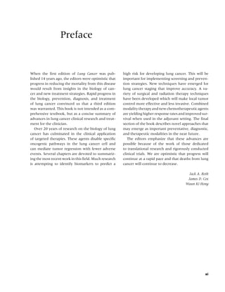
Table 1.1 Health consequences of smoking. annual number of deaths from breast, colon, and
prostate cancer combined [15]. Recent advances in
Cancer Acute myeloid leukemia
technology have enabled earlier diagnoses, and ad-
Bladder
vances in surgery, radiation therapy, imaging, and
Cervical
Esophageal chemotherapy have produced improved responses
Gastric rates. However, despite these efforts, overall sur-
Kidney vival has not been appreciably affected in 30 years,
Laryngeal and only 12–15% of patients with lung cancer are
Lung being cured with current treatment approaches
Oral cavity and pharyngeal
[16]. The prognosis of lung cancer depends largely
Pancreatic
on early detection and immediate, premetastasis
Cardiovascular Abdominal aortic aneurysm
stage treatment [17]. Prevention of lung cancer
diseases Coronary heart disease (angina pectoris, is the most desirable and cost-efficient approach
ischemic heart disease, myocardial
to eradicating this deadly condition. Numerous
infarction, sudden death)
epidemiologic studies consistently define smoking
Cerebrovascular disease (transient
as the major risk factor for lung cancer (e.g. [18–
ischemic attacks, stroke)
20]). The causal role of cigarette smoking in lung
Peripheral arterial disease
cancer mortality has been irrefutably established
Pulmonary Acute respiratory illnesses in longitudinal studies, one of which lasted as long
diseases Pneumonia
as 50 years [21]. Tobacco smoke, which is inhaled
Chronic respiratory illnesses either directly or as second-hand smoke, contains
Chronic obstructive pulmonary
an estimated 4000 chemical compounds, including
disease
over 60 substances that are known to cause cancer
Respiratory symptoms (cough,
[22]. Tobacco irritants and carcinogens damage the
phlegm, wheezing, dyspnea)
cells in the lungs, and over time the damaged cells
Poor asthma control
may become cancerous. Cigarette smokers have
Reduced lung function in infants lower levels of lung function than nonsmokers
exposed (in utero) to maternal
[9,23], and quitting smoking greatly reduces
smoking
cumulative risk for developing lung cancer [24].
Reproductive Reduced fertility in women
The association of smoking with the development
effects Pregnancy and pregnancy outcomes of lung cancer is the most thoroughly documented
Premature rupture of membranes
causal relationship in biomedical history [25]. The
Placenta previa
Placental abruption
link was first observed in the early 1950s through
Preterm delivery the research of Sir Richard Doll, whose pioneering
Low infant birth weight research has, perhaps more so than any other epi-
Infant mortality (sudden infant death demiologist of his time, altered the landscape of dis-
syndrome) ease prevention and consequently saved millions of
Other Cataract lives worldwide. In two landmark US Surgeon Gen-
effects Osteoporosis (reduced bone density in erals’ reports published within a 20-year interval (in
postmenopausal women, increased risk 1964 [26] and in 2004 [9]), literature syntheses fur-
of hip fracture) ther documented the strong link between smoking
Periodontitis and cancer. Compared to never smokers, smokers
Peptic ulcer disease (in patients who are have a 20-fold risk of developing lung cancer, and
infected with Helicobacter pylori) more than 87% of lung cancers are attributable to
Surgical outcomes smoking [27]. The risk for developing lung cancer
Poor wound healing increases with younger age at initiation of smoking,
Respiratory complications greater number of cigarettes smoked, and greater
number of years smoked [11]. Women smoking the
Source: Reference [9].](https://image.slidesharecdn.com/lungcancer3rded-120126215311-phpapp01/85/Lung-cancer-3rd-ed-13-320.jpg)
![Smoking Cessation 3
same amount as men experience twice the risk of Smoking among lung
developing lung cancer [28,29]. cancer patients
Tobacco use among patients with cancer is a se-
Second-hand smoke and rious health problem with significant implications
lung cancer for morbidity and mortality [36–39]. Evidence in-
dicates that continued smoking after a diagnosis
While active smoking has been shown to be the with cancer has substantial adverse effects on treat-
main preventable cause of lung cancer, second- ment effectiveness [40], overall survival [41], risk
hand smoke contains the same carcinogens that are of second primary malignancy [42], and increases
inhaled by smokers [30]. Consequently, there has the rate and severity of treatment-related complica-
been a concern since release of the 1986 US Sur- tions such as pulmonary and circulatory problems,
geon General’s report [31] concluding that second- infections, impaired would healing, mucositis, and
hand smoke causes cancer among nonsmokers and Xerostomia [43,44].
smokers. Although estimates vary by exposure lo- Despite the strong evidence for the role of smok-
cation (e.g., workplace, car, home), the 2000 Na- ing in the development of cancer, many cancer pa-
tional Household Interview Survey estimates that a tients continue to smoke. Specifically, about one
quarter of the US population is exposed to second- third of cancer patients who smoked prior to their
hand smoke [32]. Second-hand smoke is the third diagnoses continue to smoke [45] and among pa-
leading cause of preventable deaths in the United tients received surgical treatment of stage I nonsmall
States [33], and it has been estimated that expo- cell lung cancer [46] found only 40% who were ab-
sure to second-hand smoke kills more than 3000 stinent 2 years after surgery. Davison and Duffy [47]
adult nonsmokers from lung cancer [34]. Accord- reported that 48% of former smokers had resumed
ing to Glantz and colleagues, for every eight smok- regular smoking after surgical treatment of lung
ers who die from a smoking-attributable illness, one cancer. Therefore, among patients with smoking-
additional nonsmoker dies because of second-hand related malignancies, the likelihood of a positive
smoke exposure [35]. smoking history at and after diagnosis is high.
Since 1986, numerous additional studies have Patients who are diagnosed with lung cancer may
been conducted and summarized in the 2006 US face tremendous challenges and motivation to quit
Surgeon General’s report on “The Health Conse- after a cancer diagnosis can be influenced by a range
quences of Involuntary Exposure of Tobacco Smoke.” The of psychological variables. Schnoll and colleagues
report’s conclusions based on this additional ev- [48] reported that continued smoking among pa-
idence are consistent with the previous reports: tients with head and neck and lung cancer is asso-
exposure to second-hand smoke increases risk of ciated with lesser readiness to quit, having relatives
lung cancer. More than 50 epidemiologic stud- who smoke at home, greater time between diag-
ies of nonsmokers’ cigarette smoke exposure at noses and assessment, greater nicotine dependence,
the household and/or in the workplace showed lower self-efficacy, lower risk perception, fewer per-
an increased risk of lung cancer associated with ceived pros and greater cons to quitting, more fa-
second-hand smoke exposure [34]. This means that talistic beliefs, and higher emotional distress. Lung
20 years after second-hand smoke was first es- cancer patients should be advised to quit smoking,
tablished as a cause of lung cancer in lifetime but once they are diagnosed, some might feel that
nonsmokers, the evidence supporting smoking ces- there is nothing to be gained from quitting [49].
sation and reduction of second-hand smoke expo- Smoking cessation should be a matter of special
sure continues to mount. Eliminating second-hand concern throughout cancer diagnosis, treatment,
smoke exposure at home, in the workplaces, and and the survival continuum, and the diagnosis of
other public places appears to be essential for re- cancer should be used as a “teachable moment”
ducing the risk of lung cancer development among to encourage smoking cessation among patients,
nonsmokers. family members, and significant others [37]. The](https://image.slidesharecdn.com/lungcancer3rded-120126215311-phpapp01/85/Lung-cancer-3rd-ed-14-320.jpg)
![4 Chapter 1
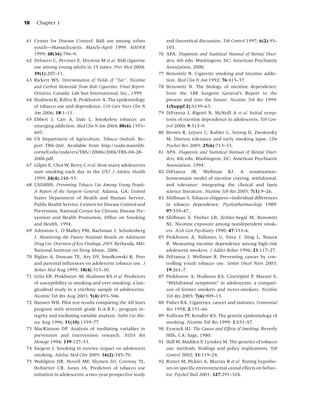
Table 1.2 Percentage of current cigarette smokersa aged ≥18 years, by selected characteristics—National Health
Interview Survey, United States, 2005.
Characteristic Category Men (n = 13,762) Women (n = 17,666) Total (n = 31,428)
Race/ethnicityb White, non-Hispanic 24.0 20.0 21.9
Black, non-Hispanic 26.7 17.3 21.5
Hispanic 21.1 11.1 16.2
American Indian/Alaska Native 37.5 26.8 32.0
Asianc 20.6 6.1 13.3
Educationd 0–12 years (no diploma) 29.5 21.9 25.5
GEDe (diploma) 47.5 38.8 43.2
High school graduate 28.8 20.7 24.6
Associate degree 26.1 17.1 20.9
Some college (no degree) 26.2 19.5 22.5
Undergraduate degree 11.9 9.6 10.7
Graduate degree 6.9 7.4 7.1
Age group (yrs) 18–24 28.0 20.7 24.4
25–44 26.8 21.4 24.1
45–64 25.2 18.8 21.9
≥65 8.9 8.3 8.6
Poverty levelf At or above 23.7 17.6 20.6
Below 34.3 26.9 29.9
Unknown 21.2 16.1 18.4
Total 23.9 18.1 20.9
a
Persons who reported having smoked at least 100 cigarettes during their lifetime and at the time of the interview reported
smoking every day or some days; excludes 296 respondents whose smoking status was unknown.
b
Excludes 314 respondents of unknown or multiple racial/ethnic categories or whose racial/ethnic category was unknown.
c
Excludes Native Hawaiians or other Pacific Islanders.
d
Persons aged ≥25 years, excluding 339 persons with unknown level of education.
e
General Educational Development.
f
Calculated on the basis of US Census Bureau 2004 poverty thresholds.
Source: Reference [7].
medical, psychosocial, and general health benefits proportion of the population continues to smoke.
of smoking cessation for cancer patients provide a In 2005, an estimated 45.1 million adult Americans
clear rationale for intervention. (20.9%) were current smokers; of these, 80.8% re-
ported to smoking every day, and 19.2% reported
smoking some days [7]. The prevalence of smoking
Forms of tobacco varies considerably across populations (Table 1.2),
with a greater proportion of men (23.9%) than
Smoked tobacco women (18.1%) reporting current smoking. Per-
Cigarettes have been the most widely used form of sons of Asian or Hispanic origin exhibit the low-
tobacco in the United States for several decades [51], est prevalence of smoking (13.3 and 16.2%, respec-
yet in recent years, cigarette smoking has been de- tively), and American Indian/Alaska natives exhibit
clining steadily among most population subgroups. the highest prevalence (32.0%). Also, the preva-
In 2005, just over half of ever smokers reported be- lence of smoking among adults varies widely across
ing former smokers [3]. However, a considerable the United States, ranging from 11.5% in Utah to](https://image.slidesharecdn.com/lungcancer3rded-120126215311-phpapp01/85/Lung-cancer-3rd-ed-15-320.jpg)
![Smoking Cessation 5
28.7% in Kentucky [51]. Twenty-three percent of less tobacco than standard cigarettes, bidis expose
high school students report current smoking, and their smokers to considerable amounts of hazardous
among boys, 13.6% report current use of smoke- compounds. A smoking machine-based investiga-
less tobacco, and 19.2% currently smoke cigars [52]. tion found that bidis deliver three times the amount
These figures are of particular concern, because of carbon monoxide and nicotine and almost five
nearly 90% of smokers begin smoking before the times the amount of tar found in conventional
age of 18 years [53]. cigarettes [63].
Other common forms of burned tobacco in the
United States include cigars, pipe tobacco, and bidis. Smokeless tobacco
Cigars represent a roll of tobacco wrapped in leaf to- Smokeless tobacco products, also commonly called
bacco or in any substance containing tobacco [54]. “spit tobacco,” are placed in the mouth to allow ab-
Cigars’ popularity has somewhat increased over the sorption of nicotine through the buccal mucosa. Spit
past decade [55]. The latter phenomenon is likely tobacco includes chewing tobacco and snuff. Chew-
to be explained by a certain proportion of smok- ing tobacco, which is typically available in loose leaf,
ers switching cigarettes for cigars and by adoles- plug, and twist formulations, is chewed or parked in
cents’ experimentation with cigars [56]. In 1998, the cheek or lower lip. Snuff, commonly available as
approximately 5% of adults had smoked at least one loose particles or sachets (resembling tea bags), has
cigar in the past month [57]. The nicotine content a much finer consistency and is generally held in
of cigars sold in the United States ranged from 5.9 the mouth and not chewed. Most snuff products
to 335.2 mg per cigar [58] while cigarettes have a in the United States are classified as moist snuff.
narrow range of total nicotine content, between 7.2 The users park a “pinch” (small amount) of snuff
and 13.4 mg per cigarette [59]. Therefore, one large between the cheek and gum (also known as dip-
cigar, which could contain as much tobacco as an ping) for 30 minutes or longer. Dry snuff is typically
entire pack of cigarettes is able to deliver enough sniffed or inhaled through the nostrils; it is used less
nicotine to establish and maintain physical depen- commonly [64].
dence [59]. In 2004, an estimated 3.0% of Americans 12 years
Pipe smoking has been declining steadily over the of age and older had used spit tobacco in the past
past 50 years [60]. It is a form of tobacco use seen month. Men used it at higher rates (5.8%) than
among less than 1% of Americans [60]. Bidi smok- women (0.3%) [60]. The prevalence of spit tobacco
ing is a more recent phenomenon in the United is the highest among 18- to 25-year-olds and is sub-
States. Bidis are hand-rolled brown cigarettes im- stantially higher among American Indians, Alaska
ported mostly from Southeast Asian countries. Bidis natives, residents of the southern states, and ru-
are wrapped in a tendu or temburni leaf [61]. Visually, ral residents [61,66]. The consumption of chew-
they somewhat resemble marijuana joints, which ing tobacco has been declining since the mid-1980s;
might make them attractive to certain groups of conversely, in 2005, snuff consumption increased by
the populations. Bidis are available in multiple fla- approximately 2% over the previous year [66], pos-
vors (e.g., chocolate, vanilla, cinnamon, strawberry, sibly because tobacco users are consuming snuff in-
cherry, mango, etc.), which might make them par- stead of cigarettes in locations and situations where
ticularly attractive to younger smokers. A survey smoking is banned.
of nearly 64,000 people in 15 states in the United
States revealed that young people (18–24 years of
age) reported higher rates of ever (16.5%) and Factors explaining tobacco use
current (1.4%) use of bidis then among older adults
(ages 25 plus years). With respect to sociodemo- Smoking initiation
graphic characteristics, the use of bidis is most com- In the United States, smoking initiation typically
mon among males, African Americans, and con- occurs during adolescence. About 90% of adult
comitant cigarette smokers [62]. Although featuring smokers have tried their first cigarette by 18 years](https://image.slidesharecdn.com/lungcancer3rded-120126215311-phpapp01/85/Lung-cancer-3rd-ed-16-320.jpg)
![6 Chapter 1
of age and 70% of daily smokers have become tine acts on the brain to produce a number of ef-
regular smokers by that age [67,68]. Because most fects [77,78] and immediately after exposure, nico-
adolescents who smoke at least monthly continue tine induces a wide range of central nervous sys-
to smoke into adulthood, youth-oriented tobacco tem, cardiovascular, and metabolic effects. Nicotine
preventions and cessation strategies are warranted stimulates the release of neurotransmitters, in-
[67,68]. Since the mid-1990s, by 2004, the past- ducing pharmacologic effects, such as pleasure
month prevalence had decreased by 56% in 8th and reward (dopamine), arousal (acetylcholine,
graders, 47% in 10th graders, and 32% in 12th norepinephrine), cognitive enhancement (acetyl-
graders [69]. In recent years, however, this down- choline), appetite suppression (norepinephrine),
ward trend has decelerated [69]. The downward learning and memory enhancement (glutamate),
trend is unlikely to be sustained without steady and mood modulation and appetite suppression (sero-
systematic efforts by health care providers in pre- tonin), and reduction of anxiety and tension
venting initiation of tobacco use and assisting young (β-endorphin and GABA) [78]. Upon entering the
smokers in quitting. brain, a bolus of nicotine activates the dopamine re-
A wide range of sociodemographic, behavioral, ward pathway, a network of nervous tissue in the
personal, and environmental factors have been ex- brain that elicits feelings of pleasure and stimulates
amined as potential predictors of tobacco exper- the release of dopamine.
imentation and initiation of regular tobacco use Although withdrawal symptoms are not the only
among adolescents. For example, it has been sug- consequence of abstinence, most cigarette smok-
gested that the prevalence of adolescent smok- ers do experience craving and withdrawal on ces-
ing is related inversely to parental socioeconomic sation [79], and, therefore, relapse is common [80].
status and adolescent academic performance [68]. The calming effect of nicotine reported by many
Other identified predictors of adolescent smoking users is usually associated with a decline in with-
include social influence and normative beliefs, neg- drawal effects rather than direct effects on nicotine
ative affect, outcome expectations associated with [53]. This rapid dose-response, along with the short
smoking, resistance skills (self-efficacy), engaging in half-life of nicotine (t 1/2 = 2 h), underlies tobacco
other risk-taking behaviors, exposure to smoking in users’ frequent, repeated administration, thereby
movies, and having friends who smoke [70–75]. perpetuating tobacco use and dependence. Tobacco
Although numerous studies have been successful users become proficient in titrating their nicotine
in identifying predictors of smoking initiation, few levels throughout the day to avoid withdrawal
studies have identified successful methods for pro- symptoms, to maintain pleasure and arousal, and
moting cessation among youth, despite the finding to modulate mood. Withdrawal symptoms include
that in 2005, more than half of high school cigarette depression, insomnia, irritability/frustration/anger,
smokers have tried to quit smoking in the past year anxiety, difficulty concentrating, restlessness, in-
and failed [52]. These results confirm the highly creased appetite/weight gain, and decreased heart
addictive nature of tobacco emphasizing the need rate [81,82].
for more effective methods for facilitating cessation The assumption that heavy daily use (i.e., 15–
among the young. 30 cigarettes per day), is necessary for dependence
to develop is derived from observations of “chip-
Nicotine addiction pers,” adult smokers who have not developed de-
Nicotine has come to be regarded as a highly addic- pendence despite smoking up to five cigarettes per
tive substance. Judging by the current diagnostic cri- day for many years [83,84]. Chippers do not tend
teria, tobacco dependence appears to be quite preva- to differ from other smokers in their absorption and
lent among cigarette smokers; more than 90% of metabolism of nicotine, causing some investigators
smokers meet the DSM-IV (Diagnostic and Statisti- to suggest that this level of consumption may be too
cal Manual of Mental Disorders) criteria for nicotine low to cause nicotine dependence. However, these
dependence [76]. Research has shown that nico- atypical smokers are usually eliminated from most](https://image.slidesharecdn.com/lungcancer3rded-120126215311-phpapp01/85/Lung-cancer-3rd-ed-17-320.jpg)
![Smoking Cessation 7
studies, which are routinely limited to smokers of smoked, number of cigarettes smoked per day) will
at least 10 cigarettes per day [83]. be more similar for persons who are related geneti-
Signs of dependence on nicotine have been re- cally (i.e., biologically) than for persons who are not
ported among adolescent smokers, with approx- related genetically. Hence, one would expect to ob-
imately one fifth of them exhibiting adult-like serve greater similarities between children and their
dependence [85]. Although, lengthy and regular biological parents and siblings than would be ob-
tobacco use has been considered necessary for served between children and their adoptive parents
nicotine dependence to develop [68], recent re- or adopted siblings. Indeed, research has demon-
ports have raised concerns that nicotine depen- strated stronger associations (i.e., higher correlation
dence symptoms can develop soon after initiation, coefficients) between biologically-related individu-
and that these symptoms might lead to smoking als, compared to nonbiologically-related individu-
intensification [79,86]. Adolescent smokers, who als, for the reported number of cigarettes consumed
use tobacco regularly, tend to exhibit high craving [90]. In recent years, it has become more difficult
for cigarettes and substantial levels of withdrawal to conduct adoption studies, because of the reduced
symptoms [87]. number of intranational children available for adop-
tion [91]. Additionally, delayed adoption (i.e., time
elapsed between birth and entry into the new fam-
Genetics of tobacco use and ily) is common with international adoptions and
dependence might lead to an overestimation of genetic effects
if early environmental influences are attributed to
As early as 1958, Fisher hypothesized that the link genetic influences [92].
between smoking and lung cancer could be ex- In twin studies, identical (monozygotic) twins
plained at least in part by shared genes that predis- and fraternal (dizygotic) twins are compared. Iden-
pose individuals to begin smoking as young adults tical twins share the same genes; fraternal twins,
and to develop lung cancer later in adulthood [88]. like ordinary siblings, share approximately 50% of
More recently, tobacco researchers have begun to their genes. If a genetic link exists for the phe-
explore whether genetic factors do in fact contribute nomenon under study, then one would expect to
toward tobacco use and dependence. see a greater concordance in identical twins than
Tobacco use and dependence are hypothesized to in fraternal twins. Thus, in the case of tobacco
result from an interplay of many factors (includ- use, one would expect to see a greater proportion
ing pharmacologic, environmental and physiologic) of identical twins with the same tobacco use be-
[77]. Some of these factors are shared within fam- havior than would be seen with fraternal twins.
ilies, either environmentally or genetically. Studies Statistically, twin studies aim to estimate the per-
of families consistently demonstrate that, compared centage of the variance in the behavior that is
to family members of nonsmokers, family members due to (1) genes (referred to as the “heritability”),
of smokers are more likely to be smokers also. How- (2) shared (within the family) environmental ex-
ever, in addition to shared genetic predispositions, periences, and (3) nonshared (external from the
it is important to consider environmental factors family) environmental experiences [91]. A num-
that promote tobacco use—siblings within the same ber of twin studies of tobacco use have been con-
family share many of the same environmental in- ducted in recent years. These studies have largely
fluences as well as the same genes. To differentiate supported a genetic role [91,93]; higher concor-
the genetic from the environmental influences, epi- dance of tobacco use behavior is evident in identical
demiologists use adoption, twin, twins reared apart, twins than in fraternal twins. The estimated aver-
and linkage study designs [89]. age heritability for smoking is 0.53 (range, 0.28–
Key to the adoption studies is the assumption that 0.84) [93,94]; approximately half of the variance
if a genetic link for tobacco use exists, then tobacco in smoking appears to be attributable to genetic
use behaviors (e.g., smoking status, number of years factors.](https://image.slidesharecdn.com/lungcancer3rded-120126215311-phpapp01/85/Lung-cancer-3rd-ed-18-320.jpg)
![8 Chapter 1
Recent advances in the mapping of the human metabolism via the cytochrome P450 liver enzymes
genome have enabled researchers to search for (specifically, CYP2A6 and CYP2D6).
genes associated with specific disorders, including In summary, each of these types of study designs
tobacco use. Using a statistical technique called link- supports the hypothesis that genetics influence the
age analysis, it is possible to identify genes that pre- risk for a wide range of tobacco-related phenotypes,
dict a trait or disorder. This process is not based on such as ever smoking, age at smoking onset, level
prior knowledge of a gene’s function, but rather it of smoking, ability to quit, and the metabolic path-
is determined by examining whether the trait or ways of nicotine (e.g., see [45,89,95–99]). But given
disorder is coinherited with markers found in spec- that there are many predictors of tobacco use and
ified chromosomal regions. Typically, these types dependence, of which genetic predisposition is just
of investigations involve collection of large family one piece of a complex puzzle, it is unlikely that so-
pedigrees, which are studied to determine inheri- ciety will move toward widespread genotyping for
tance of the trait or disorder. This method works early identification of individuals who are at risk
well when a single gene is responsible for the out- for tobacco use. Perhaps a more likely use of ge-
come; however, it becomes more difficult when netics as related to tobacco use is its potential for
multiple genes have an impact, such as with to- improving our treatment for dependence [91]. If
bacco use. In linkage studies of smoking, it is com- genetic research leads to new knowledge regarding
mon for investigators to identify families, ideally the mechanisms underlying the development and
with two or more biologically-related relatives that maintenance of dependence, it is possible that new,
have the trait or disorder under study (referred to more effective medications might be created. Fur-
as affected individuals, in this case, smokers) and thermore, through pharmacogenomics research we
other unaffected relatives. For example, data from might gain improved knowledge as to which pa-
affected sibling pairs with parents is a common de- tients, based on their genetic profiles, would be best
sign in linkage analysis. A tissue sample (typically treated with which medications. Researchers are be-
blood) is taken from each individual, and the sample ginning to examine how DNA variants affect health
undergoes genotyping to obtain information about outcome with pharmacologic treatments, with a
the study participant’s unique genetic code. If a goal of determining which genetic profiles respond
gene in a specific region of a chromosome is as- most favorably to specific pharmaceutical aids for
sociated with smoking, and if a genetic marker is cessation (e.g. [98,100–103]).
linked (i.e., in proximity), then the affected pairs
(such as affected sibling pairs) will have increased
odds for sharing the same paternal/maternal gene Benefits of quitting
[91].
As genetic research moves forward, new clues The reports of the US Surgeon General on the
provide insight into which genes might be promis- health consequences of smoking, released in 1990
ing “candidates” as contributors to tobacco use and and 2004, summarize abundant and significant
dependence. Currently, there are two general lines health benefits associated with giving up tobacco
of research related to candidate genes for smoking. [9,104]. Benefits noticed shortly after quitting (e.g.,
One examines genes that affect nicotine pharmaco- within 2 weeks to 3 months), include improvements
dynamics (the way that nicotine affects the body) in pulmonary function and circulation. Within
and the other examines genes that affect nicotine 1–9 months of quitting, the ciliary function of
pharmacokinetics (the way that the body affects the lung epithelium is restored. Initially, patients
nicotine). A long list of candidate genes are being might experience increased coughing as the lungs
examined—some of the most extensively explored clear excess mucus and tobacco smoke particu-
involve (a) the dopamine reward pathway (e.g., lates. In several months, smoking cessation results
those related to dopamine synthesis, receptor acti- in measurable improvements of lung function. Over
vation, reuptake, and metabolism) and (b) nicotine time, patients experience decreased coughing, sinus](https://image.slidesharecdn.com/lungcancer3rded-120126215311-phpapp01/85/Lung-cancer-3rd-ed-19-320.jpg)
![Smoking Cessation 9
congestion, fatigue, shortness of breath, and risk for positive influence on survival rates. Although many
pulmonary infection and 1 year postcessation, the smoking cessation interventions are aimed at pri-
excess risk for coronary heart disease is reduced to mary prevention of cancer, these results indicate
half that of continuing smokers. After 5–15 years, that there can be substantial medical benefits for
the risk for stroke is reduced to a rate similar to individuals who quit smoking after they are diag-
that of people who are lifetime nonsmokers, and nosed with cancer.
10 years after quitting, an individual’s chance of
dying of lung cancer is approximately half that of Smoking cessation interventions
continuing smokers. Additionally, the risk of devel-
oping mouth, larynx, pharynx, esophagus, bladder, Effective and timely administration of smoking ces-
kidney, or pancreatic cancer is decreased. Finally, sation interventions can significantly reduce the risk
15 years after quitting, a risk for coronary heart dis- of smoking-related disease [110]. Recognizing the
ease is reduced to a rate similar of that of people who complexity of tobacco use is a necessary first step
have never smoked. Smoking cessation can also lead in developing effective interventions and trials for
to a significant reduction in the cumulative risk for cessation and prevention. The biobehavioral model
death from lung cancer, for males and females. of nicotine addiction and tobacco-related cancers
Smokers who are able to quit by age 35 can be presents the complex interplay of social, psycho-
expected to live an additional 6–9 years compared logical, and biological factors that influence tobacco
to those who continue to smoke [105]. Ossip-Klein use and addiction (Figure 1.1). These factors in turn
et al. [106] recently named tobacco use a “geriatric mediate dependence, cessation, and relapse in most
health issue.” Indeed, a considerable proportion of individuals, and treatment has been developed to
tobacco users continue to smoke well into their 70s address many of the factors noted in the model [38].
and 80s, despite the widespread knowledge of the
tobacco health hazards. Elderly smokers frequently The health care provider’s role
claim that the “damage is done,” and it is “too late and responsibility
to quit;” however, a considerable body of evidence Health care providers are uniquely positioned to
refutes these statements. Even individuals who assist patients with quitting, having both access to
postpone quitting until age 65 can incur up to four quitting aids and commanding a level of respect that
additional years of life, compared with those who renders them particularly influential in advising pa-
continued to smoke [24,106]. Therefore, elderly tients on health-related issues. To date, physicians
smokers should not be ignored as a potential target have received the greatest attention in the scien-
for cessation efforts. Health care providers ought to tific community as providers of tobacco cessation
remember that it is never too late to advise their treatment. Although less attention has been paid to
elderly patients to quit and to incur health benefits. other health care providers such as pharmacists and
A growing body of evidence indicates that con- nurses, they too are in a unique position to serve
tinued smoking after a diagnosis of cancer has the public and situated to initiate behavior change
substantial adverse effects. For example, these among patients or complement the efforts of other
studies indicate that smoking reduces the over- providers [64,111].
all effectiveness of treatment, while causing com- Fiore and associates conducted a meta-analysis
plications with healing as well as exacerbating of 29 investigations in which they estimated that
treatment side effects, increases risk of developing compared with smokers who do not receive an in-
second primary malignancy, and decreases over- tervention from a clinician, patients who receive
all survival rates [36–38,107–109]. On the other a tobacco cessation intervention from a physician
hand, the medical, health, and psychosocial bene- clinician or a nonphysician clinician are 2.2 and
fits of smoking cessation among cancer patients are 1.7 times as likely to quit smoking at 5 or more
promising. Gritz et al. [37] indicated that stopping months postcessation, respectively [112]. Although
smoking prior to diagnosis and treatment can have a brief advice from a clinician has been shown to](https://image.slidesharecdn.com/lungcancer3rded-120126215311-phpapp01/85/Lung-cancer-3rd-ed-20-320.jpg)
![10 Chapter 1
Social factors
• Culture
• Socio-economic status
• Media/peer/family influences
• Politics
Psychological factors
• Comorbidity Tobacco use,
• Personality Behavioral, Dependence,
Cancer
• Stress Neurochemical, Cessation, Relapse
Physiological factors
Biological factors
• Genetics
• Nutrition
Figure 1.1 Biobehavioral model of nicotine addiction and tobacco-related cancers. (Adapted from [38].)
lead to increased likelihood of quitting, more in- from self-help materials to individual cognitive–
tensive counseling leads to more dramatic increases behavioral therapy, enable individuals to more ef-
in quit rates [112]. Because the use of pharma- fectively recognize high-risk smoking situations, de-
cotherapy agents approximately doubles the odds velop alternative coping strategies, manage stress,
of quitting [7,112], smoking cessation interventions improve problem-solving skills, and increase social
should consider combining pharmacotherapy with support [113]. The Clinical Practice Guideline out-
behavioral counseling. lines a five-step framework that clinicians can apply
To assist clinicians and other health care providers when assisting patients with quitting. Health care
in providing cessation treatment, the US Public providers should: (a) systematically identify all to-
Health Service has produced a Clinical Practice Guide- bacco users, (b) strongly advise all tobacco users to
line for the Treatment of Tobacco Use and Dependence quit, (c) assess readiness to make a quit attempt, (d)
[112]. The Guideline is based on a systematic re- assist patients in quitting, and (e) arrange follow-up
view and analysis of scientific literature which yields contact. The steps have been described as the 5 A’s:
a series of recommendations and strategies to as- Ask, Advise, Assess, Assist, and Arrange follow-up
sist health care providers in delivering smoking (Table 1.3). Due to the possibility of relapse, health
cessation treatment. The Guideline emphasizes the care providers should also provide patients with
importance of systematic identification of tobacco brief relapse prevention treatment. Relapse preven-
users by health care workers and offering at least tion reinforces the patient’s decision to quit, reviews
brief treatment interventions to every patient who the benefits of quitting, and assists the patient in re-
uses tobacco. Among the most effective approaches solving any problems arising from quitting [112].
for quitting are behavioral counseling and pharma- The outlined strategy has been termed the 5 R’s
cotherapy, used alone or, preferably, in combination (Table 1.3): Relevance, Risks, Rewards, Roadblocks,
[112]. and Repetition. In the absence of time or expertise
for providing more comprehensive counseling, clin-
Behavioral counseling icians are advised to (at a minimum), ask about to-
Behavioral interventions play an integral role in bacco use, advise tobacco users to quit, and refer
smoking cessation treatment, either alone or in con- these patients to other resources for quitting, such
junction with pharmacotherapy. These interven- as a toll-free tobacco cessation quitline (1-800-QUIT
tions, which include a variety of methods ranging NOW, in the US).](https://image.slidesharecdn.com/lungcancer3rded-120126215311-phpapp01/85/Lung-cancer-3rd-ed-21-320.jpg)
![Smoking Cessation 11
Table 1.3 The 5 A’s and 5 R’s for smoking cessation interventions.
5 A’s Ask about tobacco use Identify and document tobacco use status for every patient at every visit
Advise to quit Urge every tobacco user to quit in a clear, strong, and personalized manner
Assess readiness to make a Assess whether or not the tobacco user is ready to make a quit attempt in
quit attempt the next 30 days
Assist in quit attempt Use counseling and/or pharmacotherapy with the patient willing to make a
quit attempt to help him or her quit
Arrange follow-up Schedule follow-up contact, preferably within the first week after the quit
date
5 R’s Relevance Encourage the patient to indicate why quitting is personally relevant,
being specific as possible
Risk Ask the patient to identify the negative consequences of tobacco use,
including acute risks (e.g., short breath), long-term risks (e.g., cancer, and
environmental risks, e.g., cancer among family)
Rewards Request that the patient identify potential benefits of stopping tobacco
use (e.g., improved health)
Roadblocks Ask the patient to identify barriers or impediments to quitting and note
the elements of treatment that could address such barriers (e.g.,
withdrawal symptoms, fear of failure, lack of support)
Repetition Repeat the motivational intervention every time an unmotivated patient
visits the clinic setting
Adapted from [112].
Pharmaceutical aids for nicotine oral inhaler), sustained-release bupropion,
smoking cessation and varenicline tartrate. These are described in brief
below, and summaries of the prescribing informa-
According to the Clinical Practice Guideline [112], tion for each medication are provided in Table 1.4.
all patients attempting to quit should be encour-
aged to use one or more effective pharmacother- Nicotine replacement therapy
apy agents for cessation except in the presence of In clinical trials, patients who use NRT products are
special circumstances. These recommendations are 1.77 times as likely to quit smoking than are those
supported by the results of more than 100 controlled who receive placebo [7]. The main mechanism of
trials demonstrating that patients receiving pharma- action of NRT products is thought to be a stimula-
cotherapy are approximately twice as likely to re- tion of nicotine receptors in the ventral tegmental
main abstinent long-term (greater than 5 mo) when area of the brain, which results in dopamine release
compared to patients receiving placebo (Figure 1.2). in the nucleus accumbens. The use of NRT is to re-
Although one would argue that pharmacotherapy duce the physical withdrawal symptoms and to al-
is costly and might not be a necessary component leviate the physiologic symptoms of withdrawal, so
of a treatment plan for each patient, it is the most the smoker can focus on the behavioral and psy-
effective known method for maximizing the odds chological aspects of quitting before fully abstaining
of success for any given quit attempt, particularly nicotine. Key advantages of NRT are that patients
when combined with behavioral counseling [112]. are not exposed to the carcinogens and other toxic
Currently, seven marketed agents have an FDA- compounds found in tobacco and tobacco smoke,
approved indication for smoking cessation in the and NRT provides slower onset of action than nico-
US: five nicotine replacement therapy (NRT) for- tine delivered via cigarettes, thereby eliminating the
mulations (nicotine gum, nicotine lozenge, trans- near-immediate reinforcing effects of nicotine ob-
dermal nicotine patches, nicotine nasal spray, and tained through smoking (Figure 1.3). NRT products](https://image.slidesharecdn.com/lungcancer3rded-120126215311-phpapp01/85/Lung-cancer-3rd-ed-22-320.jpg)
![12
Table 1.4 FDA-approved medications for smoking cessation.
Nicotine replacement therapy (NRT) formulations
Bupropion SR Varenicline
Chapter 1
Gum Lozenge Transdermal preparations1 Nasal spray Oral inhaler
2 2 2 3 3 2
Nicorette , Generic Commit , Generic Nicoderm CQ Generic Patch Nicotrol NS Nicotrol inhaler Zyban , Generic Chantix3
OTC OTC OTC OTC/Rx Rx Rx Rx Rx
2 mg, 4 mg; 2 mg, 4 mg 24-hour release (formerly Habitrol) Metered spray 10 mg cartridge 150 mg sustained-release 0.5 mg, 1 mg tablet
original, FreshMint2, Fruit Chill2, mint 7 mg, 14 mg, 21 mg 24-hour release 0.5 mg nicotine in 50 L delivers 4 mg inhaled tablet
Product
mint, orange2 7 mg, 14 m g, 21 mg aqueous nicotine solution nicotine vapor
≥25 cigarettes/day: 4 mg 1st cigarette ≤30 minutes after >10 cigarettes/day : >10 cigarettes/day: 1–2 doses/hour 6–16 cartridges/day; 150 mg po q AM × 3 days, Days 1–3:
<25 cigarettes/day: 2 mg waking: 4 mg 21 mg/day × 6 weeks 21 mg/day × 4 weeks (8–40 doses/day) individualized dosing then increase to 150 mg 0.5 mg po q AM
1st cigarette >30 minutes after 14 mg/day × 2 weeks 14 mg/day × 2 weeks One dose = 2 sprays (one in pobid Days 4 –7:
Week 1–6: waking: 2 mg 7 mg/day × 2 weeks 7 mg/day × 2 weeks each nostril); each spray • Initially, use at least 0.5 mg po bid
1 piece q 1–2 hours delivers 0.5 mg of nicotine to 6 cartridges/day • Do not exceed 300 mg/day Weeks 2–12:
Week 7–9: Week 1–6: ≤10 cigarettes/day: ≤10 cigarettes/day: the nasal mucosa 1 mg po bid
• Best effects with • Treatment should be
1 piece q 2–4 hours 1 lozenge q 1–2 hours 14 mg/day × 6 weeks 14 mg/day × 6 weeks continuous puffing for initiated while patient is
Week 10–12: Week 7–9: 7 mg/day × 2 weeks 7 mg/day × 2 weeks • Maximum • Patients should begin
20 minutes still smoking
1 piece q 4–8 hours 1 lozenge q 2–4 hours – 5 doses/hour therapy 1 week prior to
Week 10–12: • Nicotine in cartridge is • Set quit date 1–2 weeks quit date
• May wear patch for 16 hours • May wear patch for 16 – 40 doses/day depleted after 20 minutes after initiation of therapy
• Maximum, 24 pieces/day 1 lozenge q 4–8 hours • Take dose after eating
if patient experiences hours if patient • For best results, initially use of active puffing Allow at least 8 hours
• Chew each piece slowly sleep disturbances experiences sleep at least 8 doses/day
• with a full glass of water
• Maximum, 20 lozenges/day • Patient should inhale into between doses
• Park between cheek and (remove at bedtime) disturbances Patients should not sniff, • Dose tapering is not
• Allow to dissolve slowly • back of throat or puff in • Avoid bedtime dosing to
gum when peppery or (remove at bedtime) swallow, or inhale through necessary
(20–30 min) • Duration: 8–10 weeks short breaths minimize insomnia
tingling sensation appears Nausea and insomnia are
Dosing
the nose as the spray is • Do not inhale into the •
(~15–30 chews) • Nicotine release may cause • Duration: 8 weeks being administered • Dose tapering is not side effects that are
a warm, tingling sensation lungs (like a cigarette) but necessary
• Resume chewing when taste Duration: 3 –6 mo usually temporary
• “puff’’ as if lighting a pipe
or tingle fades • Do not chew or swallow • Can be used safely with • Duration: 12 weeks; an
• Open cartridge retains NRT
• Repeat chew/park steps until • Occasionally rotate to additional 12 week course
potency for 24 hours
most of the nicotine is gone different areas of the mouth • Duration: 7–12 weeks, may be used in selected
(taste or tingle does not • Duration: up to 6 months with maintenance up to patients
• No food or beverages 15
return; generally 30 min) minutes before or during use 6 months in selected
• Park in different areas of Duration: up to 12 weeks patients
•
mouth
• No food or beverages 15 min
before or during use
• Duration: up to 12 weeks
• Mouth/jaw soreness • Nausea • Local skin reactions (erythema, pruritus, burning) • Nasal and/or throat irritation • Mouth and/or throat • Insomnia • Nausea
• Hiccups • Hiccups • Headache (hot, peppery, or burning irritation • Dry mouth • Sleep disturbances
• Dyspepsia • Cough • Sleep disturbances (insomnia) or abnormal/vivid sensation) • Unpleasant taste • Nervousness/difficulty (insomnia, abnormal
• Hypersalivation • Heartburn dreams (associated with nocturnal nicotine • Rhinitis • Cough concentrating dreams)
• Effects associated with • Headache absorption) • Tearing • Rhinitis • Rash • Constipation
incorrect chewing • Flatulence • Sneezing • Dyspepsia • Constipation • Flatulence
technique: • Insomnia • Cough • Hiccups • Seizures (risk is 1/1000 • Vomiting
Adverse effects
Lightheadedness • Headache • Headache [0.1%])
Nausea/vomiting
Throat and mouth
irritation](https://image.slidesharecdn.com/lungcancer3rded-120126215311-phpapp01/85/Lung-cancer-3rd-ed-23-320.jpg)
![Nicotine replacement therapy (NRT) formulations
Gum Lozenge Transdermal preparations Nasal spray Oral inhaler Bupropion SR Varenicline
Nicoderm CQ Generic Patch
• Gum use might satisfy oral • Lozenge use might satisfy • Provides consistent nicotine levels over 24 hours • Patients can titrate therapy • Patients can titrate • Easy to use; oral • Easy to use; oral
cravings oral cravings • Easy to use and conceal to manage withdrawal therapy to manage formulation might be formulation might be
• Gum use may delay weight • Patients can titrate therapy • Once-a-day dosing associated with fewer symptoms withdrawal symptoms associated with fewer associated with fewer
gain to manage withdrawal compliance problems • Mimics hand-to-mouth compliance problems compliance problems
• Patients can titrate therapy symptoms ritual of smoking • Can be used with NRT • Offers a new
to manag e withdrawal • Might be beneficial in mechanism of action for
Advantages
symptoms patients with depression patients who have failed
other agents
• Gum chewing may not be • Gastrointestinal side effects • Patients cannot titrate the dose • Nasal/throat irritation may • Initial throat or mouth • Seizure risk is increased • May induce nausea in
socially acceptable (nausea, hiccups, • Allergic reactions to adhesive might occur be bothersome irritation can be • Several up to one third of
• Gum is difficult to use with heartburn) might be • Patients with dermatologic conditions should not use • Dependence can result bothersome contraindications and patients
dentures bothersome the patch • Patients must wait 5 • Cartridges should not be precautions preclude • Post-marketing
• Patients must use proper minutes before driving or stored in very warm use (see Precautions, surveillance data not yet
chewing technique to operating heavy machinery conditions or used in above) available
minimize adverse effects • Patients with chronic nasal very cold conditions
Disadvantages
disorders or severe reactive • Patients with underlying
airway disease should not bronchospastic disease
use the spray must use the inhaler
with caution
2 mg: $3.28–$6.57 (9 pieces) 2 mg: $3.66–$5.26 (9 pieces) $2.24–$3.89 $1.90–$2.94 $3.67 $5.29 $3.62–$6.04 $4.00–$4.22
4 mg: $4.31 –$6.57 (9 pieces) 4 mg: $3.66–$5.26 (9 pieces) (1 patch) (1 patch) (8 doses) (6 cartridges) (2 tablets) (2 tablets)
Cost/day4
From Rx for Change: [8] Copyright © 1999--2007, with permission.
1
Transdermal patch formulations previously marketed, but no longer available: Nicotrol 5 mg, 10 mg, 15 mg delivered over 16 hours (Pfizer) and generic patch (formerly Prostep) 11 mg and 22 mg delivered over 24 hours.
2
Marketed by GlaxoSmithKline.
3
Marketed by Pfizer.
4
Average wholesale price from 2006 Drug Topics Redbook. Montvale, NJ: Medical Economics Company, Inc., June 2007.
Abbreaviations : Hx, history; MAO, monoamine oxidase; NRT, nicotine replacement therapy; OTC, (over-the-counter) non-prescription product; Rx, prescription product.
For complete prescribing information, please refer to the manufacturers' package inserts.
Smoking Cessation
13](https://image.slidesharecdn.com/lungcancer3rded-120126215311-phpapp01/85/Lung-cancer-3rd-ed-24-320.jpg)
![14 Chapter 1
30
Active drug
25 23.9
Placebo
21.4
19.5 20.0
20
Percent quit
16.4 17.1
15 14.6
11.5 11.8
10.2
10 8.6 8.8 9.1 7.9
5
0
Nicotine gum Nicotine Nicotine Nicotine Nicotine Bupropion Varenicline
patch lozenge nasal spray inhaler
Figure 1.2 Long-term (≥6 mo) quit rates for FDA-approved medications for smoking cessation. (Data adapted from
[4–7].) (From Rx for Change: [114] Copyright c 1999–2007, with permission.)
should be used with caution in patients who have are classified by the Food and Drug Administration
underlying serious arrhythmias, serious or worsen- as pregnancy category D agents. Yet despite these
ing angina pectoris, or a recent (within 2 weeks) concerns, most experts perceive the risks of NRT to
myocardial infarction [112]. Animal data suggest be small relative to the risks of continued smoking.
that nicotine is harmful to the developing fetus, Use of NRT may be appropriate in patients with un-
and as such prescription formulations of nicotine derlying cardiovascular disease or in women who
Cigarette
25
Moist snuff
20
Plasma nicotine (mcg/L)
Nasal spray
15
Inhaler
10
Lozenge (2 mg)
5
Gum (2 mg)
0
0 10 20 30 40 50 60
Time (min) Patch
Figure 1.3 Plasma nicotine concentrations for various nicotine-containing products. (From Rx for Change: [114];)
Copyright c 1999–2007, with permission.)](https://image.slidesharecdn.com/lungcancer3rded-120126215311-phpapp01/85/Lung-cancer-3rd-ed-25-320.jpg)
![Smoking Cessation 15
are pregnant if these patients are under medical su- mal teratogenic or embryocidal effects, but there are
pervision [112]. Patients with temporomandibular no controlled studies in women, or (b) no studies are
joint disease should not use the nicotine gum, and available in either animals or women. Correspond-
patients smoking fewer than 10 cigarettes daily ingly, the manufacturer recommends that this agent
should initiate NRT with caution and generally at be used during pregnancy only if clearly necessary
reduced dosages [112]. The safety and efficacy of [118].
NRT have not been established in adolescents, and
currently none of the NRT products are indicated Varenicline tartrate (Chantix)
for use in this population [112,115]. The efficacy of varenicline, a partial agonist selec-
tive for the a4b2 nicotinic acetylcholine receptor
Sustained-release bupropion (Zyban) [120,121), is believed to be the result of sustained,
Initially marketed as an atypical antidepressant, low-level agonist activity at the receptor site com-
sustained-release bupropion is hypothesized to pro- bined with competitive inhibition of nicotine bind-
mote smoking cessation by inhibiting the reuptake ing. The partial agonist activity induces mod-
of dopamine and norepinephrine in the central est receptor stimulation, which leads to increased
nervous system [116] and acting as a nicotinic dopamine levels, thereby attenuating the symptoms
acetylcholine receptor antagonist [117]. These neu- of nicotine withdrawal. In addition, by competi-
rochemical effects are believed to modulate the tively blocking the binding of nicotine to nicotinic
dopamine reward pathway and reduce the cravings acetylcholine receptors in the central nervous sys-
for nicotine and symptoms of withdrawal [112]. tem, varenicline inhibits the surges of dopamine
Because seizures are a dose-related toxicity release that occur following the inhalation of to-
associated with bupropion, this medication is bacco smoke. The latter effect might be effective in
contraindicated in patients with underlying seizure preventing relapse by reducing the reinforcing and
disorders and in patients receiving concurrent ther- rewarding effects of smoking [120]. The FDA classi-
apy with other forms of bupropion (Wellbutrin, fies varenicline as a pregnancy category C drug, and
Wellbutrin SR, and Wellbutrin XL). Bupropion also the manufacturer recommends that this medication
is contraindicated in patients with anorexia or bu- be used during pregnancy only if the potential ben-
limia nervosa and in patients who are undergo- efit justifies the potential risk to the fetus [121].
ing abrupt discontinuation of alcohol or sedatives
(including benzodiazepines) due to the increased Summary
risk for seizures. The concurrent administration Tobacco use remains prevalent among the popula-
of bupropion and a monoamine oxidase (MAO) tion and represents a matter of special public health
inhibitor is contraindicated and at least 14 days concern. It is the primary risk factor for the devel-
should elapse between discontinuation of an MAO opment of lung cancer. It has been shown to cause
inhibitor and initiation of treatment with bupro- malignancies in other locations, as well as numerous
pion [118]. Although seizures were not reported in other diseases. The body of knowledge of various
the smoking cessation clinical trials, the incidence aspects of smoking behavior has largely increased
of seizures with the sustained-release formulation over the past two decades. Studies of factors predis-
(Wellbutrin) used in the treatment of depression posing to smoking initiation among youth may pro-
was 0.1% among patients without a previous his- vide important clues for the development of feasi-
tory of seizures [119]. For this reason, bupropion ble and effective smoking prevention activities. The
should be used with extreme caution in patients knowledge of biobehavioral factors leading to devel-
with a history of seizure, cranial trauma, patients opment of nicotine dependence may assist in pro-
receiving medications known to lower the seizure viding more effective treatments to patients who use
threshold, and patients with underlying severe hep- tobacco products. The five A’s approach (Ask about
atic cirrhosis. Bupropion is classified as a pregnancy tobacco use, Advise patients to quit, Assess readiness
category C drug, meaning that either (a) animal to quit, Assist with quitting, and Arrange follow-
studies have demonstrated that the drug exerts ani- up) is described in the US Public Health Service](https://image.slidesharecdn.com/lungcancer3rded-120126215311-phpapp01/85/Lung-cancer-3rd-ed-26-320.jpg)
![16 Chapter 1
Clinical Practice Guideline for Treating Tobacco Use and 15 Spiro SG, Silvestri GA. One hundred years of lung
Dependence. Health care providers are encouraged to cancer. Am J Respir Crit Care Med 2005; 172:523–
implement at least brief interventions at each en- 39.
counter with a patient who uses tobacco. 16 Knop C. Lung Cancer. Boston, MA: Hones and Barlett,
2005.
17 Pastorino U. Early detection of lung cancer. Thematic
References Rev Ser 2006; 73:5–13.
18 Thun M, Day-Lally C, Myers D et al. Trends in Tobacco
1 USDHHS. Cancer.A Report of the Surgeon General. Smoking and Mortality from Cigarette Use in Cancer Pre-
Rockville, MD: Office of Smoking and Health, 1982. vention Studies I (1959 through 1965) and II (1982 through
2 Mokdad AH, Marks JS, Stroup DF, Gerberding JL. Ac- 1988). Washington, DC: National Cancer Institute,
tual causes of death in the United States, 2000 [Spe- 1997.
cial Communication]. JAMA 2004; 291(10):1238–45. 19 Thun MJ, Lally CA, Flannery JT, Calle EE, Flanders
3 Centers for Disease Control and Prevention. Tobacco WD, Heath CW, Jr. Cigarette smoking and changes in
use among adults—United States, 2005. MMWR 2006; the histopathology of lung cancer. J Natl Cancer Inst
55:1145–1148. 1997; 89:1580–6.
4 Gonzales D, Rennard SI, Nides M et al. Varenicline, an 20 Wynder E, Muscat JE. The changing epidemiology
a4β2 nicotinic acetylcholine receptor partial agonist, of smoking and lung cancer. Environ Health Perspect
vs sustained-release bupropion and placebo for smok- 1995; 103(Suppl 8):143–8.
ing cessation: a randomized controlled trial. JAMA 21 Stampfer M. New insights from British doctors study.
2006; 296:47–55. Br Med J 2004; 328(7455):1507.
5 Hughes JR, Stead LF, Lancaster T. Antidepressants 22 National Cancer Institute. Cigarette Smoking and Can-
for smoking cessation; Art. No. CD000031. DOI: cer. Questions and Answers, 2004 [cited October
10.1002/14651858.CD000031.pub3, 2004. 12, 2006]. Available from http://www.cancer.gov/
6 Jorenby DE, Hays JT, Rigotti NA et al. for Vareni- cancertopics/factsheet/Tobacco/cancer.
cline Phase 3 Study Group. Efficacy of varenicline, an 23 Kamholz SL. Pulmonary and cardiovascular con-
a4β2 nicotinic acetylcholine receptor partial agonist, sequences of smoking. Med Clin North Am 2004;
vs placebo or sustained-release bupropion for smok- 88:1415–30.
ing cessation: a randomized controlled trial. JAMA 24 Peto R, Darby S, Deo H et al. Smoking, smoking ces-
2006; 296:56–63. sation, and lung cancer in the UK since 1950: com-
7 Silagy C, Lancaster T, Stead L, Mant D, Fowler G. bination of national statistics with two case–control
Nicotine replacement therapy for smoking cessation. studies. BMJ 2000; 321(7257):329.
Cochrane Database Syst Rev 2004; 3:CD000146. 25 Albert A, Samet J. Epidemiology of lung cancer. Chest
8 CDC. Annual smoking-attributable mortality, years of 2003; 123(Suppl 1):21S–49S.
potential life lost, and economic costs—United States, 26 US Public Health Service. Surgeon General’s Advisory
1995–1999. MMWR 2002; 51:300–3. Committee on Smoking and Health. Washington, DC: US
9 USDHHS. The Health Consequences of Smoking: A Report Public Health Service, 1964.
of the Surgeon General. Bethesda, MD: US Department 27 American Cancer Society. Cancer Facts and Figures. At-
of Health and Human Services, Centers for Disease lanta, GA: American Cancer Society, 2004.
Control and Prevention, National Center for Chronic 28 Mackay J, Amos A. Women and tobacco. Respirology
disease Prevention and Health Promotion, 2004. 2003; 8:123–30.
10 Center for Disease Control. Cigarette smoking at- 29 Siegfriend J. Woman and lung cancer: does estrogen
tributable mortality—United States. MMWR 2003; play a role? Lancet Oncol 2001; 2(8):606–13.
52(35):842–4. 30 US Environmental Protection Agency. Respiratory
11 Gotay C. Behavior and cancer prevention. J Clin Oncol Health Effects of Passive Smoking: Lung Cancer and Other
2005; 23:301–10. Disorders. Washington, DC: US EPA, 1992. Report No.:
12 Peto R, Lopez AD, Thurn M, Heath C, Doll R. Mortal- Publication EPA/600/6-90/006F.
ity from smoking worldwide. BMJ 1996; 52:12–21. 31 USDHHS. The Health Consequences of Using Smokeless
13 Yoder L. Lung cancer epidemiology. Medsurg Nurs Tobacco. A Report of the Advisory Committee to the Sur-
2006; 15(3):171–5. geon General. NIH Publication No 86-2874 1986 [cited
14 Jemel A, Siegel R, Ward E et al. Cancer statistics 2006. May 8, 2006]. Available from http://profiles.nlm.nih.
Cancer J Clin 2006; 56(2):106–30. gov/NN/B/B/F/C/ /nnbbfc.pdf.](https://image.slidesharecdn.com/lungcancer3rded-120126215311-phpapp01/85/Lung-cancer-3rd-ed-27-320.jpg)
![Smoking Cessation 17
32 Center for Disease Control. State-specific prevalence 47 Davison A, Duffy M. Smoking habits of long-term
of cigarette smoking among adults, and policies and survivors of surgery for lung cancer. Thorax 1982;
attitudes about second-hand smoke—United States, 37:331–3.
2000. MMWR 2001; 50(49):1101–6. 48 Schnoll R, Malstrom M, James C et al. Correlates of
33 Evans WN, Crankshaw E, Nimsch C et al. Media and tobacco use among smokers and recent quitters diag-
secondhand smoke exposure: results from a national nosed with cancer. Patient Educ Couns 2002; 46:137–
survey. Am J Health Behav 2006; 30(1):62–71. 45.
34 USDHHS. The Health Consequences of Involuntary Expo- 49 Dresler C, Bailey M, Roper C, Patterson G, Cooper
sure to Tobacco Smoke: A Report of the Surgeon General. J. Smoking cessation and lung cancer resection. Chest
Atlanta, GA: USDHHS, 2006. 1996; 110:1199–202.
35 Glantz S, Parmley W. Passive smoking and heart 50 Thun MJ, Henley SJ, Calle EE. Tobacco use and can-
disease: epidemiology, physiology and biochemistry. cer: an epidemiologic perspective for geneticists. Onco-
Circulation 1991; 83(1):1–12. gene 2002; 21:7307–25.
36 Cox L, Patten C, Ebbert J et al. Tobacco use outcomes 51 Center for Disease Control and Prevention (CDC).
among patients with lung cancer treated for nicotine State-specific prevalence of cigarette smoking among
dependence. J Clin Oncol 2002; 20:3461–9. adults and secondhand smoke rules and policies in
37 Gritz E, Fingeret M, Vidrine D, Lazev A, Mehta N, homes and workplaces—United States, 2005. MMWR
Reece G. Successes and failures of the teachable mo- 2006; 55:1148–51.
ment. Cancer 2005; 106:17–27. 52 Center for Disease Control. Youth risk behavior
38 Gritz E, Vidrine D, Lazev A. Smoking Cessation in Cancer surveillance, United States, 2005. MMWR 2006;
Patients: Never too Late to Quit. New York, NY: Springer 55:SS–55.
Publishing, 2003. 53 USDHHS. Tobacco Addiction. Atlanta, GA: USDHHS,
39 Schnoll R, Rothman R, Wielt D et al. A randomized pi- 2006. (Printed 1998, reprinted 2001, Revised 2006)
lot study of cognitive–behavioral therapy versus basic 54 Baker F, Ainsworth S, Dye JT et al. Health risks
health education for smoking cessation among cancer associated with cigar smoking. JAMA 2000; 284:
patients. Ann Behav Med 2004; 30(1):1–11. 735–40.
40 Vander Ark W, DiNardo LJ, Oliver D. Factors affect- 55 US Department of Agriculture. Tobacco Outlook. Report
ing smoking cessation in patients with head and neck TBS-258, 2005 [cited April 22, 2005]. Available from
cancer. Laryngoscope 1997; 107:888–92. http://www.ers.usda.gov/publications/soview.aspf=
41 DeBoer M, Van den Borne B, Pruyne J et al. Psychoso- speciality/tbs-bb/.
cial and physical correlates of survival and recurrence 56 National Cancer Institute. Cigars. Health Effects
in patients with head and neck carcinoma: results of and Trends. Bethesda, MD: National Cancer Insti-
a 6-year longitudinal study. Cancer 1998; 83:2567– tute, 1998. Report No.: NIH Publication No. 98–
629. 4302.
42 Tucker M, Murray N, Shaw E et al. Second primary 57 CDC. State-specific prevalence of current cigarette
cancers related to smoking and treatment of small- and cigar smoking among adults—United States,
cell lung cancer. J Natl Cancer Inst 1997; 89:1782–8. 1998. MMWR Morb Mortal Wkly Rep 1999; 48(45):
43 Benowitz NL. Pharmacologic aspects of cigarette 1034–9.
smoking and nicotine addiction. N Engl J Med 1988; 58 Henningfield JE, Fant R, Radzius A et al. Nicotine con-
319:1318–30. centration, smoke pH and whole tobacco aqueous pH
44 Moller A, Villebro N, Pedersen T, Tonnesen H. of some cigar brands and types popular in the US.
Effects of preoperative smoking intervention on Nicotine Tob Res 1999; 1(2):163–8.
post-operative complications: a randomized clinical 59 Kozlowski LT, Mehta N, Sweeney C et al. Filter ven-
trial. Lancet 2002; 359:1114–7. tilation and nicotine content of tobacco in cigarettes
45 Spitz MR, Fueger JJ, Chamberlain R, Goepfert H, from Canada, The United Kingdom, and the United
Newell G. Cigarette smoking patterns in patients af- States. Tob Control 1998; 7(4):369.
ter treatment of upper aerodigestive tract cancers. J 60 U.S. Department of Health and Human Services, Sub-
Cancer Educ 1990; 5:109–13. stance Abuse and Mental Health Services Administra-
46 Gritz ER, Nisenbaum R, Elashoff RE, Holmes EC. tion. Results from the 2004 National Survey on Drug Use
Smoking behavior following diagnosis in patients and Health: National Findings (Office of Applied Stud-
with stage I non-small cell lung cancer. Cancer Causes ies, NHSDA Seris H-28, DHHS Publication No. SMA
Control 1991; 2:105–12. 05–4062), 2005.](https://image.slidesharecdn.com/lungcancer3rded-120126215311-phpapp01/85/Lung-cancer-3rd-ed-28-320.jpg)
![Smoking Cessation 19
93 Hughes JR. Genetics of smoking: a brief review. Behav 107 Pinto B, Trunzo J. Health behaviors during and after
Ther 1986; 17:335–45. cancer diagnosis. Cancer 2005; 104(Suppl 11):2614–
94 Carmelli D, Swan GE, Robinette D, Fabsitz R. Genetic 23.
influences on smoking—a study of male twins. N Engl 108 Schnoll R, Rothman R, Wielt D et al. A random-
J Med 1992; 327:829–33. ized pilot study of cognitive–behavioral therapy ver-
95 Heath A, Kirk K, Meyer J, Martin N. Genetic and sus basic health education for smoking cessation
social determinants of initiation and age at onset among cancer patients. Ann Behav Med 2005; 30(1):1–
of smoking in Australian twins. Behav Genet 1999; 11.
29:395–407. 109 Walker MC, Larsen R, Zona D, Govindan R, Fisher
96 Koudsi N, Tyndale R. Genetic influences on smoking. E. Smoking urges and relapse among lung cancer
Drug Monit 2005; 27(6):704–9. patients: findings from a preliminary retrospective
97 Lerman C, Tyndale R, Patterson F et al. Nicotine study. Prev Med 2004; 39:449–57.
metabolite ratio predicts efficacy of transdermal nico- 110 Prokhorov AV, Hudmon KS, Gritz ER. Promot-
tine for smoking cessation. Clin Pharmacol Ther 2006; ing smoking cessation among cancer patients: a
79:600–8. behavioral model. Oncology (Huntingt) Dec 1997;
98 Swan GE, Hops H, Wilhelmsen KC et al. A genome- 11(12):1807–13; discussion 13–4.
wide screen for nicotine dependence susceptibility 111 Corelli R, Hudmon K. Pharmacologic interventions
loci. Am J Med Genet B Neuropsychiartr Genet June 5, for smoking cessation. Crit Care Nurs Clin N Am 2006;
2006; 141(4):354–60. 18:39–51.
99 Xian H, Scherrer J, Madden P et al. The heritability 112 Fiore MC, Bailey WC, Cohen SJ et al. Treating To-
of failed smoking cessation and nicotine withdrawal bacco Use and Dependence: Clinical Practice Guideline.
in twins who smoked and attempted to quit. Nicotine Rockville, MD: US Department of Health and Human
Tob Res 2003; 5:245–54. Services, 2000.
100 Berrettini WH, Lerman CE. Pharmacotherapy and 113 West R, McNeill A, Raw M. Smoking cessation guide-
pharmacogenetics of nicotine dependence. Am J Psy- lines for health professionals: an update. Thorax 2000;
chiatry 2005; 152:1441–51. 55:987–99.
101 Lerman C, Jepson C, Wileyto EP et al. Role of func- 114 Rx for Change: Clinician-Assisted Tobacco Cessation.
tional genetic variation in the dopamine D2 receptor San Francisco, CA: University of California San
(DRD2) in response to bupropion and nicotine re- Francisco, University of Southern California, and
placement therapy for tobacco dependence: results Western University of Health Sciences, 1999–
of two randomized clinical trials. Neuropsychopharma- 2007.
cology [online] 2006; 31(1):231–42. 115 Prokhorov AV, Hudmon KS, Stancic N. Adolescent
102 Lerman C, Shields PG, Wileyto EP et al. Pharmaco- smoking: epidemiology and approaches for achieving
genetic investigation of smoking cessation treatment. cessation. Paediatr Drugs 2003; 5(1):1–10.
Pharmacogenetics 2002; 12(8):627–34. 116 Ascher JA, Cole JO, Colin J et al. Bupropion: a re-
103 Munafo MR, Lerman C, Niaura R, Shields AE, Swan view of its mechanism of antidepressant activity. J
GE. Smoking cessation treatment: pharmacogenetic Clin Psychiatry 1995; 56:395–401.
assessment. Curr Opin Mol Ther 2005; 7:202–8. 117 Slemmer JE, Martin BR, Damaj MI. Bupropion is
104 USDHHS. The Health Benefits of Smoking Cessation: A Re- a nicotinic antagonist. J Pharmacol Exp Ther 2000;
port of the Surgeon General. Atlanta, GA: United States 295:321–7.
Department of Health and Human Services, Public 118 GlaxoSmithKline Inc. Zyban Package Insert. Research
Health Service, Centers for Disease Control, Center Triangle Park, NC: GlaxoSmithKline Inc., 2006.
for Chronic Disease Prevention and Health Promo- 119 Dunner DL, Zisook S, Billow AA, Batey SR, John-
tion, 1990. Office on Smoking and Health, United ston JA, Ascher JA. A prospective safety surveillance
States Government Printing Office, CDC Publication study for bupropion sustained-release in the treat-
No.: 90–8416. ment of depression. J Clin Psychiatry 1998; 59:366–
105 Taylor DH, Jr, Hasselblad V, Henley J, Thus MJ, Sloan 73.
FA. Benefits of smoking cessation for longevity. Am J 120 Foulds J. The neurobiological basis for partial agonist
Public Health 2002; 92(6):990–6. treatment of nicotine dependence: varenicline. Int J
106 Ossip-Klein D, Pearson T, McIntosh S et al. Smoking Clin Pract 2006; 60:571–6.
is a geriatric disease. Nicotine Tob Res 1999; 1(4):299– 121 Pfizer Inc. Chantix Package Insert. New York, NY: Pfizer
300. Inc., 2006.](https://image.slidesharecdn.com/lungcancer3rded-120126215311-phpapp01/85/Lung-cancer-3rd-ed-30-320.jpg)
![CHAPTER 2
Lung Cancer Susceptibility Genes
Joan E. Bailey-Wilson
Introduction of lung cancer (for review see [3,4,25–27]) with a
recent prospective European study estimating that
After heart disease, cancer is the most common between 16 and 24% of lung cancers in nonsmokers
cause of death and lung cancer is the most common and long-term ex-smokers were attributable to ETS
cause of cancer death in the United States [1]. From [28]. A recent meta-analysis of 22 studies showed
1950 to 1988, lung cancer experienced the largest that exposure to workplace ETS increased risk of
increase in mortality rate of all the cancers and lung lung cancer in workers by 24% and that this risk
cancer caused an estimated 146,000 deaths in the was highly correlated with duration of exposure
United States in 1992 [2]. Lung cancer became the [29]. These environmental risk factors cannot be
leading cause of cancer death in men in the early reviewed in detail here. There is little doubt that
1950s and in women in 1987. the majority of lung cancer cases are attributable to
Cancer of the lung has frequently been cited as an (i.e., would not occur in the absence of) cigarette
example of a malignancy that is solely determined smoking and other behavioral and environmental
by the environment [3,4] and the risks associated risk factors [2,7,25,30]. However, some investiga-
with cigarette smoking [3–7] and certain occupa- tors have long hypothesized that individuals differ
tions, such as mining [8], asbestos exposure, ship- in their susceptibility to these environmental in-
building, and petroleum refining [9–14], are well sults (e.g. [31–34]). It is well known that muta-
established. Most lung cancers are attributable to tions and loss of heterozygosity at genetic loci such
cigarette smoking (e.g. [15]). Dietary studies have as oncogenes and tumor suppressor genes are in-
found reduction in risk associated with high com- volved in lung carcinogenesis (see [35,36] for re-
pared to low consumption of carotene-containing views) but most of these changes are thought to
fruits and vegetables (for reviews see [16–19]). At be accumulated at the somatic cell level. However,
least one recent, very large meta-analysis [20] has evidence has been mounting that certain allelic vari-
found significant protective effects of increased lev- ants at some genetic loci may affect susceptibil-
els of dietary β-cryptoxanthin although recent trials ity to lung cancer, although these effects may be
of beta-carotene and vitamin A supplements have small. Furthermore, mounting epidemiologic evi-
not shown any significant reduction in lung cancer dence has suggested lung cancer may show famil-
risk; instead they showed an increased risk of lung ial aggregation after adjusting for cigarette smok-
cancer death in the treated group [21–24]. Environ- ing and other risk factors, and that differential sus-
mental tobacco smoke (ETS, passive smoking) has ceptibility to lung cancer may be inherited in a
also been shown to be associated with increased risk Mendelian fashion. There is evidence that both lung
cancer and smoking-associated cancer in general
Lung Cancer, 3rd edition. Edited by Jack A. Roth, James D. Cox,
have an inherited genetic component, but the exis-
and Waun Ki Hong. c 2008 Blackwell Publishing, tence of such a genetic component has not been def-
ISBN: 978-1-4051-5112-2. initely proven. This chapter will detail the evidence
20](https://image.slidesharecdn.com/lungcancer3rded-120126215311-phpapp01/85/Lung-cancer-3rd-ed-31-320.jpg)
![Lung Cancer Susceptibility Genes 21
suggesting the existence of inherited major sus- disease or trait such as lung cancer has a genetic
ceptibility loci for lung cancer risk, and will relate susceptibility, one asks three major questions:
these risks to the well-known risks due to environ- 1 Does the disease (lung cancer) cluster in families?
mental risk factors, particularly personal cigarette If some risk for lung cancer is inherited, then one
smoking. would expect to see clustering of that trait in some
families above what would be expected by chance.
2 If the aggregation of lung cancer does occur in
Inhalation of tobacco smoke some families, can the observation be explained by
shared environmental/cultural risk factors? In this
The association between cigarette smoking and lung disease, one needs to assess whether the familial
cancer is strong and well established (e.g. [3,5,6,37– clustering of lung cancer is solely due to clustering
42]). The incidence of lung cancer is correlated with of smoking behaviors or other environmental expo-
the cumulative amount and duration of cigarettes sures within families.
smoked in a dose–response relationship [7,38,43] 3 If the excess clustering in families is not explained
and smoking cessation results in decreased risk of by measured environmental risk factors, is the pat-
the disease, with the amount of decrease being re- tern of disease consistent with Mendelian transmis-
lated to time elapsed since the individual stopped sion of a major gene (i.e., of transmission through
smoking [7,44]. Lung cancer rates and smoking some families of a moderately high penetrance risk
rates are also highly correlated in different geo- allele) and can this gene(s) be localized and identi-
graphic regions [45]. In 1991, Shopland et al. [46] fied in the human genome.
showed that the relative risk of lung cancer for male
smokers versus nonsmokers is 22.36 and that for fe- Evidence for familial aggregation of
male smokers versus female nonsmokers is 11.94. lung cancer
They also estimated that 90% of lung cancers in Tokuhata and Lilienfeld [48,49] provided epidemi-
men and 78% in women were directly attributable ologic evidence for familial aggregation of lung can-
to tobacco smoking. Kondo et al. [47] showed a cer over 40 years ago. After accounting for personal
significant ( p < 0.001) dose–response relationship smoking, their results suggested the possible inter-
between number of cigarettes smoked and the fre- action of genes, shared environment, and common
quency of p53 mutations in tumors of lung cancer life-style factors in the etiology of lung cancer. In
patients, suggesting that somatic p53 mutations may their study of 270 lung cancer patients and 270 age-,
be caused in some way by exposure to a carcino- sex-, race-, and location-matched controls and their
gen/mutagen in tobacco smoke or its metabolites. A relatives, they found a relative risk of 2–2.5 for mor-
review by Anberg and Samet [30] discusses this evi- tality due to lung cancer in cigarette-smoking rel-
dence of the role of cigarette smoking in lung cancer atives of cases as compared to smoking relatives
causation in more detail. None of the evidence given of controls. Nonsmoking relatives of lung cancer
below for genetic susceptibility loci should be con- cases were also at higher risk when compared to
strued as suggesting that cigarette smoking is not nonsmoking relatives of controls. Smoking was a
the main cause of lung cancer. more important risk factor for males but family his-
tory was the more important risk factor for females.
They also noted a synergistic interaction between
Biologic risk factors familial and smoking factors on the risk of lung
cancer in relatives, with smoking relatives of lung
In general, all studies suggesting genetic suscepti- cancer patients having much higher risk of lung
bility have also shown strong risk due to cigarette cancer than either nonsmoking relatives of patients
smoking and often have shown an interaction of or smoking relatives of controls. They observed a
high-risk genotype and smoking on lung cancer substantial increase in mortality due to noncancer-
risk. When trying to determine whether a complex ous respiratory diseases in relatives of patients as](https://image.slidesharecdn.com/lungcancer3rded-120126215311-phpapp01/85/Lung-cancer-3rd-ed-32-320.jpg)
![22 Chapter 2
compared to relatives of controls, suggesting that total duration of smoking, cigarette pack-years, and
the case relatives have a common susceptibility to a cumulative index of occupational/industrial ex-
respiratory diseases. However, they found no signif- posures. Parents of probands had a fourfold risk of
icant differences between the spouses of the lung having developed lung cancer as opposed to parents
cancer cases and controls for lung cancer mortality, of spouses, after adjusting for the effects of age, sex,
mortality from noncancerous respiratory diseases, smoking, and occupational exposures. Females over
or smoking habits. 40 years old who were relatives of probands were
The major weakness of this study was that smok- at nine times higher risk than similar female rela-
ing status alone was used, as no measures of amount tives of spouses, even among nonsmokers who had
or duration of smoking in the relatives were avail- not reported excessive exposure to hazardous occu-
able. Therefore, some of the familial aggregation pations. Among female heavy smokers who were
could be due to familial correlation in smoking lev- relatives of probands, the risk was increased four-
els or age at starting smoking. However, nonsmok- to sixfold. Overall, male relatives of probands had a
ing relatives of cases were at higher risk than non- greater risk of lung cancer than their female coun-
smoking relatives of controls. terparts. After controlling for the confounding ef-
Since this time, many other studies have shown fects of the measured environmental risk factors,
evidence of familial aggregation of lung cancer. In relationship to a proband remained a significant de-
1975, Fraumeni et al. [50] reported an increased terminant of lung cancer, with a 2.4 odds in favor
risk of lung cancer mortality in siblings of lung can- of relatives of probands.
cer probands. In 1982, Goffman et al. [51] reported These same families were reanalyzed [55] to de-
families with excess lung cancer of diverse histo- termine if nonlung cancers exhibited similar familial
logic types. Lynch et al. [52] reported evidence for aggregation. When analyzing the number of cancers
increased risk of cancer at all anatomic sites for rel- at any site that occurred in a family, proband fam-
atives of lung cancer patients but no significant in- ilies were found to be 1.67 times more likely than
creased risk for lung cancer alone in these relatives. spouse families to have one family member (other
Leonard et al. [53] reported that survivors of famil- than the proband) with cancer, and 2.16 times more
ial retinoblastoma may also be at increased risk for likely to have two family members with cancer. For
small cell lung cancer. three cancers and four or more cancers, the relative
In southern Louisiana, our retrospective case– risk increased to 3.66 and 5.04, respectively. Each
control studies reported an increased familial risk for risk estimate was significant at the 0.01 level. The
lung cancer [54] and nonlung cancers [55] among most striking differences in cancer prevalence be-
relatives of lung cancer probands after allowing for tween proband and control families were noted for
the effects of age, sex, occupation, and smoking. cancer of the nasal cavity/sinus, mid-ear, and lar-
In these two studies, familial aggregation analy- ynx (odds ratio, OR = 4.6); trachea, bronchus and
ses were performed on a set of 337 lung cancer lung (OR = 3.0); skin (OR = 2.8); and uterus, pla-
probands (cases), their spouse controls, and the par- centa, ovary, and other female organs (OR = 2.1).
ents, siblings, half-siblings, and offspring of both After controlling for age, sex, cigarette smoking, and
the probands and the controls. The probands were occupational/industrial exposures, relatives of lung
male and female Caucasians who died from lung cancer probands maintained an increased risk of
cancer during the period 1976–1979 in a 10-parish nonlung cancer ( p < 0.05) when compared to rel-
(county) area of southern Louisiana, a region noted atives of spouse controls.
for its high lung cancer mortality rates. There were A family case–control study, drawn from
about 3.5 male probands to every female lung can- a population-based registry in Saskatchewan,
cer proband in the dataset. A strong excess risk for Canada, was reported by McDuffie [56]. A total
lung cancer was detected among first-degree rela- of 359 cases and 234 age- and gender-matched
tives of probands compared to relatives of spouse community controls were included in the study.
controls, after adjusting for age, sex, smoking status, Most families reported at least one member with a](https://image.slidesharecdn.com/lungcancer3rded-120126215311-phpapp01/85/Lung-cancer-3rd-ed-33-320.jpg)
![Lung Cancer Susceptibility Genes 23
history of neoplastic disease exclusive of the lung cancer risk associated with lung cancer in
proband (62% of patients’ families and 57% of con- relatives.
trol families). However, the families of the lung can- Cannon-Albright et al. [58] examined the degree
cer cases were more likely (30%) to have two or of relatedness of all pairs of lung cancer patients in
more family members affected with any cancer than the Utah Population Database. By comparing this to
the families of the controls. The case families were the degree of relatedness in sets of matched controls,
also significantly more likely to have two or more they showed that lung cancer exhibited excess fa-
relatives with lung cancer than were the control miliality, and three of four histological tumor types
families. In addition, a higher percentage of all pri- still showed excess familiality when considered sep-
mary tumors were lung tumors (16.5%) in patients’ arately. In the same population, but using different
relatives as compared to controls’ relatives (10%). methodology, Goldgar et al. [59] studied lung can-
The progression of increased risks for observing 1, cer probands and controls who had died in Utah
2, 3, and 4+ affected relatives in case families ver- and their first-degree relatives. They found that 2.55
sus control families was slightly smaller than in the times more lung cancers occurred in first-degree rel-
Sellers et al. study [55] but showed the same type of atives of lung cancer probands than expected based
progression. on rates in control relatives. When they stratified
Family history data from an incident case–control by gender, they observed higher relative risks for
study in Texas were analyzed for evidence of familial female relatives of female probands (FRR = 4.02)
aggregation by Shaw et al. [57]. A total of 943 histo- versus male relatives of male probands (FRR = 2.5).
logically confirmed lung cancer cases and 955 age-, No adjustment was made in these analyses for per-
gender-, vital-status-, and ethnicity-matched con- sonal smoking or other environmental risk factors,
trols were interviewed regarding smoking, alcohol so these results may simply reflect the familiality of
use, cancer in first-degree relatives, medical history, smoking behaviors. However, this is a largely non-
and demographic characteristics. After adjusting for smoking population and Utah has the lowest smok-
personal smoking status, passive smoking exposure ing rates of any state in the United States.
(ever/never), and gender, participants with at least The number of lung cancers observed in some
one first-degree relative with lung cancer had a lung twin studies have been too small to draw con-
cancer risk of 1.8 compared to those with no rela- clusions regarding familiality of lung cancer [60]
tives with lung cancer. Lung cancer risk increased as although possible aggregation of bronchoalveolar
the number of relatives with cancer increased and carcinoma has been suggested in twin and family
was highest when only relatives with lung cancer studies [61,62]. However, this effect may be due to
were considered (odds ratios of 1.7 and 2.8 for one aggregation of cigarette smoking as risk of this can-
and two or more relatives with lung cancer, respec- cer is linked to tobacco consumption [63]. In 1995,
tively). Lung cancer was diagnosed at a significantly a study using a large twin registry, the National
younger age among cases who had first-degree rel- Academy of Sciences—National Research Council
atives with lung cancer than among those who had Twin Registry, Braun et al. [64] reported that the
no relatives with lung cancer. However, no such age observed concordance rates of monozygotic (MZ)
difference was seen between cases who had first- twins for death from lung cancer compared to that
degree relatives with any cancer versus those who of dizygotic (DZ) twins was 1.1 (95% confidence
had no relatives with cancer. This study also exam- interval, 0.6–1.9) although this did not adjust for
ined histologic subtypes of lung cancer cases and smoking behaviors in the twins. These results sug-
found that for each histologic type, there were sig- gest that, as expected, on a population level, smok-
nificant risks associated with having any relatives ing behavior is probably a much stronger risk factor
with lung cancer, with odds ratios of 2.1 for ade- than inherited genetic susceptibility.
nocarcinoma, 1.9 for squamous cell carcinoma, and Studies of familial risk of lung cancer in non-
1.7 for small cell lung cancer. Finally, in this study, smokers [65–67] have also shown increased risk
only current and former smokers had an increased of lung cancer associated with a family history of](https://image.slidesharecdn.com/lungcancer3rded-120126215311-phpapp01/85/Lung-cancer-3rd-ed-34-320.jpg)
![24 Chapter 2
lung cancer. The study by Schwartz et al. [65] found that first-degree relatives of lung cancer patients
increased risk of lung cancer among relatives of were at significantly increased risk for lung cancer
younger, nonsmoking lung cancer cases as com- compared to the same relatives of controls. They
pared with relatives of younger controls after adjust- also observed that families of the lung cancer pa-
ing for smoking, occupational and medical histories tients were significantly more likely to have three or
of each family member, suggesting increased sus- more affected relatives than were control families.
ceptibility to lung cancer among relatives of early- A series of studies using the Swedish Family-
onset nonsmoking lung cancer patients. Wu et al. Cancer Database [72–75], which totals over
[66] found an increased risk of lung cancer in per- 10.2 million individuals, found that a high propor-
sons with a history of lung or aerodigestive tract can- tion of lung cancers diagnosed before the age of 50
cer in first-degree relatives after adjustment for ETS appear to be heritable, and that lung cancer patients
exposure, which was significant for affected moth- with a family history of lung cancer were at a sig-
ers and sisters. Mayne et al. [67], in a population- nificantly increased risk of subsequent primary lung
based study of nonsmokers (45% never smokers cancers.
and 55% former smokers who had quit at least In the United Kingdom, a case–control study of
10 years prior to diagnosis or interview; 437 lung lung cancer prevalence in first-degree relatives of
cancer cases and 437 matched population controls) 1482 female lung cancer cases and 1079 female
in New York State, found that after adjusting for age controls [76] was performed, adjusting for age and
and smoking status (yes, no) in the relatives, a posi- tobacco exposure (pack-years) in the cases and
tive history in first-degree relatives of any cancer or controls. They found that lung cancer in any first-
lung cancer or aerodigestive tract cancer or breast degree relative was associated with a significant in-
cancer were each associated with significantly in- crease in lung cancer risk, and that the increase in
creased risk of lung cancer. risk was stronger in relatives of cases with onset less
In 2000, Bromen et al. [68], in a population-based than 60 years or cases with three or more affected
case–control study in Germany, showed that lung relatives. However, this study was not able to adjust
cancer in parents or siblings was significantly as- for personal smoking in the relatives since these data
sociated with an increased risk of lung cancer and were not available.
that this risk was much stronger in younger partic- A study of white and black relatives of early-onset
ipants. In 2003, Etzel et al. [69] evaluated whether lung cancer cases and of 773 frequency-matched
first-degree relatives of lung cancer cases were at in- controls in Detroit, Michigan [77], showed that
creased risk for lung cancer and for other smoking- smokers with a family history of early-onset lung
related cancers (bladder, head and neck, kidney, and cancer had a higher risk of lung cancer with increas-
pancreas). They studied 806 hospital-based lung ing age than smokers without a family history, and
cancer patients and 663 controls matched on age, that relatives of black cases were at higher risk than
sex, ethnicity, and smoking history, all from the relatives of white cases, after adjusting for age, sex,
Houston, Texas, area. After adjustment for smok- pack-years of cigarette smoking, pneumonia, and
ing history of patients and their relatives, there COPD.
was significant evidence for familial aggregation of A recent study [78] utilizing the Icelandic Can-
lung cancer and of smoking-related cancers. How- cer Registry calculated risk ratios of lung cancer in
ever, they did not find increased aggregation in first-, second-, and third-degree relatives of 2756
the families of young onset (less than or equal to lung cancer patients diagnosed between 1955 and
age 55) lung cancer cases or in families of never- 2002. Relative risks were significantly elevated for
smokers. all three classes of relatives, and this increased risk
Two studies in China [70,71] found, after adjust- was stronger in relatives of early-onset lung cancer
ing for age, sex, birth order, residence, family size, patients (age at onset less than or equal to 60 years).
chronic obstructive pulmonary disease (COPD), The effect did not appear to be solely due to the
smoking and cumulative index of smoky coal ex- effects of smoking in all relative types, except for
posure or occupational/industrial exposure index, cousins and spouses.](https://image.slidesharecdn.com/lungcancer3rded-120126215311-phpapp01/85/Lung-cancer-3rd-ed-35-320.jpg)
![Lung Cancer Susceptibility Genes 25
A review in 2005 by Matakidou et al. [79] of 28 22% of all lung cancers in persons up to age 70, a
case–control, 17 cohort and 7 twin studies of the re- reflection of an increasing proportion of noncarriers
lationship between family history and risk of lung succumbing to the effects of long-term exposure to
cancer and a meta-analysis of risk estimates, con- tobacco [80,87].
cluded that the case–control and cohort studies con- Additional segregation analysis of these families
sistently show an increased risk of lung cancer given was performed to determine whether evidence ex-
a family history of lung cancer, and that risk appears ists for a major gene that increases susceptibility to
to be increased given a history of early-onset lung a group of smoking-associated cancers rather than
cancer or of multiple affected relatives. However, just lung cancer alone. The trait was defined as
the results of the twin studies and the observed in- unaffected or affected with cancer at any of the
creased risk of disease in spouses highlighted the following sites: lung, lip, oral cavity, esophagus, na-
importance of environmental risk factors, such as sopharynx, trachea, bronchus, larynx, cervix, blad-
smoking, in this disease. der, kidney, colon/rectum, and pancreas. The results
were compatible with segregation of a major gene
that influences age-of-onset of cancer. The hypothe-
Segregation analyses of lung- and ses of Mendelian dominant, codominant, and reces-
smoking-associated cancers sive inheritance could not be rejected but, according
Given the evidence for familial aggregation of lung to Akaike’s Information Criterion [88], Mendelian
and other smoking-associated cancers, after ac- codominant inheritance provided the best fit to
counting for personal tobacco use and occupa- the data [89]. Additional analyses suggest that bet-
tional/industrial risk factors, segregation analyses ter fit of Mendelian inheritance of an allele that
have been performed to determine whether pat- acts with smoking to influence the risk of can-
terns of transmission consistent with at least one cer is obtained if a somewhat different cluster of
major, high-penetrance genetic locus may be in- smoking-associated cancers is considered “affected”:
volved in lung cancer risk. lung, oral cavity, esophagus, nasopharynx, lar-
Sellers et al. [80] performed genetic segregation ynx, pancreas, bladder, kidney, and uterine cervix
analyses on the lung cancer proband families of Ooi [90].
et al. [54] described above. The trait was expressed Segregation analyses of these data ([91] and
as a dichotomy, affected or unaffected with lung our unpublished results) using Class A regressive
cancer. The analyses used the general transmission models showed significant evidence for a poly-
probability model [81], which allows for variable genic/multifactorial component in addition to the
age of onset of the lung cancer [82–84]. The likeli- major gene component described above. Inclusion
hood of the models was calculated using a correction of this polygenic/multifactorial component signifi-
factor appropriate for single ascertainment [85,86], cantly improved the fit of the model to the data
i.e., conditioning the likelihood of each pedigree on without changing the basic conclusions of the pre-
the probands being affected by their ages at exami- vious analyses, i.e., that evidence exists for a major
nation or death. locus with a codominant susceptibility allele that
Age of onset of lung cancer was assumed to fol- acts in conjunction with smoking, and that all mod-
low a logistic distribution that depended on pack- els excluding such a major gene effect were rejected.
years of cigarette consumption and its square, an Gauderman et al. [92] reanalyzed these same data
age coefficient and a baseline parameter. Results in- using a Gibbs sampler method to examine gene by
dicated compatibility of the data with Mendelian environment interactions and found evidence for a
codominant inheritance of a rare major autosomal major dominant susceptibility locus that acts in con-
gene that produces earlier age of onset of the can- junction with cigarette smoking to increase risk; this
cer. Segregation at this putative locus could account model was very similar to the previous results since
for 69 and 47% of the cumulative incidence of lung the codominant Mendelian models predicted very
cancer in individuals up to ages 50 and 60, respec- small numbers of homozygous susceptibility allele
tively. The gene was predicted to be involved in only carriers.](https://image.slidesharecdn.com/lungcancer3rded-120126215311-phpapp01/85/Lung-cancer-3rd-ed-36-320.jpg)
![26 Chapter 2
Yang and her coworkers performed complex seg- and demonstrate statistical evidence for at least one
regation analysis on the families of nonsmoking major gene that acts in conjunction with personal
lung cancer probands in metropolitan Detroit [93]. smoking and possibly chronic bronchitis to increase
Evidence was found for Mendelian codominant in- risk of lung cancer.
heritance with modifying effects of smoking and
chronic bronchitis in families of nonsmoking cases Weaknesses of familial aggregation
diagnosed at ages 40–59. The estimated risk allele and segregation analyses
frequency was 0.004. While homozygous individu- While most of these studies included measures of
als with the risk allele are rare in the study popula- personal smoking on the cases (or probands) and
tion, penetrance was very high for early-onset lung controls in the models, some of the aggregation
cancer (85% in males and 74% in females by age studies did not include measures of amount of
60). The probability of developing lung cancer by cigarette smoking in the relatives and only one in-
age 60 in individuals heterozygous for the rare al- cluded measures of passive smoking. The segrega-
lele was low in the absence of smoking and chronic tion analyses did not include passive smoking or oc-
bronchitis (7% in males and 4% in females) but in cupational risk factors in the models, and only one
the presence of these risk factors it increased to 85% of these three studies collected data on history of
in males and 74% in females, which was the same chronic bronchitis. Furthermore, segregation anal-
level predicted for homozygotes. The attributable yses are not sufficient to prove the existence of a
risk associated with the high-risk allele declines with major locus because only a subset of all possible
age, when the role of tobacco smoking and chronic models can be tested. These segregation analyses
bronchitis become more important. did not, for example, model the large number of
Wu et al. [94] performed complex segregation possible oligogenic cases where two or three major
analysis of families of 125 female, nonsmoking loci act in conjunction with smoking to affect risk of
lung cancer probands in Taiwan. These lung cancer lung cancer.
probands were diagnosed with lung cancer between
1992 and 2002 at two hospitals in Taiwan. Complete Oncogenes and tumor suppressor
data on patients, spouses and first-degree relatives genes
were collected for 108 families. Data collected on the In addition to epidemiological evidence, experimen-
patients and their relatives included demographic, tal evidence of the role of genes in lung cancer cau-
life-style, and medical history variables. Complex sation has been accumulating. First, it seems prob-
segregation analysis using logistic models for age at able that genetic changes are responsible for the
onset, including pack-years of cigarette smoking in pathogenesis of most, if not all, human malignan-
the model was performed on 58 of these families. cies [95]. In particular, lung carcinogenesis is the re-
An ascertainment correction was made using the sult of a series of genetic mutations that accumulate
phenotype of the probands, but this may have been progressively in the bronchial epithelium, first gen-
inadequate since the 58 families were a subset of erating histologically identifiable premalignant le-
the 108 families where there was at least one addi- sions and finally resulting in an invasive carcinoma.
tional affected relative in the family. The Mendelian The premalignant genetic changes may occur many
codominant model that included risk due to per- years before the appearance of invasive carcinoma.
sonal smoking fit the data best, significantly better Cytogenetic and molecular studies have shown
than the sporadic or purely environmental mod- that mutations in protooncogenes and tumor sup-
els. This model was not rejected against the general pressor genes (TSGs) are critical in the multistep de-
model in an early-onset (less than 60 years) subset velopment and progression of lung tumors. Allele
of the families but was rejected in the later-onset loss analyses have implicated the presence of other
families and the total dataset. tumor suppressor genes involved in lung tumorige-
Taken together, the Taiwan, Detroit, and nesis. These studies revealed frequent occurrences
Louisiana studies share remarkably similar results of chromosomal deletions including regions of 3p,](https://image.slidesharecdn.com/lungcancer3rded-120126215311-phpapp01/85/Lung-cancer-3rd-ed-37-320.jpg)
![Lung Cancer Susceptibility Genes 27
5q, 8p, 9p, 9q, 11p, 11q, and 17q. These studies are with lung cancer. Cancers were verified by medical
outside the scope of this chapter (see e.g. [96–98] records, pathology reports, cancer registry records,
for reviews). or death certificates for 69% of individuals affected
with either lung or throat cancer (LT), and by reports
of multiple family members for the other 31% of
Linkage analysis of lung cancer family members affected with LT. Of these families,
only 52 had enough biospecimens available to make
Linkage analysis is a statistical analysis of pedi- them informative for linkage analyses. DNA isolated
gree data that looks for evidence of cosegregation from blood was genotyped at the Center for Inher-
through the generations in human pedigrees of al- ited Disease Research (CIDR, a National Institutes of
leles at a genetic “susceptibility” locus and some Health-supported core research facility), and DNA
known genetic “marker” locus (usually a DNA poly- from buccal cells and archival tissue and sputum
morphism). Linkage analysis is a very powerful were genotyped at the University of Cincinnati, for
method for detecting genetic loci that are highly a panel of 392 microsatellite (short tandem repeat
penetrant (after adjusting for environmental risk polymorphism, STRP) marker loci. The data were
factors). However, power decreases as the suscep- checked for errors and then analyzed using para-
tibility allele becomes more common and less pene- metric and nonparametric linkage methods. Marker
trant. Since cigarette smoking is an extremely strong allele frequencies were calculated separately and
risk factor for lung cancer (e.g., 4), it is important linkage analyses were performed separately for the
that one looks for a major gene after controlling for white American and African American families,
at least personal smoking, as this will increase the with the results combined in overall tests of linkage.
power to detect linkage. Our primary analytical approach assumed a
Bailey-Wilson et al. [99] published the first ev- model with 10% penetrance in carriers and 1% pen-
idence of linkage of a putative lung cancer sus- etrance in the noncarriers. This analytical approach
ceptibility locus to a region of chromosome 6q. weights information only from the affected subjects.
Data were collected at eight recruitment sites of the For this analysis we used FASTLINK for two-point
Genetic Epidemiology of Lung Cancer Consortium analysis and SIMWALK2 for multipoint analysis. We
(GELCC): the University of Cincinnati, University of chose this linkage model as our primary analytical
Colorado, Johns Hopkins School of Public Health, approach because of uncertainty about the strength
Karmanos Cancer Institute, Saccomanno Research of relationship between smoking behavior and lung
Institute, Louisiana State University Health Sciences cancer risk in the high-risk families we are study-
Center, Mayo Clinic, and Medical College of Ohio. ing, and because the complex “gene + environ-
Of the 26,108 lung cancer cases screened at GELCC ment” models from the published segregation anal-
sites for this study, 13.7% had at least one first- yses were not currently available in any multipoint
degree relative with lung cancer. Following the ini- linkage analysis program. In addition, since about
tial family history screening process, we collected 90% of the affected family members in our studies
additional information from the 3541 families with smoked, weighting only the affected individuals in
at least one first-degree relative with lung cancer. our simple dominant, low penetrance model has the
We interviewed probands and/or their family rep- effect of jointly allowing for smoking status, while
resentatives to collect data regarding additional per- ignoring information from unaffected subjects. We
sons affected with any cancers in the extended fam- allowed for genetic heterogeneity (different families
ily, vital status of affected individuals, availability of having different genetic causation) in the analysis.
archival tissue, and willingness of family members Secondary analyses used more complex models that
to participate in the study. Further pedigree devel- included age and pack-years of cigarette smoking to
opment and biospecimen collection (blood, buccal modify the penetrances. Our standard for this anal-
cells, or fixed tissue) were performed on 771 fami- ysis was LODLINK, which uses the genetic regres-
lies with three or more first-degree relatives affected sive model, obtained from segregation analyses by](https://image.slidesharecdn.com/lungcancer3rded-120126215311-phpapp01/85/Lung-cancer-3rd-ed-38-320.jpg)
![28 Chapter 2
Chromosome 6 multipoint HLOD linkage results
5.00
4.00
3.00
hlod-52
markers
LOD
2.00
hlod-38
hlod-23
1.00
0.00
0.00 50.00 100.00 150.00 200.00 250.00
cM
Figure 2.1 Plot of chromosome 6 parametric multipoint HLOD scores (lung cancer affected only analysis) from
SIMWALK2 in all 52 families, in the 38 families with 4 or more affected individuals, and in the 23 multigenerational
families with 5 or more affected individuals.
Sellers et al. [80] and Bailey-Wilson et al. [91]. The estimated to be linked to this region. Our non-
current implementation of LODLINK only permit- parametric analyses and the two-point parametric
ted two-point analysis when a covariate is included analyses that used the Sellers et al. model [80,91]
and it is well known that two-point linkage is less all provided additional support for linkage to this
powerful in general than multipoint linkage analy- region.
sis. Nonparametric analyses were also performed as Additional families are now being collected by the
secondary analyses with variance components mod- GELCC to attempt to confirm this linkage result in
els using SOLAR (binary trait option) and mixed ef- an independent sample and to narrow the critical
fects Cox regression models, in which time to onset region that may contain a susceptibility gene. In ad-
of disease is modeled as a quantitative trait. dition, several other regions showed suggestive ev-
Multipoint parametric linkage under the sim- idence of linkage and these are being pursued.
ple dominant low-penetrance affected-only model
(Figure 2.1) yielded a maximum heterogeneity LOD
score (HLOD) of 2.79 at 155 cM (marker D6S2436) Association of common alleles of
on chromosome 6q23–25 in the 52 families, with small effect (polymorphisms) with
67% of families estimated to be linked. Multipoint lung cancer risk
analysis of a subset of 38 families with four affected
relatives gave an HLOD of 3.47 at this same loca- Results of hundreds of studies using association
tion, with 78% of families estimated to be linked, analysis to evaluate the effects of various polymor-
whereas for the 23 highest risk families (five or more phisms, in metabolic genes, growth factors, growth
affected in two or more generations), the multipoint factor receptors, markers of DNA damage and re-
HLOD score was 4.26, with 94% of these families pair and genomic instability, and in oncogenes and](https://image.slidesharecdn.com/lungcancer3rded-120126215311-phpapp01/85/Lung-cancer-3rd-ed-39-320.jpg)
![Lung Cancer Susceptibility Genes 29
tumor suppressor loci have been published. Many at several other regions of the genome supports the
of these studies have yielded inconsistent results. possibility of locus heterogeneity in lung cancer.
The effects of risk alleles at these loci are expected If, through linkage and positional cloning tech-
to be individually small, and they may interact with niques, a genetic locus or loci that contributes to
smoking and/or other loci to increase lung cancer inheritable risk for lung cancer can be identified, or
risk. These association studies are beyond the scope one of the candidate loci suggested to modify risk
of this chapter. Two recent reviews [100,101] can by association studies can be confirmed as a sus-
help the reader obtain an overview of these studies. ceptibility locus, then the effects of the alleles at
this locus and its interaction with cigarette smok-
ing and the other well-known environmental risk
Discussion factors for lung cancer can be elucidated with much
more accuracy than presently possible and our un-
All these lines of evidence suggest that there may derstanding of lung carcinogenesis in general may
be one or several genes causing inherited increased be increased.
risk to lung cancer in the general population. While
association studies have given evidence that alle-
les at various genetic loci may influence lung can- Acknowledgments
cer risk, there has frequently been disagreement be-
tween studies. The first linkage study of lung cancer This work was supported by the intramural program
has given significant evidence of linkage to a region of the National Human Genome Research Institute,
on chromosome 6q. If a susceptibility locus is iden- NIH.
tified in this region, it will be of major public health
importance as it will allow identification of individ-
uals at especially high risk who can then be targeted References
for intensive efforts at environmental risk reduc-
tion. In addition, identification of such a gene will 1 Ries LAG, Harkins D, Krapcho M et al. (eds). SEER
lead to better understanding of the mechanism of Cancer Statistics Review, 1975–2003. Bethesda, MD:
carcinogenesis in general, perhaps eventually lead- National Cancer Institute. http://seer.cancer.gov/csr/
ing to better methods of prevention and treatment. 1975 2003/, based on November 2005 SEER data
Confirmation of a genetic predisposition for lung submission, posted to the SEER web site, 2006.
2 Beckett WS. Epidemiology and etiology of lung can-
cancer can be obtained by finding evidence for link-
cer. Clin Chest Med 1993; 14:1–15.
age of the putative susceptibility gene(s) to genetic
3 Doll R, Peto R. The Causes of Cancer: Quantitative Es-
marker loci in a specific chromosomal region(s). timates of Avoidable Risks of Cancer in the United States
One potential problem in the search for such a link- Today. New York: Oxford University Press, 1981.
age is heterogeneity. There are different types of 4 Doll R, Peto R. The Causes of Cancer. Oxford: Oxford
heterogeneity of this disease and of its etiological University Press, 1981.
factors: (1) there is heterogeneity at the level of 5 Burch PR. Smoking and lung cancer. Tests of a causal
histological types of lung cancer, (2) there is het- hypothesis. J Chron Dis 1980; 33:221–38.
erogeneity at the level of exposure to a variety of 6 Carbone D. Smoking and cancer. Am J Med 1992;
environmental risk factors, and (3) there could be 93(1A):13S–17S.
heterogeneity at the level of inherited susceptibil- 7 Doll R, Peto R, Wheatley K, Gray R, Sutherland I.
Mortality in relation to smoking: 40 years’ observa-
ity loci, i.e., there could be one locus involved in
tions on male British doctors. BMJ 1994; 309:901–11.
susceptibility for one family and a different locus
8 Seaton A. Occupational pulmonary neoplasms. In:
involved in susceptibility for another family. All of Occupational Lung Disease. Philadelphia: Saunders,
these types of heterogeneity could possibly con- 1975.
found the identification of a susceptibility locus (or 9 Seaton A. Occupational pulmonary neoplasms. In:
loci) for lung cancer. The suggestive evidence in the Morgan WKC, Seaton A (eds). Occupational Lung Dis-
published linkage study [99] for susceptibility loci eases, 2nd edn. Philadelphia: Saunders, 1984:657–75.](https://image.slidesharecdn.com/lungcancer3rded-120126215311-phpapp01/85/Lung-cancer-3rd-ed-40-320.jpg)
![30 Chapter 2
10 Blot WJ, Fraumeni JF. Geographic patterns of lung 26 Fontham ETH, Correa P, Chen VW. Passive smoking
cancer: industrial correlations. Am J Epidemiol 1976; and lung cancer. J La State Med Soc 1993; 145:133–6.
103:539–50. 27 Subramanian J, Govindan R. Lung cancer in never
11 Blot WJ, Harrington JM, Toledo A et al. Lung cancer smokers: a review. J Clin Oncol 2007; 25(5):561–70.
after employment in shipyards during World War II. 28 Vineis P, Hoek G, Krzyzanowski M et al. Lung can-
N Engl J Med 1979; 229:620–4. cers attributable to environmental tobacco smoke and
12 Gottlieb MS, Steadman R. Lung cancer in Shipbuild- air pollution in non-smokers in different European
ing and related industries in Louisiana. South Med J countries: a prospective study. Environ Health 2007;
1979; 72:1099–101. 6:7.
13 Whitesell PL, Drage CW. Occupational lung cancer. 29 Stayner L, Bena J, Sasco AJ et al. Lung cancer risk
Mayo Clin Proc 1993; 68:183–8. and workplace exposure to environmental tobacco
14 Cullen MR, Barnett MJ, Balmes JR et al. Predictors smoke. Am J Public Health 2007; 97(3):545–51.
of lung cancer among asbestos-exposed men in the 30 Anberg A, Samet J. Epidemiology of lung cancer. In:
{beta}-carotene and retinol efficacy trial. Am J Epi- Kane M, Kelly K, Miller Y, Bunn P (eds). The Biology
demiol 2005; 161(3):260–70. of Lung Cancer. Volume 122 of the Lung Biology in Health
15 Mattson ME, Pollack ES, Cullen JW. What are the and Disease Series. New York: Marcel Dekker, 1998.
odds that smoking will kill you? Am J Public Health 31 Parnell RW. Smoking and cancer [Letter]. Lancet
1987; 77:425–31. 1951; 1:963.
16 Fontham ETH. Protective dietary factors and lung 32 Heath CW: Differences between smokers and non-
cancer. Int J Epidemiol 1990; 19:S32–42. smokers. Arch Intern Med 1958; 101:377–88.
17 Donaldson MS. Nutrition and cancer: a review of the 33 Friberg L. Smoking habits of monozygotic and dizy-
evidence for an anti-cancer diet. Nutr J Oct 20, 2004; gotic twins. Br Med J 1959; 1:1090–2.
3:19. 34 Motulsky AG. Drug reactions, enzymes, and bio-
18 Gonzalez CA. Nutrition and cancer: the current epi- chemical genetics. JAMA 1957; 165:835–7.
demiological evidence. Br J Nutr 2006; 96(Suppl 1): 35 Niklinski J, Niklinska W, Chyczewski L, Becker HD,
S42–5. Pluygers E. Molecular genetic abnormalities in pre-
19 Divisi D, Di Tommaso S, Salvemini S, Garramone malignant lung lesions: biological and clinical impli-
M, Crisci R. Diet and cancer. Acta Biomed Aug 2006; cations. Eur J Cancer Prev 2001; 10(3):213–26.
77(2):118–23. 36 Panani AD, Roussos C. Cytogenetic and molecular as-
20 Mannisto S, Smith-Warner SA, Spiegelman D et al. pects of lung cancer. Cancer Lett 2006; 239(1):1–9.
Dietary carotenoids and risk of lung cancer in a 37 Hammond EC. Smoking in relation to the death rates
pooled analysis of seven cohort studies. Cancer Epi- of one million men and women. Natl Cancer Inst
demiol Biomarkers Prev 2004; 13:40–8. Monogr 1966; 19:127–204.
21 Omenn GS, Goodman G, Thornquist M et al. The 38 Doll R, Peto R. Cigarette smoking and bronchial car-
beta-carotene and retinol efficacy trial (CARET) for cinoma: dose and time relationships among regular
chemoprevention of lung cancer in high risk popula- smokers and lifelong non-smokers. J Epidemiol Com-
tions: smokers and asbestos-exposed workers. Cancer munity Health 1978; 32:303–13.
Res 1994; 54:2038s–43s. 39 Garfinkel L. Selection, follow-up, and analysis in the
22 Marwick C. Trials reveal no benefit, possible harm of American Cancer Society prospective studies. Natl
beta carotene and vitamin A for lung cancer preven- Cancer Inst Monogr 1985; 67:49–52.
tion [News]. JAMA 1996; 275:422. 40 Department of Health and Human Services, Public
23 Smigel K. Beta carotene fails to prevent cancer in two Health Service. Health Consequences of Smoking, Cancer:
major studies; CARET intervention stopped [News]. A Report of the Surgeon General. Washington, DC: US
J Natl Cancer Inst 1996; 88:145. Government Printing Office, 1972.
24 Goodman GE, Thornquist MD, Balmes J et al. The 41 Department of Health and Human Services, Pub-
Beta-Carotene and Retinol Efficacy Trial: incidence lic Health Service. Reducing the Health Consequences of
of lung cancer and cardiovascular disease mortality Smoking: 25 Years of Progress; A Report of the Surgeon
during 6-year follow-up after stopping beta-carotene General. Washington, DC: US Government Printing
and retinol supplements. J Natl Cancer Inst 2004; Office, 1989 (Publication No. CDC 89-8411).
96(23):1743–50. 42 International Agency for Research on Cancer. To-
25 Davila DG, Williams DE. The etiology of lung cancer. bacco smoking. IARC Monogr Eval Carcinog Risks Hum
Mayo Clin Proc 1993; 68:170–82. 1986; 38:15–395.](https://image.slidesharecdn.com/lungcancer3rded-120126215311-phpapp01/85/Lung-cancer-3rd-ed-41-320.jpg)
![32 Chapter 2
74 Li X, Hemminki K. Familial multiple primary lung 88 Akaike H. A new look at the statistical model iden-
cancers: a population-based analysis from Sweden. tification. IEEE Trans Automatic Control 1974; 19:
Lung Cancer 2005; 47(3):301–7. 716–23.
75 Hemminki K, Li X. Familial risk for lung cancer by 89 Chen PL, Sellers TA, Bailey-Wilson JE, Rothschild
histology and age of onset: evidence for recessive in- H, Elston RC. Segregation analysis of smoking-
heritance. Exp Lung Res 2005; 31(2):205–15. associated malignancies; Evidence for Mendelian in-
76 Matakidou A, Eisen T, Bridle H, O’Brien M, Mutch R, heritance. Am J Hum Genet 1991; 49(Suppl):15.
Houlston RS. Case–control study of familial lung can- 90 Sellers TA, Chen PL, Potter JD, Bailey-Wilson
cer risks in UK women. Int J Cancer 2005; 116(3):445– JE, Rothschild H, Elston RC. Segregation analy-
50. sis of smoking-associated malignancies; evidence for
77 Cote ML, Kardia SL, Wenzlaff AS, Ruckdeschel JC, Mendelian inheritance. Am J Med Genet 1994; 52:308–
Schwartz AG. Risk of lung cancer among white and 14.
black relatives of individuals with early-onset lung 91 Bailey-Wilson JE, Elston RC, Sellers TA, Roth-
cancer. JAMA 2005; 293(24):3036–42. schild H. Segregation analysis of lung cancer using
78 Jonsson S, Thorsteinsdottir U, Gudbjartsson DF et al. Class A regressive models. Am J Hum Genet 1992;
Familial risk of lung carcinoma in the Icelandic pop- 51(Suppl):A145.
ulation. JAMA 2004; 292(24):2977–83. [Erratum in: 92 Gauderman WJ, Morrison JL, Carpenter CL, Thomas
JAMA 2005; 293(2):163.] DC. Analysis of gene-smoking interaction in lung
79 Matakidou A, Eisen T, Houlston RS. Systematic re- cancer. Genetic Epidemiol 1997; 14:199–214.
view of the relationship between family history and 93 Yang P, Schwartz AG, McAllister AE, Swanson GM,
lung cancer risk. Br J Cancer 2005; 93(7):825–33. Aston CE. Lung cancer risk in families of nonsmok-
80 Sellers TA, Bailey-Wilson JE, Elston RC, Wilson AF, ing probands: heterogeneity by age at diagnosis. Genet
Ooi WL, Rothschild H. Evidence for mendelian in- Epidemiol 1999; 17(4):253–73.
heritance in the pathogenesis of lung cancer. J Natl 94 Wu PF, Lee CH, Wang MJ et al. Cancer aggregation and
Cancer Inst 1990; 82:1272–9. complex segregation analysis of families with female
81 Elston RC, Stewart J. A general model for the genetic non-smoking lung cancer probands in Taiwan. Eur J
analysis of pedigree data. Hum Hered 1971; 21:523– Cancer 2004; 40(2):260–6.
42. 95 Croce CM. Genetic approaches to the study of the
82 Elston RC, Yelverton KC. General models for segre- molecular basis of human cancer. Cancer Res 1991;
gation analysis. Am J Hum Genet 1975; 27:31–45. 51(Suppl):5015s–8s.
83 Bonney GE. Regressive logistic models for familial dis- 96 Thomas RK, Weir B, Meyerson M. Genomic ap-
ease and other binary traits. Biometrics 1986; 42:611– proaches to lung cancer. Clin Cancer Res 2006; 12(14,
25. Pt 2):4384s–91s.
84 Elston RC, George VT. Age of onset, age at exami- 97 Breuer RH, Postmus PE, Smit EF. Molecular pathol-
nation, and other covariates in the analysis of family ogy of non-small-cell lung cancer. Respiration 2005;
data. Genet Epidemiol 1989; 6:217–20. 72(3):313–30.
85 Cannings C, Thompson EA. Ascertainment in the 98 Bunn PA, Jr. Molecular biology and early diagnosis in
sequential sampling of pedigrees. Clin Genet 1977; lung cancer. Lung Cancer 2002; 38(1):S5–8.
12:208–12. 99 Bailey-Wilson JE, Amos CI, Pinney SM et al. A major
86 Elston RC, Sobel E. Sampling considerations in the lung cancer susceptibility locus maps to chromosome
gathering and analysis of pedigree data. Am J Hum 6q23-25. Am J Hum Genet 2004; 75(3):460–74.
Genet 1979; 31:62–9. 100 Schwartz AG, Prysak GM, Bock CH, Cote ML. The
87 Sellers TA, Bailey-Wilson JE, Elston RC, Rothschild molecular epidemiology of lung cancer. Carcinogenesis
H. Evidence for Mendelian factors in early-onset lung Dec 20, 2006 [Epub ahead of print].
cancer. In: Origins of Human Cancer: A Comprehensive Re- 101 Kiyohara C, Otsu A, Shirakawa T, Fukuda S, Hopkin
view. New York: Cold Spring Harbor Laboratory Press, JM. Genetic polymorphisms and lung cancer suscep-
1991:775–80. tibility: a review. Lung Cancer 2002; 37(3):241–56.](https://image.slidesharecdn.com/lungcancer3rded-120126215311-phpapp01/85/Lung-cancer-3rd-ed-43-320.jpg)
![CHAPTER 3
Lung Cancer Susceptibility and Risk
Assessment Models
Xifeng Wu, Hushan Yang, Jie Lin, and Margaret R. Spitz
Introduction Cancer (IARC) [6] with “sufficient evidence for
carcinogenicity” in humans or lab animals. While
In 2007, it is estimated that there will be 213,380 smokers are at higher risk of LC than those who
new cases of lung cancer (LC) and 160,390 LC- have never smoked, there is substantial variation
related deaths in the United States [1]. These deaths in LC risk among smokers [7,8]. Peto et al. [7]
represent 31% of total mortality from all cancers in related UK national trends in smoking, smoking
US men and 26% in women. While tobacco smok- cessation, and LC to the contrasting results from
ing is the predominant cause of LC, a variety of two case–control studies of smoking and LC in the
other exposures, such as family history of LC, var- United Kingdom [9]. Results showed large increases
ious chronic respiratory diseases, and environmen- in cumulative LC risk among continuing smokers
tal tobacco smoke (ETS), are also linked to elevated in 1990 data, reflecting prolonged smoking expo-
LC risk. Host susceptibility may also be involved in sure. Among both men and women in 1990, former
LC risk, since only a fraction of smokers develops LC smokers had lower LC rates than continuing smok-
[2–5]. Because carcinogenesis is a multistep process, ers, with the reduction increasing substantially for
multiple molecular events during this process ac- increased time since quitting. This study stresses the
count for the malignant transformation upon initial importance of quitting smoking at an earlier age
carcinogenic exposure. Understanding that multi- to avoid subsequent risk of LC. The lifetime risk
ple components contributing to LC can lead to the of developing LC remains high for former smok-
identification of high-risk subgroups, who may ben- ers, no matter how long the period of abstinence;
efit from targeted screening or other interventions. however, longer duration of abstinence is associated
Here, we provide a summary of recent advances in with greater reductions in risk [7]. The relationship
the molecular epidemiology of LC. between ETS exposure and LC has been extensively
evaluated in many epidemiologic studies. Results
Epidemiologic risk factors in LC from several meta-analyses report a positive asso-
ciation [10,11].
Smoking
Cigarette smoking
Family history
Cigarette smoke contains >80 carcinogens evalu-
There have been a number of published studies
ated by the International Agency for Research on
showing familial aggregation of LCs in first-degree
Lung Cancer, 3rd edition. Edited by Jack A. Roth, James D. Cox,
relatives of probands with LC [12–18]. Schwartz
and Waun Ki Hong. c 2008 Blackwell Publishing, et al. [17] showed that the LC risk in a first-degree
ISBN: 978-1-4051-5112-2. relative was associated with a 7.2-fold (95%
33](https://image.slidesharecdn.com/lungcancer3rded-120126215311-phpapp01/85/Lung-cancer-3rd-ed-44-320.jpg)
![34 Chapter 3
confidence interval (CI), 1.3–39.7) increased risk Environmental and occupational
among nonsmokers with early age at onset (40– exposures
59 year old group). The association between an in- Asbestos, arsenic, bischloromethylether,chromium,
creased risk of LC among first-degree relatives has nickel, polycyclic aromatic compounds, radon, and
been confirmed in other studies [19,20]. On the vinyl chloride have all been implicated in LC etiol-
other hand, Kreuzer et al. [21] reported no evi- ogy, and have been reviewed extensively before.
dence of familial risk. Familial aggregation only pro-
vides indirect evidence for the genetic influence, Nutrition and dietary patterns
and could be due to common genetic profiles among
the family members, shared smoking patterns, or by Fruits and vegetables
a combination of both factors. Observational studies strongly suggest that in-
creased vegetable and fruit intake is associated
with reduced risk of LC [30–33]. In the European
Prior respiratory diseases Prospective Investigation into Cancer and Nutrition
LC risk may be modified by a prior history of res- (EPIC), with data collected from 478,021 subjects, a
piratory diseases such as asthma, bronchitis, em- significant inverse association between LC risk and
physema, and hay fever. It has been reported fruit consumption was observed after adjusting for
that there was a significant protective effect in smoking and other confounders (RR = 0.60; 95%
the association between hay fever and LC (odds CI, 0.46–0.98 for the highest quintile compared to
ratio (OR) = 0.58; 95% CI, 0.48–0.70), and a the lowest); however, there was no such associa-
significantly increased risk associated with prior tion with vegetable consumption [33]. In a large
physician-diagnosed emphysema (OR = 2.87; 95% prospective Danish cohort study comprising 54,158
CI, 2.20–3.76) [22]. A significantly lower frequency participants, the incidence rate of LC was highest
of hay fever was observed among patients with ma- in the lowest quartile of plant food intake (fruit,
lignancies of lung, colon, bladder, and prostate as vegetable, legumes, and potatoes) [34]. Neuhouser
compared to controls [23]. It was suggested that et al. [35] in a pooled analysis of eight prospective
the protective effects were attributed to enhanced studies with a total of 3206 incident LC cases oc-
immune surveillance resulting from better detec- curring among 430,281 individuals followed for 6–
tion and destruction of malignant cells [16,23–27]; 16 years reported that compared to the lowest quin-
also possibly, anti-inflammatory agents used to treat tile of consumption, the RRs of the highest quintile
hay fever might contribute to this protection. In consumption for total fruits, total fruits and vegeta-
contrast, Talbot-Smith et al. [24] and Osann [16] bles, and total vegetables were 0.77 (0.67–0.87; p <
found no association between hay fever and LC 0.001), 0.7 (0.69–0.90; p = 0.001), and 0.88 (0.78–
risk. 1.00; p = 0.12), respectively. They concluded that
In another large case–control study comprised of elevated fruit and vegetable consumption, mostly
2854 cases and 3116 controls from seven different due to fruit intake, is associated with a modest re-
European countries, a history of eczema was in- duction in LC risk [35]. Overall, the association
versely associated with LC risk with an OR of 0.61 between high intake of fruit and vegetables and re-
(0.5–0.8) [28]. In a meta-analysis, asthma was a duced LC risk appears conclusive but what subtypes
significant risk factor for LC among never smok- of fruits and vegetables and which micronutrients
ers with a pooled risk ratio (RR) of 1.9 (1.4–2.5) contribute to this protection remain controversial.
when adjusted for ETS exposure [29]. Currently,
there is no consensus on the role of prior respiratory Carotenoids
diseases, other than emphysema, in LC and the em- Carotenoids are red and yellow fat-soluble pig-
pirical evidence, which is not entirely consistent, ments found in many fruits and vegetables.
has been largely derived from observational epi- Numerous studies have shown that a diet high
demiologic data. in total carotenoids is protectives, but results are](https://image.slidesharecdn.com/lungcancer3rded-120126215311-phpapp01/85/Lung-cancer-3rd-ed-45-320.jpg)
![Susceptibility and Risk Assessment Models 35
inconsistent in the association between individual ever smokers. Tests for interaction between smok-
carotenoids and LC. In a pooled analysis of seven ing and β-carotene intake were significant [44].
cohort studies in North America and Europe based
on a follow-up of 7–16 years with 3155 incident LC Folate, isothiocyanates, and phytoestrogens
cases among 399,765 participants, Mannisto et al. Folate deficiency is implicated in in vitro studies
[36] reported that only β-cryptoxanthin intake in alterations in DNA methylation, DNA synthesis,
was inversely associated with LC risk with a RR of and disruption of DNA repair activities [45]; how-
0.76 (0.67–0.86) even after controlling for intake of ever, observational studies have yielded inconsis-
vitamin C, folate, other carotenoids, multivitamin tent results [46–49]. In the New York State [46] and
use, and smoking status. In the Alpha-Tocopherol, Netherland Cohort Studies [48], an inverse associa-
β-carotene Cancer Prevention Study (ATBC) [37], tion between folate intake and LC risk was reported.
with 1644 incident LC cases, during 14 years Shen et al. [49] also reported that dietary folate in-
of follow-up, lower risks were observed for the take was associated with a 40% reduction in LC risk
highest versus the lowest quintiles of lycopene among former smokers. In contrast, data from the
(28% reduction), lutein/zeaxanthin (17%), β- Nurse’s Health Study revealed a lack of association
cryptoxanthin (15%), total carotenoids (16%), [47].
serum β-carotene (19%), and serum retinol (27%), Isothiocyanates (ITCs) are nonnutrient com-
while intakes of β-carotene, α-carotene, and retinol pounds in cruciferous vegetables with anticarcino-
were not associated with significant reduction. In genic properties. One possible mechanism for their
a pooled analysis of the Nurse’s Health Study and protective action is through downregulation of cy-
the Health Professional Follow-up Study (HPFS), tochrome P-450 biotransformation enzyme levels
Michaud et al. [38] reported that only α-carotene and induction of phase II enzymes [50,51]. ITCs can
and lycopene intakes were significantly associated also induce apoptosis, cell cycle arrest, and cell dif-
with lower risk of LC. In overall analyses of all ferentiation [52]. Brennan et al. [53] showed that
carotenoids combined, LC risk was significantly weekly consumption of cruciferous vegetables pro-
lower in subjects with high total carotenoid intake tected against LC in those who were GSTM1 null
with RR of 0.68 (0.49–0.94). Inadequate adjust- (OR = 0.67; 95% CI, 0.49–0.91), GSTT1 null (0.63,
ment for confounding, especially smoking factors, 0.37–1.07), or both (0.28, 0.11–0.67). Similar pro-
and the lack of consideration of multicollinearity tective results were noted for consumption of spe-
between individual carotenoids may be responsible cific cruciferous vegetables. However, Spitz et al. [54]
for inconsistent results across studies. reported that current smokers with both low ITC
Data generated by the Beta-Carotene and Retinol dietary intake and the GSTM1 null genotype or the
Efficacy Trial (CARET) and ATBC trials failed to con- GSTT1 null genotype exhibited increased LC risk,
firm the protective role of β-carotene in smokers with ORs of 2.22 (1.20–4.10) and 3.19 (1.54–6.62),
[35,37]. Contrary to the expectation and observa- respectively. This association was confirmed by Gao
tional epidemiologic evidence, supplementation of et al. [55]. The comparable OR in the presence of
β-carotene resulted in a surprisingly increased over- both null genotypes was 5.45 (1.72–17.22). Results
all LC incidence and higher total mortality among in former smokers were not statistically significant.
current smokers [39–41]. Debate has been focused Dietary phytoestrogens are plant-derived nons-
on dosage, duration of trials, and the difference teroidal compounds with weak estrogen-like activ-
between dietary intake and supplement use [42]. ity. A significant reductions in risk of LC with in-
Preclinical data provide biologic plausibility for this creased phytoestrogen intake was observed [56].
adverse interaction between cigarette smoking and The highest quartile of intake of total phytoestro-
β-carotene [43]. Recently, in a cohort of French gens from food sources was associated with a 46%
Women [44], high intake of β-carotene was signifi- reduction in risk (OR = 0.54; 95% CI, 0.42–0.70;
cantly protective against LC among never smokers, p < 0.001 for trend). Several studies in Asian pop-
but was associated with increased LC incidence in ulations, whose diet contains large quantities of](https://image.slidesharecdn.com/lungcancer3rded-120126215311-phpapp01/85/Lung-cancer-3rd-ed-46-320.jpg)
![36 Chapter 3
phytoestrogens, also reported reduced risk of LC increasing risk of LC with increasing number of the
associated with high intakes of phytoestrogen variant allele of the CYP1A1 Ile462Val SNP [63].
[57–61].
CYP1B1
CYP1B1 is an extrahepatic xenobiotic enzyme ex-
Genetic variations in pressed in the human lung, and is inducible upon
LC risk assessment exposure to tobacco smoking and B[a]P [64]. A
nonsynonymous SNP (nsSNP) results in the substi-
Individual susceptibility could be modulated by ge- tution of leucine by valine in exon 3. Though no sig-
netic variants in genes involved in many cellu- nificant main effects of this polymorphism and LC
lar processes such as carcinogen metabolism, DNA risk have been found, subgroup associations have
repair, cell cycle checkpoint control, apoptosis, been reported [65,66].
telomere integrity and microenvironment control.
Numerous molecular epidemiological studies have CYP2A6
been conducted to evaluate the associations of com- CYP2A6 transforms nicotine into 3-hydroxycoti-
mon sequence variants of these genes with LC risk nine, the primary urinary nicotine metabo-
but the results are conflicting for most polymor- lite in tobacco smokers. It is also involved in
phisms. The following section will focus on some the metabolism of nitrosamine 4-(methylnitros-
consistent results as well as those contradictory re- amino)-1-(3-pyridyl)-1-butanone (NNK), N -nitro-
sults that merit further investigation. sodimethylamine (NNN), and N-nitrosodiethyla-
mine (NDEA), major nitrosamine carcinogens
Carcinogen metabolism resulting from tobacco combustion. Considerable
Tobacco carcinogens are metabolized by phase I and interindividual genetic variation exists in the
phase II xenobiotic enzymes. Phase I enzymes (the CYP2A6 gene, many variants of which have been
cytochrome P450 (CYP) family members) are in- associated with altered carcinogen metabolizing ca-
volved in the activation of carcinogens to form elec- pacity. Particularly interesting, those homozygous
trophilic metabolites that are further processed by for CYP2A6∗ 4C, a whole-gene deletion polymor-
phase II detoxification enzymes through the conju- phism [67], exhibited lower LC risk only in smokers
gation of hydrophobic or electrophilic compounds. [68–70], supporting the notion that subjects with
Several groups of enzymes are involved in these reduced activity of phase I metabolism enzymes may
steps. have lower cancer risk. It was also suggested that in-
dividuals with the variant allele and hence reduced
CYP1A1 CYP2A6 activity are less likely to become addicted
CYP1A1 is the most extensively studied phase I car- to nicotine, making it is easier to quit smoking and
cinogen metabolizing enzyme involved in bioacti- leading to reduced LC risk [71].
vation of a wide spectrum of carcinogens including
benzo[a]pyrene (B[a]P), one of the most abundant CYP2A13
polycyclic aromatic hydrocarbon (PAH) carcinogens CYP2A13, the primary CYP isoenzyme to activate
derived from tobacco-smoking. Two CYP1A1 sin- NNK, is highly expressed in the human respiratory
gle nucleotide polymorphisms (SNPs) have been as- tract [72–74]. Wang et al. [75] showed that variant
sessed in LC association studies. In a meta-analysis genotypes of the Arg257Cys SNP of CYP2A13 were
of 22 studies with 1441 Caucasian cases and 1779 associated with substantially decreased LC risk with
controls, when compared to the common homozy- an OR of 0.41 (0.23–0.71). Furthermore, Zhang et al.
gotes, the rare homozygote variant T3801C geno- [76] reported that individuals homozygous for the
type carriers had a 2.28-fold increased LC risk (0.98– variant allele exhibited a 2-fold decrease in NNK ac-
5.28) [62]. In a pooled analysis of 11 studies, Le tivation efficiency, and heterozygotes had 37–56%
Marchand et al. noted a dose–response effect of lower metabolic activity.](https://image.slidesharecdn.com/lungcancer3rded-120126215311-phpapp01/85/Lung-cancer-3rd-ed-47-320.jpg)
![Susceptibility and Risk Assessment Models 37
CYP2C9 Common variants in the GST family have been ex-
CYP2C9 is involved in the activation of PAH tensively investigated but the results have been in-
and heterocyclic aromatic amines. The CYP2C9∗ 2 consistent. A recent meta-analysis compiling 119
(Arg144Cys) is the most intensively studied SNP, but previous studies with 19,729 cases and 25,921 con-
results have been inconsistent due to small study trols reported a 1.18-fold increased LC risk (1.14–
sizes [77–79]. 1.23) associated with the null genotype [97]. How-
ever, when the analysis was restricted to five large
CYP2D6 studies (3436 cases and 3897 controls) with >500
CYP2D6 is responsible for the metabolism of NNK. cases in each to avoid potential publication bias,
In a meta-analysis of 13 studies, no association was only a slight increase in LC risk was identified with
noted with the variant genotype [80]. This was con- an OR of 1.04 (0.95–1.14). For the whole-gene
firmed by studies in different ethnic groups [81,82]. deletion polymorphism of GSTT1, a meta-analysis
with 9632 cases and 12,322 controls from 44 previ-
CYP2E1 ous studies reported a 1.09-fold increase in LC risk
CYP2E1 metabolizes a wide variety of carcinogens (1.02–1.16) [97]; however, restricting the analysis
including NNK. There is an Rsa I polymorphism in to four large studies with >500 cases eliminated this
the 5 flanking region which has been shown to af- effect with an OR of 0.99 (0.86–1.11). Publication
fect gene transcription [83], and individuals in var- bias and study heterogeneity were not detected in
ious populations with the variant allele exhibited a this GSTT1 meta-analysis, suggesting limited poten-
significant reduction in LC risk [84–88]. Phenotypic tial of this polymorphism alone as a risk factor for
assays suggest that the variant allele is associated LC. Two SNPs (Ile105Val and Ala114Val) have been
with impaired host capacity to process chlorzoxa- commonly studied for the GSTP1 gene. A reduction
zone to its active metabolites [89]. However, a few in GSTP1 enzyme activity has been reported to be
studies reported null results for this polymorphism associated with the variant allele of both SNPs [98].
and LC risk [79]. Similar conflicting results were However, a meta-analysis with 6221 cases and 7602
also reported for the Dra I polymorphism located in controls from 25 previous studies, and with 1251
intron 6 [83,90–92]. cases and 1295 controls from 4 studies did not reveal
any significant association with LC risk for either
MPO variant [97]. GSTM3 has a 3bp-deletion polymor-
Myeloperoxidase (MPO) transforms a broad range phism in intron 6, which has been investigated in
of tobacco smoking-derived procarcinogens such as different populations but again with mixed results.
B[a]P. A promoter SNP (–463G>A) has been asso- A meta-analysis of five studies with 1238 cases and
ciated with reduced gene expression and enzymatic 1179 controls did not note any significant associa-
activity, as well as reduced level of tobacco-derived tion with LC risk with an OR of 1.05 (0.89–1.23)
DNA adducts [93,94]. A meta-analysis of 2686 cases [97].
and 3325 controls [95] reported a nonsignificantly
reduced LC risk associated with the variant allele NAT family
with an OR of 0.86 (0.67–1.1). This finding is con- Human N-acetyltransferases (NATs) are generally
sistent with another study [96]. classified into two categories, slow and fast acetyla-
tors, depending on the effect on protein enzymatic
GST family activity by the polymorphisms. So far, the studies of
GST is a family of soluble detoxification enzymes LC risk with NAT polymorphisms have yielded in-
that mainly catalyzes the conjugation of GSH with consistent results. For NAT1, significant gene–dose
intermediate cytotoxic compounds metabolized by effects were observed between slow acetylators and
phase I enzymes. There are four classes of GST increased risk of LC in one study [99]. Conversely,
enzymes, namely, GST alpha (GSTA), GST mu a German study [100] demonstrated that the NAT1
(GSTM), GST pi (GSTP), and GST theta (GSTT). fast acetylators had an increased risk of lung](https://image.slidesharecdn.com/lungcancer3rded-120126215311-phpapp01/85/Lung-cancer-3rd-ed-48-320.jpg)
![38 Chapter 3
adenocarcinoma, but not squamous cell carcinoma, EPHX1
which was consistent with the observation that fast There are two commonly studied SNPs (Try113His
NAT1 acetylators exhibited a higher level of chro- and His139Arg) in the microsomal epoxide hydro-
mosomal aberrations as well as increased LC risk lase 1 (EPHX1) gene that encodes a phase II pro-
in smokers [101]. Similarly, discordant conclusions tein involved in the hydrolysis of reactive aliphatic
have been noted for NAT2 polymorphisms with LC and arene epoxides derived from PAH and aromatic
risk. A recent meta-analysis of 16 studies with a total amines [110]. Mixed results were derived from a
of 3865 cases and 6077 controls failed to detect any number of small case–control studies evaluating
statistically significant difference in NAT2 acetylator their associations with LC risk. A pooled analysis of
status and LC risk [102]. 986 cases and 1633 controls from eight studies re-
ported a decreased LC risk associated with the vari-
ant allele of Try113His polymorphism with OR of
UGT family 0.70 (0.51–0.96) [111], which was further validated
UDP-glucuronosyltransferase (UGT) superfamily by an Austrian study [112]. In both studies, the rare
catalyzes the conjugation of a glucuronic acid moi- allele of His139Arg was associated with an increased
ety to the nucleophilic substrate resulting from LC risk that did not reach statistical significance.
the phase I bioactivation. A small Japanese study
showed that the UGT1A7∗ 3 polymorphism was as- GPX1
sociated with a 4.02-fold (1.57–10.30) increased LC Glutathione peroxidase I (GPX1) is a selenium-
risk compared with the wild-type UGT1A7∗ 1 allele dependent phase II antioxidant enzyme which plays
[103]. a crucial role in detoxifying organic peroxides and
hydrogen peroxides [113]. A nsSNP (Pro198Leu)
has been widely assessed for an association with LC
SULT family risk, but the results appear to be conflicting in dif-
The sulfotransferase (SULT) family catalyzes the ferent populations [114–116].
sulfonation of a wide spectrum of exogenous and
endogenous compounds. SULT1A1 G638A is the DNA damage and repair
most studied SNP and the variant allele has been The capacity to repair DNA damage from endoge-
associated with reduced sulfotransferase activity nous and exogenous sources is crucial to maintain-
[104]. Compared with the wild-type, the variant- ing genomic integrity. Nucleotide-excision repair
containing genotypes exhibited a significantly in- (NER), base-excision repair (BER), double-strand-
creased LC risk with an OR of 1.85 (1.44–2.37) in a break repair (DSBR), and mismatch repair (MMR)
Chinese population [105], which was in line with a are four key DNA repair pathways that operate on
Caucasian study showing a 1.41-fold increased LC different types of DNA damage. Common genetic
risk (1.04–1.91) [106]. variants in genes involved in these repair path-
ways have been reported in many LC susceptibility
studies [117].
NQO1
NAD(P)H:quinono oxidoreductase 1 (NQO1) is a Genetic polymorphisms in DNA repair genes
cytosolic protein that metabolizes quinoid com- NER
pounds and derivatives. In vitro studies demon- NER is capable of removing a wide class of helix-
strated a significantly reduced enzyme activity asso- distorting lesions that interfere with base pair-
ciated with the variant allele of a Pro187Ser SNP in ing and generally disrupt transcription and normal
a dose-dependent fashion [107,108]. Nonetheless, replication resulting from tobacco smoking such as
a meta-analysis of 19 studies with a total of 6980 benzo[a]pyrene diol-epoxide (BPDE) [118]. NER is
cases and 8080 controls reported no significant LC the most versatile DNA repair pathway and includes
risk association [109]. ∼30 proteins involved in DNA damage recognition,](https://image.slidesharecdn.com/lungcancer3rded-120126215311-phpapp01/85/Lung-cancer-3rd-ed-49-320.jpg)
![Susceptibility and Risk Assessment Models 39
incision, DNA ligation, and resynthesis [119]. A lower DRC than the wild-type homozygotes [127].
growing number of studies have examined the as- PAT+/+ subjects were at significantly increased risk
sociation of polymorphisms in NER genes and LC for LC with OR of 1.60 (1.01–2.55) in a Spanish pop-
risk. ulation [128]. Hu et al. showed that the minor Val
allele of Ala499Val was associated with increased
ERCC2/XPD risk of LC in a Chinese population [129].
Two SNPs in the XPD gene have been partic-
ularly well studied: Asp312Asn and Lys751Gln. ERCC1
Manuguerra et al., in a meta-analysis, showed that ERCC1, a highly conserved enzyme, is required for
the two homozygote variant genotypes were as- the incision step of NER [118,119]. Two common
sociated with increased risk of LC with ORs of polymorphisms, C8092A and Asn118Asn, are as-
1.25 (1.04–1.51) and 1.24 (1.05–1.47), respectively sociated with altered ERCC1 mRNA stability and
[120]. Spitz et al. reported that both polymorphisms mRNA levels [130,131]. Zhou et al. reported no
might modulate NER capacity in LC patients [121]; overall association between these two SNPs and LC
both cases and controls with the wild-type genotype risk [132]; however, Zienolddiny et al. showed that
exhibited the most proficient DNA repair capacity the rare homozygous genotype of Asn118Asn poly-
(DRC) as measured by the host cell reactivation as- morphism was associated with a significantly in-
say. A statistically significant difference in allele fre- creased risk of nonsmall cell LC (NSCLC) (OR =
quency between different ethnic groups has been 3.11; 95% CI, 1.82–5.30) [125].
observed for these two SNPs [120].
XPG
XPA XPG functions as a structure-specific endonucle-
A polymorphic site (–4A>G) in the 5 UTR region of ase that cleaves the damaged DNA strand in
the XPA gene has been evaluated in several studies, NER [118,119]. Two studies investigating the
with most identifying the G allele as being associated His1104Asp polymorphism came to the similar con-
with reduced LC risk [122–124]. One study, how- clusion that the rare homozygote genotype had a
ever, suggested that the A allele had a protective role protective role in LC [133,134].
in LC [125]. Wu et al. reported that control subjects
with one or two copies of the G allele demonstrated BER
more efficient DRC than did those with the homozy- In parallel with NER proteins which primarily op-
gous A allele as measured by the host cell reactiva- erate on bulky lesions, the BER proteins mainly
tion assay [122]. This finding is consistent with the repair damaged DNA bases arising from endoge-
putative association reported with reduced LC risk nous oxidative processes and hydrolytic decay of
in Caucasians. DNA. OGG1, APE1, and XRCC1 are three key play-
ers in BER. OGG1 is a base-specific glycosylase that
XPC initiates repair by releasing the modified base, 8-
XPC binds to HR23B, and the XPC-HR23B com- oxoguanine, and creating abasic sites. APE1 is an
plex functions as an early DNA damage detector in endonuclease that incises the DNA strand at the aba-
NER [118,119]. Lee et al. studied the association of sic site. XRCC1 functions as a scaffold protein in BER
seven XPC polymorphisms (–449G>C, –371G>A, – by bringing DNA polymerase and ligase together at
27G>C, Ala499Val, PAT −/+, IVS11 –5C>A, and the site of repair [118]. Genetic polymorphisms in
Lys939Gln) with LC risk in a Korean population and these three genes have also been implicated in LC
found that only the –27C allele was associated with risk.
a significantly increased risk for LC with an OR of
1.97 (1.22–3.17) [126]. The PAT−/+ is a biallelic OGG1
poly (AT) insertion/deletion polymorphism in in- A high incidence of spontaneous lung adenoma
tron 9. PAT+ homozygotes exhibited significantly and carcinoma was found in OGG1-knockout mice,](https://image.slidesharecdn.com/lungcancer3rded-120126215311-phpapp01/85/Lung-cancer-3rd-ed-50-320.jpg)
![40 Chapter 3
which suggested that OGG1 acts as a suppressor of ATM
LC [135]. Four studies concluded that the homozy- In human cells, ATM is required for the early re-
gous variant genotype of Ser326Cys was associated sponse to ionizing radiation. ATM senses genomic
with significantly increased risk of LC [125,136– damage and initiates DNA repair through interact-
138], while one reported a borderline significance ing with the MRN (MRE11, RAD50, and NBS1)
[139]. A meta-analysis also found increased risk complex and subsequently activating a series of
among subjects carrying the homozygous variant downstream signaling mediators [153]. In a Korean
genotype (OR = 1.24; 95% CI, 1.01–1.53) [140]. study, an SNP in intron 62 (IVS62 +50G>A) ex-
These findings are consistent with experimental hibited a significantly increased LC risk with an OR
evidence that this isoform exhibits lower enzyme of 1.6 (1.1–2.1) [154], and higher risks for haplo-
activity [141]. types and diplotypes containing the variant allele
with ORs of 7.6 (1.7–33.5) and 13.2 (3.1–56.1), re-
APE1 spectively. The close proximity of this SNP to ATM
Four case–control studies investigated the associa- PI3K and FAT domains suggests a potential func-
tion between Asp148Glu SNP and LC risk all report- tional impact on ATM kinase activity.
ing no significant association [125,142–144]. Sub-
NBS1
jects who had the rare homozygote of Ile64Val had
In the HR pathway, the first event is the resec-
reduced risk of NSCLC with an OR of 0.10 (0.01–
tion of the DNA to yield single-strand overhangs
0.81). No significant association was observed for
[118]. NBS1 is part of an exonuclease complex that
the Gln51His polymorphism [125].
takes part in this step. Zienolddiny et al. and Mat-
ullo et al. showed no association between the NBS1
XRCC1 Glu185Gln polymorphism and LC risk [125,155],
There are three extensively studied SNPs of but Lan et al. reported that homozygotes for this al-
XRCC1, Arg194Trp, Arg280His, and Arg399Gln. The lele had an increased risk of LC with an OR of 2.53
Arg194Trp variant has been shown to be associated (1.05–6.08) [156].
with BPDE sensitivity in vitro [145]. Most studies
have reported a trend for reduced risk for the Trp al- XRCC3
lele in LC [125,139,146,147], as did a meta-analysis XRCC3 is an RAD51-related protein involved in cat-
of tobacco-related cancers with an OR of 0.86 (0.77– alyzing the DNA strand exchange reaction during
0.95), based on 4895 cases and 5977 controls from HR [118]. Five epidemiological studies found no
16 studies [140]. The results for the Arg280His poly- association between the thr241Met polymorphism
morphism were contradictory [139,143,146]. Most with LC risk [125,143,148,155,157].
studies showed no significant association between
LIG4
Arg399Gln and LC risk [125,139,142–144,146,148–
In the NHEJ pathway, LIG4 has an important role in
151] and this finding has been confirmed by a
linking the ends of a double-strand break together
meta-analysis with 6120 LC cases and 6895 controls
[118]. Matullo et al. found no association between
[152].
two LIG4 polymorphisms (Ala3Val and Thr9Ile) and
LC risk [155]. Sakiyama et al. reported that the vari-
DSB ant allele of the Ile658Val polymorphism of LIG4
DSB is considered the most detrimental to cellu- was associated with a reduced risk of squamous cell
lar DNA damage as both DNA strands are affected. carcinoma with an OR of 0.4 (0.1–0.8) [158].
DSBs arise from a number of mechanisms, including
ionizing radiation, X-ray, certain chemotherapeutic MMR
agents and replication errors [118]. Homologous re- The MMR system maintains the stability of the
combination (HR) and nonhomologous end-joining genome during repeated duplication, by repairing
(NHEJ) are two complementary pathways in DSBR. base–base mismatches, caused not only by errors](https://image.slidesharecdn.com/lungcancer3rded-120126215311-phpapp01/85/Lung-cancer-3rd-ed-51-320.jpg)
![Susceptibility and Risk Assessment Models 41
of DNA polymerases that escape their proofreading Comet assay
function, but also by insertion/deletion loops that The comet assay is a single-cell gel electrophoresis
result from slippage during replication of repetitive method used to measure DNA damage in individ-
sequences or during recombination [118]. So far, ual cells. It is a sensitive and versatile method with
only a few studies have investigated the connection high throughout potential [168,169]. The alkaline
between MMR and LC. version (pH > 13) of the comet assay can detect
MLH1 –93G>A polymorphism was studied for its DNA damage such as single-strand breaks, double-
association with risk of LC and no overall associa- strand breaks, and alkaline labile sites [170]. Com-
tion was identified [159]; Jung et al. investigated the mon mutagens used in this assay include BPDE,
association of MSH2 –118T>C, IVS1 +9G>C, IVS10 bleomycin, and γ-radiation. Wu et al. found that
+12A>G, and IVS12 –6T>C genotypes with LC risk higher γ-radiation- and BPDE-induced olive tail
[160] and found that the presence of at least one moments, one of the parameters for measuring
IVS10 +12G allele was associated with a decreased DNA damage, were significantly associated with
risk of adenocarcinoma as compared with the IVS10 2.32- and 4.49-fold risks of LC, respectively [171].
+12AA genotype with OR of 0.59 (0.40–0.88), and Rajaee-Behbahani et al. reported lower repair rate of
the presence of at least one IVS12 –6C allele was as- bleomycin-induced DNA damage using the alkaline
sociated with an increased risk of adenocarcinoma comet assay in LC patients compared with controls
as compared with the IVS12 –6TT genotype with an [172].
OR of 1.52 (1.02–2.27) [160].
DNA adducts
Using 32 P postlabeling techniques, two studies by
DNA damage and repair phenotypic assays the same group indicated a significant association
The phenotypic assays for DNA damage and repair between the level of in vitro BPDE-induced DNA
include measuring: (a) DNA damage/repair after a adducts and risk for LC [173,174], suggesting sub-
chemical or physical mutagen challenge (such as the optimal ability to remove the BPDE-DNA adduct re-
mutagen sensitivity, comet, and induced adduct as- sulted in increased susceptibility to tobacco carcino-
says); (b) unscheduled DNA synthesis; (c) cellular gen exposure [174].
ability to remove DNA lesions from plasmid trans-
fected into lymphocyte cultures in vitro by expres- Host cell reactivation assay
sion of damaged reporter genes (the host–cell reac- The host cell reactivation assay measures global NER
tivation assay); (d) activity of DNA repair enzyme as a biomarker for LC susceptibility [175–177], by
(repair activity assay for 8-OH-Guanine) [161,162]. quantifying the activity of a reporter gene (CAT
or LUC gene) in undamaged lymphocytes trans-
fected with BPDE-treated plasmids. Because a single
Mutagen sensitivity unrepaired BPDE-induced DNA adduct can block
The mutagen sensitivity assay quantifies chromatid reporter gene transcription [178], the measured
breaks induced by mutagens in cultured lympho- reporter gene activity reflects the ability of the trans-
cytes in vitro as an indirect measure of DRC fected cells to remove the adducts from the plasmid.
[163,164]. Bleomycin is a clastogenic agent that Reduced capacity to repair adducts is observed in
mimics the effects of radiation by generating free cases compared to controls and is associated with
oxygen radicals capable of producing DNA single- an increased risk of LC with evidence of a signifi-
and double-strand breaks that initiate BER and DSB cant dose–response association between decreased
repair [165]. Wu et al. showed that higher BPDE and DRC and risk of LC [175–177].
bleomycin sensitivities were independently signifi-
cantly associated with increased risks of LC, a find- 8-OGG assay
ing that has been confirmed by other studies [3– The enzyme 8-oxoguanine DNA N-glycosylase is
5,166,167]. encoded by the OGG1 gene and initiates the BER](https://image.slidesharecdn.com/lungcancer3rded-120126215311-phpapp01/85/Lung-cancer-3rd-ed-52-320.jpg)
![42 Chapter 3
pathway. The OGG activity assay monitors the abil- compared to the haplotype with wild-type alleles at
ity of OGG to remove an 8-oxoguanine residue from all three loci. This result was supported by func-
a radiolabeled synthetic DNA oligonucleotide, gen- tional studies showing that the variant-harboring
erating two DNA products that can be distinguished haplotypes exhibited a reduced apoptotic index and
on the basis of size [179]. Paz-Elizur et al. showed reduced DNA repair capacity [187]. Moreover, the
that OGG activity was significantly lower in periph- association with the intron 3 polymorphism was
eral blood mononuclear cells from LC patients than confirmed in a recent large-scale European study,
in those from controls. Individuals in the lowest ter- which reported a 2.98-fold [194] increased LC risk
tile of OGG activity exhibited an increased risk of for the homozygous variant genotype. Although
NSCLC compared with those in the highest tertile most studies suggest a positive association between
(OR = 4.8; 95% CI, 1.5–15.9) [179]. Gackowski et al. these p53 polymorphisms and LC risk, disagree-
also reported that the repair activity of OGG was ments exist including a recent meta-analysis of
significantly higher in blood leukocytes of healthy 13 LC studies showing no LC risk association for
volunteers than in LC patients [180]. any of these polymorphisms [195].
Cell cycle control p73
The intricate cell cycle regulatory network is essen- p73 may activate p53 down-stream transcriptional
tial for cells to undergo replication, division, prolif- effectors such as p21 to control cell cycle progression
eration, and differentiation. Anomalies of cell cycle and apoptosis [196]. A dinucleotide polymorphism
regulation genes are frequently observed in a vari- in the 5 UTR of p73 is associated with an increased
ety of human malignancies including LC, and are risk of LC in a Caucasian population but a protec-
considered to be one of the most critical early-stage tive effect in a Chinese population [197,198], sug-
events in carcinogenesis [181–184]. gesting the possible existence of ethnic-specific risk
differentiation. Furthermore, a gene–dosage effect
Genetic polymorphisms in cell by combining both p53 and p73 variant alleles to-
cycle-related genes gether was demonstrated [199].
p53
p53 is the most important tumor suppressor gene of MDM2
the genome defense system regulating pivotal cel- MDM2, a ubiquitin ligase, negatively regulates p53
lular activities such as DNA damage response, DNA activity either by binding to the transactivation do-
repair, cell cycle control, and apoptosis. Three poly- main of p53 protein and inhibiting its transcrip-
morphisms of the p53 gene have been commonly tional activation of p21, or by targeting p53 pro-
studied in cancer susceptibility. Weston et al. first re- tein to ubiquitin-mediated proteasome degradation
ported the association between the Arg72Pro nsSNP [200]. A T to G transversion in the intronic promoter
in exon 4 and increased LC risk [185], which was region of MDM2 was associated with increased LC
confirmed by a number of subsequent studies in risk in Chinese [190], Koreans [201], and Euro-
various populations [186–191]. Functional assays peans [202]. Other studies exhibited similarly el-
corroborated this finding by demonstrating the as- evated risk, although not reaching statistical sig-
sociation between the variant allele and increased nificance [203,204]. The agreement amongst these
p53 mutations in tumor tissues, as well as a reduced studies recapitulates the in vivo observation that
rate of apoptosis in white blood cells of LC patients the variant allele upregulates MDM2 expression and
[189,192,193]. Wu et al. reported an association thus reduces p53 protein level [205].
with the variant genotype of both the intron 3 16-bp
deletion/insertion and the intron 6 polymorphisms HRAD9
[187]. Analyses of haplotypes reconstructed using HRAD9 is a phosphorylation target of ATM kinase
these three polymorphisms demonstrated an in- that plays a crucial role in DNA repair and cell cycle
creased LC risk for the variant-harboring haplotypes arrest in response to DNA damage [206]. A nsSNP](https://image.slidesharecdn.com/lungcancer3rded-120126215311-phpapp01/85/Lung-cancer-3rd-ed-53-320.jpg)
![Susceptibility and Risk Assessment Models 43
(His239Arg) in exon 8 was reported to be over- G2/M cell percentages as well as a lower apopto-
represented in LC patients in a Japanese popula- sis rate [217]. Moreover, the change in p53 pro-
tion [207]. Moreover, the high homology of the tein level correlated with the G2/M delay and
wild-type allele across species and computational chromatid breaks upon gamma exposure, indi-
predictions that the variant allele might adversely cating that possible defective cell cycle check-
affect protein function support this association point functions in cancer patients might be asso-
[207,208]. ciated with p53-dependent DNA damage response
and DRC. These findings were elaborated in a
CCND1 larger case–control study by Wu et al. reporting
CCND1 is the most important cyclin that drives the that the γ-radiation-induced delay of both S and
G1-S transition in conjunction with CDK4/CDK6. G2/M phases might also predict LC susceptibility
Overexpression of CCND1 is associated with in- [171]. Similar conclusions were drawn by Zheng
creased cell proliferation, deviated apoptosis, ele- et al. [218].
vated cancer risk and poor survival [209]. A G807A
SNP in exon 4 affects mRNA alternative splicing Apoptosis
and the A allele has been shown to be associated
with higher nuclear protein level and poor NSCLC Genetic polymorphisms in
patient survival compared with the C allele [210]. apoptotic pathways
Qiuling et al. reported an association of the A al- Apoptosis (programmed cell death) is an essential
lele with early onset and increased LC risk in a Chi- cellular defense mechanism. Two principal signal-
nese population [211], and Hung et al. noted that ing pathways, the intrinsic pathway and the ex-
individuals with the A allele-containing genotype trinsic pathway, are implicated in the coordination
exhibited an increased LC risk in individuals with of the apoptotic process. In the extrinsic apopto-
prior X-ray exposure [194], confirming the observa- sis pathway, polymorphisms influencing the FASL–
tion that elevated nuclear CCND1 expression might FAS interaction might affect LC predisposition. In
promote carcinogenesis in a milieu of high genomic a Chinese case–control study, two promoter SNPs
instability [212]. of FAS (−1377G >A) and FASL (−844T >C) [219]
were associated with increased LC risks with ORs
STK15 of 1.59 (1.21–2.10) and 1.79 (1.26–2.52), respec-
STK15 is a serine/theronine protein kinase modu- tively, when the rare homozygotes were compared
lating G2/M cell cycle progression through its regu- to the common homozygotes. A multiplicative in-
lation of mitotic spindle formation and centrosome teractive effect was noted. In the intrinsic apop-
duplication [196,213]. Dysfunction of STK15 might tosis pathway, CASP9 is the only gene that has
result in aberrant chromosome replication and seg- been assessed for a role in LC development. In a
regation, a potential early event in LC [196,214]. A Korean study, Park et al. found that two CASP9
nsSNP (Phe31Ile) exhibited a protective effect on LC promoter SNPs (−1263A >G and −712C >T) were
risk [215], a finding discordant with its roles in the associated with significantly altered LC risk with
carcinogenesis of most other malignancies [216], OR’s of 0.64 (0.42–0.98) and 2.32 (1.09–4.94),
suggesting that this SNP might modulate tumori- respectively, when their homozygous variant geno-
genesis in a cancer type-specific manner. types were compared to the homozygous wild-type
reference group [220]. Furthermore, the haplo-
Cell cycle phenotypic assays type composed of the G allele of −1263A >G and
Two types of cell cycle arrest phenotypic assays the C allele of −712C>T was associated with a
have been developed to assess LC risk. Using flow significantly decreased risk. This was consistent
cytometry, Zhao et al. showed that when com- with the results from single SNP analysis and
pared to control subjects, LC patients exhibited was functionally validated by a promoter-luciferase
significantly less γ-radiation-elicited increases in assay.](https://image.slidesharecdn.com/lungcancer3rded-120126215311-phpapp01/85/Lung-cancer-3rd-ed-54-320.jpg)
![44 Chapter 3
Phenotypic assays in apoptotic pathways Compared to the common homozygotes, the rare
Two groups have reported that impaired mutagen- homozygotes exhibited reduced telomerase activity.
induced apoptotic capacity was associated with in-
creased risk of LC [217] using the TUNEL (Terminal Tumor microenvironment
transferase dUTP nick end labeling) method [221].
Biros et al. [192] reported that in LC patients, indi- Microenvironmental factors
viduals with the variant allele of p53 Pro72Arg poly- Matrix metalloproteins (MMPs) degrade a range of
morphism exhibited a lower level of apoptotic white extracellular matrix and nonmatrix proteins. Since
blood cells. This finding was consistent with the re- MMP expression level has been implicated in can-
sults from Wu et al. [187] who showed that the hap- cer development, several polymorphisms in the pro-
lotype containing the wild-type alleles of the three moter regions of MMPs that might affect gene ex-
p53 polymorphisms (intron 3, exon 4, and intron pression have been evaluated.
6) was associated with higher apoptotic index than
those with at least one variant allele. MMP1
MMP1 is a highly expressed interstitial collagenase,
Telomere and telomerase which degrades fibrillar collagens. MMP1 is upregu-
Telomeres are TTAGGG repeat complexes bound by lated by tobacco exposure [226] and overexpression
specialized nucleoproteins at the ends of chromo- of MMP1 in tumors has been linked to tumor inva-
somes in eukaryotic cells. By capping the ends of sion and metastasis [227,228]. A 1G/2G polymor-
chromosomes, telomeres prevent nucleolytic degra- phism in the MMP1 promoter was associated with
dation, end-to-end fusion, irregular recombination altered gene expression [229]. Promoters contain-
and other events lethal to cells [222]. Wu et al. [223] ing the 2G allele displayed higher transcriptional ac-
measured telomere length in the peripheral blood tivity than those with the 1G allele. An increased LC
lymphocytes of LC patients and age-matched con- risk was identified with the 2G/2G genotype by Zhu
trols and found significantly shorter telomere length et al. [230]. Two other studies, Su et al. [231] and
in LC cases. Fang et al. [232] both noted an increase in LC risk
To date, only two studies have indicated that com- with the 2G allele, but this did not reach statistical
mon sequence variants in the TERT genomic region significance.
might predispose to LC. TERT is the protein moi-
ety of telomerase, the key enzyme in the mainte- MMP2
nance of telomere length by synthesizing TTAGGG MMP2 is a gelatinase whose substrates include
nucleotide repeats. In the majority of human can- gelatins, collagens, and fibronectin. The expression
cers, telomerase is activated and cells overcome levels of MMP2 have been commonly used to pre-
senescence and become immortalized. Wang et al. dict cancer prognosis. A promoter SNP (−1306C>T)
[224] showed that a polymorphic tandem repeat has been associated with reduced activity due to
minisatellite (MNS16A) in the promoter region of a possible interference with the SP1-binding site
an antisense transcript of the TERT gene regulates [233]. Interestingly, compared to the variant allele
the antisense transcript expression. Cells with short associated with lower gene expression, the wild-
tandem repeats displayed higher promoter activity type allele exhibited an association with LC risk
compared to those with longer tandem repeats. A with an OR of 2.18 (1.70–2.79) in a Chinese pop-
subsequent case–control study showed that the long ulation [234]. Another promoter SNP (−735C>T),
tandem repeat variant was associated with a more which was linked with −1306C>T, was also associ-
than 2-fold increase in LC risk in a recessive pat- ated with risk for the wild-type allele with an OR of
tern, supporting the conjecture that the antisense 1.57 (1.27–1.95) [235] which retains the SP1 bind-
transcript might serve as a tumor suppressor gene ing site as well as a higher transcriptional activa-
inhibiting the expression of TERT [224]. Another tion efficiency [236]. Moreover, it was noted that an
polymorphism was recently identified in the Ets2 even higher risk was associated with the haplotype
binding site of the TERT promoter region [225]. containing the wild-type alleles at both loci and this](https://image.slidesharecdn.com/lungcancer3rded-120126215311-phpapp01/85/Lung-cancer-3rd-ed-55-320.jpg)
![Susceptibility and Risk Assessment Models 45
risk showed a multiplicative interaction effect with was associated with higher LC risk among never
smoking [235]. smokers in comparison to haplotypes containing
−82G and 1082A [238].
MMP3
MMP3 is a stromelysin whose substrates include Inflammation
collagens, gelatin, aggrecan, fibronectin, laminin, Airway inflammation may promote tumorigenesis
and casein. The most commonly studied MMP3 through multiple mechanisms such as inducing ox-
polymorphism is a promoter variant located at idative stress and lipid peroxidation [243]. To date,
−1171 nucleotide, containing either five or six only a few polymorphisms in inflammation genes
adenosines that may affect promoter transcription have been evaluated for their roles in lung tumori-
activity [237]. In a Caucasian population, a hap- genesis and the results have been mostly discrepant.
lotype containing the 6A allele exhibited a higher
LC risk in never smokers [238]. However, in a Chi- Anti-inflammatory genes
nese study, Fang et al. reported that smokers with The major functions of anti-inflammation genes
the MMP3 5A allele had a 1.68-fold (1.04–2.70) in- such as IL4, IL10, IL13, and PPARs are to resolve the
creased risk to develop NSCLC [232]. acute inflammatory reactions. Among these, IL10 is
produced by monocytes and lymphocytes and ex-
MMP7 hibits multiple functions in the regulation of cell-
MMP7 is a matrilysin whose substrates include col- mediated immunity, inflammation, and angiogene-
lagens, aggrecan, decorin, fibronectin, elastin, and sis [244]. Three SNPs in the promoter region of IL10
casein. MMP7 is highly expressed in lungs of patients have been identified (−1082A>G, −819C>T, and
with pulmonary fibrosis and other conditions asso- −592C>A). In a Chinese study, the variant allele
ciated with airway and alveolar injury. The variant of the −1082A>G SNP was associated with a 5.26-
allele of a promoter SNP, –181A>G, might lead to fold (2.65–10.4) increased LC risk [245], which was
higher promoter activity and increased mRNA lev- in agreement with another study in small cell LC
els [239]. Consistently, the variant-harboring geno- [246]. The variant allele has been shown to affect
types, when compared to the common homozy- IL10 protein level through regulating gene tran-
gotes, have been proven to predispose to risk of scription [247–249].
NSCLC [240].
Proinflammatory genes
MMP9 Engels et al. [250] systematically evaluated a panel
MMP9 is a gelatinase and the major structural com- of 59 single nucleotide polymorphisms (SNP) in 37
ponent of the basement membrane. Hu et al. re- inflammation-related genes among non-Hispanic
ported that two common nsSNPs, Arg279Gln and Caucasian lung cancer cases (N = 1,553) and con-
Pro574Arg, might confer LC susceptibility in a dose- trols (N = 1,730). They found that Interleukin 1
dependent fashion [241]. beta (IL1B) C3954T was associated with increased
risk of lung cancer and that one IL1A-IL1B hap-
MMP12 lotype, containing only the IL1B 3954T allele, was
MMP12 is a metalloelastase required for associated with elevated lung cancer risk. These as-
macrophage-mediated extracellular matrix pro- sociations were stronger in heavy smokers, partic-
teolysis and tissue invasion. A promoter SNP ularly for IL 1B C3954T. IL1B activates a mixture
(−82A>G) might regulate MMP12 expression of inflammatory signaling mediators including NF
through modulating the binding affinity of tran- Kappa B, leading to an amplified proinflammatory
scription activation protein 1 [242]. Another effect. A variable number of tandem repeats (VNTR)
nsSNP (1082A>G) leads to the substitution of polymorphism in intron 2 of the IL1RN gene [251]
serine for asparagine. Although no significant LC influences the expression of both IL1B and IL1RN
risk associations were identified for either SNP, a [252,253]. Lind et al. [251] observed an increased LC
haplotype containing the −82A and 1082G alleles risk in individuals with both the IL1RN ∗ 1 and the](https://image.slidesharecdn.com/lungcancer3rded-120126215311-phpapp01/85/Lung-cancer-3rd-ed-56-320.jpg)
![46 Chapter 3
IL1B-31T alleles, indicating a possible interacting ef- regulate cellular growth [265]. High plasma levels
fect between the two polymorphisms. In addition, of IGF1 were associated with increased risk of LC in
compared to IL1RN∗ 2, the IL1RN∗ 1 allele carriers a dose-dependent manner [266]. To date, only two
exhibited a 2-fold increased level of DNA adducts SNPs were reported to predispose to LC. The ho-
in normal lung tissues, consistent with its associ- mozygous variant at the −202 nucleotide position
ation with elevated cancer risk [251]. TNFA stim- of IGFBP3 promoter region was reported to be neg-
ulates tumor formation through activating multi- atively correlated to LC susceptibility in a Korean
ple pathways leading to an enhanced inflammatory population [267]. This was supported by the ob-
microenvironment [254,255]. In a Chinese study servation that IGFBP3 may have a dual role in the
[256], the rare allele of the −308G>A polymor- biosynthesis of IGFs [268], and serum-circulating
phism, which was associated with increased TNF IGFBP3 protein might prolong the half-life of IGF
expression [257,258], exhibited a 3.75-fold (2.38– through influencing its interaction with the mem-
5.92) excess cancer risk compared with the com- brane receptors [269]. A recent study evaluating
mon allele; while the variant allele of the −238G>A 1476 nsSNPs of cancer-related genes identified a sig-
polymorphism, which was associated with reduced nificantly altered LC risk associated with Trp138Arg
TNF transcription [259], was found to be signifi- of IGFBP5 [270]. Using the Pathway Assist software,
cantly protective with an OR of 0.26 (0.13–0.50). 11 out of 1476 SNPs exhibiting significant LC risk
IL6 induces the expression of numerous down- association were mapped to the GH–IGF axis [270],
stream signaling effectors of acute-phase inflamma- indicating the importance of this pathway in LC
tion including COX2 and NFKB [260]. A −634C>G development.
SNP in the promoter region has been associated
with increased LC risk only in nonsmoking Chi- EGF
nese women with asthma/atopy [261], suggesting Epidermal growth factor (EGF), a small molecule
a potential interplay between inflammation poly- ligand that activates receptor tyrosine kinase (RTK),
morphisms and chronic inflammatory conditions. mediates signal transduction pathways. An A to G
Another IL6 promoter SNP, −174G>C, which influ- transition in the 5 UTR of EGF gene has been asso-
ences the protein levels of IL6 and C-reactive pro- ciated with reduced LC risk in a Korean population
tein, was associated with increased risk of squamous [271].
cell but not adenocarcinoma [262]. Cyclooxygenase
2 (COX2) is an essential enzyme in the biosynthe-
sis of inflammation-promoting prostaglandins and VEGF
is overexpressed in many human cancers including Vascular endothelial growth factor (VEGF) is a
LC [263]. A 3 UTR SNP (C8473T) was associated proangiogenesis protein implicated in carcinogene-
with elevated LC risk in a dose-dependent manner sis and metastasis of many cancers. Three common
[262]. It was suggested that the variant allele might polymorphisms in the promoter region (−634G>C,
lead to more stabilized COX2 mRNA and therefore, −1154G>A, and −2578C>A) regulate VEGF pro-
a stronger inflammatory effect [262]. tein level, vascular density, as well as vascularization
status of tumor tissues from NSCLC patients [272].
Growth factor However, no study has assessed their implications
in LC risk.
IGFs
Both insulin growth factors (IFG1 and IGF2) play Methylation-related genes
a major role in fostering cell proliferation, survival, Aberrant methylation of pivotal cell growth-related
migration and inhibiting apoptosis [264]. The inter- genes may lead to carcinogenesis through regulating
actions between IGFs and IGFRs are regulated by their protein expression and common genetic vari-
IGF binding proteins (IGFBPs) functioning in both ants in methylation maintenance genes may also
IGF-dependent and IGF-independent manners to impact cancer susceptibility.](https://image.slidesharecdn.com/lungcancer3rded-120126215311-phpapp01/85/Lung-cancer-3rd-ed-57-320.jpg)
![Susceptibility and Risk Assessment Models 47
DNMT 3B lysine 9 in histone 3. A1624G>C SNP in the 3
DNMT3B is responsible for the generation of ge- UTR region was associated with a 2.63-fold (1.10–
nomic methylation patterns. To date, three DNMT3B 6.29) increased risk of LC in ever smokers when
polymorphisms have been evaluated in LC suscep- variant-containing genotypes were compared to
tibility. Wang et al. reported that a C to T single homozygous wild-types [279]. In vitro assays
base substitution in the promoter region was as- showed a more than 2-fold higher transcript level
sociated with enhanced promoter activity [273]. for the variant allele, indicating that this SNP
Genotypes encompassing the variant allele were as- might be the causal agent functioning through
sociated with a 1.88-fold excess of LC risk com- influencing protein expression.
pared to the common homozygotes in Caucasians
[274]. In a Korean population, Lee et al. noted that
the variant alleles of another two promoter poly- LC risk assessment models
morphisms (−283C>T and −579G>T) were both
associated with reduced risk for LC [275]. The re- Statistical models relating multiple risk factors to
sults of the these studies were in concordance as cancer risk can identify high-risk subsets of smokers.
both reported that the allele leading to enhanced There are three criteria to evaluate the performance
DNMT3B expression was associated with increased of risk assessment models: calibration (reliability),
cancer risk. discrimination, and accuracy [280]. Calibration as-
sesses the ability of a model to predict the num-
MBD1 ber of endpoint events in subgroups of the popula-
MBD1 is a mediator of the DNA methylation- tion and is evaluated by using the goodness-of-fit
induced gene silencing. Jang et al. reported that statistic. Discrimination is a measure of a model’s
the wild-type allele of a −634G>A SNP in the pro- ability to distinguish between those who will and
moter region was associated with LC risk with OR will not develop disease, and is quantified by cal-
of 3.10 (1.24–7.75) in a Korean population [276]. culating the concordance statistic, or area under a
For another two SNPs (−501delT and Pro401Ala), receiver operating characteristic (ROC) curve. Ac-
the wild-type alleles were correlated with increased curacy including positive and negative predictive
risk of adenocarcinoma but not with other LC values refers to the model’s ability to categorize spe-
subtypes. Luciferase assays demonstrated that the cific individuals. The best-known cancer prediction
haplotype containing the risk-conferring alleles ex- model is the Gail model for breast cancer [281]. It
hibited higher promoter activity, indicating the has been validated in several populations [282–285]
presence of a negative correlation between MBD1 and appears to give accurate predictions for women
expression and LC development. undergoing routine mammographic screening but
probably overestimates the risk for young women
MTHFR not undergoing routine mammography [286]. The
MTHFR gene encodes an essential enzyme involved modest discrimination ability of the Gail model calls
in the production of the S-adenosylmethionine in- for the incorporation of promising biological fac-
termediate for DNA methylation [277]. Besides a tors [287–290]. Prediction models for other cancers
role in DNA methylation, MTHFR is also important (melanoma [291,292], colorectal cancer [293], and
in maintaining normal cellular folate levels. So far, LC [7,8]) have also emerged.
two nsSNPs (677C>T and 1298A>C) have been as- The few published LC risk assessment models
sessed in studies with inconsistent results [278]. mainly focus on smoking behavior and demo-
graphic characteristics. Bach et al. [8] used data col-
SUV39H2 lected from CARET, a large, randomized trial of LC
Suppressor of variegation 3–9 homolog 2 prevention, to derive a LC risk prediction model.
(SUV39H2) is a site-specific histone methyl- The model used the subject’s age, sex, asbestos ex-
transferase responsible for the methylation of posure history, and smoking history to predict LC](https://image.slidesharecdn.com/lungcancer3rded-120126215311-phpapp01/85/Lung-cancer-3rd-ed-58-320.jpg)
![48 Chapter 3
risk and was derived by use of data from five CARET not be replicated even with key study parameters
study sites and then validated by assessing the ex- similar to the original ones. Beyond a possible effect
tent it could predict events in the sixth study site. from population heterogeneity, shortcomings in ex-
The model was then applied to evaluate the risk of perimental design and statistical methodology such
LC among smokers enrolled in a study of LC screen- as small sample size, lack of control for confound-
ing with computed tomography (CT). The model ing, selection bias, and multiple comparisons may
identified smoking variables (duration of smoking, account for a large part of these discrepancies. Since
average number of cigarettes smoked per day, dura- cancer is a multistep and multifactorial disease, the
tion of abstinence), age, asbestos exposure and the influence of individual variants identified from most
study drug, β-carotene and retinyl palmitate as sig- candidate gene approach studies on overall cancer
nificant predictors of LC. The model provided strong risk might be minimal. Moreover, many cancer risk-
evidence that LC risk varies greatly among smok- associated genetic variants lack functional valida-
ers and was internally validated and well calibrated tion. To circumvent these caveats, pathway-based
with a cross-validated concordance index of 0.72. approaches have been exploited that simultane-
Bach’s model is most applicable to heavy smokers ously analyze the impact of multiple variants in
aged between 50 and 75 years. Recently, Spitz et al. the same carcinogenesis-related signaling or func-
[294] developed lung cancer risk models for never, tion pathway on cancer predisposition. This strategy
former, and current smokers, respectively. In their might amplify the effect from single variants; how-
models, factors with strong etiological roles, e.g., en- ever, the pathway-based approach also depends on
vironmental tobacco smoke, family history of can- a priori knowledge from basic investigations sug-
cer, dust exposure, prior respiratory disease, and gesting the involvement of the pathway in tumori-
smoking history variables were all identified as sig- genesis. A haplotype-based genome scan approach
nificant predictors of lung cancer risk. The models has also been proposed to identify causal variants
were internally validated with cross-validated con- in the whole-genome scale without any presump-
cordance statistics for the never, former, and current tion based on prior knowledge, as has been success-
smoker models of 0.57, 0.63, and 0.58, respectively. fully applied to isolate causal polymorphisms in a
The computed 1-year absolute risk of lung cancer variety of common human diseases. This approach
for a hypothetical male current smoker with an esti- mandates stringent study designs, adequate sample
mated relative risk close to 9 was 8.68%. The ordinal size, and statistical power. In addition, high-power
risk index performed well in that true-positive rates computational methodologies of data analysis and
in the designated high-risk categories were 69% error shooting should be developed to probe the vast
and 70% for current and former smokers, respec- amount of interactions amongst genetic and envi-
tively. When externally validated, this risk assess- ronmental factors, and molecular function assays
ment procedure could use easily obtained clinical should be carried out to determine the genotype–
information to identify individuals who may benefit phenotype correlations and validate the biological
from increased screening surveillance for lung can- significance of the identified high risk alleles.
cer. In summary, current LC risk prediction models
have been focused on smoking variables and there
is potential to develop more accurate models by col- References
lecting more data and incorporating additional risk
factors. Moreover, external validation of existing 1 American Cancer Society: Cancer Facts & Figures.
Atlanta, GA: American Cancer Society, 2007.
models to independent populations is important.
2 Hsu TC, Spitz MR, Schantz SP. Mutagen sensitivity: a
biological marker of cancer susceptibility. Cancer Epi-
demiol Biomarkers Prev 1991; 1(1):83–9.
Concluding remarks 3 Spitz MR Hsu TC, Wu X, Fueger JJ, Amos CI, Roth
JA. Mutagen sensitivity as a biological marker of lung
The results of many reported associations of single cancer risk in African Americans. Cancer Epidemiol
polymorphism analyses are incongruent and could Biomarkers Prev 1995; 4(2):99–103.](https://image.slidesharecdn.com/lungcancer3rded-120126215311-phpapp01/85/Lung-cancer-3rd-ed-59-320.jpg)
![Susceptibility and Risk Assessment Models 55
157 David-Beabes GL, Lunn RM, London SJ. No associ- exposure to DNA-damaging agents and effect of con-
ation between the XPD (Lys751G1n) polymorphism founding factors. Cancer Epidemiol Biomarkers Prev
or the XRCC3 (Thr241Met) polymorphism and lung 2000; 9(10):1005–15.
cancer risk. Cancer Epidemiol Biomarkers Prev 2001; 171 Wu X, Roth JA, Zhao H et al. Cell cycle checkpoints,
10(8):911–2. DNA damage/repair, and lung cancer risk. Cancer Res
158 Sakiyama T, Kohno T, Mimaki S et al. Association of 2005; 65(1):349–57.
amino acid substitution polymorphisms in DNA re- 172 Rajaee-Behbahani N, Schmezer P, Risch A et al. Al-
pair genes TP53, POLI, REV1 and LIG4 with lung can- tered DNA repair capacity and bleomycin sensitivity
cer risk. Int J Cancer 2005; 114(5):730–7. as risk markers for non-small cell lung cancer. Int J
159 Park SH, Lee GY, Jeon HS et al. −93G→A polymor- Cancer 2001; 95(2):86–91.
phism of hMLH1 and risk of primary lung cancer. Int 173 Li D, Wang M, Cheng L, Spitz MR, Hittelman WN,
J Cancer 2004; 112(4):678–82. Wei Q. In vitro induction of benzo(a)pyrene diol
160 Jung CY, Choi JE, Park JM et al. Polymorphisms in epoxide-DNA adducts in peripheral lymphocytes as
the hMSH2 gene and the risk of primary lung cancer. a susceptibility marker for human lung cancer. Can-
Cancer Epidemiol Biomarkers Prev 2006; 15(4):762–8. cer Res 1996; 56(16):3638–41.
161 Potter JD, Goode E, Morimoto L. AACR special 174 Li D, Firozi PF, Wang LE et al. Sensitivity to DNA
conference: the molecular and genetic epidemiol- damage induced by benzo(a)pyrene diol epoxide and
ogy of cancer. Cancer Epidemiol Biomarkers Prev 2003; risk of lung cancer: a case–control analysis. Cancer Res
12(8):803–5. 2001; 61(4):1445–50.
162 Spitz MR, Wei Q, Dong Q, Amos CI, Wu X. Genetic 175 Shen H, Spitz MR, Qiao Y et al. Smoking, DNA repair
susceptibility to lung cancer: the role of DNA dam- capacity and risk of nonsmall cell lung cancer. Int J
age and repair. Cancer Epidemiol Biomarkers Prev 2003; Cancer 2003; 107(1):84–8.
12(8):689–98. 176 Wei Q, Cheng L, Amos CI et al. Repair of tobacco
163 Hsu TC, Johnston DA, Cherry LM et al. Sensitivity carcinogen-induced DNA adducts and lung cancer
to genotoxic effects of bleomycin in humans: possi- risk: a molecular epidemiologic study. J Natl Cancer
ble relationship to environmental carcinogenesis. Int Inst 2000; 92(21):1764–72.
J Cancer 1989; 43(3):403–9. 177 Wei Q, Cheng L, Hong WK, Spitz MR. Reduced DNA
164 Wu X, Gu J, Amos CI, Jiang H, Hong WK, Spitz MR. A repair capacity in lung cancer patients. Cancer Res
parallel study of in vitro sensitivity to benzo[a]pyrene 1996; 56(18):4103–7.
diol epoxide and bleomycin in lung carcinoma cases 178 Koch KS, Fletcher RG, Grond MP et al. Inactiva-
and controls. Cancer 1998; 83(6):1118–27. tion of plasmid reporter gene expression by one
165 Burger RM, Peisach J, Horwitz SB. Mechanism of benzo(a)pyrene diol-epoxide DNA adduct in adult
bleomycin action: in vitro studies. Life Sci 1981; rat hepatocytes. Cancer Res 1993; 53(Suppl 10):
28(7):715–27. 2279–86.
166 Wei Q, Gu J, Cheng L et al. Benzo(a)pyrene diol 179 Paz-Elizur T, Krupsky M, Blumenstein S, Elinger D,
epoxide-induced chromosomal aberrations and risk Schechtman E, Livneh Z. DNA repair activity for ox-
of lung cancer. Cancer Res 1996; 56(17):3975–9. idative damage and risk of lung cancer. J Natl Cancer
167 Strom SS, Wu S, Sigurdson AJ et al. Lung can- Inst 2003; 95(17):1312–9.
cer, smoking patterns, and mutagen sensitivity in 180 Gackowski D Speina E, Zielinska M et al. Products
Mexican-Americans. J Natl Cancer Inst Monogr 1995; of oxidative DNA damage and repair as possible
(18):29–33. biomarkers of susceptibility to lung cancer. Cancer Res
168 Kassie F, Parzefall W, Knasmuller S. Single cell gel 2003; 63(16):4899–902.
electrophoresis assay: a new technique for human 181 Vincenzi B, Schiavon G, Silletta M et al. Cell cycle
biomonitoring studies. Mutat Res 2000; 463(1):13–31. alterations and lung cancer. Histol Histopathol 2006;
169 Tice RR, Agurell E, Anderson D et al. Single cell 21(4):423–35.
gel/comet assay: guidelines for in vitro and in vivo 182 Au NH, Cheang M, Huntsman DG et al. Evaluation
genetic toxicology testing. Environ Mol Mutagen 2000; of immunohistochemical markers in non-small cell
35(3):206–21. lung cancer by unsupervised hierarchical clustering
170 Moller P, Knudsen LE, Loft S, Wallin H. The comet analysis: a tissue microarray study of 284 cases and
assay as a rapid test in biomonitoring occupational 18 markers. J Pathol 2004; 204(1):101–9.](https://image.slidesharecdn.com/lungcancer3rded-120126215311-phpapp01/85/Lung-cancer-3rd-ed-66-320.jpg)
![CHAPTER 4
The Molecular Genetics of Lung Cancer
David S. Shames, Mitsuo Sato, and John D. Minna
A brief history of cancer genetics these sequences are “proto-oncogenes” ready to be
activated during cancer pathogenesis.
That cancer is a genetic disease was first understood Around the same time Alfred Knudson used
near the turn of the last century [1]. After the dis- statistical inference to devise the complementary
covery that DNA was the genetic material, cytoge- concept of recessive anti-oncogenes. In a classic pa-
netic studies showed that neoplasms were nearly per, Knudson postulated that if the overall muta-
always clonal with respect to karyotype and chro- tion rate between patients with the inherited form
mosomal pattern. These early genetic studies led to of retinoblastoma versus the sporadic version were
the concept of the clonal inheritance of somatically similar, then the frequent incidence of multifocal
acquired genetic abnormalities in cancer pathogen- or bilateral retinoblastomas in familial cases must
esis [2]. occur on the background of a germline mutation
By the late 1960s, there were two competing hy- in a critical gene [5]. The implication of this study
potheses both of which derived from the observa- was that, at least in retinoblastoma, two mutations
tion that retroviral-like sequences of DNA and RNA were sufficient for the onset of disease: the so-called
were frequently found in tumor cells: the idea popu- “two-hit hypothesis.” Several years later, the gene
larized by Temin involved retrotranscription of viral that is responsible for familial retinoblastoma was
genes into host cell DNA (this hypothesis eventu- cloned [6]. In the two decades since retinoblastoma
ally led to a Nobel Prize for Temin and his postdoc- was first cloned and characterized, several hundred
toral researcher David Baltimore); the other idea de- genes—either oncogenes or tumor suppressors or
veloped by Huebner and Todaro led to the concept their accomplices—have been implicated in cancer
of the oncogene—genes that promote the develop- pathogenesis [7,8].
ment of cancer [3,4]. The distinction between these The brief overview presented above suggests that
two ideas was the source of the oncogenic element: there are at least two distinct genetic components
in Temin’s view, the carcinogen derived from an in- to cellular transformation: there are large, clonal
fectious agent, whereas for Todaro and Huebner, the chromosome aberrations (aneuploidy) including
source was an endogenous, vertically transmitted, translocations, amplifications, and deletions, and
retroviral-like gene. Both proposals turned out to there are alterations that occur at the level of
partially correct. By directly testing these compet- the gene, which often include point mutations,
ing hypotheses, Nobel laureates, Varmus and Bishop small amplifications, and deletions. By studying the
were able to show that normal cells contain gene genetic lesions that frequently occur in primary
sequences that are homologous to viral oncogenes; tumor material, cancer geneticists have made sig-
nificant inroads into a general understanding of the
Lung Cancer, 3rd edition. Edited by Jack A. Roth, James D. Cox,
mechanisms of cancer pathogenesis [9]. Modern
and Waun Ki Hong. c 2008 Blackwell Publishing, molecular biology techniques including cloning, the
ISBN: 978-1-4051-5112-2. polymerase chain reaction, and genome-wide DNA
61](https://image.slidesharecdn.com/lungcancer3rded-120126215311-phpapp01/85/Lung-cancer-3rd-ed-72-320.jpg)
![62 Chapter 4
microarrays have increased the rate with which or those shed into the blood as aids for very early
new genes are discovered and disease associations diagnosis or learning who is at the highest risk for
determined. The next step in the biotechnology developing cancer? These patients would be can-
revolution will be to translate our growing under- didates for extensive screening and early detection
standing of cancer genetics into rational diagnos- efforts. Could some of the changes be targets for de-
tic and drug development platforms, and ultimately veloping tumor-specific vaccines or targeting drugs
into better treatment strategies. This chapter dis- to specific molecular abnormalities for therapeutic
cusses the genetic basis of lung cancer in light of the purposes? A molecular diagnostic platform was re-
ongoing translational and clinical challenges these cently approved for use in breast cancer, and similar
diseases present to physicians, and describes new designs must be developed for use in suspected lung
approaches to developing molecularly targeted ther- cancer cases. Possible targets for these platforms in-
apies to treating lung cancer. clude altered gene expression patterns, serum pro-
tein profiles, and aberrantly methylated DNA.
Although it seems intuitive that the large mass
Overview of lung cancer etiology, of tumor cells that make up the bulk of the tu-
incidence, and treatment mor should be the target of cancer drugs, recent
evidence suggests that this bulk tumor cell popu-
Pathologists have described various different his- lation may be less important to tumor progression
tologies of lung cancer. There are two major sub- than a rare cancer stem cell that can self-renew,
types: small cell lung cancer (SCLC), and nonsmall initiate invasion, and propagate metastases. These
cell lung cancer (NSCLC). SCLC accounts for 25% of cancer stem cells are often less sensitive to cytotoxic
lung cancer cases in the United States, and NSCLC chemotherapy than the bulk primary tumor, and
accounts for the remaining 75%. NSCLCs can be evade first-line therapy as a result. The key to tar-
further subdivided into several subtypes: adenocar- geting these rare cells is the development of molec-
cinoma (Ad), squamous cell carcinoma (SCC), large ularly targeted therapies based on the profile of the
cell carcinoma (LCC), bronchioalveolar carcinoma individual tumors.
(BAC), and various mixed subtypes. While this clas- Individual tumors exhibit significant phenotypic
sification system is based on histology, there are sig- and epigenetic variation, yet they are normally
nificant molecular differences between SCLC and clonal with respect to crucial genetic alterations.
NSCLC. Thus, there is an ongoing effort to describe This means that the evolution of a particular tumor
these differences in terms of mRNA expression pro- is driven, at least in part, by the oncogenic changes it
files as well as the acquired genetic and epigenetic has acquired, and suggests that the continued prop-
changes between the different subtypes. There are agation of the tumor also depends on the activ-
significant clinical differences in terms of prognosis ity of the oncogenes it contains. Bernard Weinstein
and treatment strategies for the different subtypes. likened this effect of oncogene dependence to an
Therefore, another effort is directed at determin- Achilles “heal” for the tumor: he proposed that be-
ing if specific molecular abnormalities predict stage cause tumors are “addicted” to the presence of a par-
and prognosis as well as the different responses to ticular oncogene, they might be uniquely sensitive
chemotherapy and radiation therapy well described to compounds or natural products (antibodies) that
in patients. specifically target the function of the activated onco-
We need to understand the molecular differences gene [10]. Similarly, a given tumor with a mutation
between tumors arising in current smokers, for- in a “gatekeeper” tumor suppressor gene whose loss
mer smokers, and lifetime never smokers. Are there of function is absolutely required for a particular tu-
different acquired molecular abnormalities in lung mor to develop may be hypersensitive to replacing
cancers arising in women and men or in persons of the activity of that tumor suppressor gene (TSG).
different ethnicity or age? Can we use the molec- These concepts are the basis for rational, or molec-
ular abnormalities found in lung tissue, sputum, ularly targeted, therapeutic approaches. Several](https://image.slidesharecdn.com/lungcancer3rded-120126215311-phpapp01/85/Lung-cancer-3rd-ed-73-320.jpg)
![The Molecular Genetics of Lung Cancer 63
Table 4.1 Molecularly targeted therapies that may be of value in lung cancer treatment.
Gene Type of alteration Drug or therapeutics targeting abnormalities
EGFR Mutation and amplification Tyrosine kinase inhibitors (gefitinib, elrotinib)
Chimeric IgG monoclonal antibody (cetuximab)
HER2 Mutation and amplification Pan-ERBB tyrosine kinase inhibitor (CI-1033)
Humanized monoclonal antibody (trastuzumab)
c-KIT Overexpressed Tyrosine kinase inhibitor (imatinib)
SRC Constitutively activated Src inhibitor (dasatinib)
BRAF Mutation Raf kinase inhibitor (sorafenib)
RAS Mutation Farnesyl transferase inhibitors (tipifarnib, lonafarnib)
MEK Constitutively activated Inhibitor of MEK (CI-1040, PD325901)
PI3K/AKT/mTOR Constitutively activated PI3K inhibitor (LY294002)
mTOR (rapamycin) and its derivatives (CCI-779, RAD001, AP23576)
BCL2 Overexpressed Antisense oligonucleotide (oblimersen sodium)
Inhibitor of BCL2 (ABT-737)
p53 Mutation and deletion p53 adenoviral vector (Advexin)
FUS1 Loss of protein expression FUS1 nanoparticles (DOTAP:Chol-FUS1)
VEGF Overexpressed Humanized monoclonal antibody (bevacizumab)
VEGFR-2 and EGFR inhibitor (ZD6474)
Telomerase Overexpressed Telomerase template antagonist (GRN163L)
EGFR, epidermal growth factor receptor; VEGF, vascular endothelial growth factor.
(Adapted from Ref [11])
examples of this new therapeutic approach are now Molecular genetics of lung cancer
available and are having a significant impact in the
clinic (Table 4.1). Tobacco smoke and lung cancer
To fully exploit the potential targets in human It is well known that tobacco smoke is the major
cancer cells for rational drug design, an understand- cause of lung cancer. Smokers are 14-fold more
ing of the mutational repertoire of human cancer likely to develop lung cancer than nonsmokers
is necessary. Recently there have been several re- [14]. There are more than 60 carcinogens in to-
ports on large-scale sequencing of candidate genes bacco smoke, many of which are activated by the
in cancer cells (such as all tyrosine kinases) that p450 enzymes in the cytosol and then interact co-
have identified mutations in several genes that drug valently with DNA, forming DNA adducts [15].
targets such as PI3 kinases [12]. Following this, the Human cells have evolved specialized mechanisms
NIH, in collaboration with the Broad Institute at MIT that repair different types of DNA adducts, as well
and Johns Hopkins University among others, has as a specific DNA polymerase (DNA polymerase)
begun to collect data for The Cancer Genome At- that can bypass the most common types of DNA
las (TCGA). Over the next decade, this project will mutations. Benzopyrene and 5-methylchrysene (as
produce a wealth of information that will need to well as other components of tobacco smoke)
be analyzed and put into biological context to be form large adducts that cannot be bypassed by
exploited for pharmaceutical development [13]. DNA polymerase eta, and need to be removed](https://image.slidesharecdn.com/lungcancer3rded-120126215311-phpapp01/85/Lung-cancer-3rd-ed-74-320.jpg)
![64 Chapter 4
by nucleotide excision repair (NER). Enzymes in next section, we will discuss the genes frequently
this DNA repair pathway remove large adducts affected by mutational events in human lung can-
by cleaving the DNA helix where adducts have cer, and how knowledge of their function will trans-
formed, replacing the affected base, and then ligat late into novel, effective therapeutics. While we dis-
the broken DNA chain. This pathway also repairs cuss genomic instability and loss or gain of gene
inter- and intra-strand DNA cross-links. Another function in different sections, it is important to re-
important family of tobacco smoke carcinogens, the alize that these factors are not mutually exclusive
N-nitrosamines, frequently induce miscoding mu- and both contribute to cellular transformation in
tations. The most common type of miscoding mu- complex and cooperative ways. The consequences
tation involves alkylation of guanine at the 6 O of specific alterations in DNA sequence, be they
position, and 6 O-methylguanine methyltransferase large-scale translocations or single-point mutations,
repairs this particular change. are rarely binary events; rather, it is the accrued ef-
Several studies have explored the nature of to- fects of multiple, sequential genetic alterations over
bacco smoke-induced mutations in lung cancer pa- time that gives each tumor its idiosyncratic clinical
tients and have found that the most common type of course and outcome.
mutation is a G-T transversion. When the profile of It has been argued that the term genetic insta-
tumor-acquired point mutations in the p53 tumor bility properly refers to the rate at which genetic al-
suppressor gene is compared between smokers and terations occur [17]. Vogelstein and others correctly
nonsmokers with lung cancer, there are clear dis- argue that the rate of genetic change cannot be in-
tinctions in the position and type of mutation that ferred from the extant alterations in a given sam-
occur. In smokers, the most frequent type of muta- ple, but rather should be determined experimen-
tion is G:C>T:A transversion, whereas in lung can- tally. As a result, here we will distinguish between
cer patients with no smoking history, the most com- the terms genomic instability and genetic instability,
mon type of mutation is G:C>A:T transition at CpG and use the term genomic instability to refer only to
sites (the cytosine in the CpG dinucleotides is partic- the fact of alterations in chromosome number (ane-
ularly susceptible to spontaneous deamination re- uploidy) or gross alterations in chromosome struc-
sulting in a conversion to thymine) [15,16]. Fur- ture through translocation, amplification, and dele-
ther distinctions are apparent when the positions of tion (chromosomal instability). Genomic instability
G-T transversions are compared between smoking- can involve LOH, particularly in the context of tu-
related lung cancer and other common types of can- mor suppressor genes. In this case, one allele has a
cer [15]. mutation or epigenetic change inactivating one al-
lele while the other wild-type allele is lost along with
Chromosomal instability, aneuploidy, many other genes leaving the cell with a completely
and loss of heterozygosity inactive tumor suppressor gene. This commonly oc-
There are several types of genetic damage that con- curs in the case of the well-known TSGs p53, p16,
tribute to lung cancer pathogenesis: (i) changes in and RB.
chromosome number; (ii) changes in chromosome LOH refers to the loss of one allele of a given lo-
structure; (iii) allelic alterations and loss of het- cus, but says nothing about the number of copies
erozygosity (LOH); and (iv) sequence alterations in of that locus. This distinction is important be-
the form of point mutations or small amplifications cause tumor cells frequently duplicate their chro-
or deletions [17]. The first three types of genetic mosome complement on a background of LOH
damage fall under the rubric of genomic instability such that one parental allele of a chromosome
and can occur anywhere in the genome, whereas is lost, but the other is duplicated. The net ef-
the final type involves mutations in protein coding fect is that daughter cells are hemizygous for
sequences. In this section, we will discuss genomic a given allele, but retain a normal karyotype
instability in the context of chromosomal instabil- for that particular chromosome. The mechanisms
ity, aneuploidy, and loss of heterozygosity. In the that cause genomic instability include exposure](https://image.slidesharecdn.com/lungcancer3rded-120126215311-phpapp01/85/Lung-cancer-3rd-ed-75-320.jpg)
![The Molecular Genetics of Lung Cancer 65
to carcinogens, hypoxia, hypomethylation of het- on chromosome 3p, suggesting the presence of one
erochromatic DNA, loss of mitotic checkpoint con- or more tumor suppressor genes in this chromo-
trols, defective DNA repair, and telomere shortening some region. Although LOH by itself is not sufficient
[18–20]. to indicate the presence of a tumor suppressor lo-
Karyotypic studies were the first to shed light on cus, subsequent, high-resolution analyses showed
the genetic complexity of cancer pathogenesis, and that in some SCLCs, several genes in the minimally
one of the first observations was that cancer cells of- deleted 3p21.3 and 3p14.2 were deleted on both the
ten exhibit significant aneuploidy. Solid tumors fre- maternal and paternal alleles and thus completely
quently undergo genome duplication early in their gone from the cancer cell genome, a so-called ho-
evolution, and many malignancies exhibit a hy- mozygous deletion [24]. Homozygous deletions are
potetraploid genotype. Genome duplication occurs rare in cancer cell genomes, and are taken as a
during mitosis, and may involve centrosome am- strong indication that a tumor suppressor gene ex-
plification and the formation of multipolar spindles ists in the affected region. Other homozygous dele-
prior to cytokinesis [21]. Genome duplication prob- tions common in lung cancer occur on 9p21 and
ably occurs in normal cells, but functional mitotic 17p13. These loci turned out to include the tumor
checkpoints and sentinel DNA damage response suppressor genes p16 and p53, respectively. Subse-
proteins such as p53 and ATM detect aberrant spin- quent work showed the 3p14.2 region to include the
dle formation and either induce apoptosis or repair TSG fragile histidine triad, FHIT, while the 3p21.3
the damage. In preneoplastic cells with mutations region encodes several closely linked TSGs includ-
in p53 or other crucial genes, this type of damage ing RASSF1A, FUS1, NPRG2, 101F6, SEMA3B, and
can go undetected. SEMA3F [24,25].
Karyotypic studies also yielded the first informa- Another common type of genomic instability in
tion about large genetic alterations in lung cancer. lung cancer primarily affects short repetitive se-
A major step to achieve lung cancer chromosome quences of DNA, which are called microsatellites.
analysis occurred with the ability to grow lung These microsatellites, while polymorphic can un-
cancer cells in tissue culture, which allowed prepa- dergo tumor-specific (compared to normal DNA
ration of cancer cell metaphases for analysis [22]. from the same patient) alterations in length as a
Indeed, karyotypic studies where the first to demon- result of insertion or deletion of the repeating units.
strate genetic similarities and differences between Tumors vary significantly in the rate of microsatel-
NSCLC and SCLC [23]. Frequent sites of chromoso- lite instability (MSI), which may be due to differ-
mal losses in SCLC include 3p, 5q, 13q, and 17p. ences in extant DNA repair pathways as is the case
These occur together with double minutes asso- in colon cancer [17]. MSI can be measured by us-
ciated with amplification of the myelocytomatosis ing a series of microsatellite markers in polymerase
viral oncogene homolog (MYC), particularly the c- chain reaction (PCR)-based assays. The overall fre-
Myc, family of genes. In NSCLCs, deletions of 3p, quencies of MSI from 13 studies are 35% for SCLCs
9q, and 17p; +7, i(5)(p10), and i(8)(q10) are com- and 22% for NSCLCs [26]. However, it remains to
mon [23]. Molecular cytogenetic methods includ- be determined whether MSI is a cause or corollary
ing array-based comparative genomic hybridization of lung tumorigenesis.
(CGH), microsatellite marker analysis, and single-
nucleotide polymorphism (SNP) studies have con-
firmed and extended earlier work. CGH analysis Preneoplasia and the early
incorporates whole genome-scale analyses with detection of lung cancer
relatively high-resolution quantitative information
and revealed gains in 5p, 1q24, and Xq26, and dele- As discussed above, lung cancer results from the
tions in 22q12.1–13.1, 10q26, and 16p11.2 [23]. accumulated effects of genetic and epigenetic al-
Comparative genomic studies led to the finding terations over time. Strong evidence for this posi-
that nearly all SCLCs and many NSCLCs suffer LOH tion derives from molecular genetic studies which](https://image.slidesharecdn.com/lungcancer3rded-120126215311-phpapp01/85/Lung-cancer-3rd-ed-76-320.jpg)
![66 Chapter 4
show that some genetic alterations found in frank which contained defined tumor suppressor genes
tumors can also be identified in preneoplastic lung or oncogenes. Loss of tumor suppressor gene func-
cells. Using a series of microsatellite markers and tion and activation of oncogenes contribute to the
precise microdissection of cancer and lung preneo- initiation, development, and maintenance of lung
plastic lesions in smoking-damaged lung epithelium cancer by conferring six distinct properties, called
as well as primary lung cancers, several groups have the “hallmarks of cancer” [9]. The hallmarks in-
shown that as cells progress histologically from hy- clude self-sufficiency in growth signals (activation
perplastic epithelium through dysplasia, carcinoma of oncogenes), insensitivity to growth-inhibitory
in situ, to invasive carcinomas, they acquire more signals (inactivation of TSGs), evading apoptosis,
frequent and extensive genetic alterations [27,28]. immortalization, sustained angiogenesis, and tissue
The earliest genetic change that has been identified invasion and metastases. In the following section,
in preneoplastic bronchial epithelial cells often in- we will discuss the genes involved in conferring
volves the short arm of chromosome 3. The specific these “hallmarks” on lung cancer cells.
region is a 630-kb minimal homozygously deleted
portion of cytoband 3p21.3 [24]. This locus encom-
passes approximately 20 genes, including RASSF1A, Epigenetic basis of lung
FUS1, and SEMA3B, which are discussed in the next cancer—DNA methylation
section. and tumor suppressor
The most common genetic alterations and the rel- gene inactivation
ative timing of their appearance during lung tumori-
genesis are of particular interest because knowledge Lung cancers turn out to have at least as many
of their occurrence can be potentially used for risk epigenetic alterations as genetic changes. Epige-
assessment of who is the most likely to develop lung netic phenomena are heritable characteristics (phe-
cancer. However, these changes primarily represent notypes) that cannot be explained by differences
a “full defect” induced by cigarette smoking and in the primary structure of DNA. In normal cells,
only rarely do sites of these changes progress to full- genomic DNA is packaged into chromatin. Chro-
fledged cancer. matin regulates the spatial arrangement and acces-
In exposure-related cancers such as lung cancer, sibility of DNA to transcription factors in the nu-
progenitor epithelial cell clones frequently undergo cleus. DNA methylation is an important component
epigenetic and genetic alterations that expand into of epigenetic gene regulation in normal cells and its
“fields” of cells, exacerbating the problem of clonal dysregulation is crucial to cellular transformation
instances of genetic damage. The presence of spe- on at least two levels: genome-wide hypomethyla-
cific genetic changes such as a defined mutation can tion and gene-specific promoter hypermethylation.
be used to track clonally-related cells. In one such Genome-wide hypomethylation affects heterochro-
study, a group of pathologists examined 10 widely matic regions of the genome, which do not ordinar-
dispersed sites in the tracheobronchial tree of a pa- ily code for protein. These regions were believed to
tient who died of severe atherosclerosis and found be transcriptionally inert, or “junk” DNA, but recent
patches of cells with the identical p53 point muta- evidence suggests that the transcriptional capacity
tion in seven of these sites [29]. While there was genome has been underestimated, and thus could
no evidence of cancer in any organ at autopsy, the encode sequences important for cancer [20].
presence of this mutation indicated that a lung cell Genome-wide hypomethylation has several im-
with the stem-like properties existed and migrated plications in preneoplastic cells, affecting both
throughout the lung. transcription and genetic integrity. Transcriptional
The combination of chronic exposure to cigarette effects include loss of imprinting, re-expression
smoke and chromosomal instability lead to LOH of genes involved in fetal development, and
in 3p21.3 (several genes), 9p21 (p16), and 17p.13 transcriptional activation of repetitive elements
(p53) and frequent amplifications in eight (c-Myc), [19,30,31]. The genetic effects are indirect and](https://image.slidesharecdn.com/lungcancer3rded-120126215311-phpapp01/85/Lung-cancer-3rd-ed-77-320.jpg)
![The Molecular Genetics of Lung Cancer 67
involve larger-scale processes such as overall chro- gene RASSF1A, which has a prominent CpG island
matin architecture, aneuploidy, and DNA replica- are nearly always inactivated by LOH and promoter
tion [20,32,33]. hypermethylation in both SCLC and NSCLC. Thus, a
There is overwhelming evidence that tumor- curious feature of aberrant promoter hypermethy-
acquired promoter hypermethylation, leading to lation is that it does not appear to affect all genes
loss of expression of the associated gene, is a com- with equal probability. An even more conspicuous
mon event during the multistep pathogenesis of hu- example of this phenomenon is evidenced by the
man lung cancer [26,34–37]. Over the past decade, difference between p16 and RB; the protein prod-
nearly 150 genes have been identified that show ucts of these two genes interact directly and inac-
tumor-specific methylation in primary tumor sam- tivation of one or the genes (and thus this regu-
ples, including many in lung cancer (Table 4.2) [38]. latory pathway) is nearly universal in tumors. In-
Certain loci are preferentially methylated in certain terestingly in SCLC, RB (13q14) is nearly univer-
cancer types [39,40]. Gene-specific promoter hyper- sally inactivated, whereas in NSCLC, it is usually
methylation is an early event in tumorigenesis and p16 (9p21) that is lost. Both genes have large CpG
occurs in conjunction with transcriptional silencing islands in their promoter regions, but only p16 is
of the associated gene. In addition, aberrant pro- methylated with significant frequency, whereas in-
moter hypermethylation often coincides with loss activation of RB almost always occurs through DNA
of heterozygosity resulting in complete loss of ex- mutations. This suggests tumor-acquired promoter
pression and thus function of the affected locus hypermethylation is nonrandom, and that there is
[16,37]. However, the molecular mechanisms that something about certain loci that makes them par-
drive tumor-acquired promoter hypermethylation ticularly susceptible to aberrant methylation or to
in cancer progression are not yet known [41]. mutation [37,45].
DNA methylation-dependent transcriptional si-
lencing frequently affects genes that are involved
in transcriptional regulation, DNA repair, negative Tumor suppressor genes
regulation of the cell cycle, as well as growth reg-
ulatory signaling pathways (Table 4.2). Similar to Several key tumor-suppressor pathways are fre-
genetic changes, promoter hypermethylation in- quently inactivated in lung cancer. These include
creases during tumor progression. However, in- the p53 and the p16INK4a —CyclinD1-CDK4-RB
creasing promoter hypermethylation also occurs pathways.
with increasing age and with carcinogen exposure-
related cancers such as that of the colon and lung The p53 pathway
[42]. In the lung, a continuum of increasing methy- The tumor suppressor gene p53 is the most fre-
lation from hyperplasia through invasive carcinoma quently mutated gene in human cancer, and p53
is evident [27,28,34,43,44]. Aberrant promoter hy- is inactivated by mutation in ∼90% of SCLCs and
permethylation has been found in a variety of pre- ∼50% of NSCLCs, respectively [26,46]. Most in-
neoplastic lesions, which supports the hypothesis activating mutations in p53 are caused by point
that this epigenetic alteration is an early event in mutations in the DNA-binding domain (missense
carcinogenesis. This observation has resulted in sub- mutation, 70–80%) of one parental allele and LOH
stantial interest from the medical community in (deletion) of the other. Occasionally homozygous
that detection of methylation in sputum, blood, or deletions are observed. p53 is located at chromo-
bronchial washings may have utility in the early de- some 17p13.1, and codes for a protein that functions
tection of cancer. as a key transcription factor. The transcriptional tar-
Some genes, such as the important TSG p53, are gets of p53 include a number of cell cycle regula-
never inactivated by promoter hypermethylation tory proteins such as p21 and MYC, as well as many
because they do not have a promoter region CpG proteins involved in apoptosis such as BAX, 14-3-
islands. Other genes, such as the tumor suppressor 3σ, and GADD45. p53 regulation occurs primarily at](https://image.slidesharecdn.com/lungcancer3rded-120126215311-phpapp01/85/Lung-cancer-3rd-ed-78-320.jpg)
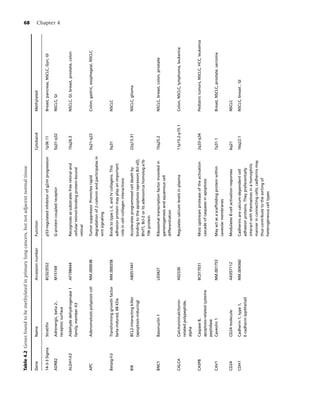
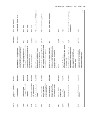
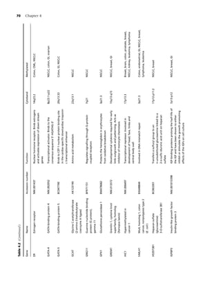
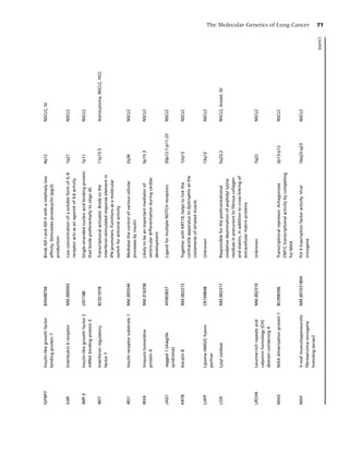
![Table 4.2 (Continued )
74
Gene Name Accession number Function Cytoband Methylated
SOX15 SRY (sex determining AB006867 Transcription factor. Binds to the 5’-aacaat-3’ 17p13 NSCLC, breast, prostate, colon
region Y)-box 15 sequence
Chapter 4
SPARC Secreted protein, acidic, NM 003118 Regulates cell growth through interactions 5q31.3-q32 NSCLC, pancreas
cysteine-rich (osteonectin) with the extracellular matrix and cytokines
TBX1 T-box 1 AF373867 Transcriptional regulator involved in 22q11.21 NSCLC
developmental processes
TFPI2 Tissue factor pathway AK129833 Regulation of plasmin-mediated matrix 7q22 NSCLC
inhibitor 2 remodeling. Inhibits trypsin, plasmin, factor
VIIa/tissue factor and weakly factor XA
TIMP3 TIMP metallopeptidase AB051444 Complexes with metalloproteinases (such as 22q12.1-q13.2 Brain, kidney, NSCLC, breast, colon
inhibitor 3 (Sorsby fundus collagenases) and irreversibly inactivates
dystrophy, them
pseudoinflammatory)
TJP2 Tight junction protein 2 AB209630 Plays a role in tight junctions and adherens 9q13-q21 NSCLC, pancreas
(zona occludens 2) junctions
TMEFF2 Transmembrane protein DQ133599 May be a novel survival factor for 2q32.3 Colon, NSCLC, breast, esophegeal
with EGF-like and two hippocampal and mesencephalic neurons
follistatin-like domains 2
TWIST1 Twist homolog 1 NM 000474 Transcription factor involved in the negative 7p21.2 NSCLC, breast, glioma
(acrocephalosyndactyly 3; regulation of cellular determination and in
Saethre-Chotzen syndrome) the differentiation of several lineages
(Drosophila) including myogenesis, osteogenesis, and
neurogenesis. Necessary for breast cancer
metastasis
uPA Plasminogen activator, NM 002658 Potent plasminogen activator 10q24 Breast, NSCLC, prostate
urokinase
ZNF22 Zinc finger protein 22 NM 006963 Binds DNA through the consensus sequence 10q11 NSCLC
(KOX 15) 5’-caatg-3’. May be involved in
transcriptional regulation and play a role in
tooth formation
NSCLC, nonsmall cell lung cancer; HCC, hepatocellular carcinoma; Gyn, gynecological tumors including cervical, ovarian, and endometrial cancers; Gl, gastroin-
testinal tumors; AML/CML, acute and chronic myelogenous leukemia; CNS, central nervous system.
(Adapted from Shames et al. [37].)](https://image.slidesharecdn.com/lungcancer3rded-120126215311-phpapp01/85/Lung-cancer-3rd-ed-85-320.jpg)
![The Molecular Genetics of Lung Cancer 75
the level of protein stability. p53 controls transcrip- clinical trials of p53 gene replacement therapy. The
tion of MDM2, an E3 ubiquitin ligase, which in turn results from preclinical and early-stage clinical trials
regulates p53 stability in a feedback loop. This par- of p53 gene replacement therapy using a replication
ticular connection in the p53 pathway is a frequent incompetent retrovirus p53 expression vector in pa-
target of dysregulation in tumor cells. tients with NSCLCs, show evidence of antitumor
The p53 pathway is activated in response cel- activity and the feasibility and safety of gene ther-
lular stress and DNA damage induced by gamma- apy [47]. INGN 201 (Ad5CMV-p53, AdvexinTM ),
irradiation, ultraviolet light, DNA damaging drugs, a replication-impaired p53 adenoviral vector has
and carcinogens. p53 stabilization results in the been evaluated in clinical trials, and is both safe and
expression of downstream genes, which induces effective for the treatment of several different types
either cell cycle arrest to permit DNA repair, or of cancer [48]. This treatment has been approved in
programmed cell death when there is too much China for the treatment of primary head and neck
damage. Loss of p53 function allows cells to di- cancers in combination with radiation therapy and
vide in spite of genetic damage, which can result is currently undergoing phase III trials in head and
the clonal expansion of premalignant cells. In most neck cancer in the United States.
cases, only mutant, missense p53 is present because
of LOH involving the wild-type p53 allele. However, The RB pathway
in some cases, mutant p53 proteins can form het- The RB pathway plays a central role in G1/S
erodimers with wild-type p53 inactivating its tumor cell transition. Hypophosphorylated RB exerts its
suppressive function even before LOH. These “gain- growth suppressive effect by binding to and inhibit-
of-function” mutations contribute to increased tu- ing the E2F transcription factor, which promotes
morigenicity and invasiveness of several types of cells through the G1/S transition. RB is phosphory-
cancers [26,46]. However, despite large-scale stud- lated by the CyclinD1/CDK4 complex. Once these
ies, it is not clear whether NSCLCs with p53 muta- kinases phosphorylate RB, it releases E2F, resulting
tions have impaired survival compared to lung can- in transition from G1 to S. Thus, loss of RB function
cers with only wild-type p53. though deletion or mutation leads to loss of the G1/S
There are two important upstream regulators checkpoint, and is a common event in lung cancer,
in the p53 pathway: MDM2 and p14ARF . MDM2 particularly SCLCs (>90%), while inactivation of
functions as an oncogene by reducing p53 levels RB is found in 15–30% of NSCLCs [26].
through enhancing proteasome-dependent degra- The activity of the CDK4/Cyclin D1 complex is
dation. Amplifications of MDM2 were reported in regulated by p16. p16 keeps RB hypophosphory-
∼7% (2/30) of NSCLCs, resulting in loss of p53 lated (and growth suppressing mode) by preventing
function [46]. p14ARF derives from the p16 locus CDK4 from phosphorylating RB. Thus, loss of p16
with an alternatively spliced 5-exon that results in function results in loss of function of the RB path-
an alternative reading frame for translation. p14 en- way. By contrast to RB, p16 is more frequently in-
codes a protein that binds to MDM2 thereby inhibit- activated in NSCLCs (∼70%) than in SCLCs (10%)
ing its ubiquitination activity, which leads to the sta- [26]. Inactivation of p16 is caused by LOH coupled
bilization of p53. Immunohistochemistry analyses with deletion, intragenic mutations or promoter
of p14ARF on lung cancers have shown that p14ARF hypermethylation of the remaining allele. In lung
protein expression was lost in ∼65% of SCLCs and cancer, promoter methylation is the most frequent
∼40% of NSCLCs. Thus, through p53 mutation or method of inactivation of p16.
changes in MDM2 or p14, the p53 pathway is inac- Overexpression of either CDK4 or Cyclin D1
tivated in the majority of all lung cancers. inhibits RB pathway function by saturating the
Lung cancer cells are addicted to loss of p53 func- growth suppressive activity of p16. CDK4 is am-
tion. When wild-type p53 is re-expressed in lung plified in some cases of NSCLCs, but cyclin D1 is
cancer cells with mutant or deleted p53, the tumor overexpressed in more than 40% of NSCLCs as as-
cells undergo apoptosis. These findings have led to sessed by immunohistochemistry [26,49]. Recently,](https://image.slidesharecdn.com/lungcancer3rded-120126215311-phpapp01/85/Lung-cancer-3rd-ed-86-320.jpg)
![76 Chapter 4
overexpression of Cyclin D1 in normal-appearing cell growth in vitro and in vivo. These preclinical
bronchial epithelial of patients with NSCLCs has studies provide a basis for FUS1 gene therapy clin-
been reported to be associated with smoking and to ical trials for the treatment of lung tumors using
predict shorter survival, suggesting the possible util- FUS1-nanoparticles [54,55].
ity of Cyclin D1 as a molecular marker to identify Two other 3p21.3 candidate tumor suppressor
high-risk individuals [50]. Thus through changes in genes, Semaphorin 3B (SEMA3B) and a family
either RB, p16, CDK4, or cyclin D1, this important member SEMA3F, are extracellular secreted mem-
growth regulatory pathway is inactivated and dis- bers of the semaphorin family, and are impor-
rupted in the large majority of lung cancers. tant in axonal guidance. Wild-type SEMA3B, but
not missense mutant SEMA3B, induces apopto-
3p tumor suppressor genes sis when re-expressed in lung cancers or added
Allele loss in 3p, including LOH and homozygous as a soluble molecule [56,57]. Overexpression of
deletion, occurs in nearly 100% of SCLCs and more SEMA3F in tissue culture results in inhibition of
than 90% of NSCLCs and is one of the earliest tumor cell growth and tumor cell invasion. Both
events in lung cancer development. Because of the SEMA3B and SEMA3F are soluble, secreted pro-
early changes in chromosome region 3p21.3 (oc- teins, and therefore are promising candidates for
curring in histologically normal lung epithelium) drug development.
the presence of 3p allele loss and inactivation of Two other 3p genes with evidence to support
expression of these 3p TSGs can be of use in de- their candidacy as tumor suppressors are FHIT and
termining smoking related field effects. Three dis- retinoic acid receptor beta (RARβ). FHIT is located
creet regions of 3p loss have been identified by in 3p14.2, one of the most common fragile sites of
allelotyping in lung cancers, including, a 600-kb the human genome. FHIT is either homozygously
segment in 3p21.3, the 3p14.2 (FHIT/FRAB3), and deleted or expresses aberrant transcripts in more
the 3p12 (ROBO1/DUTT1) regions. The 3p21.3 re- than 50% of lung cancers [58]. In addition, FHIT
gion has been analyzed most extensively and 25 overexpression induces apoptosis in lung cancer
genes were identified from this region. cells. RARβ is located at 3p24 and functions as a
One of the best studied genes in this region is receptor for retinoic acid (RA). Although the RARβ
RASSF1A, which is rarely mutated in lung cancer gene is not mutated in lung cancer, it undergoes
but whose expression is frequently lost by tumor methylation in 72% of SCLCs and 41% of NSCLCs,
acquired promoter methylation [51,52]. RASSF1A leading to loss of its expression [59]. Re-expression
is involved in multiple pathways critical to can- of RARβ in lung cancer cell lines suppresses their
cer pathogenesis, including cell cycle, apoptosis, growth in the culture and nude mice [60].
and microtubule stability. RASSF1A is methylated
in ∼90% of SCLCs and ∼40% of NSCLCs and has
the ability to suppress the growth of lung cancer
cell lines in tissue culture and as xenografts in nude Oncogenes and the pathways
mice [51,52]. they regulate
FUS1 is located next to RASSF1A and one of the
two alleles of the gene is often lost in lung cancers. While there are multiple components to each of the
FUS1 is rarely mutated in lung cancers, does not un- growth signaling pathways involved in lung can-
dergo promoter hypermethylation, yet the protein cer, we will focus the discussion on those proteins
product of this gene is frequently lost in lung can- that are frequently affected by genetic abnormalities
cer compared to normal lung tissues [53]. Wild-type in cancer. It has become clear that these mutated
FUS1 but not tumor-acquired mutant FUS1 induces proteins, while driving cells toward transformation,
G1 growth arrest and apoptosis [53]. Administration also “addict” the cells to their abnormal function.
of FUS1 with in DOTAP:cholesterol (DOTAP:Chol) This concept is referred to as “oncogene addiction”
nanoparticles (FUS1-nanoperticles) inhibits cancer and represents a cellular physiologic state where the](https://image.slidesharecdn.com/lungcancer3rded-120126215311-phpapp01/85/Lung-cancer-3rd-ed-87-320.jpg)
![The Molecular Genetics of Lung Cancer 77
continued presence of the abnormal function, while 19 (44% of all mutations) and missense mutations
oncogenic, also becomes required for the tumor to in exon 21 (41% of all mutations) are the most fre-
survive [61]. This means that if the function is re- quent, accounting for more than 80% of all muta-
moved or inhibited, for example, by a targeted drug, tions [67]. Importantly, the presence of mutations
the tumor cells die. By contrast, bystander normal in TK domain correlates with the drug sensitivity to
cells, which are not “addicted” to the mutant pro- TKIs [65,66]. An intriguing characteristic of EGFR
tein, are much less sensitive to the drug; thus, the mutations is that they occur in a highly selected
targeted drugs have great tumor cell specificity. The subpopulation: female East-Asian never smokers
most important example of this concept for lung with adenocarcinoma histology [68]. Notably, be-
cancer is EGFR. Tumors with mutations in EGFR are fore EGFR mutations were discovered, all of same
dependent on survival signals transduced by mu- clinicopathological factors were found to be associ-
tant EGFR, and thus are particularly sensitive to ty- ated with tumor responses to TKIs [69,70].
rosine kinase inhibitors (TKIs) [62]. These findings Although several studies have confirmed the re-
have led to massive genome-wide sequencing ef- lationship between the presence of mutant EGFR
forts (discussed above) targeting thousands of genes and the response to TKIs [65,66,71], a subset of
to find additional mutated oncogene targets for ra- NSCLC patients with mutant EGFRs do not respond
tional therapeutics design. to TKIs. These tumors often (>50%) have a “sec-
ond” TK domain mutation (T790M) usually found
in patients who relapse after TKI treatment, suggest-
Receptor tyrosine kinases ing its contribution to acquired resistance to TKIs
[72,73]. However, several examples of the T790M
The EGFR family mutations occur in lung tumors not treated with
The EGFR family of receptors are transmembrane EGFR TKIs, and often the mutation is only in a small
TK receptors and are composed of EGFR, HER2, subset of the tumor cells. This contrasts with the
HER3, and HER4 and each has unique proper- other EGFR TK domain mutations, which are in all
ties. For example, HER2 lacks a functional ligand- tumor cells. Also, a germline EGFR T790M mutation
binding domain and HER3 lacks kinase activity [63]. was reported to be associated with familial NSCLC,
Upon ligand binding, these EGFR family members suggesting that this mutation could predispose peo-
form active homo- and hetero-dimers, leading to ple to lung cancer [74]. Fortunately, there are EGFR
autophosphorylation and activation of intracellular TKIs that inhibit EGFR with the T790M mutation,
signaling cascades. EGFR is overexpressed in ∼70% and these drugs are currently under clinical evalu-
of NSCLCs but rarely expressed in SCLCs [64]. There ation [75].
are several drugs targeting EGFR or HER2 currently Some patients without EGFR mutation also re-
available including the small molecule TKIs, gefi- spond to TKIs, and several predictive markers other
tinib, erlotinib, and the monoclonal antibodies, ce- than EGFR mutation have been reported to corre-
tuximab (targeting EGFR), and trastuzumab (target- late with TKI response, including EGFR amplifica-
ing HER2). tion, elevated EGFR protein, HER2 amplification,
Recently, several mutations in the TK domain of HER3 amplification, and activation of AKT [76–
EGFR have been described, and are not infrequent in 80]. In fact, KRAS mutations and EGFR mutations
NSCLC (10–20%), but never occur in SCLC [65,66]. are mutually exclusive. KRAS mutations are asso-
Of interest is that TK domain mutations are almost ciated with cigarette smoking, while EGFR muta-
exclusive to lung cancer, whereas intracellular re- tions generally occur in never smokers [81]. These
gion mutations are found in glioblastomas. In lung studies suggest that other biological features be-
cancer, these mutations are limited to the first four sides EGFR mutation status determine TKI response.
exons of the TK domain and are categorized into Among biologic predictors, EGFR mutation and
three different types (deletions, insertions, and mis- amplification by fluorescence in situ hybridization
sense point mutations). Inframe deletions in exon are highly correlated with TKI response while](https://image.slidesharecdn.com/lungcancer3rded-120126215311-phpapp01/85/Lung-cancer-3rd-ed-88-320.jpg)
![78 Chapter 4
EGFR protein expression gives conflicting results lial cells adjacent to tumors with EGFR mutations.
[65,66,71,76,82]. There is also the possibility that The discovery of EGFR mutations could be used
tumors with EGFR mutations are associated with as an early detection marker and chemoprevention
better survival independent of TKI treatment. Thus, target [94]. Transgenic mice with either EGFR point
all survival studies after TKI treatment need to have mutations or deletion mutations develop lung ade-
molecular analyses for comparison [80,83,84]. Two nocarcinomas with similar histology to those seen
well-controlled phase III studies were conducted for in patients [95,96]. When the mutant gene was
these drugs. The results of these studies showed that “turned-off” in the mice through controlled gene
erlotinib prolonged survival of previously treated expression the lung tumors all regressed indicating
NSCLC patients by 2 months (BR21 trial), while that mutant EGFR is required for both initiation and
gefitinib failed to show survival benefit (Iressa Sur- maintenance of the tumors.
vival Evalulation in Lung Cancer (ISEL)) [86,87].
Despite positive preclinical studies of the combi- c-KIT
nation of TKI and chemotherapy, several phase SCLC but not NSCLC frequently express (40–70%)
III studies have failed to show a survival bene- both the receptor c-KIT and its ligand, stem cell fac-
fit of adding erlotinib or gefitinib to conventional tor (SCF) suggesting an autocrine loop may promote
chemotherapy [88,89]. Finally, lung cancers with the growth of the SCLC cells [97]. However, unlike
EGFR mutations are more sensitive to ionizing ra- gastrointestinal stromal tumors which frequently
diation than those without EGFR mutations, which contain c-KIT mutations, activating c-KIT mutations
potentially provides a molecular basis for combined are very rare in lung cancer [98,99]. While imatinib,
modality treatment involving TKIs and radiother- an inhibitor of c-KIT kinase, inhibits cell growth in
apy [90]. some c-KIT expressing SCLC cell lines in vitro, two
While standard criteria for selecting patients with phase II clinical studies and a mouse xenograft study
NSCLC for TKI therapy are being developed, in prac- failed to show tumor regression in SCLC by imatinib
tice, East-Asian female patients with tumors that monotherapy [100–103].
have EGFR mutations or EGFR amplification and
that are never smokers often receive TKI therapy. To RAS/RAF/MEK/ERK pathway
address this issue, prospective clinical trials designed The RAS family of proto-oncogenes (HRAS, KRAS,
to incorporate the patient’s clinicopathological data and NRAS) are 21-kD plasma membrane-associated
as well as molecular biological features (EGFR mu- G-proteins that regulate key signal transduction
tation and/or amplification) of the tumors are cur- pathways involved in normal cellular differenti-
rently underway. ation, proliferation, and survival [104]. Multiple
HER2 mutations occur in 2% of NSCLCs. All re- studies have shown that oncogenic KRAS (e.g.,
ported HER2 mutations are in-frame insertions in KRASV12 mutant) activates cell signaling pathways
exon 20 and target the corresponding TK domain important to cellular transformation [105]. As a
region as in EGFR insertion mutations and occur in result, KRAS abnormalities represent an impor-
the same subpopulation as those with EGFR muta- tant therapeutic target. RAS mutations (nearly al-
tions (adenocarcinoma, never smoker, East Asian, ways KRAS mutations in lung cancer) are found in
and woman) [68,91,92]. So far no small molecule 15–20% of NSCLCs, especially in adenocarcinomas
inhibitors show similar potency against HER2 muta- (20–30%), but never in SCLCs [26]. The mutations
tions as seen with EGFR TKIs and studies are needed occur in codons 12, 13, and 61, all of which in-
to see if mutant HER2 lung cancers respond to the fluence intrinsic GTPase activity [104]. A number
anti-HER2 antibody trastuzumab. HER4 mutations of drugs that target different aspects of RAS func-
were found in (2.3%) NSCLC tumor samples from tion and metabolism have been developed and are
Asian patients including male smokers [93]. currently under clinical investigation [104]. These
EGFR mutations occur as preneoplastic lesions include the farnesyl transferase inhibitors tipifarnib
occurring in histologically normal bronchial epithe- and lonafarnib, which are now being tested in the](https://image.slidesharecdn.com/lungcancer3rded-120126215311-phpapp01/85/Lung-cancer-3rd-ed-89-320.jpg)
![The Molecular Genetics of Lung Cancer 79
combination with cytotoxic drugs in phase III clini- 9 Hanahan D, Weinberg RA. The hallmarks of cancer.
cal trials [106]. Cell Jan 7, 2000; 100(1):57–70.
BRAF protein serine/threonine kinase is a down- 10 Weinstein IB. Cancer. addiction to oncogenes—the
stream effecter of the Ras pathway and mutations Achilles heal of cancer. Science Jul 5, 2002; 297(5578):
of BRAF occur in ∼70% melanoma, but in only 3% 63–4.
11 Sato M, Shames DS, Gazdar AF, Minna JD. A Trans-
of lung cancers [107–109]. However, for those rare
lational View of the Molecular Pathogenesis of Lung
lung cancers, mutated BRAF protein is a potentially
Cancer. J Thorac Oncol 2007; 2(4):327–43.
important and specific therapeutic target. An orally 12 Sjoblom T, Jones S, Wood LD et al. The consensus cod-
administered Raf kinase inhibitor, BAY 43-9006 (so- ing sequences of human breast and colorectal cancers.
rafenib), is currently being tested in phase I and Science Oct 13, 2006; 314(5797):268–74.
phase II trials in lung cancer [110–111]. 13 Varmus H. The new era in cancer research. Science
Activated BRAF phosphorylates and activates May 26, 2006; 312(5777):1162–5.
MEK1 and MEK2, which in turn phosphorylate 14 Amos CI, Xu W, Spitz MR. Is there a genetic basis
and activate ERK1 and ERK2. However, MEK or for lung cancer susceptibility? Recent Results Cancer Res
ERK gene amplification or mutations have not been 1999; 151:3–12.
found in lung cancers. Nevertheless, ERK1/ERK2 15 Pfeifer GP, Denissenko MF, Olivier M, Tretyakova N,
Hecht SS, Hainaut P. Tobacco smoke carcinogens,
are constitutively activated in a subset of lung can-
DNA damage and p53 mutations in smoking-
cers and MEK and ERK remain therapeutic tar-
associated cancers. Oncogene Oct 21, 2002; 21(48):
gets for lung cancer treatment using an oral MEK 7435–51.
inhibitor CI-1040 and its derivative PD03255901 16 Jones PA, Baylin SB. The fundamental role of epi-
[112]. genetic events in cancer. Nat Rev Genet Jun 2002;
3(6):415–28.
17 Lengauer C, Kinzler KW, Vogelstein B. Genetic in-
References stabilities in human cancers. Nature Dec 17, 1998;
396(6712):643–9.
1 Mazin B, Qumsiyeh YY. Molecular methods in oncol- 18 Ma Y, Jacobs SB, Jackson-Grusby L et al. DNA CpG
ogy: cytogenetics. In: Vincent T, Devita SH, Rosen- hypomethylation induces heterochromatin reorgani-
berg SA (eds). Cancer: Principles and Practice of Oncology, zation involving the histone variant macroH2A. J Cell
7th edn. Philadelphia: Lippincott, Williams & Wilkins, Sci Apr 15, 2005; 118(Pt 8):1607–16.
2005. 19 Holm TM, Jackson-Grusby L, Brambrink T, Yamada Y,
2 Nowell PC. The clonal evolution of tumor cell popu- Rideout WM, III, Jaenisch R. Global loss of imprint-
lations. Science Oct 1, 1976; 194(4260):23–8. ing leads to widespread tumorigenesis in adult mice.
3 Varmus HE. Nobel lecture. Retroviruses and onco- Cancer Cell Oct 2005; 8(4):275–85.
genes. I. Biosci Rep Oct 1990; 10(5):413–30. 20 Jaenisch R, Bird A. Epigenetic regulation of gene
4 Todaro GJ, Huebner RJ. The viral oncogene hypoth- expression: how the genome integrates intrinsic
esis: new evidence. Proc Natl Acad Sci U S A 1972; and environmental signals. Nat Genet Mar 2003;
69:1009–15. 33(Suppl):245–54.
5 Knudson AG, Jr. Mutation and cancer: statistical 21 Fukasawa K. Centrosome amplification, chromo-
study of retinoblastoma. Proc Natl Acad Sci U S A Apr some instability and cancer development. Cancer Lett
1971; 68(4):820–3. Dec 8, 2005; 230(1):6–19.
6 Friend SH, Bernards R, Rogelj S et al. A human DNA 22 Whang-Peng J, Kao-Shan CS, Lee EC et al. Specific
segment with properties of the gene that predisposes chromosome defect associated with human small-cell
to retinoblastoma and osteosarcoma. Nature Oct 16– lung cancer; deletion 3p(14-23). Science Jan 8, 1982;
22, 1986; 323(6089):643–6. 215(4529):181–2.
7 Futreal PA, Coin L, Marshall M et al. A census of hu- 23 Testa JR, Liu Z, Feder M et al. Advances in the analysis
man cancer genes. Nat Rev Mar 2004; 4(3):177–83. of chromosome alterations in human lung carcino-
8 Sjoblom T, Jones S, Wood LD et al. The consensus cod- mas. Cancer Genet Cytogenet May 1997; 95(1):20–32.
ing sequences of human breast and colorectal cancers. 24 Lerman MI, Minna JD. The 630-kb lung cancer ho-
Science Oct 13, 2006; 314(5797):268–74. mozygous deletion region on human chromosome](https://image.slidesharecdn.com/lungcancer3rded-120126215311-phpapp01/85/Lung-cancer-3rd-ed-90-320.jpg)
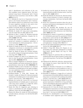
![The Molecular Genetics of Lung Cancer 81
from the 3p21.3 homozygous deletion region leads to gefitinib. N Engl J Med May 20, 2004; 350(21):2129–
G1 arrest and growth inhibition of lung cancer cells. 39.
Oncogene Sep 27, 2001; 20(43):6258–62. 66 Paez JG, Janne PA, Lee JC et al. EGFR mutations in
54 Ito I, Ji L, Tanaka F et al. Liposomal vector mediated lung cancer: correlation with clinical response to gefi-
delivery of the 3p FUS1 gene demonstrates potent tinib therapy. Science Jun 4, 2004; 304(5676):1497–
antitumor activity against human lung cancer in vivo. 500.
Cancer Gene Ther Nov 2004; 11(11):733–9. 67 Shigematsu H, Gazdar AF. Somatic mutations of epi-
55 Uno F, Sasaki J, Nishizaki M et al. Myristoylation of dermal growth factor receptor signaling pathway in
the fus1 protein is required for tumor suppression lung cancers. Int J Cancer Jan 15, 2006; 118(2):257–
in human lung cancer cells. Cancer Res May 1, 2004; 62.
64(9):2969–76. 68 Shigematsu H, Lin L, Takahashi T et al. Clinical and
56 Castro-Rivera E, Ran S, Thorpe P, Minna JD. biological features associated with epidermal growth
Semaphorin 3B (SEMA3B) induces apoptosis in lung factor receptor gene mutations in lung cancers. J Natl
and breast cancer, whereas VEGF165 antagonizes Cancer Inst Mar 2, 2005; 97(5):339–46.
this effect. Proc Natl Acad Sci U S A Aug 3, 2004; 69 Fukuoka M, Yano S, Giaccone G et al. Multi-
101(31):11432–7. institutional randomized phase II trial of gefitinib for
57 Tomizawa Y, Sekido Y, Kondo M et al. Inhibition of previously treated patients with advanced non-small-
lung cancer cell growth and induction of apoptosis af- cell lung cancer (The IDEAL 1 Trial) [corrected]. J Clin
ter reexpression of 3p21.3 candidate tumor suppres- Oncol Jun 15, 2003; 21(12):2237–46.
sor gene SEMA3B. Proc Natl Acad Sci U S A Nov 20, 70 Kris MG, Natale RB, Herbst RS et al. Efficacy of gefi-
2001; 98(24):13954–9. tinib, an inhibitor of the epidermal growth factor re-
58 Sozzi G, Veronese ML, Negrini M et al. The FHIT gene ceptor tyrosine kinase, in symptomatic patients with
3p14.2 is abnormal in lung cancer. Cell Apr 5, 1996; non-small cell lung cancer: a randomized trial. JAMA
85(1):17–26. Oct 22, 2003; 290(16):2149–58.
59 Virmani AK, Rathi A, Zochbauer-Muller S et al. Pro- 71 Pao W, Miller V, Zakowski M et al. EGF receptor gene
moter methylation and silencing of the retinoic acid mutations are common in lung cancers from “never
receptor-beta gene in lung carcinomas. J Natl Cancer smokers” and are associated with sensitivity of tumors
Inst Aug 16, 2000; 92(16):1303–7. to gefitinib and erlotinib. Proc Natl Acad Sci U S A Sep
60 Houle B, Rochette-Egly C, Bradley WE. Tumor- 7, 2004; 101(36):13306–11.
suppressive effect of the retinoic acid receptor beta in 72 Kobayashi S, Boggon TJ, Dayaram T et al. EGFR mu-
human epidermoid lung cancer cells. Proc Natl Acad tation and resistance of non-small-cell lung cancer
Sci U S A Feb 1, 1993; 90(3):985–9. to gefitinib. N Engl J Med Feb 24, 2005; 352(8):786–
61 Weinstein IB. Cancer. Addiction to oncogenes— 92.
the Achilles heal of cancer. Science Jul 5, 2002; 73 Pao W, Miller VA, Politi KA et al. Acquired resistance
297(5578):63–4. of lung adenocarcinomas to gefitinib or erlotinib is
62 Sordella R, Bell DW, Haber DA, Settleman J. associated with a second mutation in the EGFR kinase
Gefitinib-sensitizing EGFR mutations in lung can- domain. PLoS Med Mar 2005; 2(3):e73.
cer activate anti-apoptotic pathways. Science Aug 20, 74 Bell DW, Gore I, Okimoto RA et al. Inherited sus-
2004; 305(5687):1163–7. ceptibility to lung cancer may be associated with the
63 Rowinsky EK. The erbB family: targets for thera- T790M drug resistance mutation in EGFR. Nat Genet
peutic development against cancer and therapeu- Dec 2005; 37(12):1315–6.
tic strategies using monoclonal antibodies and tyro- 75 Kobayashi S, Ji H, Yuza Y et al. An alternative in-
sine kinase inhibitors. Annu Rev Med 2004; 55:433– hibitor overcomes resistance caused by a mutation of
57. the epidermal growth factor receptor. Cancer Res Aug
64 Franklin WA, Veve R, Hirsch FR, Helfrich BA, Bunn 15, 2005; 65(16):7096–101.
PA, Jr. Epidermal growth factor receptor family in 76 Cappuzzo F, Hirsch FR, Rossi E et al. Epidermal growth
lung cancer and premalignancy. Semin Oncol Feb factor receptor gene and protein and gefitinib sensi-
2002; 29(1, Suppl 4):3–14. tivity in non-small-cell lung cancer. J Natl Cancer Inst
65 Lynch TJ, Bell DW, Sordella R et al. Activating muta- May 4, 2005; 97(9):643–55.
tions in the epidermal growth factor receptor under- 77 Cappuzzo F, Magrini E, Ceresoli GL et al. Akt phos-
lying responsiveness of non-small-cell lung cancer to phorylation and gefitinib efficacy in patients with](https://image.slidesharecdn.com/lungcancer3rded-120126215311-phpapp01/85/Lung-cancer-3rd-ed-92-320.jpg)
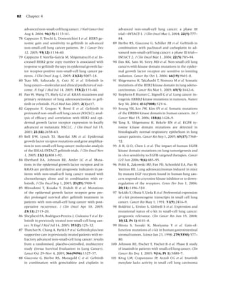
![The Molecular Genetics of Lung Cancer 83
expressing c-kit protein: a phase II clinical trial. Cancer 107 Brose MS, Volpe P, Feldman M et al. BRAF and RAS
May 15, 2005; 103(10):2128–31. mutations in human lung cancer and melanoma. Can-
102 Wolff NC, Randle DE, Egorin MJ, Minna JD, Ilaria RL, cer Res Dec 1, 2002; 62(23):6997–7000.
Jr. Imatinib mesylate efficiently achieves therapeu- 108 Davies H, Bignell GR, Cox C et al. Mutations of the
tic intratumor concentrations in vivo but has limited BRAF gene in human cancer. Nature Jun 27, 2002;
activity in a xenograft model of small cell lung cancer. 417(6892):949–54.
Clin Cancer Res May 15, 2004; 10(10):3528–34. 109 Naoki K, Chen TH, Richards WG, Sugarbaker DJ,
103 Wang WL, Healy ME, Sattler M et al. Growth inhibi- Meyerson M. Missense mutations of the BRAF gene
tion and modulation of kinase pathways of small cell in human lung adenocarcinoma. Cancer Res Dec 1,
lung cancer cell lines by the novel tyrosine kinase in- 2002; 62(23):7001–3.
hibitor STI 571. Oncogene Jul 20, 2000; 19(31):3521– 110 Liu B, Barrett T, Choyke P et al. A phase II study of BAY
8. 43-9006 (Sorafenib) in patients with relapsed non-
104 Downward J. Targeting RAS signalling pathways small cell lung cancer (NSCLC) [Abstract 17119]. Pro
in cancer therapy. Nat Rev Cancer Jan 2003; 3(1): Am Soc Clin Onc 2005; 24:18S.
11–22. 111 Tuveson DA, Weber BL, Herlyn M. BRAF as a poten-
105 Stacey DW, Kung HF. Transformation of NIH 3T3 cells tial therapeutic target in melanoma and other malig-
by microinjection of Ha-ras p21 protein. Nature Aug nancies. Cancer Cell Aug 2003; 4(2):95–8.
9–15, 1984; 310(5977):508–11. 112 Hoshino R, Chatani Y, Yamori T et al. Constitutive ac-
106 Isobe T, Herbst RS, Onn A. Current management of tivation of the 41-/43-kDa mitogen-activated protein
advanced non-small cell lung cancer: targeted ther- kinase signaling pathway in human tumors. Oncogene
apy. Semin Oncol Jun 2005; 32(3):315–28. Jan 21, 1999; 18(3):813–22.](https://image.slidesharecdn.com/lungcancer3rded-120126215311-phpapp01/85/Lung-cancer-3rd-ed-94-320.jpg)
![CHAPTER 5
Molecular Biology of Preneoplastic
Lesions of the Lung
Ignacio I. Wistuba and Adi F. Gazdar
Introduction of the distant airway of the lung. Squamous cell car-
cinomas and SCLCs usually arise centrally, whereas
Lung cancer is the leading cause of cancer deaths in adenocarcinomas and large cell carcinomas usually
the United States and worldwide [1]. The high mor- arise peripherally [3]. However, the specific respira-
tality of this disease is primarily due to the fact that tory epithelial cell type from which each lung can-
the majority of the lung cancers are diagnosed at ad- cer type develops has not been established. As with
vanced stages when the options for treatment are other epithelial malignancies, it is believed that lung
mostly palliative. Experience with other epithelial cancers arise after a series of progressive patholog-
tumors has shown that if neoplastic lesions can be ical changes, known as preneoplastic or premalig-
detected and treated at their intraepithelial stage the nant lesions [4,5]. Although the sequential preneo-
chances for survival can be improved significantly. plastic changes have been defined for centrally aris-
Thus, to reduce the mortality rate of lung cancer, ing squamous carcinomas of the lung [6], they have
new techniques and approaches must be developed been poorly documented for the other major forms
to identify, diagnose, and treat preinvasive lesions. of lung cancers [4,5].
However, the early diagnosis of lung cancer repre- Although many molecular abnormalities have
sents an enormous challenge. been described in clinically evident lung cancers
From histopathological and biological perspec- [2], relatively little is known about the molecular
tives, lung cancer is a highly complex set of dif- events preceding the development of lung carcino-
ferent but related neoplasms [2], probably having mas and the underlying genetic basis of lung car-
multiple preneoplastic pathways. Lung cancer con- cinogenesis. In the past decade, several studies have
sists of several histological types, including small provided information regarding the molecular char-
cell lung carcinoma (SCLC) and nonsmall cell lung acterization of the preneoplastic changes involved
carcinoma (NSCLC) types of squamous cell carci- in the pathogenesis of lung cancer, especially squa-
noma, adenocarcinoma (including the noninvasive mous cell carcinoma and adenocarcinoma [7–10].
type of bronchioloalveolar carcinoma, BAC), and Many of these molecular changes have been de-
large cell carcinoma [3]. Lung cancers may arise tected in the histologically normal respiratory mu-
from the major bronchi (central tumors) or small cosa of smokers [11,12]. The high-risk population
bronchi, bronchioles, or alveoli (peripheral tumors) targeted for early detection efforts are heavy smok-
ers and patients who have survived a cancer of the
upper aerodigestive tract. However, conventional
Lung Cancer, 3rd edition. Edited by Jack A. Roth, James D. Cox,
morphologic methods for the identification of pre-
and Waun Ki Hong. c 2008 Blackwell Publishing, malignant cell populations in the lung airways have
ISBN: 978-1-4051-5112-2. important limitations. This has led to research in
84](https://image.slidesharecdn.com/lungcancer3rded-120126215311-phpapp01/85/Lung-cancer-3rd-ed-95-320.jpg)
![Preneoplastic Lesions of the Lung 85
biological properties, including molecular and ge- Cancer (IASLC) histological classification of
netic changes, of the respiratory epithelium and its preinvasive lesions of the lung lists three main
corresponding preneoplastic cells and lesions. morphologic forms of preneoplastic lesions in the
Although several studies have provided rele- lung [3]: (a) squamous dysplasia and carcinoma in
vant information regarding the molecular charac- situ (CIS); (b) atypical adenomatous hyperplasia
terization of the premalignant changes involved (AAH); and (c) diffuse idiopathic pulmonary neu-
in the pathogenesis of lung cancer, especially for roendocrine cell hyperplasia (DIPNECH). While
squamous cell carcinoma [10], that information is the sequential preneoplastic changes have been
not sufficient to identify with certainty molecular defined for centrally arising squamous carcinomas,
pathogenetic pathways or molecular markers use- they have been poorly documented for large cell
ful for risks assessment, targeted chemoprevention carcinomas, adenocarcinomas and SCLCs [4,5].
or treatment, and early detection of lung prema- Mucosal changes in the large airways that may
lignant lesions. Further research in this area may precede invasive squamous cell carcinoma include
provide new methods for assessing the likelihood squamous dysplasia and CIS [4,5]. Adenocarcino-
of developing invasive lung cancer in smokers and mas may be preceded by morphological changes
allow for early detection and monitoring of their including AAH in peripheral airway cells [4,15].
response to chemopreventive regimens. Attempts While DIPNECH are thought to be precursor lesions
to better define the pathogenesis of lung premalig- for carcinoids of the lung, for SCLC there is no
nancy have been thwarted by the relative invisibil- specific preneoplastic change have been identified.
ity of the cellular lesions and their random distribu-
tion throughout the respiratory airway field, and Squamous cell carcinoma
new methodologies, including computed tomog- preneoplastic lesions
raphy (CT) imaging [13] and fluorescence bron- Mucosal changes in the large airways that may pre-
choscopy [14], has been introduced to better iden- cede or accompany invasive squamous cell carci-
tify and visualize lung premalignant lesions. noma include hyperplasia, squamous metaplasia,
In this chapter we summarize the current infor- squamous dysplasia, and CIS [4,5] (Figure 5.1).
mation on lung cancer molecular and histopatho- There are no squamous cells in the normal airways.
logic pathogenesis and discuss the complexity of The progenitor or stem cells for the squamous meta-
the identification of novel molecular mechanisms plastic epithelium of the proximal airway is not
involved in the development of the lung prema- known, but it is presumed that the basal cells are
lignant disease, and their relevance to the devel- pluripotent and can give rise to metaplastic and dys-
opment of new strategies for early detection and plastic squamous cells, which function as precursors
chemoprevention. In addition, we describe the rec- of squamous cell carcinomas.
ognized preneoplastic lesions for major types of lung Dysplastic squamous lesions may be graded in of
cancers and review the current concepts of early different intensities (i.e., mild, moderate, and se-
pathogenesis and the progression of the most im- vere); however, these lesions represent a contin-
portant histologic types of lung cancer. uum of cytologic and histologic atypical changes
that may show some overlapping between cate-
gories. Whereas mild squamous dysplasia is char-
Pathology of lung cancer acterized by minimal architectural and cytological
preneoplastic lesions disturbance, moderate dysplasia exhibits more cy-
tological irregularity, which is even higher in severe
Lung cancers are believed to arise after a series of dysplasia and is accompanied by considerable cellu-
progressive pathological changes (preneoplastic or lar polymorphism. In a subset of squamous dysplas-
precursor lesions) in the respiratory mucosa. The tic changes, the basal membrane thickens and there
recent 2004 World Health Organization (WHO) is vascular budding in the subepithelial tissue that
International Association for the Study of Lung results in papillary protrusions of the epithelium,](https://image.slidesharecdn.com/lungcancer3rded-120126215311-phpapp01/85/Lung-cancer-3rd-ed-96-320.jpg)
![86 Chapter 5
Squamous cell carcinoma
Normal bronchial Basal cell Squamous Carcinoma Squamous cell
epithelium hyperplasia metaplasia Dysplasia in situ carcinoma
Adenocarcinoma
Normal alveolar AAH BAC
epithelium
Adenocarcinoma
Normal bronchiolar
epithelium
Small cell carcinoma
Normal bronchial Basal cell
SCLC
epithelium hyperplasia
Figure 5.1 Summary of histopathologic changes involved which seems to be the precursor for a subset of lung ade-
in the pathogenesis of lung cancer. The sequence of pre- nocarcinomas, those with bronchioloalveolar carcinoma
neoplastic lesions involved in the development of squa- (BAC) features. No preneoplastic lesion has been recog-
mous cell carcinoma of the lung has been elucidated. nized for SCLC (microphotographs of histology sections
For adenocarcinoma histology, the only known preneo- stained with hematoxylin and eosin).
plastic lesion is AAH (atypical adenomatous hyperplasia),
lesions that have been termed angiogenic squamous bronchoscopy, such as lung-imaging fluorescent en-
dysplasia (ASD) [16]. These lesions indicate that doscopy (LIFE), greatly increases the sensitivity for
angiogenesis commences at a relatively early pre- detection of squamous dysplastic and CIS lesions
neoplastic stage. CIS demonstrates extreme cytolog- [14]. Little is known about the rate and risks of pro-
ical aberrations with almost complete architectural gression from squamous dysplasia to CIS and ulti-
disarray, but with an intact basement membrane mately to invasive squamous cell carcinoma.
and absence of stromal invasion. Foci of CIS usu-
ally arise near bifurcations in the segmental bronchi, Adenocarcinoma precursor lesions
subsequently extending proximally into the adja- It has been suggested that peripherally arising
cent lobar bronchus and distally into subsegmental adenocarcinomas may be preceded by AAH in
branches. These lesions are often not detected by peripheral airway cells [4,15]; however, the res-
conventional white-light bronchoscopy or gross ex- piratory structures and the specific epithelia
amination. However, the utilization of fluorescent cell types involved in the origin of most lung](https://image.slidesharecdn.com/lungcancer3rded-120126215311-phpapp01/85/Lung-cancer-3rd-ed-97-320.jpg)
![Preneoplastic Lesions of the Lung 87
adenocarcinomas are unknown (Figure 5.1). AAH is However, a rare lesion called DIPENECH has been
considered a putative precursor of adenocarcinoma associated with the development of other neuroen-
[4,15]. AAH is a discrete parenchymal lesion aris- docrine tumors of the lung, typical and atypical
ing in the alveoli close to terminal and respiratory carcinoids [4,24]. DIPENECH lesions include local
bronchioles. Because of their size, AAH are usually extraluminal proliferations in the form of tumorlets.
incidental histological findings, but they may be de- Carcinoid tumors are arbitrarily separated from tu-
tected grossly, especially if they are 0.5 cm or larger. morlets if the neuroendocrine proliferation is 0.5 cm
The increasing use of high resolution CT scans for or larger.
screening purposes has led to an increasing aware-
ness of this entity, as it remains one of the most im-
portant differential diagnoses of air filled peripheral Molecular pathogenesis of
lesions (called “ground glass opacities”) [17]. AHH lung cancer
maintains an alveolar structure lined by rounded,
cuboidal, or low columnar cells. Although our current knowledge of the molecular
The postulated progression of AAH to nonin- pathogenesis of lung cancer is still meager, during
vasive BAC adenocarcinoma with, apparent from the last decade, there are several important lessons
the increasingly atypical morphology, is supported learnt on the molecular pathogenesis of this tu-
by morphometric, cytofluorometric, and molecu- mor, including the following: (a) There are several
lar studies [5,15]. Invasion may follow, especially histopathologic and molecular pathways associated
at the fibrous centers of these lesions, giving rise with the development of the major types of NSCLC.
to adenocarcinomas having mixed features of BAC (b) Although there is a field effect phenomenon for
and tubular or papillary adenocarcinomas. Distinc- lung preneoplastic lesions, recent data suggest that
tion between highly atypical AAH and nonmuci- there are at least two distinct lung airways com-
nous BAC is sometimes difficult. However, as these partments (central and peripheral) for lung can-
lesions have very high 5-year survival rates after re- cer pathogenesis. (c) Inflammation may play an
section provided they are <2 cm in size, the distinc- important role in lung cancer development and
tion may be academic. Somewhat arbitrarily, BAC it could be an important component of the field
are considered generally >10 mm in size, with more effect phenomenon. (d) For lung adenocarcinoma,
cellular atypia than their AAH counterparts. The at least two smoking and nonsmoking-related path-
origin of AAH is still unknown, but the differentia- ways have been identified. Most of the molecu-
tion phenotype derived from immunohistochemical lar and histopathologic changes in the respiratory
and ultrastructural features suggests an origin from epithelium associated to lung cancer pathogenesis
the progenitor cells of the peripheral airways, such have been related to smoking [10]. However, the
Clara cells and type II pneumocytes [18,19]. recent discovery of frequent EGFR gene mutations
There is an increasing body of evidence to sup- in lung cancer and adjacent normal epithelium in
port the concept of AAH as precursor of at least never or light smokers suggests the presence of at
a subset of adenocarcinomas. AAH is most fre- least two distinct molecular pathogenesis for lung
quently detected in lung from patients bearing lung cancer, smoking and nonsmoking-related [25,26].
cancers (9–20%), especially adenocarcinomas (up As a relatively small subset of smokers develop lung
to 40%) compared to squamous cell carcinomas cancer, attention has been focused in the identifica-
(11%) [5,20–22]. By contrast, autopsy studies have tion of specific molecular and histopathologic path-
reported AAH in ∼3% of noncancer patients [23]. ways that can predict lung cancer development in
high-risk populations. One of those key pathways,
Precursor lesions of neuroendocrine currently under intense investigation, is the activa-
tumors tion of inflammation-related pathways.
As stated above, the precursor lesions for the most Several studies have revealed that multiple ge-
common type of neuroendocrine carcinoma of the netic changes are found in clinically evident lung
lung, the SCLC, are unknown [4,5] (Figure 5.1). cancers, and involve known and putative tumor](https://image.slidesharecdn.com/lungcancer3rded-120126215311-phpapp01/85/Lung-cancer-3rd-ed-98-320.jpg)
![88 Chapter 5
suppressor genes as well as several dominant onco- least one bronchial epithelial site from 44% of lung
genes [2]. Lung cancers arise after a series of cancer patients and cancer-free smokers. A recent
molecular changes that commence in histologically nested case–control study [37] of incident lung can-
normal epithelium and demonstrate a specific se- cer cases from an extremely high-risk cohort for
quence [7,10]. There is a preferred order of these evaluating promoter methylation of 14 genes in
allele loss changes with 3p allele loss (several 3p sputum showed that the prevalence for methylation
sites) followed by 9p (p16INK4a locus) as the earli- of gene promoters increased as the time to lung can-
est changes occurring in histologically normal ep- cer diagnosis decreased. Six (p16INK4a , MGMT, DAPK,
ithelium [7,9,27]. Telomerase activation has been RASSF1A, PAX5β, and GATA5) of 14 genes were as-
also implicated as an early event in lung cancer sociated with a >50% increased lung cancer risk. In
pathogenesis [28,29]. Telomerase shortening repre- addition, in the same study, the concomitant methy-
sents an early genetic abnormality in bronchial car- lation of three or more of these six genes was associ-
cinogenesis, preceding telomerase expression and ated with a 6.5-fold increased risk and a sensitivity
p53/Rb inactivation, which predominate in high- and specificity of 64%. Of interest, the methylation
grade squamous preinvasive lesions [30]. Precise patterns of adenocarcinomas arising in smokers and
microscopic-based microdissection of epithelial tis- never smokers are different, and may be related to
sue followed by allelotyping of smoking damaged the major genetic changes in these tumors (KRAS
lung from lung cancer patients or current or former and EGFR mutations, respectively) [38].
smokers without lung cancer revealed multiple le- Considerable attention has been given to the
sions containing clonal abnormalities of allele loss, identification of the 3p genes involved in lung can-
occurring in both histologically normal as well as cer pathogenesis including RARβ at 3p24, FHIT at
mildly abnormal (hyperplasia and squamous meta- 3p14.2, RASSF1A, BLU, FUS1, and SEMA3B located
plasia) and preneoplastic (dysplasia) respiratory ep- at 3p21.3, and potentially ROBO1 at 3p12 [9,32,39].
ithelium [31]. While those changes are found in Their expression is frequently lost in lung cancer,
the lungs of current and former smokers without usually by promoter methylation [40]. However,
lung cancer they are almost never found in lifetime specific roles of the genes undergoing activation or
never smokers [11,12]. Interestingly, these clonal inactivation and the order of cumulative molecular
changes persist for decades after smoking cessation changes that lead to the development of each lung
[11]. tumor histologic type remain to be elucidated.
Similar evidence exists for multiple promoter Profiling studies using high-throughput tech-
methylation changes in smoking-damaged lung ep- nologies for the identification of molecular signa-
ithelium and sputum specimens [32,33]. Results tures associated to the development and progres-
on methylation analysis of several genes, including sion of lung cancer precursor lesions are extremely
RARβ-2, H-cadherin, APC, p16INK4a , and RASFF1A in- difficult to perform because usually they are small
dicate that abnormal gene methylation is a relatively size lesions that need histological confirmation by
frequent (one or more genes in 48%) in oropharyn- tissue fixation and histopathologic processing. Al-
geal and bronchial epithelial cells in heavy smokers though some profiling studies have been performed
with evidence of sputum atypia [33]. Methylation in using in vitro cultured human normal bronchial ep-
one or more of three genes tested (p16INK4a , GSTP1, ithelial cells [41,42], recently a specific pattern of
and DAPK) has been demonstrated in bronchial protein expression using proteomic methodology
brush specimens in about one third of smokers [34]. of the airway epithelium that accurately classified
Results from another study indicated that aberrant bronchial and alveolar tissue with normal histology
promoter hypermethylation of the p16INK4a gene from preinvasive bronchial lesions and from inva-
occurs frequently in the bronchial epithelium of sive lung cancer was reported [41]. Although these
lung cancer cases and smokers without cancer and findings need to be further validated, this represent
persists after smoking cessation [35,36]. Aberrant a first step toward a new proteomic characterization
promoter methylation of p16INK4a was seen in at of the human model of lung cancer development.](https://image.slidesharecdn.com/lungcancer3rded-120126215311-phpapp01/85/Lung-cancer-3rd-ed-99-320.jpg)
![Preneoplastic Lesions of the Lung 89
LOH: 3p21 and 9p21
Normal epithelium Microsatellite instability
Telomerase dysregulation
Hyperplasia
Cell proliferation
Angiogenesis (VEGF-VEGFR)
Squamous metaplasia Change in cell differentiation
Methylation of multiple genes:
P16INK4a , RAR- , H-Cad, DAPK
EGFR overexpression
Dysplasia LOH 8p22
FHIT (3p14) inactivation
LOH 5q22-APC and 13q14-RB
TP53 mutation
Invasive
carcinoma
Figure 5.2 Molecular pathogenesis of squamous cell carcinoma of the lung. Several sequential molecular abnormalities
have been recognized in the multistep pathogenesis of squamous cell carcinoma of the lung, which have been detected
in high-risk individuals.
Pathogenesis of squamous field effect or field cancerization [7,9,11,12]. (c)
cell carcinoma Mutations follow a sequence, with progressive al-
The current working model of the sequential molec- lelic losses at multiple 3p (3p21, 3p14, 3p22-24,
ular abnormalities in the pathogenesis of squamous and 3p12) chromosome sites and 9p21 (p16INK4a )
cell lung carcinoma indicates that: (a) Genetic ab- as the earliest detected changes. Later changes
normalities commence in histologically normal ep- include 8p21-23, 13q14 (RB), and 17p13 (TP53)
ithelium and increase with increasing severity of [7,9,27]. p16INK4a methylation has been also de-
histologic changes [7] (Figure 5.2). (b) Molecular tected at an early stage of squamous preinva-
changes in the respiratory epithelium are exten- sive lesions with a frequency that increases dur-
sive and multifocal throughout the bronchial tree ing histopathologic progression (24% in squamous
of smokers and lung cancer patients, indicating a metaplasia and 50% in CIS) [35]. (d) Multiple clonal](https://image.slidesharecdn.com/lungcancer3rded-120126215311-phpapp01/85/Lung-cancer-3rd-ed-100-320.jpg)
![90 Chapter 5
and subclonal patches of molecular abnormalities The recent developments in molecular biology
not much larger in size than the average bronchial have increased our knowledge of critical biolog-
biopsy obtained by fluorescent bronchoscopy, esti- ical pathways that are deregulated in lung can-
mated to be approximately 40,000–360,000 cells, cer cells and they have provided rationale for the
can be detected in the normal and slightly abnor- development of targeted therapy in human tu-
mal bronchial epithelium of patients with lung can- mors, including lung. Activation of tyrosine kinases
cer [31]. Despite encouraging results from isolated (TK), particularly receptor TK is increasingly rec-
studies [37], most of these findings have not been ognized as a common cause for deregulation of
useful for the development of successful strategies these pathways, and inhibiting TK has proven to
for lung cancer risk assessment, early detection, and be an effective strategy for a number of malignan-
chemoprevention. cies, including lung cancer [46]. Thus, the possi-
Interestingly, in a subset of squamous metaplas- ble activation of signaling pathways early in the
tic and dysplastic changes, the basal membrane be- pathogenesis of lung cancer have created an op-
comes thickened and there is vascular budding in portunity for the design of targeted chemopre-
the subepithelial tissues that results in papillary pro- vention strategies [47]. Of interest, most impor-
trusions of the epithelium, lesions termed ASD [16]. tant signaling pathways that are being targeted in
ASD lesions are more frequently detected using lung cancer have been shown to be also deregu-
fluorescent bronchoscopy compared to white-light lated in lung cancer preneoplastic lesions, mostly
conventional bronchoscopy [43]. In the bronchial in the squamous cell carcinoma pathway, in-
biopsies with these lesions microvessel density is el- cluding, among others, the inflammation-related
evated in comparison to normal mucosa but not in polyunsaturated fatty acid metabolic pathways [48],
comparison to other forms of hyperplasia or dys- retinoic acid signaling [49], and pathways involv-
plasia. ASD thus represents a qualitatively distinct ing Ras [15,50,51], EGFR [26,52], phosphoinositide
form of angiogenesis in which there is architectural 3-kinase (PI3K)/Akt [53,54], insulin-like growth
rearrangement of the capillary microvasculature. (IGF) factor axis [55], and mTOR [50].
Genetic analysis of surface epithelium in a subset
of lesions revealed loss of heterozygosity (LOH) at Pathogenesis of lung adenocarcinoma
chromosome 3p in 53% of lesions, and compared Several molecular changes frequently present in
with normal epithelium, proliferative activity was lung adenocarcinomas are also present in AAH le-
markedly elevated in ASD lesions. ASD occurs in ap- sions, and they are further evidence that AAH may
proximately 19% of high-risk smokers without car- represent true preneoplastic lesions (Figure 5.3).
cinoma who underwent fluorescence bronchoscopy The most important finding is the presence of KRAS
[44] and was not present in biopsies from 16 nor- (codon 12) mutations in up to 39% of AAHs,
mal nonsmoker control subjects [16]. The presence which are also a relatively frequent alteration in
of this lesion in high-risk smokers suggests that aber- lung adenocarcinomas [15,56]. Other molecular
rant patterns of microvascularization may occur at alterations detected in AAH are overexpression
an early stage of bronchial carcinogenesis. The find- of Cyclin D1 (∼70%), p53 (ranging from 10 to
ing of vascular endothelial growth factor (VEGF) 58%), survivin (48%), and HER2/neu (7%) pro-
isoforms and VEGF receptors (VEGFR) by semi- teins overexpression [15,57,58]. Some AAH le-
quantitative reverse transcriptase-PCR confirmed sions have demonstrated LOH in chromosomes 3p
by immunohistochemistry in bronchial squamous (18%), 9p (p16INK4a , 13%), 9q (53%), 17q, and
dysplastic compared to normal bronchial epithe- 17p (TP53, 6%), changes that are frequently de-
lia [45] supports the notion that angiogenesis de- tected in lung adenocarcinomas [59,60]. A study
velops early in lung carcinogenesis and that these on lung adenocarcinoma with synchronous multi-
abnormalities provide rationale for the develop- ple AAHs showed frequent LOH of tuberous scle-
ment of targeted antiangiogenic chemoprevention rosis complex (TSC)-associated regions (TSC1 at 9q,
strategies. 53%, and TSC2 at 16p, 6%), suggesting that these](https://image.slidesharecdn.com/lungcancer3rded-120126215311-phpapp01/85/Lung-cancer-3rd-ed-101-320.jpg)
![Preneoplastic Lesions of the Lung 91
LOH: 3p21 and p21
Microsatellite instability Normal Normal EGFR mutation
epithelium epithelium
Telomerase dysregulation
Cell proliferation
KRAS mutation
p53, cyclin D1 and survivin expression
AAH ?
LKB1 inactivation
LOH 9q-TSC1 and 16p-TSC2
Smokers BAC
Never smokers
TP53 mutation
Invasive
carcinoma
Figure 5.3 Molecular pathogenesis of adenocarcinoma of the lung. At least two molecular pathways have been
identified in the development of lung adenocarcinoma, smoking, and nonsmoking-related (AAH, atypical adenomatous
hyperplasia; BAC, bronchioloalveolar carcinoma).
are candidate loci for tumor suppressor gene in a tumors, and to test novel chemopreventive agents
subset of adenocarcinomas of the lung [60]. Ac- [63]. The KRAS oncogenic mouse model is charac-
tivation of telomerase expressed by expression of terized for the development of peripheral alveolar
human telomerase RNA component (hTERC) and type of proliferations, including AAH, adenoma,
telomerase reverse transcriptase (hTERT) mRNA, and adenocarcinoma [63]. Using this mouse model,
has been detected in 27–78% of AAH lesions, de- several important findings that need to be further
pending in their atypia level [61]. Recently, it has validated in human tissues have been reported.
been shown that loss of LKB1, a serine/threonine Kim et al. [64] identified the potential stem cell
kinase that functions as a tumor suppressor gene, is population (expressing Clara cells-specific protein
frequent in lung adenocarcinomas (25%) and AAH and surfactant protein-C, termed bronchioalveolar
(21%) with severe cytological atypia, while it is rare stem cell, BASC) that maintains the bronchiolar
in mild atypical AAH lesions (5%), suggesting that Clara cells and alveolar cells of the distal respiratory
LKB1 inactivation may play a role in the AAH pro- epithelium and which could be considered the
gression to malignancy [62]. precursors of lung KRAS neoplastic lesions in
Several mouse models have been developed to mice. Wislez et al. [50] provided evidence that
better study various oncogenic molecular signaling the expansion of lung adenocarcinoma precursors
pathways and the sequence of molecular events induced by oncogenic KRAS requires mammalian
involved in the pathogenesis of peripheral lung target of rapamycin (mTOR)-dependent signaling](https://image.slidesharecdn.com/lungcancer3rded-120126215311-phpapp01/85/Lung-cancer-3rd-ed-102-320.jpg)
![92 Chapter 5
and, most importantly, that inflammation-related the respiratory epithelium has been mutagenized,
host factors, including factors derived from presumably from exposure to tobacco-related car-
macrophages, play a critical role in mice adenocar- cinogenesis [67]. Several studies performed in the
cinoma progression. Recent findings reported by respiratory epithelium of lung cancer patients and
Collado et al. [65], suggest that KRAS oncogene- smokers individuals have demonstrated that mul-
induced senescence may help to restrict tumor tiple molecularly altered foci of bronchial epithe-
progression of lung peripheral lesions in mice. They lium are present throughout the airway [7,8]. A
discovered that a substantial number of cells in detailed analysis of premalignant and malignant ep-
mice premalignant alveolar type of lesions undergo ithelium from squamous cell carcinoma patients in-
oncogene-induced senescence, but the cells that dicated that multiple, sequentially occurring allele-
escape senescence by loss of oncogene-induced specific chromosomal deletions (LOH) commence in
senescence effectors, such as p16INK4a or p53, widely dispersed, apparently clonally independent
progress to malignancy. Thus, senescence is a foci, early in the multistage pathogenesis of squa-
defining feature of premalignant lung lesions, but mous cell carcinomas of the lung [8]. These observa-
not invasive tumors. tions were extended to former and current smokers
[11,12], whose bronchial epithelium demonstrate
Pathogenesis of SCLC multiple foci of genetic changes similar to those
As stated before, no phenotypically identifiable ep- found in lung cancers and may persist for many
ithelial lesion has been identified as a precursor for years after smoking cessation [11].
SCLC (Figure 5.1). A study comparing the molec-
ular changes (LOH at several chromosomal sites
and microsatellite instability) occurring in histolog- Inflammation and lung cancer
ically normal and mildly abnormal (hyperplastic)
centrally located bronchial epithelia accompanying Accumulating evidence suggests that tumor pro-
SCLCs and NSCLCs tumors demonstrated a signifi- gression is governed not only by genetic changes
cantly higher incidence of genetic abnormalities in intrinsic to cancer cells but also by epigenetic and
bronchial epithelia accompanying SCLC than those environmental factors. Chronic inflammation has
adjacent to NSCLC (squamous cell carcinoma and been hypothesized as one of the most important
adenocarcinoma) [8]. These findings indicate that epigenetic and environmental factors contributing
more widespread and more extensive genetic dam- to epithelial cancer development and tumor pro-
age is present in bronchial epithelium in patients gression [68]. A chronic inflammatory process en-
with SCLC. The finding that some specimens of hances cell proliferation, cell survival, and cell mi-
normal or mildly abnormal epithelia accompany- gration in epithelial cells, as well as angiogenesis
ing SCLCs have a high incidence of genetic changes in the adjacent stroma, thereby promoting epithe-
suggests that SCLC may arise directly from histo- lial tumor development [68]. In the last decades,
logically normal or mildly abnormal epithelium, inflammation and related pathways have been sug-
without passing through a more complex histologic gested to play an important role in the pathogene-
sequence. sis of lung cancer, particularly in smoking-damaged
respiratory epithelium [69,70]. However, the mech-
anisms involved are not well understood.
Field defect phenomenon in lung The specific cellular and molecular pathways
cancer pathogenesis that link such inflammatory responses to malignant
transformation vary depending on the microorgan-
Current information suggests that lung premalig- ism, target organ, and tumor subtype [68,70]. How-
nant lesions are frequently extensive and multifocal ever, despite these differences, several common fea-
throughout the respiratory epithelium, indicating a tures exist, including the release of free radicals
field effect [66]. This phenomenon is called field that contribute to malignant transformation of ep-
defect or field cancerization, by which much of ithelial cells by peroxidating lipids and inducing](https://image.slidesharecdn.com/lungcancer3rded-120126215311-phpapp01/85/Lung-cancer-3rd-ed-103-320.jpg)
![Preneoplastic Lesions of the Lung 93
genetic mutations [68]. Such damage to epithelial phenomenon in the pathogenesis of lung cancer, be-
cells stimulates apoptotic cell death and reactive ing frequently detected in bronchial squamous dys-
epithelial hyperproliferation that promotes further plastic changes and peripheral lung AAH lesions in
mutation. Moreover, inflammation-related carcino- lung cancer patients [77], and in a limited num-
genesis results from the stimulation of angiogenesis ber of squamous dysplasias obtained from smokers
and from inflammatory cells and mediators that act without cancer [78]. NF-κB has recently identified
directly on epithelial cells and indirectly on stromal as a molecular link between chronic inflammation
cells and extracellular matrix components [68]. and cancer [79,80], suggesting that NF-κB exerts
The association between chronic inflammatory its oncogenic effects in both the tumor and the mi-
conditions of the lung and cancer has been stud- croenvironment, promoting the survival of prema-
ied extensively [70]. As stated above, several stud- lignant epithelial cells [81]. NF-κB has shown to
ies have found that smoker with chronic obstruc- suppress apoptosis and induce expression of proto-
tive pulmonary disease (COPD) have an increased oncogenes such as c-myc and cyclin D1, which di-
risk of lung cancer compared to smokers without rectly stimulate cell proliferation [82]. In addition,
COPD [70]. In COPD, at the level of the alveoli, NF-κB regulates the expression of various molecules
inflammation leads to protease release and oxida- important in tumorigenesis, such as matrix met-
tive inactivation of antiproteases by inflammatory alloproteinases, COX-2, inducible nitric oxide syn-
cells contributing to degradation of the extracel- thase, chemokines, and inflammatory cytokines, all
lular matrix [71,72]. At the level of the conduct- of which promote tumor cell invasion and angio-
ing airways, there is metaplasia of the airway genesis [83].
epithelium to a mucus-secreting phenotype, thick- The eicosanoid pathway, specifically COX-2, has
ening of the airway wall from the increased de- been involved in the pathogenesis of lung cancer.
position of matrix molecules and the proliferation COX-2, an intermediate early response gene in-
of mesenchymal cells, and narrowing from fibro- duced by growth factors, oncogenes, carcinogens,
sis [71,72]. These changes are also present in the and tumor-promoter phorbol esters [84], has been
lungs of smokers without COPD but they are not as shown to be overexpressed in lung adenocarcinoma
severe [73]. COPD patients with 40 or more pack- and squamous cell carcinoma [85]. Cyclooxyge-
years of smoking history have demonstrated a high nase catalyzes the synthesis of prostaglandins from
prevalence of premalignant dysplasia (24% severe arachidonic acid, and both arachidonic acid and
and CIS) detectable through sputum cytology [74]. eicosanoids are potent inflammatory and growth
Compared with men, women smokers have a lower agents. Both preclinical and clinical trials of the ef-
prevalence of high-grade preinvasive lesions in the fect of celecoxib on lung cancer prevention have
observed airways (14% versus 31%), and women shown a marked reduction in PGE2 production
with preinvasive lesions had fewer such lesions. The [86]. COX-2 immunohistochemical expression has
prevalence of preinvasive lesions did not change shown to be highly expressed in bronchial squa-
substantially for more than 10 years after cessation mous dysplasias, especially those having high-grade
of smoking. Lung function was associated with the histology (severe dysplasia and CIS) [87]. Recent
prevalence of preinvasive lesions, but the associa- findings suggest that the COX-2 inhibitor celecoxib
tion was weaker in women than in men [75]. may be capable of modulating the proliferation in-
A number of lines of evidence suggest that dices and apoptotic balance in bronchial tissue of
chronic inflammation contributes to the process of active smokers [88].
lung carcinogenesis through activation of a number However, it is currently unknown whether
of molecular pathways, including the nuclear fac- NF-κB or COX-2 activity itself plays a causal role
tor kappa B (NF-κB) [69,70]. In NSCLC cell lines, in the initiation event leading to lung cancer or
it has been demonstrated that tobacco components whether it may participate in tumor promotion and
stimulate NF-κB-dependent survival [76]. It has progression. Clearly, despite recent advances, the
been recently demonstrated that NF-κB p65 pro- role of inflammation in lung cancer pathogenesis
tein nuclear overexpression is an early and frequent still remains an open question.](https://image.slidesharecdn.com/lungcancer3rded-120126215311-phpapp01/85/Lung-cancer-3rd-ed-104-320.jpg)
![94 Chapter 5
Nonsmoking-related pathways such mutations. The finding of relatively infrequent
EGFR mutations in AAH lesions (3 out of 40 ex-
Although most lung cancers are smoking-related tu- amined) [101,102], and the finding of no muta-
mors, a subset of NSCLCs arises in never smoker pa- tion [95] or relatively low frequency of mutation
tients. Adenocarcinoma histology is the tumor type in true BACs of the lung, support the concept that
most frequently detected in never smoker popula- genetic abnormalities of EGFR are no relevant in the
tion. Recently, somatic mutations of EGFR and HER- pathogenesis of alveolar-type lung neoplasia. Thus,
2/NEU, TK members of the ErbB family, have been two different molecular pathways have been iden-
reported in a subset of lung adenocarcinoma pa- tified in the pathogenesis of lung adenocarcinoma,
tients having never or light smoker status, female a smoking-associated activation of KRAS signaling,
gender and East Asian ethnicity [89–96]. The EGFR and nonsmoking-associated activation of EGFR sig-
mutations are clinically relevant because most of naling, the latter detected in histologically normal
them have been associated with sensitivity of lung bronchial and bronchiolar epithelium (Figure 5.3).
adenocarcinoma to small molecule TK inhibitors
(gefitinib and erlotinib) [89–91,97]. Over 80% of
the mutations detected in EGFR are in-frame dele- Summary
tions in exon 19 and a single missense mutation in
exon 21 (L858R) [89–92,94,95]. It has been pro- Lung cancer results from the accumulation of mul-
posed that lung cancer cells with mutant EGFR tiple genetic and epigenetic changes and different
might become physiologically dependent on the patterns of molecular alterations have been detected
continued activity of the gene for the maintenance among the major lung cancer histology types. There
of their malignant phenotype, leading to acceler- are three main morphologic forms of preneoplas-
ated development of lung adenocarcinoma [25]. tic lesions recognized in the lung: squamous dys-
Recent studies have demonstrated that tumor cell plasias, AAH, and DIPENECH. However, these le-
high EGFR copy number, identified by fluorescent sions account for the development of only a subset
in situ hybridization (FISH) technique, may also be of lung cancers. For squamous cell carcinoma of the
predictor for response to EGFR TK inhibitors [98– lung, the current working model indicates a step-
100] and may involved in the pathogenesis of lung wise sequence of molecular and histopathological
adenocarcinoma. changes, with the molecular abnormalities starting
To better understand the pathogenesis of EGFR in histologically normal and mildly abnormal ep-
mutant lung adenocarcinomas, the presence of ithelia. AAH is considered a putative precursor of a
EGFR mutations in the normal bronchial and bron- subset of lung adenocarcinoma, and they demon-
chiolar epithelium adjacent to mutant tumors has strate similar molecular changes than invasive tu-
been investigated. EGFR mutations have been de- mors. At least, two different molecular pathways
tected in normal appearing peripheral respiratory have been detected in lung adenocarcinoma patho-
epithelium in 9 out 21 (44%) adenocarcinoma pa- genesis: smoking-related pathways associated with
tients, but not in patients without mutations in the KRAS mutations and nonsmoking-related pathways
tumors [26]. The findings of more frequent EGFR associated with EGFR mutations; the latter are
mutations in normal epithelium within the tumor detected in histologically normal respiratory ep-
(43%) than in adjacent sites (24%) suggest a lo- ithelium. Molecular changes detected in lung tu-
calized field effect phenomenon, probably affecting mors and associated preneoplastic lesions have been
preferentially the peripheral lung airway compart- detected in smoking-damaged epithelium of smok-
ment, for this abnormality in the respiratory ep- ers, including histologically normal bronchial ep-
ithelium of the lung. Although the cell type having ithelium. A number of lines of evidence suggest
those mutations is unknown, it has been hypothe- that chronic inflammation contributes to the pro-
sized that stem or progenitor cells of the bronchial cess of lung carcinogenesis through activation of
and bronchiolar epithelium are the cell type bearing a number of molecular pathways. Molecular and](https://image.slidesharecdn.com/lungcancer3rded-120126215311-phpapp01/85/Lung-cancer-3rd-ed-105-320.jpg)
![Preneoplastic Lesions of the Lung 95
histopathological changes in the respiratory epithe- 13 Mulshine JL. Current issues in lung cancer screening.
lium are extensive and multifocal throughout the Oncology (Williston Park) Nov 2005; 19(13):1724–30;
bronchial tree of smokers and lung cancer patients, discussion 30–1.
indicating a field effect phenomenon. 14 Lam S, MacAulay C, leRiche JC, Palcic B. Detection
and localization of early lung cancer by fluorescence
bronchoscopy. Cancer Dec 1, 2000; 89(Suppl 11):
References 2468–73.
15 Westra WH. Early glandular neoplasia of the lung.
1 Jemal A, Murray T, Ward E et al. Cancer statistics, Respir Med 2000; 1:163–9.
2005. CA Cancer J Clin Jan–Feb 2005; 55(1):10–30. 16 Keith RL, Miller YE, Gemmill RM et al. Angiogenic
2 Minna JD, Gazdar A. Focus on lung cancer. Cancer Cell squamous dysplasia in bronchi of individuals at high
2002; 1:49–52. risk for lung cancer. Clin Cancer Res 2000; 6(5):1616–
3 Travis WD, Brambilla E, Muller-Hermelink HK, Harris 25.
CC. Tumours of the lung. In: Travis WD, Brambilla E, 17 Ohtsuka T, Watanabe K, Kaji M, Naruke T,
Muller-Hermelink HK, Harris CC (eds). Pathology and Suemasu K. A clinicopathological study of resected
Genetics: Tumours of the Lung, Pleura, Thymus and Heart. pulmonary nodules with focal pure ground-glass
Lyon: International Agency for Research on Cancer opacity. Eur J Cardiothorac Surg Jul 2006; 30(1):160–3.
(IARC), 2004:9–124. 18 Kitamura H, Kameda Y, Ito T, Hayashi H. Atypical ade-
4 Colby TV, Wistuba II, Gazdar A. Precursors to pul- nomatous hyperplasia of the lung. Implications for
monary neoplasia. Adv Anat Pathol 1998; 5(4):205– the pathogenesis of peripheral lung adenocarcinoma
15. [see comments]. Am J Clin Pathol 1999; 111(5):610–
5 Kerr KM. Pulmonary preinvasive neoplasia. J Clin 22.
Pathol 2001; 54:257–71. 19 Osanai M, Igarashi T, Yoshida Y. Unique cellular fea-
6 Franklin W, Wistuba I, Geisinger K et al. Squamous tures in atypical adenomatous hyperplasia of the
dysplasia and carcinoma in situ. In: Travis W, Bram- lung: ultrastructural evidence of its cytodifferentia-
billa E, Muller-Hermelink HK, Harris CC (eds). Pathol- tion. Ultrastruct Pathol 2001; 25:367–73.
ogy and Genetics Tumors of the Lung, Pleura, Thymus and 20 Nakanishi K. Alveolar epithelial hyperplasia and ade-
Heart. Lyon: International Agency for Research on nocarcinoma of the lung. Arch Pathol Lab Med 1990;
Cancer (IARC), 2004:68–72. 114(4):363–8.
7 Wistuba II, Behrens C, Milchgrub S et al. Sequential 21 Chapman AD, Kerr KM. The association between
molecular abnormalities are involved in the multi- atypical adenomatous hyperplasia and primary lung
stage development of squamous cell lung carcinoma. cancer. Br J Cancer 2000; 83:632–6.
Oncogene 1999; 18:643–50. 22 Koga T, Hashimoto S, Sugio K et al. Lung adenocarci-
8 Wistuba II, Berry J, Behrens C et al. Molecular changes noma with bronchioloalveolar carcinoma component
in the bronchial epithelium of patients with small cell is frequently associated with foci of high-grade atyp-
lung cancer. Clin Cancer Res 2000; 6:2604–10. ical adenomatous hyperplasia. Am J Clin Pathol 2002;
9 Wistuba II, Behrens C, Virmani AK et al. High res- 117:464–70.
olution chromosome 3p allelotyping of human lung 23 Yokose T, Doi M, Tanno K, Yamazaki K, Ochiai A.
cancer and preneoplastic/preinvasive bronchial ep- Atypical adenomatous hyperplasia of the lung in au-
ithelium reveals multiple, discontinuous sites of 3p topsy cases. Lung Cancer 2001; 33:155–61.
allele loss and three regions of frequent breakpoints. 24 Armas OA, White DA, Erlanson RA, Rosai J. Diffuse
Cancer Res 2000; 60(7):1949–60. idiopathic pulmonary neuroendocrine cell prolifera-
10 Wistuba II, Mao L, Gazdar AF. Smoking molecular tion presenting as interstitial lung disease. Am J Surg
damage in bronchial epithelium. Oncogene Oct 21, Pathol 1995; 19:963–70.
2002; 21(48):7298–306. 25 Gazdar AF, Shigematsu H, Herz J, Minna JD. Muta-
11 Wistuba II, Lam S, Behrens C et al. Molecular damage tions and addiction to EGFR: the Achilles “heal” of
in the bronchial epithelium of current and former lung cancers? Trends Mol Med Oct 2004; 10(10):481–
smokers. J Natl Cancer Inst 1997; 89:1366–73. 6.
12 Mao L, Lee JS, Kurie JM et al. Clonal genetic alter- 26 Tang X, Shigematsu H, Bekele BN et al. EGFR ty-
ations in the lungs of current and former smokers. J rosine kinase domain mutations are detected in
Natl Cancer Inst 1997; 89:857–62. histologically normal respiratory epithelium in lung](https://image.slidesharecdn.com/lungcancer3rded-120126215311-phpapp01/85/Lung-cancer-3rd-ed-106-320.jpg)
![96 Chapter 5
cancer patients. Cancer Res Sep 1, 2005; 65(17):7568– 39 Lerman MI, Minna JD. The 630-kb lung cancer ho-
72. mozygous deletion region on human chromosome
27 Wistuba II, Behrens C, Virmani AK et al. Allelic 3p21.3: identification and evaluation of the resident
losses at chromosome 8p21-23 are early and frequent candidate tumor suppressor genes. The International
events in the pathogenesis of lung cancer. Cancer Res Lung Cancer Chromosome 3p21.3 Tumor Suppressor
1999; 59(8):1973–9. Gene Consortium. Cancer Res 2000; 60(21):6116–33.
28 Yashima K, Milchgrub S, Gollahon LS et al. Telom- 40 Zabarovsky ER, Lerman MI, Minna JD. Tumor sup-
erase enzyme activity and RNA expression during the pressor genes on chromosome 3p involved in the
multistage pathogenesis of breast carcinoma. Clin Can- pathogenesis of lung and other cancers. Oncogene
cer Res 1998; 4(1):229–34. Oct 7, 2002; 21(45):6915–35.
29 Miyazu YM, Miyazawa T, Hiyama K et al. Telomerase 41 Rahman SM, Shyr Y, Yildiz PB et al. Proteomic pat-
expression in noncancerous bronchial epithelia is a terns of preinvasive bronchial lesions. Am J Respir Crit
possible marker of early development of lung cancer. Care Med Dec 15, 2005; 172(12):1556–62.
Cancer Res Nov 1, 2005; 65(21):9623–7. 42 Jorgensen ED, Dozmorov I, Frank MB, Centola M,
30 Lantuejoul S, Soria JC, Morat L et al. Telomere short- Albino AP. Global gene expression analysis of human
ening and telomerase reverse transcriptase expres- bronchial epithelial cells treated with tobacco con-
sion in preinvasive bronchial lesions. Clin Cancer Res densates. Cell Cycle Sep 2004; 3(9):1154–68.
Mar 1, 2005; 11(5):2074–82. 43 Hirsch FR, Franklin WA, Gazdar AF, Bunn PA, Jr.
31 Park IW, Wistuba II, Maitra A et al. Multiple clonal Early detection of lung cancer: clinical perspectives
abnormalities in the bronchial epithelium of patients of recent advances in biology and radiology. Clin Can-
with lung cancer [in process citation]. J Natl Cancer cer Res Jan 2001; 7(1):5–22.
Inst 1999; 91(21):1863–8. 44 Hirsch FR, Prindiville SA, Miller YE et al. Fluorescence
32 Zochbauer-Muller S, Fong KM, Maitra A et al. 5 CpG versus white-light bronchoscopy for detection of pre-
island methylation of the FHIT gene is correlated with neoplastic lesions: a randomized study. J Natl Cancer
loss of gene expression in lung and breast cancer. Can- Inst Sep 19, 2001; 93(18):1385–91.
cer Res 2001; 61(9):3581–5. 45 Merrick DT, Haney J, Petrunich S et al. Overex-
33 Zochbauer-Muller S, Lam S, Toyooka S et al. Aberrant pression of vascular endothelial growth factor and
methylation of multiple genes in the upper aerodiges- its receptors in bronchial dysplasia demonstrated by
tive tract epithelium of heavy smokers. Int J Cancer quantitative RT-PCR analysis. Lung Cancer Apr 2005;
Nov 20, 2003; 107(4):612–6. 48(1):31–45.
34 Soria JC, Rodriguez M, Liu DD, Lee JJ, Hong WK, 46 Santarpia M, Altavilla G, Salazar F, Taron M, Rosell R.
Mao L. Aberrant promoter methylation of multi- From the bench to the bed: individualizing treatment
ple genes in bronchial brush samples from former in non-small-cell lung cancer. Clin Transl Oncol Feb
cigarette smokers. Cancer Res Jan 15, 2002; 62(2):351– 2006; 8(2):71–6.
5. 47 Abbruzzese JL, Lippman SM. The convergence of can-
35 Belinsky SA, Nikula KJ, Palmisano WA et al. Aberrant cer prevention and therapy in early-phase clinical
methylation of p16(INK4a) is an early event in lung drug development. Cancer Cell Oct 2004; 6(4):321–6.
cancer and a potential biomarker for early diagnosis. 48 Hirsch FR, Lippman SM. Advances in the biology of
Proc Natl Acad Sci U S A 1998; 95(20):11891–6. lung cancer chemoprevention. J Clin Oncol May 10,
36 Belinsky SA, Palmisano WA, Gilliland FD et al. Aber- 2005; 23(14):3186–97.
rant promoter methylation in bronchial epithelium 49 Khuri FR, Cohen V. Molecularly targeted approaches
and sputum from current and former smokers. Can- to the chemoprevention of lung cancer. Clin Cancer
cer Res Apr 15, 2002; 62(8):2370–7. Res Jun 15, 2004; 10(12, Pt 2):4249s–53s.
37 Belinsky SA, Liechty KC, Gentry FD et al. Promoter 50 Wislez M, Spencer ML, Izzo JG et al. Inhibition of
hypermethylation of multiple genes in sputum pre- mammalian target of rapamycin reverses alveolar ep-
cedes lung cancer incidence in a high-risk cohort. ithelial neoplasia induced by oncogenic K-ras. Cancer
Cancer Res Mar 15, 2006; 66(6):3338–44. Res Apr 15, 2005; 65(8):3226–35.
38 Toyooka S, Tokumo M, Shigematsu H et al. Mutational 51 Wislez M, Fujimoto N, Izzo JG et al. High expres-
and epigenetic evidence for independent pathways sion of ligands for chemokine receptor CXCR2 in
for lung adenocarcinomas arising in smokers and alveolar epithelial neoplasia induced by oncogenic
never smokers. Cancer Res Feb 1, 2006; 66(3):1371–5. KRAS. Cancer Res Apr 15, 2006; 66(8):4198–207.](https://image.slidesharecdn.com/lungcancer3rded-120126215311-phpapp01/85/Lung-cancer-3rd-ed-107-320.jpg)
![CHAPTER 6
Detection of Preneoplastic Lesions
Stephen Lam
Introduction sisting of a complex branching system of conduct-
ing airways leading to peripheral gas exchange units
Currently, lung cancer survival is poor with only with a surface area of the size of a tennis court.
15% of patients surviving 5 years after diagnosis In addition, instead of a single cell type, lung can-
[1]. While new chemotherapy agents and radiother- cer consists of several cell types and they are pref-
apy have improved survival and quality of life of erentially located in different parts of the tracheo-
patients, the overall impact in the last decade has bronchial tree. There is no single method that can
been mainly on palliation rather than cure (reduc- scan the entire bronchial epithelium for preneoplas-
tion in mortality). Lung cancer survival is strongly tic lesions and allow tissue sampling for pathological
associated with stage, and the 5-year survival statis- diagnosis and molecular profiling. Several biopho-
tics reflect the stage distribution. The surveillance tonic imaging methods have been developed such
epidemiology and end results (SEER) data indicate as autofluorescence bronchoscopy and optical co-
that only 16% of lung cancer patients present with herence tomography (OCT) for localization of pre-
localized disease while the remaining presents with neoplastic lesions in areas accessible by fiberoptic
either regional or distant metastasis [2]. Improving probes. Multidetector spiral CT is a sensitive tool to
cure rates involves diagnosing patients at an earlier detect preneoplastic lesions in the peripheral lung.
stage, when the cancer is still localized. Despite more CT can serve as a virtual map to enable biopsy of
optimistic reports from tertiary care or academic peripheral lung lesions using navigational systems.
centers, population-based statistics from the SEER In this chapter, current evidence supporting the use
cancer registry (1988–2002) indicate that for local- of these methods and direction for future research
ized disease, 5-year survival is only 49.1% [2]. Thus, is discussed.
approximately half of the individuals diagnosed
with localized disease will die within 5 years, with
the vast majority of them dying from recurrence Detection of preneoplastic lesions
and progression of their disease. Clearly, strategies in central airways
to improve outcome by detecting and treating the
disease in the preinvasive stage is needed. Principles of biophotonic imaging
There are unique challenges to localize preneo- When the bronchial surface is illuminated by light,
plastic lesions in the lung. In contrast to other the light can be absorbed, reflected, back scattered,
epithelial organs, the lung is an internal organ con- or induce fluorescence [3]. These optical proper-
ties can be used for determining the structural
features as well as the biochemical composition
Lung Cancer, 3rd edition. Edited by Jack A. Roth, James D. Cox,
and functional changes in normal and abnormal
and Waun Ki Hong. c 2008 Blackwell Publishing, bronchial tissues. White-light bronchoscopy (WLB)
ISBN: 978-1-4051-5112-2. makes use of differences in specular reflection, back
99](https://image.slidesharecdn.com/lungcancer3rded-120126215311-phpapp01/85/Lung-cancer-3rd-ed-110-320.jpg)
![100 Chapter 6
scattering, and absorption properties of broadband sues. The presence of an increased concentration
visible light to define the structural features of the and distribution of hemoglobin results in increased
bronchial surface to discriminate between normal absorption of the blue excitation light and reduced
and abnormal tissues. Although it is the simplest fluorescence. For example, angiogenic squamous
imaging technique, less than 40% of carcinoma in dysplasia was found to have decreased autofluo-
situ is detectable by standard WLB [4]. Autofluo- rescence [8]. Fourthly, there is a reduction in the
rescence bronchoscopy makes use of fluorescence amount of flavins and NADH in premalignant and
and absorption properties to provide information malignant cells. Other factors such as pH and oxy-
about the biochemical composition and metabolic genation may also alter the fluorescence quantum
state of bronchial tissues. Most endogenous fluo- yield [9,10]. The extent to which these metabolic
rophores are associated with the tissue matrix or and morphologic changes will alter the fluores-
are involved in cellular metabolic processes. Col- cence signal depends on the excitation and emission
lagen and elastin are the most important struc- wavelengths used for illumination and detection in
tural fluorophores and their composition involves fluorescence imaging devices used clinically. In
cross-linking between fluorescing amino acids. Flu- bronchoscopy, the excitation wavelengths produc-
orophores involved in cellular metabolism include ing the highest tumor to normal tissue contrasts are
nicotinamide adenine dinucleotide (NADH) and between 400 and 480 nm with a peak at 405 nm
flavins. Other fluorophores include the aromatic [5,11]. The spectral differences between 500 and
amino acids (e.g., tryptophan, tyrosine, phenylala- 700 nm in normal, premalignant and malignant tis-
nine), various porphyrins, and lipopigments (e.g., sues serve as the basis for the design of several aut-
ceroids, lipofuscin). The fluorescence properties of ofluorescence endoscopic imaging devices for local-
bronchial tissue is determined by the concentration ization of early lung cancer in the bronchial tree
of these fluorophores, the distinct excitation and [3,12,13]. Recent versions of these devices usually
emission spectrum of each fluorophore, distribution use a combination of reflectance and fluorescence
of various fluorophores in the tissue, the metabolic for imaging to make use of all the optical properties
state of the fluorophores, the tissue architecture and to optimize detection of subtle preneoplastic lesions
the wavelength-dependent light attenuation due to [14–16].
the concentration as well as distribution of non-
fluorescent chromophores such as hemoglobin [3]. Autofluorescence bronchoscopy
Upon illumination by violet or blue light, normal Autofluorescence bronchoscopic (AFB) imaging
bronchial tissues fluoresce strongly in the green. As was initially developed in the early 1990s at the
the bronchial epithelium changes from normal to British Columbia Cancer Research Centre in Van-
dysplasia, and then to carcinoma in situ and inva- couver, British Columbia, as a method to localiz-
sive cancer, there is a progressive decrease in green ing high-grade dysplasia (moderate and severe dys-
autofluorescence but proportionately less decrease plasia), carcinoma in situ (CIS), and microinvasive
in red fluorescence intensity [5]. This change is due squamous cell carcinoma [17]. Prior efforts at imag-
to a combination of several factors. The autofluo- ing these lesions used porphyrin products which,
rescence yield in the submucosa is approximately while allowing better imaging, was limited by skin
10 times higher than the epithelium [3,5–7]. There photosensitivity reactions, high false-positive rates
is a decrease in extracellular matrix in the submu- as well as added costs related to the drug. These
cosa such as collagen and elastin in dysplasia and methods did not gain significant acceptance by clin-
cancer. Secondly, the increase in the number of icians [3].
cell layers associated with dysplasia or cancer de- The first FDA-approved autofluorescence bron-
creases the fluorescence measured in the bronchial choscopy device—LIFE-Lung (Novadaq Inc., Rich-
surface due to reabsorption of light by the thick- mond, BC)—was made commercially available in
ened epithelium [6,7]. Thirdly, the microvascular 1998 [12]. This system used a 442 nm light from
density is increased in dysplastic and malignant tis- a helium–cadmium laser for illumination. The red](https://image.slidesharecdn.com/lungcancer3rded-120126215311-phpapp01/85/Lung-cancer-3rd-ed-111-320.jpg)
![Detection of Preneoplastic Lesions 101
and green autofluorescent light emitted from the a fluorescence–reflectance image. The white-light
airways are captured by two image-intensified and fluorescence images can also be made displayed
charge-coupled device (CCD) cameras. A combined simultaneously [16].
color image of the relative red–green fluorescence The Olympus autofluorescence imaging bron-
intensity is generated in real-time by a computer. chovideoscope (AFI) system (Olympus Corp.,
The image is displayed green in normal areas and Tokyo, Japan) uses blue light (395–445 nm) for il-
reddish-brown in abnormal areas, due to reduced lumination. An autofluorescence image (490–700
green autofluorescence in preneoplastic and neo- nm) as well as two reflectance images, one green
plastic lesions. (550 nm) and other red (610 nm), are captured
Improvements in sensor technology, light source sequentially and integrated by a videoprocessor to
and filters make it possible to use nonimage intensi- produce a composite image [15]. Normal tissue ap-
fied CCDs for detection. Two second-generation de- pears green, abnormal tissues appear blue or ma-
vices approved by FDA make use of a combination genta.
of fluorescence and reflectance to enhance contrast
between normal and abnormal tissues. The D-Light Clinical trial results
system (Karl Storz Endoscopy of America, Culver In addition to a number of single center studies
City, California, USA) consists of an RGB CCD cam- [19–28], three multicenter clinical trials [12,18,29]
era and a filtered Xe lamp (380–460 nm). It com- and two randomized studies [30,31] using different
bines an autofluorescence image from wavelengths devices that are based on the same optical proper-
>480 nm with a blue reflectance image [14]. The ties principles showed that autofluorescence bron-
lesions appear purple against a bluish-green back- choscopy improves the detection rate of high-grade
ground. Frame averaging is used to amplify the dysplasia, carcinoma in situ and microinvasive can-
weak autofluorescence signal. cers compared to WLB by 1.4–6.3 times (Table 6.1).
The Onco-LIFE system (Novadaq Inc., Richmond, A multi-institutional trial involving 173 subjects
Canada) utilizes a combination of reflectance and and 700 biopsies acquired after white-light exam-
fluorescence imaging. Blue light (395–445 nm) and ination followed by autofluorescence examination
small amount of red light (675–720 nm) from a fil- using the LIFE-Lung device showed that fluores-
tered mercury arc lamp is used for illumination. A cence examination provided a 2.7 times increase
red reflectance image is captured in combination in relative sensitivity compared to white-light ex-
with the green autofluorescence image to enhance amination alone for localization of moderate/severe
the contrast between premalignant, malignant, and dysplasia, CIS, and invasive carcinoma [12]. When
normal tissues as well as to correct for differences invasive carcinoma was excluded, the relative sen-
in light intensities from changes in angle and dis- sitivity rose to 6.3. A second multicenter trial us-
tance of the bronchoscope from the bronchial sur- ing the D-Light device involving 293 subjects and
face [18]. Using reflected infrared red light as a ref- 821 biopsies showed a sixfold improvement in rela-
erence has the theoretical advantage over reflected tive sensitivity using autofluorescence examination
blue light in that it is less absorbed by hemoglobin [29]. A third multicenter trial using the Onco-LIFE
and hence less influenced by changes in vascularity device showed a fourfold improvement using aut-
associated with inflammation. ofluorescence examination [18].
Outside of the United States and Canada, other There are two randomized trials. One study using
systems are available for clinical use. The Pentax the LIFE-Lung device randomized the order of the
SAFE-3000 system (Pentax Corp., Tokyo, Japan) examination and randomized the operators blinded
uses a semiconductor laser diode that emits 408 nm as to the results of the other observer [30]. In this
wavelength light for illumination and detects aut- study the relative sensitivity was 3.1. A second
ofluorescence using a single high sensitivity color large, randomized, controlled multicenter trial in
CCD sensor in the fluorescence spectrum 430– Europe examined subjects who were current smok-
700 nm. Reflected blue light is used to generate ers over 40 years of age with at least 20 pack-years of](https://image.slidesharecdn.com/lungcancer3rded-120126215311-phpapp01/85/Lung-cancer-3rd-ed-112-320.jpg)
![102 Chapter 6
Table 6.1 Results of multicenter clinical trials and randomized studies.
Sensitivity (%) Specificity (%)
Number of
Reference Device subjects WLB AFB WLB AFB
Lam et al.[12]∗ LIFE-Lung 173 9 66 90 66
∗
Ernst et al. [29] D-Light 293 11 66 95 73
∗
Edell et al. [18] Onco-LIFE 170 10 44 94 75
Hirsch et al. [30]† LIFE-Lung 55 18 73 78 46
Haussinger et al. [31]† D-Light 1173 58 82 62 58
∗
Multicenter clinical trial.
†
Randomized trial.
smoking and had either symptoms suspicious for interobserver variation is to quantitate the red to
lung cancer or radiological abnormality suspicious green fluorescence intensities. An example of this
for cancer [31]. The study randomized 1173 sub- approach is illustrated by the ROC curve in Figure
jects to white light (WLB) only, or to WLB + ABF 6.1. It is possible to set a threshold for the red to
examination. The D-Light system was used. The au- green fluorescence intensity ratios for more consis-
thors reported a lower overall yield of high-grade tent scoring to reduce interobserver variation.
dysplasia or CIS (3.9%) compared to other studies In addition to interobserver variation, the relative
but reaffirmed the increased sensitivity of AFB + sensitivity between WLB and AFB is also influenced
WLB compared to WLB alone (82% versus 58%). by the type of subjects included in the study and the
A direct comparative study between WLB using mode of referral. Studies involving patients who are
fiberoptic bronchoscopes versus CCD-tipped video- referred for endobronchial therapy after diagnosis of
bronchoscopes has not been performed. A recent CIS under WLB could not expect to have any change
study showed that AFB using the LIFE-Lung system in the diagnosis with AFB for obvious reason. Par-
was more sensitive than WLB using state-of-the-art ticipants who were examined because of abnormal
videobronchoscope in the detection of high-grade sputum based on conventional sputum cytology and
dysplasia and carcinoma in situ (96% versus 72%, those with known or suspected cancer in the upper
respectively) [25]. Studies using videobronchoscope aerodigestive tract tend to have a higher yield of
for both white-light and fluorescence examinations high-grade dysplasia or CIS as well as a higher de-
instead of fiberoptic bronchoscopes showed similar tection rate under WLB [19,23,24,26,32] compared
improvements in detection rates with AFB [15,16]. to screening studies of heavy smokers without a his-
Several general observations can be made from tory of cancer in the upper aerodigestive tract [13].
the published clinical trials. A high sensitivity in ei- The detected lesions in clinical cases are probably
ther WLB or AFB is associated with a lower speci- larger and hence more obvious under WLB than
ficity (Figure 6.1). In other words, if a broncho- those discovered in screening studies.
scopist considers any bifurcation that is thickened All studies appear to show a lower specificity with
or erythematous as being abnormal, the detection AFB compare to WLB. False-positive fluorescence
rate would be higher than another bronchoscopist can occur due to the presence of inflammation,
who would only score areas that are irregular or suction, or contact trauma of the bronchial surface
granular in addition to thickening. The same bron- by the bronchoscope or coughing. Recent data on
choscopist is also more likely to score lesser degree of lesions that are positive on AFB but negative on
abnormal fluorescence as being suspicious for high- pathology (mild dysplasia or lower grade) suggests
grade dysplasia or cancer. One way to overcome the that these lesions are not entirely normal. These](https://image.slidesharecdn.com/lungcancer3rded-120126215311-phpapp01/85/Lung-cancer-3rd-ed-113-320.jpg)
![Detection of Preneoplastic Lesions 103
Figure 6.1 Receiver operating curve 100
of quantitative fluorescence imaging
using red to green intensity ratios of
areas that showed moderate dysplasia
80
(N = 50), severe dysplasia (N = 8) or
carcinoma in situ (N = 15) versus
those that were normal (N = 462),
hyperplasia (N = 1581), metaplasia 60
Sensitivity
(N = 455) or mild dysplasia (N =
549) on biopsy. The sensitivity and WLB
specificity of six reported clinical trials 40
using the LIFE-Lung device in the LIFE-Lung
white-light examination mode (WLB)
or the autofluorescence examination
20
(LIFE-Lung) mode are represented
by • or respectively. Subjective
scorings paralleled the objective
measurements with higher 0
sensitivities associated with lower 0 20 40 60 80 100
specificities and vice versa. Specificity
lesions show more genetic alterations than those been used to improve the specificity of AFB is by
with normal fluorescence suggesting they may adding optical spectroscopy [37,38]. Further stud-
have a higher potential for progression [33]. The ies are required to determine the practicality of per-
presence of multiple areas of abnormal autofluores- forming spectroscopy during a standard broncho-
cence, notwithstanding the histopathology grade, scopic procedure.
appears to be a risk factor for subsequent develop-
ment of lung cancer. Pasic et al. evaluated a group of Indications for autofluorescence
46 subjects with either previous aerodigestive can- bronchoscopy
cer or sputum atypia and reported that the pres-
ence of two or more areas of abnormal autofluores- Evaluation of patients with high-grade
cence increased the risk of developing subsequent sputum atypia
lung cancer over the next 4 years compared to sub- There is no controversy that a finding of cells sus-
jects with only one suspicious area (50% versus 8%) picious or diagnostic of malignancy on sputum cy-
[34]. Therefore, the presence of autofluorescence tology examination requires further investigation,
abnormalities in some cases may be an indicator of usually bronchoscopy and CT scanning. Sato et al.
field cancerization and increased cancer risk. reported a marked improvement in survival of pa-
False-positive fluorescence can be minimized by tients with a sputum diagnosis of squamous cell car-
the use of OCT. In principle, OCT is similar to en- cinoma and negative chest radiographs who were
doscopic ultrasound. Instead of sound waves, OCT treated following bronchoscopic localization of the
uses infrared light. Back scattered light from dif- cancer compared to a group with the same diagno-
ferent layers of the bronchial wall is captured to sis but declined treatment. The treated group had a
form an image [35,36]. High-grade dysplasia and 94.9% survival at 10 years and the untreated group
CIS appears as a multilayer structure compare to had a 33.5% survival in the same time period [26].
normal, hyperplasia or metaplasia. An example of Severe atypia on sputum cytology examination
OCT imaging is shown in Figure 6.2. The ability to has been reported in several studies to have a risk
visualize structures below the bronchial surface is of developing lung cancer within 2 years of approx-
a distinct improvement. Another approach that has imately 45% [39,40]. In the Johns Hopkins Early](https://image.slidesharecdn.com/lungcancer3rded-120126215311-phpapp01/85/Lung-cancer-3rd-ed-114-320.jpg)
![104 Chapter 6
E
BM
(a) (b)
BM
BM
(c) (d)
Figure 6.2 Optical coherence tomography of bronchial wall that shows metaplasia (a), dysplasia (b), carcinoma in situ
(c), or invasive cancer (d). BM, basement membrane; E, epithelial surface. The invasive cancer has invaded through the
basement membrane.
Lung Cancer Detection Project, moderate atypia status, was found to increase from 1.10 for mild
was also found to have an increased risk of the atypia, 1.68 for moderate atypia, 3.18 for moder-
subsequent development of lung cancer. Fourteen ate atypia or worse, and 31.4 for severe atypia or
percent of the participants with moderate atypia worse [41]. Sputum cytology of severe atypia or
developed lung cancer on long-term follow-up, worse clearly carries a risk of lung cancer that is
compared to 3% of participants without atypia [39]. high enough to warrant an aggressive diagnostic ap-
In the Colorado SPORE cohort of high-risk smokers proach with combined white-light and fluorescence
and ex-smokers with airflow obstruction, the rela- bronchoscopy.
tive risks of developing lung cancer, adjusted for age, A case may also be made for bronchoscopic
gender, recruitment year, pack-years, and smoking examination of patients with moderate atypia](https://image.slidesharecdn.com/lungcancer3rded-120126215311-phpapp01/85/Lung-cancer-3rd-ed-115-320.jpg)
![Detection of Preneoplastic Lesions 105
although the evidence is not as strong as patients rate of metachronous lesions appears even higher
with severe atypia. In a series of 79 subjects with with up to nearly 30% develop a second central
moderate sputum atypia with chest radiographs carcinoma within 4 years [40–43,47–49]. Postop-
negative for cancer, lung cancer was found at bron- erative surveillance white-light and fluorescence
choscopy in 5 (6.3%; 95% CI, 0.7–11%) [32]. Two bronchoscopy was performed in patients with com-
of the cancers were carcinoma in situ lesions and pletely resected NSCLC [34,49,50]. In 25 patients,
three were invasive. This rate of discovery of can- Weigel et al. found 12% of the patients subsequently
cer at bronchoscopy exceeds the rate of discovery of developed three lesions of moderate/severe dyspla-
colon cancer when colonoscopy is performed for a sia and one microinvasive cancer over an average
positive fecal occult blood test. of 20.5 months of follow-up [48]. Pasic et al., found
that 28% of patients with a previous lung cancer
Evaluation of patients with suspected, developed metachronous central squamous cell car-
known or previous lung cancer cinoma within a median of 47 months of follow-
Autofluorescence bronchoscopy can play a useful up [34]. The predominance of metachronous
role in both the delineation of tumor margins and lesions in the central airways in patients with pre-
to assess the presence of synchronous lesions in vious squamous cell carcinoma versus those with
patients with early lung cancer who are being as- peripheral adenocarcinoma is suggested by a study
sessed for curative surgical resection [32–35,42,43]. that showed 30% of the patients with a previously
A careful clinical–pathological study by Ikeda et al. resected squamous cell carcinoma had high-grade
in 30 patients with NSCLC who had preoperative dysplasia or worse compared to only 4% with a
AFB examination and subsequently surgical resec- previously resected adenocarcinoma [51]. Patients
tion showed more accurate delineation of the tumor with a previous curative resection for lung cancer
extent as well as finding other sites of dysplasia with are at high risk of developing second primary lung
AFB compared to WLB [17,44]. cancers. Surveillance AFB may be useful in those
Synchronous cancer can be found on autofluo- with previous squamous cell carcinoma who have
rescence bronchoscopy in up to 15% of these pa- good performance status and no significant comor-
tients and up to 44% of patients may also have other bidities.
moderate/severe dysplastic lesions that will require
bronchoscopic follow-up [19,43,45,46]. For exam- Patients with early central lung cancer
ple, Pierard et al. found that in 43 preoperative lung, eligible for curative endobronchial therapy
9.3% had a synchronous occult CIS, and 19% had When considering an early central carcinoma for
high-grade dysplasia [45]. Lam et al. reported syn- curative endobronchial therapy, autofluorescence
chronous site of CIS in 15% of 53 subjects with lung examination plays an important role in determining
cancer [19]. van Rens et al. reported the preoper- the extent of the lesion. Complete response after en-
ative evaluation of 72 NSCLC patients and found dobronchial therapy such as photodynamic therapy
3 synchronous NSCLC lesions in 3 patients (4.2%) is influenced by the surface area of the lesion and
and 13 high-grade dysplasia in 10 patients (14%) whether all margins can be visualized [52]. These
[43]. Venmans et al. detected other sites of moder- factors have an important impact on the success of
ate dysplasia or worse in 44% subjects with a known endobronchial treatment, and cannot be accurately
site of CIS referred for endobronchial therapy [46]. assessed with white-light bronchoscopy. Sutedja
The discovery of these synchronous lesions altered et al. performed AFB on 23 patients referred for in-
the therapeutic plan in these patients. traluminal therapy of NSCLC following WLB [53].
Following successful curative resection of nons- In four, CT scans showed the lesions too extensive
mall cell carcinoma, a high rate (1–3% per year) for intraluminal therapy. In the remaining 19 pa-
of second primary (metachronous) tumors is re- tients, 13 patients (68%) were found to have le-
ported [47]. In the subset of patients with prior sions too extensive for intraluminal therapy by AFB
early central squamous cell carcinoma, the reported examination.](https://image.slidesharecdn.com/lungcancer3rded-120126215311-phpapp01/85/Lung-cancer-3rd-ed-116-320.jpg)
![106 Chapter 6
Follow-up of high-grade bronchial sist or progress to invasive cancer [46,54–56]. There
intraepithelial neoplasia is only one study where patients with severe dys-
In an attempt to clarify the natural history of pre- plasia or CIS were not treated until development of
malignant lesions and CIS, longitudinal study using invasive cancer. George et al. recently reported the
serial bronchoscopy and biopsy were performed in results of observation in 22 patients [57]. Forty-one
patients with dysplasia or CIS [46,49,51,54]. These percent of these patients had previous lung cancer.
studies were small (∼50 patients or less). The du- High-grade lesions included 7 severe dysplasia and
ration of follow-up was short (<5 yr). A significant 29 CIS were found in 16 patients. The remaining
proportion of the subjects were patients with lung or patients had mild/moderate dysplasia. None of the
head and neck cancer. Overall, <2% of lesions with lesions with mild or moderate dysplasia progressed
moderate dysplasia and 11–50% of lesions with se- during the period of follow-up. Among 18 high-
vere dysplasia progressed to CIS/invasive cancer. As grade lesions that were evaluable without the influ-
high as 9% of lesions with metaplasia/mild dysplasia ence of treatment to synchronous lesion in the same
had been reported to progress to CIS/invasive can- area, 6 lesions (33%) progressed to invasive cancer
cer [55], but the number is likely to be falsely high. within 17 months; all of these were previous sites
The color ratio threshold in the LIFE-Lung device of CIS. Another 5 lesions (28%) persisted during
is optimized to detect moderate dysplasia or worse. follow-up. Three of the five patients with 6 CIS who
Therefore, the denominator for the progression rate developed invasive cancer had progressive disease
of low-grade lesions such as metaplasia/mild dyspla- despite radical radiotherapy or photodynamic ther-
sia was probably grossly underestimated. apy. Distinguishing between severe dysplasia versus
As part of several chemoprevention trials, 1881 carcinoma in situ was found to be difficult due to
heavy volunteer smokers above 45 years of age intra- and interobserver variation in the pathologi-
with ≥20 pack-years smoking history without pre- cal classification highlighting some of the problems
vious cancer in the upper aerodigestive tract were comparing studies or trying to pool data from dif-
screened by AFB (S. Lam, unpublished data). In ferent series. Repeatedly biopsies of the same site
a total of 5060 random and fluorescence bron- may also remove the lesion mechanically leading to
choscopy directed biopsies, normal/hyperplasia falsely high regression rates.
were found in 2925 sites, metaplasia/mild dyspla- The studies reviewed showed that the risk of pro-
sia in 1922, moderate dysplasia in 164, and se- gression is very high for severe dysplasia and CIS.
vere dysplasia in 49 of the biopsies. During an av- The presence of high-grade dysplasia is also a risk
erage follow-up of 4.4 years (range 1.3–12.4 yr), marker for lung cancer developing elsewhere in the
progression to CIS/invasive cancer was found in lung. Therefore, close follow-up with AFB and radi-
the same site where the initial biopsy was taken ological imaging are indicated in patients with these
in 8.2% of severe dysplasia, 1.2% of moderate lesions. Carcinoma in situ, when allowed to progress
dysplasia, 0.26% of metaplasia/mild dysplasia, and to invasive cancer can become incurable by local
0.58% of areas that were normal/hyperplasia. Al- therapy. Since endobronchial therapy such as elec-
though some lesions appear to progress from nor- trocautery treatment and cryotherapy is quick, low
mal/hyperplasia to CIS/invasive cancer within 2– cost and safe [58–60], treatment is preferable for CIS
3 years—much shorter than the traditional think- lesions instead of observation with repeated bron-
ing of 10–20 years, in general, the progression rate choscopies and biopsies.
appears to correlate with the initial pathological
change.
The progression rate of CIS to invasive cancer is Detection of preneoplastic lesions
difficult to evaluate as most centers treat these le- in peripheral lung
sions at the time of diagnosis or when the lesion
persists on repeat bronchoscopy and biopsy within 3 Atypical adenomatous hyperplasia (AAH) is
months. Overall, 39–84% of CIS were found to per- considered to be the preinvasive lesion of](https://image.slidesharecdn.com/lungcancer3rded-120126215311-phpapp01/85/Lung-cancer-3rd-ed-117-320.jpg)
![Detection of Preneoplastic Lesions 107
adenocarcinoma [61,62]. These lesions are usually Summary
less than 7 mm in diameter and are detectable on
CT scan as small, “ground glass” densities [63–66]. Rapid advances in optical imaging such as autoflu-
In resected lungs, the incidence of AAH was orescence bronchoscopy, optical coherent tomog-
estimated to be 9–21% in patients with primary raphy, and computerized tomography as well as
lung cancer and 4–10% in patients without lung an image-guided navigation biopsy devices provide
cancer [61]. Laboratory investigations demonstrate unprecedented opportunity to localize preneoplas-
that AAH cells have the ultrastructural features of tic lesions in the lung, allow biopsy to characterize
Clara cells or Type II pneumocytes [67,68] and that these lesions better and to study their natural his-
many of the molecular changes present in lung tory. Localization of these lesions also enables mini-
adenocarcinomas are present in these lesions sup- mally invasive endobronchial treatment without re-
porting the concept that AAH lesions are precursor moving adjacent normal lung tissue.
lesions of peripheral adenocarcinoma. Tumors with
a predominant ground glass CT appearance showed
a growth pattern involving replacement of alveolar
lining cells, and a better prognosis. As tumors ap- References
peared more solid on CT, they were associated with
central fibrosis, alveolar collapse, and eventually a 1 Jemal A, Siegel R, Ward E et al. Cancer statistics, 2006.
solid mass, and a less favorable outcome. The theory CA Cancer J Clin 2006; 56:106–30.
2 Surveillance, Epidemiology, and End Results (SEER)
is that there is progression from the earlier lepidic
Program (www.seer.cancer.gov) SEER*Stat Database:
growth with ground glass appearance to more solid
Incidence—SEER 17 Regs Public-Use, Nov 2005 Sub
tumors. (1973–2003 varying), National Cancer Institute, DC-
Unfortunately, the finding of ground glass den- CPS, Surveillance Research Program, Cancer Statistics
sities or small noncalcified nodules in the lung Branch, released April 2006, based on the November
is not specific for AAH, bronchioloalveolar carci- 2005 submission.
noma (BAC) or adenocarcinoma. The majority of 3 Wagnieres G, McWilliams A, Lam S. Lung cancer imag-
these small lung densities are not preneoplastic or ing with fluorescence endoscopy. In: Mycek M, Pogue
neoplastic [64,65]. Transthoracic needle aspiration B (eds). Handbook of Biomedical Fluorescence. New York:
biopsy of small lung densities ≤7 mm in size is diffi- Marcel Dekker, 2003:361–96.
cult to perform even under CT guidance. It also car- 4 Woolner L. Pathology of cancer detected cytologically.
In: Atlas of Early Lung Cancer, National Institutes of
ries a significant risk for pneumothorax. A promis-
Health, US Department of Health and Human Services.
ing approach is to use either endoscopic ultrasound
Tokyo: Igaku-Shoin, 1983:107–213.
[69], or an electromagnetic guidance system to nav- 5 Hung J, Lam S, leRiche J, Palcic B. Autofluorescence
igate a small sensor tipped catheter to the periph- of normal and malignant bronchial tissue. Laser Surg
eral lung density using virtual CT as a road map Med 1991; 11:99–105.
[70–72]. Early experience of this approach showed 6 Qu J, MacAulay C, Lam S, Palcic B. Laser-induced
improved diagnostic accuracy but the average size fluorescence spectroscopy at endoscopy: tissue optics,
of the lung nodules detected are ≥2 cm in diame- Monte Carlo modeling, and in vivo measurements. Opt
ter. Further improvement in the technology with Eng 1995; 34:3334–43.
better co-registration of the bronchoscopic image 7 Qu J, MacAulay C, Lam S, Palcic B. Optical proper-
and the virtual CT is needed to biopsy subcentime- ties of normal and carcinoma bronchial tissue. Appl
Opt 1994; 33(31):7397–405.
ter lesions. Coupling the navigation system with
8 Keith R, Miller Y, Gemmill R et al. Angiogenic squa-
OCT may allow better localization and characteriza-
mous dysplasia in bronchi of individuals at high risk
tion of small lung densities to separate preneoplas- for lung cancer. Clin Cancer Res 2000; 6:1616–25.
tic lesions and early adenocarcinoma from benign 9 Gardner C, Jacques S, Welch A. Fluorescence spec-
lesions. troscopy of tissue: recovery of intrinsic fluorescence](https://image.slidesharecdn.com/lungcancer3rded-120126215311-phpapp01/85/Lung-cancer-3rd-ed-118-320.jpg)
![Detection of Preneoplastic Lesions 109
development in the bronchial mucosa in high risk pa- 47 Johnson B. Second lung cancers in patients after treat-
tients. Lung Cancer 2003; 41(3):295–301. ment for an initial lung cancer. J Natl Cancer Inst 1998;
35 Aguirre A, Hsiung P, Ko T, Hartl I, Fujimoto J. High- 90(18):1335–45.
resolution optical coherence microscopy for high- 48 Nakamura H, Kawasaki N, Hagiwara M et al. Early hilar
speed, in vivo cellular imaging. Opt Lett 2003; 28(21): lung cancer—risk for multiple lung cancers and clinical
2064–6. outcome. Lung Cancer 2001; 33(1):51–7.
36 Tsuboi M, Hayashi A, Ikeda N et al. Optical coherence 49 Weigel TL, Yousem S, Dacic S, Kosco PJ, Siegfried
tomography in the diagnosis of bronchial lesions. Lung J, Luketich JD. Fluorescence bronchoscopic surveil-
Cancer 2005; 49:387–94. lance after curative surgical resection for non-small-
37 Bard MPL, Amelink A, Skurichina M et al. Improv- cell lung cancer. Ann Surg Oncol 2000; 7(3):176–80.
ing the specificity of fluorescence bronchoscopy for 50 Weigel TL, Kosco PJ, Dacic S, Rusch VW, Ginsberg
the analysis of neoplastic lesions of the bronchial tree RJ, Luketich JD. Postoperative fluorescence bron-
by combination with optical spectroscopy: preliminary choscopic surveillance in non-small cell lung can-
communication. Lung Cancer 2005; 47(1):41–7. cer patients [see comment]. Ann Thorac Surg 2001;
38 Zeng H, Petek M, Zorman MT, McWilliams A, Palcic B, 71(3):967–70.
Lam S. Integrated endoscopy system for simultaneous 51 Moro-Sibilot D, Fievet F, Jeanmart M et al. Clini-
imaging and spectroscopy for early lung cancer detec- cal prognostic indicators of high-grade pre-invasive
tion. Opt Lett 2004; 29:587–9. bronchial lesions. Eur Respirol J 2004; 24:24–9.
39 Frost JK, Ball WC, Jr, Levin ML et al. Sputum cy- 52 Kato H, Okunaka T, Shimatani H. Photodynamic ther-
topathology: use and potential in monitoring the apy for early stage bronchogenic carcinoma. J Clin Laser
workplace environment by screening for biological Med Surg Oct 1996; 14(5):235–8.
effects of exposure. J Occup Med 1986; 28(8):692– 53 Sutedja TG, Codrington H, Risse EK et al. Autofluores-
703. cence bronchoscopy improves staging of radiographi-
40 Risse EK, Vooijs GP, van’t Hof MA. Diagnostic signif- cally occult lung cancer and has an impact on thera-
icance of “severe dysplasia” in sputum cytology. Acta peutic strategy. Chest 2001; 120(4):1327–32.
Cytol 1988; 32:629–34. 54 Hoshino H, Shibuya K, Chiyo M et al. Biological fea-
41 Prindiville SA, Byers T, Hirsch FR et al. Sputum cyto- tures of bronchial squamous dysplasia followed up
logical atypia as a predictor of incident lung cancer in by autofluorescence bronchoscopy. Lung Cancer 2004;
a cohort of heavy smokers with airflow obstruction. 46:187–96.
Cancer Epidemiol Biomarkers Prev 2003; 12:987–93. 55 Breuer RH, Pasic A, Smit EF et al. The natural course
42 Pierard P, Faber J, Hutsebaut J et al. Synchronous le- of pre-neoplastic lesions in bronchial epithelium. Clin
sions detected by autofluorescence bronchoscopy in Cancer Res 2005; 11(2, Pt 1):537–43.
patients with high-grade preinvasive lesions and oc- 56 Bota S, Auliac JB, Paris C et al. Follow-up of bronchial
cult invasive squamous cell carcinoma of the proximal precancerous lesions and carcinoma in situ using flu-
airways. Lung Cancer 2004; 46(3):341–7. orescence endoscopy. Am J Respir Crit Care Med 2001;
43 van Rens MT, Schramel FM, Elbers JR, Lammers JW. 164(9):1688–93.
The clinical value of lung imaging fluorescence en- 57 George P, Banerjee A, Read C et al. Surveillance for
doscopy for detecting synchronous lung cancer. Lung the detection of early lung cancer in patients with
Cancer 2001; 32(1):13–8. bronchial dysplasia. Thorax 2007; 63:43–50.
44 Ikeda N, Hiyoshi T, Kakihana M et al. Histopathologi- 58 Deygas N, Froudarakis M, Ozenne G, Vergnon JM.
cal evaluation of fluorescence bronchoscopy using re- Cryotherapy in early superficial bronchogenic carci-
sected lungs in cases of lung cancer. Lung Cancer 2003; noma. Chest 2001; 120:26–31.
41(3):303–9. 59 Sutedja G, Postmus PE. Bronchoscopic treatment of
45 Pierard P, Vermylen P, Bosschaerts T et al. Synchronous lung tumors. Review article. Lung Cancer 1994; 11:1–
roentgenographically occult lung carcinoma in pa- 17.
tients with resectable primary lung cancer. Chest 2000; 60 Pasic A, Brokx HAP, Nooregraaf AV et al. Cost-
117(3):779–85. effectiveness of early interventions: comparison
46 Venmans B, vanBoxem A, Smit E, Postmus P, Sutedja between intraluminal bronchoscopic treatment and
T. Outcome of bronchial carcinoma in-situ. Chest 2000; surgical resection for T1N0 lung cancer patients. Respi-
117:1572–6. ration 2004; 71:391–6.](https://image.slidesharecdn.com/lungcancer3rded-120126215311-phpapp01/85/Lung-cancer-3rd-ed-120-320.jpg)
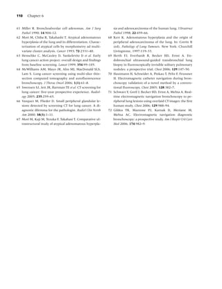
![CHAPTER 7
Treatment of Preneoplastic
Lesions of the Lung
Annette McWilliams
Introduction biopsy. Angiogenic squamous dysplasia is a recently
described histopathological change in the bronchial
Localization of intraepithelial neoplasia (IEN) (mod- epithelium of heavy smokers that is felt to be a
erate/severe dysplasia, and carcinoma in situ (CIS)) possible intermediate biomarker in bronchial car-
has been made possible by the development of cinogenesis [5]. This lesion is detected by aut-
autofluorescence imaging of the bronchial mucosa ofluorescence bronchoscopy and is a collection of
as these lesions are difficult to detect with white- capillary-sized blood vessels closely juxtaposed to
light examination even with the new generation of dysplastic epithelial cells. The capillary loops project
videoendoscopes [1]. Information about the natu- upward into the bronchial mucosa and the lesion
ral history of IEN is now emerging but considerable is associated with increased expression of prolifer-
controversy still exists regarding prognosis and in- ative markers. Although the long-term prognostic
dications for treatment. Currently, histopathology is significance is unknown, short-term follow-up at
the only available tool to predict the biological be- 1 year shows persistence of 45% of these lesions
havior of IEN and it remains the gold standard for and they were more common in subjects with a
assessment of lesions that are abnormal on autoflu- concurrent lung cancer, particularly squamous cell
orescence examination. Interobserver variation in carcinoma.
reporting the presence or degree of IEN has been In association with its improved ability to de-
an important issue but the recent development of tect IEN, autofluorescence bronchoscopy has been
new WHO classification will act to reduce this prob- shown to have reduced specificity due to the iden-
lem [2]. The development of endobronchial carci- tification of multiple false-positive lesions. How-
noma has been proposed to occur through progres- ever, this has been challenged by recent data show-
sive premalignant changes and higher degrees of ing that false-positive lesions (abnormal autoflu-
dysplasia are associated with increased risk for sub- orescence but benign pathology) have increased
sequent invasive cancer [3]. However, some lesions chromosomal aberrations [6]. The presence of aut-
may persist without progression, show nonstepwise ofluorescence abnormalities may be a marker of
progression or spontaneous regression [4]. Interpre- field carcinogenesis and overall cancer risk. Patients
tation of regression rates may be confounded by the harboring high-grade lesions appear to be at risk
removal of part or the entire lesion by bronchial for developing lung cancers at remote sites [7]. The
presence of two areas or more of abnormal autoflu-
orescence in high-risk patients, despite histopatho-
Lung Cancer, 3rd edition. Edited by Jack A. Roth, James D. Cox,
logical grade, has been shown to be associated with
and Waun Ki Hong. c 2008 Blackwell Publishing, a significantly increased risk of developing subse-
ISBN: 978-1-4051-5112-2. quent lung cancer [8].
111](https://image.slidesharecdn.com/lungcancer3rded-120126215311-phpapp01/85/Lung-cancer-3rd-ed-122-320.jpg)
![112 Chapter 7
Table 7.1 Longitudinal outcome of intraepithelial neoplasia.
Moderate
dysplasia Severe dysplasia Carcinoma in situ
Median Progression
Number of Number of follow-up Progression to Progression to to invasive
patients lesions (months) cancer cancer Persistance cancer
Venmans et al. [14] 9 9 22 – – – 67%∗
Deygas et al. [15] 35 41 NR – – – 20%†
Bota et al. [16] 104 228 24 0% ‡ 11% 75%§ –
George et al. [7] 22 53 23 0% ‡ 0% 64% 21%
Lam et al. [17] 566 208 21 1.3% 6% – –
‡
Breuer et al. [4] 52 89 17.5 9% 32% – –
Hoshino et al. [11] 50 66 6.9 1.8% 18% – –
∗
Progression occurred despite initial endobronchial treatment in 67% lesions.
†
Progression occurred despite initial cryotherapy in all lesions.
‡
Mild and moderate dysplasia reported together.
§ Persisted at 3 months and were treated.
Only mean followup duration reported.
NR, not reported.
There are presently no molecular markers to reli- stimulated discussion regarding the need to treat
ably predict the biological behavior of IEN although CIS when it is diagnosed in the bronchus. How-
work in this area is progressing. Suprabasal p53 ever, this approach does not yield information about
staining has been shown to be predictive of subse- the development or progression of the disease. Ret-
quent development of bronchial cancer in the same rospective analysis of untreated patients with ra-
lobe or bronchial spur, and may be a useful tool in diologically occult lung cancer has revealed much
combination with histopathology [9]. Telomerase lower 5-year survival rates compared to surgically
expression in nonmalignant epithelium in high- treated cases who can achieve >90% survival at 5
risk patients with treated lung cancer is associated years [13]. Certainly some subjects with significant
with an increased subsequent development of sec- comorbidities may succumb to cardiopulmonary or
ond bronchial malignancies [10]. In addition, high other disease before their lung cancer, but in health-
telomerase activity, increased Ki-67 expression and ier or younger patients who have longer expected
p53 positivity in IEN have been found to be asso- survival, ignoring the presence of carcinoma at an
ciated with persistence or progression of the lesion early curable stage is likely to be detrimental.
on longitudinal observation [11]. Longitudinal observational data using serial aut-
ofluorescence bronchoscopy and biopsy in patients
with IEN has now been reported by a number of
The natural history of high-grade centers (Table 7.1) [4,7,11,14–17]. The observed
bronchial intraepithelial neoplasia rate of progression to carcinoma of moderate dys-
plasia is up to 9% and severe dysplasia is up to
Autopsy data has revealed that some patients har- 32%. CIS is seen to either persist in >60% cases
bor radiographically occult lung cancer at the time on short- to medium-term follow-up or progress to
of their death from another cause [12]. This find- invasive cancer in more than 20–60% cases despite
ing has raised the possibility of overdiagnosis and initial treatment in some instances. Carcinoma has](https://image.slidesharecdn.com/lungcancer3rded-120126215311-phpapp01/85/Lung-cancer-3rd-ed-123-320.jpg)
![Treatment of Preneoplastic Lesions 113
also been seen to develop from nondysplastic ar- tinue observation. Reported spontaneous regres-
eas supporting the field carcinogenesis theory. Lam sion of CIS is likely influenced by diagnostic biopsy
et al. found carcinoma arising from areas of hyper- that may remove small lesions completely, thereby
plasia (0.3%) and metaplasia (0.4%), and Breuer falsely increasing the rate of regression. Historically,
et al. found the rate of progression to carcinoma from variation in histopathological reporting and differ-
metaplasia of 9% [4,17]. Breuer et al. also observed entiation between severe dysplasia and CIS may
that many sites showed nonstepwise progression also have influenced reported outcomes. These fac-
and erratic fluctuations between histologic grades tors have stimulated much debate as to whether
[4]. Bota et al. also reported carcinoma arising from these lesions should be observed or treated at ini-
areas of hyperplasia/metaplasia at a rate of 2% [16]. tial detection. However, reported data from small
The variable rates in progression of these lesions published prospective series is helpful in this situa-
may be due to the different patient populations tion (Table 7.1). These series have shown that the
evaluated and variation in histopathological inter- majority of CIS persists on short- to medium-term
pretation. Patients with a diagnosis of lung cancer follow-up and a significant proportion (21–67%)
or other aerodigestive cancers are at risk of devel- will progress to invasive cancer. In the series where
oping metachronous lung cancers, and this subset CIS lesions were observed without treatment, only
represents a very high-risk group who are likely to half of the lesions that progressed to invasive can-
have greater observed rates of progression. In Lam cer were successfully treated, raising concerns that
et al.’s reported series, with lower rates of progres- delay in treatment may have resulted in a poorer
sion of moderate/severe dysplasia to carcinoma, the outcome for the patients [7].
subjects were smokers with no clinical suspicion of There are no present biological or histopatholog-
cancer or previous history of aerodigestive cancer ical markers to assist in the assessment of these le-
[17]. In the series reported by George et al., who sions and to predict future progression or stability.
also documented lower rates of progression of IEN, Therefore, although some lesions of CIS may be in-
only 40% of study subjects had previous lung can- dolent in the short- to medium-term, a significant
cer and subjects were excluded if they had a clini- proportion is likely to eventually progress to inva-
cal suspicion of lung cancer [7]. The remainder had sive cancer. The clinical relevance will vary accord-
other risk factors such as significant smoking his- ing to individual patient characteristics and life ex-
tory, airflow limitation or asbestos exposure. Ven- pectancy, e.g., an elderly patient with severe airflow
mans et al., Deygas et al., Breuer et al., and Hoshino limitation compared to a 50-year-old patient with
et al. published series where the majority of the sub- no significant comorbidities. The risk of continued
ject population included those with prior or current observation may be the lost opportunity to treat an
aerodigestive cancers or a clinical suspicion of lung early-stage lesion and a worse final outcome due to
cancer (Table 7.1) [4,11,14,15]. bronchial wall invasion and lymph node metastasis.
There are no published guidelines for the follow-
up or treatment of moderate and severe dysplasia.
Most centers that perform autofluorescence bron- Treatment of CIS
choscopy keep these lesions under periodic surveil-
lance with autofluorescence examination although Surgery remains the mainstay of treatment of CIS
treatment of large areas of severe dysplasia may be as the traditional and proven therapy, and results in
performed. There is also considerable controversy >80–90% 5-year survival rate [18–21]. However,
regarding the appropriate management of CIS. Car- up to 30% may require bilobectomy or pneumonec-
cinoma in situ is difficult to detect without autoflu- tomy to achieve cure, and the remaining 70% usu-
orescence bronchoscopy and therefore large obser- ally require lobectomy [18,20,21]. Segmentectomy
vational series of long-term behavior do not exist has only been used in small series of selected pa-
as the imaging technique has only been developed tients with good 5-year survival rates (>90%) [19].
in the last decade. In addition, some centers treat The risk of metachronous lung cancer in patients
CIS immediately upon diagnosis rather than con- with central squamous cell carcinoma is significant,](https://image.slidesharecdn.com/lungcancer3rded-120126215311-phpapp01/85/Lung-cancer-3rd-ed-124-320.jpg)
![114 Chapter 7
ranging from 14 to 30% and synchronous lesions vasion and lymph node involvement. All margins
can be detected in up to 20% of patients [13,15, of the lesion must be visualized endoscopically, the
18–27]. In addition, surgery is not without risk in maximum diameter should be ≤10 mm and the
these patients due to comorbidities associated with whole lesion must be bronchoscopically accessible.
their smoking history and some patients will be in- Flat superficial lesions are more appropriate than
operable due to limited pulmonary reserves. There- those with a nodular or polypoid appearance. There
fore, tissue-sparing methods to conserve pulmonary should be no invasion into or beyond the cartilage
parenchyma need to be considered in the manage- layer of the bronchial wall and lymph nodes should
ment of selected small CIS, reserving surgical re- be of normal appearance and size [33]. Lesions that
section for lesions that are ineligible or have failed do not fit these criteria should be referred directly
endobronchial therapy. for curative surgery if the patient is an operable can-
There are no prospective, randomized studies didate.
comparing surgery to endobronchial therapy for Multiple technologies should be used in combi-
CIS and they are unlikely to be performed in the nation to accurately assess these lesions and assist
near future, in part due to the difficulty in obtain- in the choice of the most suitable treatment option.
ing sufficient number of cases without a very large These include autofluorescence bronchoscopy, en-
multicenter study [28]. However, outcomes of sim- dobronchial ultrasound, and computed tomography
ilar cases of early central lung cancer treated with (CT), in addition to the clinical assessment of the
endobronchial therapy or surgery are comparable patient and their comorbidities that may influence
[20,21,29]. In one cost-effectiveness analysis, the management.
total cost of treatment and follow-up of broncho-
scopically treated small stage 1A cancers in inopera- Surface characteristics
ble patients was 30% of the cost of standard surgery Autofluorescence bronchoscopy is the best modal-
in matched operable patients [29]. ity to evaluate surface features such as extent and
Endobronchial therapy is not curative for lesions size of the lesion and to accurately visualize all
that have invaded into the bronchial cartilage or margins [34,35]. These features cannot be accu-
wall and is therefore limited to ≤3 mm thickness rately assessed with white-light imaging alone. In
[22,30]. Information gained from surgical series one series, 70% of CIS cases diagnosed with white-
have been helpful in identifying lesions that have light bronchoscopy and referred for therapy were
a low risk of bronchial wall invasion and lymph not eligible for endobronchial therapy with cura-
node metastasis that may be amenable to curative tive intent when assessed by autofluorescence imag-
endobronchial therapy [19,21,31]. Bronchial wall ing, thereby resulting in a change in clinical man-
invasion and lymph node involvement were seen agement [35]. In addition, autofluorescence bron-
to be associated a number of features including: ex- choscopy has the added benefit of detecting other
tension of the lesion beyond endoscopic visibility; synchronous sites of IEN that may require surveil-
greater longitudinal dimension of the lesion (>10 lance or treatment. These are particularly common
mm length); and greater nodularity of the tumor. with central squamous cell carcinoma where up to
Clinical trials with photodynamic therapy in the ∼20% patients have a synchronous carcinoma and
treatment of small central cancers have confirmed up to ∼ 45% patients have other lesions of moderate
that a maximum diameter of 10 mm is an important or severe dysplasia [24–26,36,37].
predictor of response to therapy [32,33].
Depth of invasion and lymph
node assessment
Assessment of eligibility for Although the endoscopic features of CIS are use-
curative endobronchial therapy ful in predicting bronchial wall invasion and lymph
node involvement, a more detailed assessment is
Features predictive of response to endobronchial required. Imaging modalities to consider are endo-
therapy include surface appearance, depth of in- bronchial ultrasound (EBUS), CT scan, and positron](https://image.slidesharecdn.com/lungcancer3rded-120126215311-phpapp01/85/Lung-cancer-3rd-ed-125-320.jpg)
![Treatment of Preneoplastic Lesions 115
emission tomography (PET). Thoracic CT and PET treatment of a series of CIS, Nd-YAG laser therapy
are useful techniques for the evaluation of hilar and is not generally considered appropriate for curative
mediastinal lymph nodes and also to exclude syn- therapy due to the increased risk of perforation and
chronous peripheral tumors. Positive lymph nodes hemorrhage with the treatment of these flat lesions
either on CT or PET imaging should be investi- and the availability of safer techniques [48]. All the
gated to exclude metastases before curative endo- techniques can be used with flexible bronchoscopy
bronchial treatment is performed. under conscious sedation as the CIS lesions are small
Although true CIS is not visible with EBUS or and easily treated compared to bulkier obstructive
CT scan, as it is only cell layers thick, unexpected endoluminal disease that may require rigid bron-
deeper invasion is visible and would correctly ex- choscopy.
clude these lesions from endobronchial treatment Photodynamic therapy has the most worldwide
[38–40]. CT scan can detect bronchial wall thicken- clinical experience, mainly in Japan, but there
ing, peribronchial extension, and lymph node en- are issues of cost and photosensitivity. Electro-
largement and has been shown to alter clinical man- cautery/APC and cryotherapy are cheaper and eas-
agement in 22–35% of cases being considered for ier alternatives to PDT but large studies are not avail-
curative therapy after bronchoscopy [38,39]. able. There have been no prospective, randomized
Endobronchial ultrasonography has been eval- studies comparing these methods for curative treat-
uated prospectively in the assessment of cases of ment of CIS. There is one reported prospective series
small central squamous cell carcinoma for cura- comparing the side effects of electrocautery, PDT or
tive endobronchial treatment [39,41,42]. The sen- Nd-YAG laser in patients with similar small, intralu-
sitivity for cartilaginous involvement has been re- minal tumors treated with curative intent [30]. Sig-
ported as 86% with a specificity of 67% [42]. Results nificant airway scarring with/without stenosis was
have confirmed that the ultrasound images corre- more common with PDT or Nd-YAG laser therapy
late well with surgical resection findings and lesions than with electrocautery. This is not surprising con-
that appear intracartilaginous on EBUS have excel- sidering the deeper penetrative properties of PDT
lent long-term results after endobronchial therapy. and Nd-YAG laser compared to electrocautery. As
EBUS resulted in a change in management in 36% CIS is limited to the epithelium and only cell layers
of tumors thought to be curable by endobronchial thick it is likely that the technique itself is not the
therapy after white-light bronchoscopy and thoracic determinant of cure but that correct staging and se-
CT [39]. lection of lesions are the most important factors in
CIS lesions were not generally thought to be visi- achieving a good outcome. It is important to limit
ble with PET scan as the lesion size is often below the tissue damage in order to avoid stenosis that could
resolution threshold, but one center has reported cause further complications for the patient.
73% sensitivity and 80% specificity with PET in Many of the published studies included both stage
CT occult CIS/microinvasive carcinoma of ≤10 mm 1A as well as stage 0 (CIS) disease in both surgi-
diameter [43]. The development of new combined cally operable and inoperable patients and the size
CT/PET imaging is interesting and may provide fur- of the lesion was not always reported. Definitions of
ther advances in imaging these small lesions. complete response and duration of follow-up vary
between reports. These factors are important to con-
sider when assessing treatment outcomes and there-
Endobronchial therapies fore this review focused, where possible, on results
of treatment of either CIS or microinvasive disease
There are a number of well-described techniques ≤10 mm diameter with long-term follow-up.
that have been developed to treat endoluminal dis-
ease. These include photodynamic therapy (PDT), Photodynamic therapy
electrocautery, argon plasma coagulation (APC), Photodynamic therapy uses the interaction of a
cryotherapy, brachytherapy, and Nd-YAG laser ther- photosensitizing drug and light of a specific wave-
apy [44–47]. Despite some promising findings in the length in the presence of oxygen to produce a](https://image.slidesharecdn.com/lungcancer3rded-120126215311-phpapp01/85/Lung-cancer-3rd-ed-126-320.jpg)
![116 Chapter 7
Table 7.2 Outcome of photodynamic therapy in CIS/microinvasive disease ≤10 mm diameter.
Number of Initial Long-term
treated lesions Lesion type response response
Furukawa et al. [33] 83 CIS/microinvasive <10 mm 93% 82%
Imamura et al. [50] 19 CIS ≤10 mm 2
79% 42%
Radu et al. [51] 20 CIS (size NR) — 90%
Cortese et al. [28] 15 Superficial squamous cancer 87% 73%
<10 mm
NR, not reported.
photochemical reaction to destroy the tumor by included 15 superficial carcinoma ≤10 mm diame-
a number of different mechanisms including tu- ter in their series evaluating the role of PDT as a
mor cell apoptosis, vascular damage, and inflam- surgical alternative and achieved an initial response
matory/immune reaction. The main limitations of of 87% and a long-term response of 73%. A second
its use are the cost and size of the laser equipment, treatment to achieve cure was needed in 13% of
cost of drug and skin photosensitization, but the de- these lesions [28].
velopment of new diode laser systems and photo-
sensitizers will help to reduce these issues [32,49]. Electrocautery
The majority of clinical data has emerged from Endobronchial electrocautery uses a high-
Japan and 5-year follow-up data of the largest, most frequency alternating current that is converted to
well-described series of squamous cell carcinoma heat when passing through tissue. A small probe
<10 mm diameter has recently been published [33]. is used to focus the heat at the point of contact
In this report, 83 CIS/microinvasive lesions treated and achieve tissue destruction. It is an inexpensive
with PDT between 1980 and 2001 achieved an ini- technique with rapid and easy application and low
tial bronchoscopic cure rate of 93% that fell to 82% complication rates. There is one report of a series of
over the follow-up duration due to recurrences at 13 patients with 15 small intraluminal lesions ≤10
or near the treated site within 3–18 months after mm2 with normal CT scans. The majority of these
treatment (8/9 occurring within 12 mo) (Table 7.2) lesions were stage 1A (13/15) and the remaining
[33]. The recurrences were thought to be due to in- two lesions were CIS (stage 0). No residual disease
appropriate estimation of the peripheral margin, in- was seen at a median follow-up of 22 months in
adequate laser irradiation or unsuspected intracar- 80% lesions. The failures were found to be due to
tilaginous invasion. A smaller Japanese series of 19 more extensive disease on surgical resection, i.e.,
CIS lesions ≤10 mm2 achieved initial bronchoscopic diameter >10 mm and deeper invasion [52].
eradication in 79% lesions, but on longer follow-up A further subsequent report by the same group
some cases showed local recurrence and the final evaluating curative endobronchial treatment of
cure rate was only 42% [50]. It is uncertain as to small intraluminal, microinvasive stage 1A disease
why the results in this study are much lower than (CIS excluded) in 32 inoperable patients achieved
other series (Table 7.2). bronchoscopic eradication in >95% of treated cases.
Other studies have been performed in North Lesions were ≤10 mm in diameter and were as-
America and Europe (Table 7.2). A European study sessed to be appropriate for endobronchial ther-
reported the treatment of 20 bronchial CIS as part apy by autofluorescence imaging and CT scan. Most
of a larger series of aerodigestive cancers achiev- patients were treated with electrocautery (75%)
ing bronchoscopic eradication in 90% cases over a and the remainder with APC, PDT, or Nd-YAG
mean follow-up of 27 months [51]. The Mayo Clinic laser. A second treatment to achieve cure was](https://image.slidesharecdn.com/lungcancer3rded-120126215311-phpapp01/85/Lung-cancer-3rd-ed-127-320.jpg)
![Treatment of Preneoplastic Lesions 117
required in 9% subjects. Half the patients died in the bronchial malignancy to deliver local irradiation
mean follow-up of 5 years due to other disease or and requires the placement of an afterloading
cancers, but only one death was attributable to the polyurethane catheter into the airway during flexi-
failure of treatment of achieve cure. The remainder ble bronchoscopy. This technique is not usually used
of patients is alive without recurrent disease at the as first-line endobronchial therapy for cure of CIS
treated site after mean follow-up of 5 years [23]. or small microinvasive disease, but as second-line
Argon plasma coagulation is a more recent devel- therapy or for bulkier disease in an inoperable pa-
opment that delivers electrocautery by a noncontact tient. Brachytherapy has been evaluated as a sole
method using ionized argon gas to conduct electric- modality in a study of high dose iridium in 18 inop-
ity between the active electrode and the tissue. It erable patients with endobronchial cancers ≤10 mm
has excellent coagulative properties and is a cheap and normal CT scan, achieving 83% initial response
and easy technique [44,47]. There is little published rates that fell to 72% at 1 year follow-up [54]. Intra-
data of its use in treatment of bronchial CIS but due luminal dosage ranged between 21 and 35 Gy. How-
to its superficial coagulation properties it would also ever, 55% subjects developed asymptomatic partial
be a useful technique [53]. bronchial stenosis and 16.5% developed bronchial
wall necrosis and/or fatal hemoptysis [54]. A similar
Cryotherapy study in 34 patients with endobronchial cancer and
Cryotherapy is an inexpensive and safe technique normal CT scan was subsequently published but the
where tissue is destroyed by freezing and is partic- lesion size was not reported although it included six
ularly useful for CIS situated on the membranous CIS lesions. Similar results were obtained with 85%
portion of the trachea or main bronchi where the eradication at 2 years and 73% at 3 years follow-up
risk of perforation is greater than the cartilaginous with similar intraluminal dosages. Hemoptysis was
portion. Nitrous oxide is the most commonly used only seen in 2.9% cases [55].
agent in bronchoscopic cryotherapy. There are lim- Therefore, PDT, electrocautery, and cryotherapy
ited reports of its use for curative therapy of CIS are effective in treating small CIS/microinvasive le-
but no adverse events have been reported [15]. sions ≤10 mm diameter and achieving long-term
Deygas et al. reported the largest series of 35 pa- cure rates of between 73 and 90% in most studies.
tients with CIS/microinvasive disease detected by Brachytherapy appears similarly effective but has
white-light bronchoscopy and achieved 91% cure higher complication rates and requires repeat treat-
at 12 months follow-up. Initial size of the lesions ment fractions. Response rates may be further im-
was not reported. A second cryotherapy treatment proved in the future by the incorporation of newer
was required in 8.6% patients to achieve cure. The technologies such as autofluorescence imaging and
overall long-term cure rate was 80% as 7/35 pa- EBUS to improve eligibility assessment.
tients (20%) subsequently developed invasive dis-
ease at the previously treated sites. It is uncertain Follow-up after endobronchial
from the reported data whether the invasive disease treatment
occurred at the sites of CIS or microinvasive dis- Careful surveillance with repeat biopsies after the
ease. The lesions were only assessed with white- initial therapy is required as up to 10–15% of pa-
light bronchoscopy and thoracic CT scan and there- tients may require a second treatment to achieve
fore it is likely that some lesions may not have been cure [15,23,28]. However, this may be an overesti-
eligible for curative endobronchial therapy accord- mate of the true need for repeat treatment of CIS as
ing to the aforementioned criteria that has been sub- it arises from series that include microinvasive dis-
sequently developed [15]. ease and lesions not assessed with autofluorescence
imaging or EBUS. In surgically operable patients,
Brachytherapy if cure is not achieved with endobronchial ther-
Brachytherapy involves the insertion of a radioac- apy, then prompt referral for surgery is required to
tive source (usually Iridium192 ) near an endo- avoid progressive disease. In addition, longer-term](https://image.slidesharecdn.com/lungcancer3rded-120126215311-phpapp01/85/Lung-cancer-3rd-ed-128-320.jpg)
![118 Chapter 7
surveillance is recommended after curative therapy 2 Brambilla E, Travis WD, Colby TV, Corrin B, Shimosato
as up to 30% subjects will develop metachronous Y. The New World Health Organization classification of
disease at other sites. Follow-up bronchoscopy and lung tumors. Eur Respir J 2001; 18:1059–68.
biopsy are generally performed at 4–6 weeks after 3 McWilliams A, MacAulay C, Gazdar AF, Lam S. Inno-
the initial treatment, 3–6 monthly for the first 1–2 vative molecular and imaging approaches for the de-
tection of lung cancer and its precursor lesions. Onco-
years, and then 6–12 monthly for up to 5 years.
gene 2002; 21(45):6949–59.
4 Breuer R, Pasic A, Smit E et al. The natural course of
Conclusion preneoplastic lesions in bronchial epithelium. Clin Can-
cer Res 2005; 15:537–43.
5 Keith R, Miller Y, Gemmill R et al. Angiogenic squa-
The detection of moderate or severe dysplasia re-
mous dysplasia in bronchi of individuals at high risk
quires the consideration of longitudinal surveillance for lung cancer. Clin Cancer Res 2000; 6:1616–25.
as patients harboring these lesions are at increased 6 Helfritzsch H, Junker K, Bartel M, Scheele J. Differenti-
risk of developing malignancy either at the detected ation of positive autofluorescence bronchoscopy find-
dysplastic site or at another site in the lungs. Future ings by comparative genomic hybridization. Oncol Rep
development of biological markers may assist in the 2002; 9(4):697–701.
task of risk prediction in these patients. CIS should 7 George P, Banerjee A, Read C et al. Surveillance for
be considered for curative therapy in patients with the detection of early lung cancer in patients with
good life expectancy as there are high rates of per- bronchial dysplasia. Thorax 2007; 62:43–50.
sistence or progression to invasive carcinoma. In- 8 Pasic A, Vonk-Noordegraaf A, Risse E, Postmus P, Sut-
edja T. Multiple suspicious lesions detected by autoflu-
formation from surgical series and prospective data
orescence bronchoscopy predict malignant develop-
from clinical trials have enabled the development of
ment in the bronchial mucosa in high risk patients.
clear guidelines in the assessment of lesions for cura- Lung Cancer 2003; 41(3):295–301.
tive endobronchial therapy. There are a number of 9 Breuer R, Snijders P, Sutedja T et al. Suprabasal p53
available tools that can be used for endobronchial immunostaining in premalignant endobronchial le-
treatment that are safe and easy to use with sions in combination with histology is associated with
flexible bronchoscopy under conscious sedation. bronchial cancer. Lung Cancer 2003; 40(2):165–72.
Eligible lesions include flat, bronchoscopically ac- 10 Miyazu Y, Miyazawa T, Hiyama K et al. Telomerase ex-
cessible lesions ≤10 mm diameter with visible mar- pression in noncancerous bronchial epithelia is a possi-
gins on autofluorescence imaging, no invasion on ble marker of early development of lung cancer. Cancer
endobronchial ultrasound or CT scan and normal Res 2005; 65(21):9623–7.
11 Hoshino H, Shibuya K, Chiyo M et al. Biological fea-
lymph nodes. If the patient is a surgical candidate,
tures of bronchial squamous dysplasia followed up
curative endobronchial therapy could be discussed
by autofluorescence bronchoscopy. Lung Cancer 2004;
with the patient as a possible surgical alternative 46:187–96.
and parenchymal sparing measure in light of the 12 Manser R, Dodd M, Byrnes G, Irving L, Campbell
high risk of metachronous disease. If the patient is DA. Incidental lung cancers identified at coronial au-
not a surgical candidate, endobronchial treatment topsy: implications for overdiagnosis of lung cancer by
could be considered as first-line therapy if appro- screening. Respir Med 2005; 99(4):501–7.
priate or in combination with radiation therapy 13 Sato M, Saito Y, Endo C et al. The natural history of ra-
[50,56]. diographically occult bronchogenic squamous cell car-
cinoma: a retrospective study of overdiagnosis bias.
Chest 2004; 126(1):108–13.
References 14 Venmans B, van Boxem T, Smit E, Postmus P, Sutedja
T. Outcome of bronchial carcinoma in situ. Chest 2000;
1 Chhajed P, Shibuya K, Hoshino H et al. A compari- 117(6):1572–6.
son of video and autofluorescence bronchoscopy in 15 Deygas N, Froudarakis M, Ozenne G, Vergnon JM.
patients at high risk of lung cancer. Eur Respir J 2005; Cryotherapy in early superficial bronchogenic carci-
25(6):951–5. noma. Chest 2001; 120(1):26–31.](https://image.slidesharecdn.com/lungcancer3rded-120126215311-phpapp01/85/Lung-cancer-3rd-ed-129-320.jpg)
![CHAPTER 8
The Pathology and Pathogenesis of
Peripheral Lung Adenocarcinoma
Including Bronchioloalveolar
Carcinoma
Wilbur A. Franklin
Introduction Adenocarcinoma and the anatomy
of the peripheral airway
The histopathological classification of adenocarci-
noma of lung has received extraordinary scrutiny Adenocarcinoma is the most common tumor of the
during the past several years for several reasons. peripheral airway, an anatomic region that is not
First, improved sensitivity of thoracic imaging meth- easily accessible for tissue sampling. In this region
ods has resulted in resection of increasingly smaller the bronchioles terminate in microscopic alveolar
peripheral lung lesions, more than 70% of which sacs where gas exchange occurs. The transition from
are adenocarcinomas [1–3]. Second, high through- bronchus to alveolus is accompanied by a transition
put gene expression methods and molecular models from the pseudostratified mucociliary epithelium of
have changed molecular understanding of adeno- the distal bronchi and proximal bronchioles to non-
carcinoma. Finally, new targeted therapeutic agents mucinous epithelium in the distal bronchiole that
have provided tools that are effective in subsets of contains a mixture of ciliated and nonciliated cells,
adenocarcinomas and challenge pathologists to ac- including the Clara cells [4]. Clara cells synthesize
count for selective drug sensitivity. This chapter will numerous proteins specific for this region includ-
review the historical development of the current ing surfactant apoproteins A, B and D, tryptase,
histopathological classification pulmonary adeno- β-galactoside-binding lectin, a specific phospholi-
carcinoma and how this classification is being af- pase, and a 10 kD protein, CC10 [5]. These pro-
fected by changing concepts of pathogenesis and teins are stored in secretory granules and are re-
molecular properties of this increasingly common leased into lower airway fluid. The most abundant
tumor. protein in airway fluid is CC10, a uteroglobin-like
protein whose function has not been conclusively
established but may be to protect the lung from en-
vironmental toxins [6,7]. It serves as a marker for
distal bronchial epithelial differentiation.
At the bronchiolar terminus where the bron-
Lung Cancer, 3rd edition. Edited by Jack A. Roth, James D. Cox, chiole merges with the alveolar septum, a short
and Waun Ki Hong. c 2008 Blackwell Publishing,
segment of the airway is surfaced by a simple
ISBN: 978-1-4051-5112-2.
121](https://image.slidesharecdn.com/lungcancer3rded-120126215311-phpapp01/85/Lung-cancer-3rd-ed-132-320.jpg)
![122 Chapter 8
agranular, nonciliated cuboidal epithelium. The and support the analogy between fetal development
nondescript cuboidal cells give way distally to the and lung tumor differentiation. An incompletely ex-
flattened and thin alveolar cells that line the alve- ploited consequence of this analogy is the revela-
olar sacs. Alveolar cells are of two types, the thin tion of new biomarkers and possible targets for early
plate-like type I cells that cover more than 90% of detection and therapeutic intervention as discussed
the alveolar septa and through which gas exchange below.
occurs and the type II cells which are more numer-
ous than type I cell but cover only a small fraction
of the alveolar surface. Type I cells are only 0.1– Classification of pulmonary
0.2 microns in thickness and may not be visible by adenocarcinoma
light microscopy, but are consistently detected by
electron microscopy as a continuous lining of alve- The classification of lung cancer in general and
olar surfaces. Type II cells are more easily identi- adenocarcinoma in particular remains grounded in
fied as cuboidal cells protruding into the alveolar the careful microscopic study of human tumors in
space. These cells have several functions including hematoxylin and eosin-stained sections. This basic
secretion of surfactant, absorption of ions and fluid information has been buttressed in recent years by
from the alveolar space, and repair of alveolar injury new immunohistochemical and molecular data, but
[4]. The molecular apparatus that permits the histological diagnosis remains the sina qua non of
type II cell to accomplish these functions is ev- treatment and evaluation of outcome. At the most
ident in many pulmonary adenocarcinomas [8] basic level, adenocarcinoma is defined in the most
(see below). recent WHO monograph on lung cancer as “a ma-
In addition to mimicking normal adult tissue, lignant epithelial tumour with glandular differen-
lung tumors may recapitulate cellular and molecu- tiation or mucin production . . . ” [15]. This recent
lar properties of the embryonic lung. Lung anatomy edition, published in 2004, includes 13 types and
develops rapidly and continuously during embry- subtypes of adenocarcinoma, the most common of
onic life through five more or less distinct anatomic which is the adenocarcinoma, mixed subtype. A de-
stages (reviewed in [9]) beginning with the for- tailed histological description is provided for each of
mation of the lung bud from the foregut endo- the major subtypes of adenocarcinoma as well as for
derm in the embryonic stage (gestational age 9– some of the more unusual subtypes in order to il-
12 days). During the succeeding stages, pseudog- lustrate the diversity of histological appearances in
landular, canalicular and saccular structures, and adenocarcinoma of the lung and to provide infor-
finally alveoli develop under the strictly timed reg- mation on their differential diagnosis. The defining
ulation of transcription factors [9–11]. Distinct pat- features of each subtype are listed in Table 8.1.
terns of gene expression correspond to each stage The current classification is the result of a long
of development [12]. Similarities between gene ex- historical process in which definitions and termi-
pression profiles of developing lung and tumor have nology have been refined to accommodate ob-
been described in mouse models [13] and these servations regarding cellular structure and clinical
findings have been extrapolated to human tumors. outcome. Adenocarcinoma has been a recognized
One study suggests that adenocarcinomas recapit- tumor type since the first codified histological clas-
ulate gene expression patterns that correspond to sifications of lung cancer were introduced in the
the saccular and alveolar developmental stages of early twentieth century when lung cancer was still a
the lung while large cell and squamous carcinomas rare disease. In 1924, Marchesani produced one of
exhibit profiles that more closely match the ear- the first widely adopted histological classifications
lier pseudoglandular and canalicular stages of devel- of lung and included “zylinderzellige adenokarzi-
opment [14]. Overall comparisons of gene expres- nome” (cylindrical cell adenocarcinoma) as one of
sion profiles suggest that tumors re-express genes four main histological types of lung carcinoma. The
that are associated with embryonic development Marchesani classification was used widely without](https://image.slidesharecdn.com/lungcancer3rded-120126215311-phpapp01/85/Lung-cancer-3rd-ed-133-320.jpg)
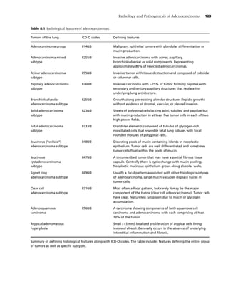
![124 Chapter 8
change for the next 25 years as the frequency of Current definitions permit no evidence of stro-
lung cancer increased. Following World War II, the mal invasion in BAC. To understand the rationale
prevalence of lung cancer grew into a major public for this definition, it is necessary to understand the
health problem and a universally applicable revised evolution of the concept of BAC.
classification was needed. In 1967, the first WHO
lung cancer classification was published, represent- The evolving definition of BAC
ing the consensus of an expert panel of pathologists. Although the hypothesis that lung tumors may orig-
The classification included three subtypes of adeno- inate in the alveoli was proposed as early as the
carcinoma, acinar, papillary, and bronchioloalveo- mid-nineteenth century [16], it was not until 1953
lar. Over the succeeding 40 years, WHO-sponsored that the thin layer of pneumocytes along the sur-
panels have convened periodically to revise the orig- face of the alveolar septae was discovered by elec-
inal classification. The most notable trend in the tron microscopy [17]. By 1960, the observation
revisions has been the increase in the number of that some well-differentiated lung cancers resem-
tumor subtypes. The increase in diagnostic cate- bled bronchiolar or alveolar epithelium and could
gories has been engendered largely by the recog- spread along the alveolar surfaces led to the in-
nition of histologically distinct categories that have troduction and general acceptance of the designa-
clinical or biological significance. The prototypical tion “bronchioloalveolar” carcinoma by Leibow in
pulmonary adenocarcinoma is bronchioloalveolar 1960. These tumors were described as “ . . . well-
carcinoma (BAC), a tumor that uniquely reflects differentiated adenocarcinomas primary in the pe-
the macroscopic and microscopic organization of the riphery of the lung beyond a grossly recognizable
lung as discussed below. bronchus, with a tendency to spread chiefly within
the confines of the lung by aerogenous and lym-
phatic routes . . . ” [18]. According to this definition,
Bronchioloalveolar carcinoma BAC could be invasive or noninvasive carcinoma.
BAC is a well-differentiated adenocarcinoma origi- The WHO classifications accepted this definition un-
nating in the peripheral lung that spreads through til 1999, when BAC was reclassified strictly as an in
the airways. Its defining histological feature is its situ tumor.
nondestructive growth along the alveolar septae, a The change in classification was based on evi-
feature that has been referred to as “lepidic” spread. dence that began to accumulate in the late 1970s
The cells that exhibit this pattern of growth are of and early 1980s. Shimosato et al. reported in 1980
two types, nonmucinous (Plate 8.1a) and mucinous that peripheral adenocarcinomas <3 cm without fi-
(Plate 8.1b). Mucinous tumors have vacuoles, usu- brosis had a significantly better prognosis than tu-
ally in the upper cytoplasm, that are large enough mors of the same size with central scarring [19].
to compress and deform the nuclei of the cell. Large Noguchi et al. later reported that tumors <2 cm had
amounts of mucin may be present in the alveoli ad- a 100% 5-year survival while the presence of fi-
jacent to tumor cells. The nonmucinous cells resem- brosis indicating stromal invasion reduced 5-year
ble Clara cells or type II pneumocytes. The Clara survival to 75% [20]. Fibrosis was strictly defined
cell-type tumors are composed of columnar cells in this study as an active fibroblastic focus and was
with eosinophilic cytoplasm and apical cytoplasmic distinguished from “collapse” in which compressed
projections (“snouts”). Type II cell tumors are more alveoli are admixed with elastic tissue. These studies
cuboidal with foamy, vacuolated cytoplasm. Nuclei highlighted the importance of fibrosis in small soli-
of all types of BAC are usually low grade with mi- tary nodules and it has been subsequently shown
totic figures few in number. In a small percent- that not only is the presence of fibrosis important
age of BAC mixtures of cell types may occur. In but the size of the fibroblastic focus also affects prog-
such cases mucin stains may be useful in confirming nosis. Tumors with areas of scarring <5–10 mm in
the mucinous or mixed mucinous/nonmucinous maximum diameter have a significantly worse out-
subtype. come that those without fibrosis [21,22]. Tumors](https://image.slidesharecdn.com/lungcancer3rded-120126215311-phpapp01/85/Lung-cancer-3rd-ed-135-320.jpg)
![Pathology and Pathogenesis of Adenocarcinoma 125
<2.0 cm with invasion at the center of a fibroblastic a result of fat or glycogen production or fixation
focus have 60% 5-year survival while tumors with artifact. Nonadenocarcinomas may produce small
more minimal fibrotic foci have 100% survival at 5 amounts of mucin and by convention, 10% tumor
years [23]. cells producing mucin is a requirement for the di-
The prognostic importance of a fibroblastic stro- agnosis of invasive adenocarcinoma.
mal response prompted a reassessment of BAC that Adenocarcinoma is graded on the basis of level
is reflected in the 1999 [24] and 2004 [15] WHO of cellular size and deformity, variation of nu-
classifications. In the revised classifications, BAC is clear size and shape, chromatin configuration, nu-
considered to be a purely in situ lesion that can clear/cytoplasmic ration, and mitotic activity. In the
spread through alveolar spaces but exhibits no stro- better-differentiated tumors, cells have finely gran-
mal invasion and does not metastasis to distant sites ular chromatin that is evenly dispersed. At the more
through blood or lymphatics. Since most tumors poorly differentiated end of the spectrum, adeno-
have a component of stromal invasion, a possible carcinoma may resemble large cell carcinoma with
effect of changing the definition of BAC could be only a few tubular structures in a background of
to reduce its frequency. By exactly how much is sheets of tumor cells. In more poorly differentiated,
not known. In Japanese studies in which the in- higher-grade tumors nuclear chromatin is coarse,
vasion exclusion was first applied, 28 of 236 soli- clumped, and irregular. Nuclear membranes are
tary tumors (12%) were classified as noninvasive well defined and may be folded and angulated. Nu-
[20]. In a more recent CT screening study in the cleoli are prominent and may be single or multiple
United States, where the in situ only criterion was and rounded or irregularly shaped.
applied, 20 of 348 tumors (6%) were classified as Precise grading criteria are not widely employed
BAC [1]. These figures are similar to older figures nor are they well defined at the present time. Gen-
to which the in situ criterion was not applied (re- erally the closer to recapitulation of normal lung
viewed in [25]). These preliminary numbers suggest tissue, the lower the grade. Histological grade, nu-
that the frequency of BAC is not likely to drastically clear grade, necrosis, and the presence of lymphatic
change as a result of changes in the criteria for the invasion or a greater than 25% papillary growth
diagnosis of BAC. The overwhelming majority of component have been associated with more aggres-
lung adenocarcinomas will continue to be classified sive behavior. However, the predictive accuracy of
as invasive, either of the acinar type or the mixed any of these features is low and generally not suf-
subtype (mixed acinar and bronchioloalveolar ficient for extrapolation to individual cases. Even
subtype). small, well-differentiated adenocarcinomas have an
8% frequency of metastasis at the time of surgery.
Invasive adenocarcinoma: acinar and There is little evidence that grade of invasive tumor
mixed subtype is of prognostic importance, the major determinant
Invasive pulmonary adenocarcinoma is character- of prognosis being stage.
ized by the formation of tubules or acini in a fi- Necrosis may be pronounced in untreated in-
brous stroma with the destruction of the underlying vasive adenocarcinomas. This feature may hinder
lung architecture (Plate 8.2). This pattern of growth efforts to microscopically assess chemotherapeutic
may occur without an associated BAC component intervention following drug administration. Levels
(acinar adenocarcinoma) or as an epicenter adja- of both necrosis pretreatment and posttreatment
cent to a BAC component (adenocarcinoma mixed should be taken into account when protocols de-
subtype). Invasive adenocarcinoma cells are colum- signed to assess the effect of chemotherapy are im-
nar or cuboidal and may produce mucin. The pro- plemented.
duction of mucin is a useful diagnostic feature and In some tumors, a recently described pattern
may be demonstrated by mucin or PAS stains. These of differentiation referred to as micropapillary and
stains help to distinguish true mucus production consisting of tufts of tumor cells without vascu-
from nonmucinous vacuolation that can occur as lar cores, may be present in variable numbers of](https://image.slidesharecdn.com/lungcancer3rded-120126215311-phpapp01/85/Lung-cancer-3rd-ed-136-320.jpg)
![126 Chapter 8
tumor cells [26]. Metastases from these tumors more characteristic of stromal or mesenchymal cells
are more frequent and the metastatic tumor de- than their cells of origin. This phenomenon is re-
posits are more frequently micropapillary than in ferred to as an epithelial–mesenchymal transition
conventional adenocarcinomas [26]. Most studies (EMT). EMT is accompanied by loss of epithelial in-
to date have suggested that this micropapillary pat- tercellular junctions and changes in the pattern of
tern of tumor growth is associated with consid- growth from the characteristic sheet-like epithelial
erably worse survival, particularly in early-stage monolayer to a single cell infiltrating pattern, char-
tumors [26–29], although at least one study has acteristic of mesenchymal cells [35]. Morphological
found no increase in survival risk for this pattern of change is accompanied by changes in patterns of
tumor growth. More data will be needed from larger gene expression. Tumor cells express mesenchyme-
cohorts to resolve this issue but for the moment associated genes such as vimentin and zeb1 at a
the micropapillary pattern of tumor cell growth is high level and epithelium-associated genes such as
best regarded carcinoma patients as a feature of E-cadherin and snail at a low level [36]. Cell lines
lung adenocarcinoma that may predict an aggres- with mesenchymal patterns of gene expression are
sive clinical course. less sensitive to EGFR blockade than those with a
The hallmark of invasive carcinoma is stromal more characteristically epithelial phenotype [37].
invasion, which is characterized by the presence How far this line of reasoning will take us is not
of active appearing fibroblasts in association with evident at the present time but the predictive edi-
tumor cells. The significance of the fibrosis has been fice provided by the EMT hypothesis may provide
debated for many years. Fibrotic scars may occur as a a theoretical rationale for drug sensitivity and early
result of inflammatory conditions such as tubercu- detection algorithms that may be clinically applica-
losis, infarction, pneumoconiosis, and many other ble in the future.
conditions. In some cases, these postinflammatory The stromal reaction in pulmonary adenocar-
scars may predate adenocarcinoma and it has been cinoma also includes leukocytic infiltrates and in
suggested that scarring may play a causative role some cases dramatic lymphocytic responses ac-
in adenocarcinoma of the lung, often in the apex companied by mononuclear dendritic cells and
of the lung [30]. Tumors associated with scarring eosinophils. Whether this infiltrate represents an
were referred to as “lung scar cancers” [31] or sim- active immunological response directed against tu-
ply “scar carcinoma.” However, by the 1980s, it was mor cell antigens or an epiphenomenon mediated
becoming evident that tumor itself could induce scar by cytokines produced by tumor cells is currently
formation [19,32] and that the cellular and molec- being debated. A detailed discussion of immuno-
ular composition of tumor-associated scars was dif- logical response to tumor is beyond the scope of
ferent than that of post-inflammatory scars with the this chapter but the presence of an inflammatory
former containing considerably greater numbers of infiltrate, particularly the presence of tumor infil-
myofibroblasts [33] and larger quantities of types I trating lymphocytes (TIL) in adenocarcinoma (Plate
[34] and V [33,34] collagen. The presence of scar- 8.3), raises hope that enhancing an immunologi-
ring in adenocarcinoma is now considered to be cal response to tumor may be a feasible treatment.
largely a host response to tumor rather than an ac- TIL concentrations estimated by simple microscopic
tive carcinogenic process. Those cases in which scar inspection of sections of nonsmall cell lung carci-
precedes tumor are now attributed to coincidental noma (NSCLC) stained for the T cell receptor CD3
occurrence of separate disease processes in the same [38,39] or dendritic cells [38], have shown a modest
anatomic location. survival advantage in those subjects with relatively
Recently, tumor–stromal relationships in adeno- high concentrations of these cells. Recent prelim-
carcinoma have been approached from an entirely inary data suggests that the subset of lymphocytes
different perspective. It has been observed that car- may predict outcome. It has been reported that CD8
cinoma cells, which are epithelium-derived, begin cells infiltrating tumor may be inactive [40] and the
to express cellular and molecular properties that are stimulation of TIL may activate CD8 cells [39,41]](https://image.slidesharecdn.com/lungcancer3rded-120126215311-phpapp01/85/Lung-cancer-3rd-ed-137-320.jpg)
![Pathology and Pathogenesis of Adenocarcinoma 127
and thus have an antitumor effect. Whether it will Tumors with abnormal mucin production—
be possible to translate these descriptive studies into mucinous (colloid) adenocarcinoma,
clinical advance remains to be determined. What mucinous cystadenocarcinoma, and signet
is clear is that the stromal and immunological re- ring adenocarcinoma
sponse to tumor is complex and that enhancement These tumors are discussed together since they
of specific responses to tumor may not only elicit an share the common feature of strikingly aberrant
antitumor response but stimulate tumor growth as production of mucin. In colloid adenocarcinoma,
well. Possible stimulatory effects from the cytokine- tumor cells are suspended in large mucinous pools
rich stromal environment of lung NSCLC [42,43] and may partially line fibrous septae within the tu-
(including adenocarcinoma) must be accounted mor (Plate 8.5). Mucinous cystadenocarcinoma is
for in immunological approaches to lung cancer usually well circumscribed with tumor cells lining
treatment. cyst-like, mucin-containing spaces. Finally, signet
ring adenocarcinoma is composed of clusters and
singly infiltrating tumor cells that contain large in-
Unusual lung cancers and tracellular mucin vacuoles [46]. They may be part
differential diagnosis of a more copious acinar tumor [47]. All of these
In addition to the relatively common types of BAC tumors are invasive tumors and resemble their
and invasive adenocarcinoma, several less common counterparts in other organs including breast and
histological patterns are encountered in pulmonary gastrointestinal tract. Immunohistochemical stains
adenocarcinoma that may present diagnostic diffi- may help to distinguish tumors originating in lung
culties. Many of these tumors resemble metastatic from those of GI origin. Pulmonary signet ring tu-
carcinoma with which they are prone to be con- mors express cytokeratin 7 and TTF-1 but not cytok-
fused. However, careful histological examination eratin 20, MUC2 or the intestine-specific homeobox
supplemented by appropriate immunohistochemi- gene CDX-2 [46,48–50], an immunohistochemical
cal profiling will usually permit precise histological profile typical of primary lung carcinoma. However,
classification. a proportion of mucinous tumor in which lung is
the only detectable site may not have immunohis-
tochemical findings that are typical of lung cancer
Papillary adenocarcinoma [50] and in these cases diagnosis of lung primary
Papillary carcinomas are tumors in which the pre- can only be made by exclusion.
dominant pattern is one in which papillary struc-
tures supported by central fibrovascular cores form Clear cell adenocarcinoma
secondary and tertiary branches (Plate 8.4). By In this tumor, cytoplasmic contents do not stain giv-
current WHO definition, this is an invasive tu- ing the cells an empty or “clear” appearance under
mor that replaces the underlying architecture of the microscope. The cytoplasmic clearing may be
the lung [44]. Tumor cells exhibit marked nu- due to the accumulation of lipid or glycogen, which
clear atypia. Papillary adenocarcinoma may contain is dissolved during conventional histological pro-
psammoma bodies and areas of necrosis. This tu- cessing. Clear cell differentiation may occur in any
mor is sometimes difficult to distinguish from BAC of the pulmonary adenocarcinoma subtypes [51].
in which there may be simple papillary structures Tumors with this morphological appearance must
but in BAC there is underlying preservation of the be distinguished from renal cell carcinoma, which
alveolar architecture. Papillary adenocarcinoma oc- is typically TTF-1 negative, and from the so-called
curs in an older population (mean age 65) and “sugar tumor,” a benign neoplasm that is thought
most patients (86%) are smokers [45]. It is impor- to arise from perivascular epithelioid cells that con-
tant to recognize this subtype of adenocarcinoma tains copious glycogen and strongly express the
since survival is poor (3.4 yr for stage I tumors) HMB45 immunohistochemical marker (reviewed in
[45]. [52]).](https://image.slidesharecdn.com/lungcancer3rded-120126215311-phpapp01/85/Lung-cancer-3rd-ed-138-320.jpg)
![128 Chapter 8
Fetal adenocarcinoma ate a characteristic CT image, the so-called “ground
Low-grade tumors resembling fetal lung have been glass opacity” [61] (Plate 8.8) [62,63]. The small pe-
distinguished in recent decades from higher-grade ripheral ground glass opacity is often a presenting
pulmonary blastoma [53–57]. These tumors have feature of BAC and is one of three major patterns
a peak incidence in the fourth decade and more of spread recognizable on CT scans described below
often occur in females than males. They typically and reviewed in [64].
occur in smokers. The characteristic feature of this 1 Solitary mass or nodule. Nodules may be “solid,”
tumor is the formation of tubules by glycogen- obscuring of the underlying lung markings, or non-
rich cells that resemble fetal bronchial tubes at 10– solid (“ground glass opacity”) in which the under-
15 weeks gestational age (Plate 8.6). The tubules lying lung architecture remains visible. The latter
contain numerous neuroendocrine cells and fre- appearance is due to the preservation of alveolar
quently form squamous morules. The stroma is air spaces and is characteristic of BAC but may
composed of myofibroblastic cells but is not a promi- also be observed in non-BAC tumors and inflam-
nent feature of this tumor, distinguishing it from matory conditions. It might be expected that all
the more aggressive pulmonary blastoma. It is im- BAC would have a ground glass appearance but
portant to recognize this tumor because of its ex- the phenomenon of collapse causes partial or com-
cellent prognosis with surgery alone, and to avoid plete opacification of tumor tissue so that histolog-
overtreatment. ical confirmation of BAC diagnosis is required for
definitive diagnosis.
2 Pneumonic form. In this form, there is a
Sclerosing hemangioma
pneumonia-like consolidation of a segment, lobe or
Pulmonary sclerosing hemangioma is a generally
entire lung that may be difficult to distinguish from
benign tumor that has a prominent epithelial com-
benign peripheral airway disease [65]. This pattern
ponent and may be mistaken for adenocarcinoma.
is due to lepidic spread of tumor in the affected lung
Typically, it is a well-circumscribed lesion that is
and partial filling of the alveoli with tumor cells,
hemorrhagic and sclerotic (Plate 8.7a) but contains
mucous, or inflammatory cells.
a mixture of cells arranged in papillary structures
3 Diffuse BAC. Diffuse BAC appears as nodular solid
in which the surface cells are strongly positive for
or ground glass opacities throughout the lungs. This
epithelial immunohistochemical markers including
form of the disease is rare.
pancytokeratin, EMA, and surfactant proteins. The
central round cells at the center of papillary struc- In general, while imaging studies may be suggestive
tures (Plate 8.7b) are less distinctly epithelial and of BAC, definitive diagnosis continues to depend on
are negative for pancytokeratin, EMA, and surfac- histological confirmation.
tant proteins. Both types of cells are TTF-1 positive
[58]. This immunophenotype profile suggests that
Cytology of BAC
sclerosing hemangioma is actually a tumor of pul-
BAC, because of its peripheral location, is rarely de-
monary progenitor cells. The clinical importance of
tected during early stages in sputum. The main ap-
the lesion is its benign clinical course.
plication for cytology in the diagnosis of BAC is in
fine needle aspiration. Because of its relatively low
Macroscopic patterns of peripheral morbidity, fine needle aspiration is often used to ac-
lung adenocarcinoma and correlation cess cells from peripheral lung nodules and other
with high-resolution imaging intrathoracic sites difficult to reach by other means.
The most sensitive current method for the detection Aspirates of BAC contain epithelial tumor cell clus-
of BAC is computed tomography since PET scans ters arranged in spheres, papillary clusters or sheets
may miss heavily mucinous or small lesions [59,60]. of uniform cells with round pale-staining nuclei
Tumor cells are often arrayed along the alveolar sep- containing grooves in the usually prominent nu-
tum as a single cell or multicell layer that can cre- clear membranes [66,67]. Aspirates usually contain](https://image.slidesharecdn.com/lungcancer3rded-120126215311-phpapp01/85/Lung-cancer-3rd-ed-139-320.jpg)
![Pathology and Pathogenesis of Adenocarcinoma 129
(a) (b)
Figure 8.1 Helical CT image showing (a) solid nodule in the left lower lobe; (b) nonsolid nodule (ground glass opacity
[GGO]) in the posterior right upper lobe. Although the GGO has been associated with BAC, some BAC exhibit central
collapse and can appear solid. (Provided by Dr Kavita Garg of the Department of Radiology, University of Colorado,
Denver, and Health Sciences Center.)
minimal inflammatory infiltrate [68]. In mucinous 20 years has provided an opportunity to examine
tumor, mucin may be visible in the aspirate [69]. small lung cancers and premalignant lesions in the
Well-differentiated invasive adenocarcinoma of the earliest stages of neoplastic transformation. Cur-
lung may have cytological features similar to BAC. rently, the most likely candidate progenitor lesion
Because it is not possible to evaluate stromal inva- for adenocarcinoma is atypical adenomatous hy-
sion by cytological examination, it is also not pos- perplasia (AAH) (also referred to as atypical alve-
sible to distinguish early invasive adenocarcinoma olar hyperplasia, bronchial adenoma). This is a
from BAC, an exclusively in situ lesion, by fine small (<5 mm) proliferation of alveolar or termi-
needle aspiration alone [70]. It may also be diffi- nal bronchial cells. In histological sections it appears
cult to unequivocally distinguish adenocarcinoma as a well-circumscribed cluster of alveoli that are
from reactive processes. A recent online survey has lined by a single layer of type II pneumocytes or
uncovered a high degree of diagnostic inaccuracy “hobnail” Clara-type cells without scarring or sig-
among even experienced cytologists in the diagnosis nificant inflammation (Figure 8.1). Involved alveoli
of lung cancer by fine needle aspiration (FNA) [71], are sometimes clustered around terminal bronchi-
an observation that suggests ancillary tests such as oles. The cells of AAH are sparsely distributed along
FISH (fluorescence in situ hybridization) [72] may the alveolar surfaces. More densely cellular lesions
ultimately prove useful in diagnosing carcinoma by may be difficult to distinguish from BAC, leading
FNA. to the suggestion that lesions under 5 mm be con-
sidered AAH with larger lesions classified as BAC
Origins of adenocarcinoma and [70].
atypical adenomatous hyperplasia AAH was originally discovered incidentally in
The application of computed tomography to the lungs resected for invasive adenocarcinoma [73].
detection of early lung cancers during the past Since the original description of AAH, many studies](https://image.slidesharecdn.com/lungcancer3rded-120126215311-phpapp01/85/Lung-cancer-3rd-ed-140-320.jpg)
![130 Chapter 8
have consistently document its presence in lung not currently available. Until this confirmatory data
cancer with reported frequency varying from 14 is available, it seems reasonable to regard AAH as
to 57% in adenocarcinoma [74–80] and 3–30% part of a continuous spectrum of neoplastic change
in squamous carcinoma [75,77–80]. This variabil- in the peripheral lung with AAH an early noninva-
ity may be in part due to the difficulty in identi- sive lesion, the histologically similar BAC a further
fying the lesions. Because of their small size they step in malignant progression and invasive tumors
are best found in lungs inflated with formalin and with mixed bronchioloalveolar and acinar subtype
thinly sectioned. Even with these precautions, AAH or purely invasive acinar carcinomas a final step in
is often macroscopically invisible and is discov- malignant progression.
ered only incidentally on microscopic examination
[78]. AAH may also be found without associated
lung cancer. In an autopsy study, 2.8% of an el-
Molecular correlates of BAC and
derly Japanese population without carcinoma had
invasive adenocarcinoma
AAH [81].
The frequent association of AAH with adenocar-
Molecular correlates have conventionally been
cinoma and BAC has suggested that AAH is a pre-
used to verify the anatomical diagnosis of adeno-
cursor of these types of tumors. The strong morpho-
carcinoma, its types and subtypes as alluded to
logical similarity to adenocarcinoma, particularly to
above. Recently, as agents targeting specific sig-
BAC, has led to the suggestion that a stepwise se-
naling pathways have been introduced, informa-
quence of premalignant changes occurs in lung sim-
tion regarding the status of those pathways has
ilar to the well-established stepwise changes that oc-
become more than a diagnostic tool. Protein expres-
cur in the colon [75]. The precursor roll of AAH is
sion, gene copy number, and mutational status have
supported by a large and growing body of morpho-
become important predictors of response to treat-
metric and molecular evidence. Cells of AAH have
ment and are being considered for use in selection
been found to be aneuploid by both morphometric
of treatment, much as HER2/neu status, is used to
analysis [82–84] and flow cytometry [84,85].
select treatment in breast cancer. Finally, molecu-
Molecular changes frequently present in carcinoma
lar studies of early lesions have been applied on a
have also been found in AAH including Ki-ras muta-
limited basis to early adenocarcinoma and premalig-
tion [86] and loss of heterozygosity in chromosomal
nant lesions in an attempt to reconstruct the molec-
regions thought to harbor tumor suppressor includ-
ular changes that lead to pulmonary adenocarci-
ing 3p and 9p [87,88], 9q [89,90], 16p [89], and 17q
noma. All these applications employ tissue biomark-
[90]. Finally, genomic and chromosomal imbalance
ers in specific contexts and for specific purposes that
has been demonstrated in AAH by comparative ge-
are listed in Table 8.2 and discussed below. The in-
nomic hybridization [91].
formation provided by these studies has changed
Despite this evidence, crucial longitudinal stud-
and will continue to affect the approach to patients
ies confirming the precursor roll of AAH in hu-
with lung cancer and has in turn changed our un-
man adenocarcinogenesis have not been possible
derstanding of the pathology and pathogenesis of
to date. Current CT screening algorithms exclude
adenocarcinoma.
nodules <5 mm, the size of most AAH, from study
[1]. Chemopreventive agents that could reduce the
malignant potential of AAH have not been tested Diagnostic molecular studies
because of the difficulty of localizing these tiny
lesions in the lungs of patients at risk and because of Immunohistochemistry
the lack of practical means to confirm the diagnosis. Immunohistochemical tests have most commonly
A high-resolution imaging or molecular signature of been applied to pulmonary adenocarcinoma in the
AAH may help to resolve this problem but these are context of distinguishing lung from nonlung origin.](https://image.slidesharecdn.com/lungcancer3rded-120126215311-phpapp01/85/Lung-cancer-3rd-ed-141-320.jpg)
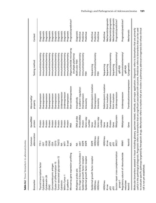
![132 Chapter 8
Table 8.3 Immunohistochemical markers useful for differential diagnosis.
CK7 CK20 TTF-1 CDX2 Calretinin
Bronchioloalveolar +++ − ++ − +
Acinar and mixed subtype +++ − ++ − +
Mucinous (“colloid,” etc.) +++ + +++ + +
Metastatic ++ ++ − +++ +
Mesothelioma ++ +++ − NA +++
Immunohistochemical biomarkers useful in the differential diagnosis of lung cancer. The reactivity scale refers to the
percentage of cases reported positive with for the marker as – (<1%), + (1–25%), ++ (25–70%), and + + + (>70%).
Results of these tests reflect a unique molecular approximately the same proportion of metastatic tu-
configuration in lung tumors that differs from that mors positive for these proteins [94–96]. An even
of histologically similar adenocarcinomas originat- smaller proportion of lung adenocarcinomas are
ing at other sites. The expected results of diagnostic positive so that expression of TTF-1 appears to be
immunohistochemical stains for BAC and invasive a better marker of lung origin than the surfactant
carcinoma are compared to expected results for tu- proteins.
mors of other types and origins in Table 8.3. Perhaps A frequent clinical problem is the distinction
the most useful immunohistochemical marker that between adenocarcinoma and mesothelioma. Sev-
has become available in recent years is thyroid tran- eral markers have been identified empirically that
scription factor 1 (TTF-1, Nkx2.1). TTF-1 is neces- address this problem. BAC and adenocarcinoma
sary for branching morphogenesis in the developing are positive for CEA, CD-15, and TAG72 but are
lung [10,92] and is first expressed early in embry- negative for calretinin and WT1 distinguishing
onic development and in the normal adult alveolar them from mesothelioma. This immunohistochem-
and thyroid cells. With the exception of the thyroid ical panel or modifications are now almost uni-
tumors and occasional nonpulmonary small carci- formly applied to address the differential diagnosis
nomas [93], its expression is confined to lung can- of mesothelioma versus adenocarcinoma.
cer. More than three quarters of pulmonary ade- Finally, several immunohistochemical biomark-
nocarcinomas express TTF-1 [94]. A high propor- ers are prognostically important. The most promi-
tion of pulmonary adenocarcinomas also express a nent of these is E-cadherin. E-cadherin forms a com-
specific pattern of cytokeratin isotypes and are posi- plex with β-catenin and other molecules at the cell
tive for CK7 but not CD20. The TTF-1 positive, CK7 surface that is critical for intercellular adhesion and
positive, and CK20 negative phenotype has become intracellular signaling (reviewed in [97]). Loss of E-
established as standard procedure in distinguish- cadherin expression may reduce intercellular adhe-
ing histologically similar pulmonary from nonpul- sion among tumor cells and render them more dis-
monary adenocarcinomas and can greatly assist in cohesive and prone to disperse into the circulation.
guiding diagnosis, staging, and treatment of adeno- In a tissue microarray study, E-cadherin has been
carcinoma. shown to be an independent prognostic indicator in
Less helpful have been immunohistochemical NSCLC including adenocarcinoma [98]. Also, there
stains for surfactant proteins. Although the expres- may be crosstalk between the E-cadherin pathway
sion of these proteins is regulated by TTF-1 [10], and EGFR in adenocarcinoma and other lung tu-
immunohistochemical stains for these proteins has mors so that intact E-cadherin signaling may be nec-
been relatively insensitive and unspecific with ap- essary for response to EGFR blockade [37]. Expres-
proximately half of all pulmonary carcinomas and sion of E-cadherin may ultimately prove useful as](https://image.slidesharecdn.com/lungcancer3rded-120126215311-phpapp01/85/Lung-cancer-3rd-ed-143-320.jpg)
![Pathology and Pathogenesis of Adenocarcinoma 133
a predictor of response to treatment, as discussed and amplifications [105,106]. However, because of
below. the high frequency of chromosome imbalance (ane-
uploidy), multicolor FISH probes have been devel-
High throughput gene expression profiling oped [107] with the aim of using aneuploidy as
Oligonucleotide microarrays have been used to a diagnostic tool. In equivocal cases, FISH can be
comprehensively assess gene expression in adeno- helpful to confirm diagnosis and may enhance the
carcinoma and to relate these patterns to outcome. sensitivity of cytological procedures [72,107–109].
Based on patterns of gene expression tumors were It may also be useful as a predictive tool in assessing
found to segregate into groups that are not pre- risk for future carcinoma in high-risk patients [110].
dictable by conventional morphological classifica- FISH may be employed more broadly in the future
tion schemes with some tumors expressing profiles as significance of aneuploidy becomes evident and
of neuroendocrine, pneumocyte, or squamoid dif- better probes are developed. Finally, FISH for spe-
ferentiation [8,99]. The prognoses of tumors that cific genetic imbalances has shown promise as a pre-
are defined by these classifier groups are statisti- dictor of response to EGFR blockade as described
cally different [8,100]. Although specific genes in below.
the classifiers may vary somewhat, raw data from
across institutions is similar and can be harmonized Predictors of response to treatment
mathematically to arrive at a common informative NSCLC has long been known to strongly express
gene list [101,102]. There is considerable overlap in EGFR (reviewed in [111]) but the molecular mech-
tumor-associated gene expression between human anism behind this overexpression has only recently
and mouse tumors [103], supporting the notion that been addressed. Nonmucinous and some mucinous
the animal tumors are driven by some of the same BAC are among the NSCLC that strongly express
molecular mechanisms as human tumors. EGFR protein [112], either with or without coex-
While these studies are of biological interest and pression of the ErbB partner, HER2/neu. In 2003,
illustrate the power of microarray analyses, the it was reported that 21% of NSCLC contain high
technology involved remains complicated and ex- copy number (>3) or amplification of chromoso-
pensive and to date most studies have been used pri- mal loci containing the EGFR1 gene [113]. Tumors
marily to supplement existing morphological lung with either true amplification consisting of clus-
cancer categorizations. Recently, an approach that tered signals or high polysomy behaved similarly
addresses changes in gene expression profiles en- in statistical analyses and tumors with either ab-
gendered by molecular pathology that is frequently normality were therefore considered together as
present in adenocarcinoma. This has been accom- high copy number tumors or “FISH positive.” FISH-
plished by examining changes in gene expression positive NSCLC with high-level protein expression
in cell lines into which genes regulating specific cell generally had a worse prognosis than FISH-negative
signaling pathways are introduced using recombi- NSCLC but adenocarcinomas were not considered
nant adenoviral constructs [104]. The resulting gene separately in this study and the overall numbers of
expression profiles correlate with clinical outcome tumors were small.
in NSCLC so that it is now possible to relate out- In 2004, point mutations in exon 18 and 21 and
come with underlying molecular pathology without deletions in exon 19, all in the ATP-binding region
reference to histology. The implications of this work of the EGFR tyrosine kinase domain were reported
for assessment of outcome and selection therapy are almost simultaneously in lung tumors by two sep-
discussed below. arate groups [114]. In one reported series [78], 16
mutations were found in 119 tumors and all but one
Fluorescence in situ hybridization of these tumors were adenocarcinomas. Slightly less
Structural chromosomal abnormalities abound in than half (49%) of the tumors were from Japanese
pulmonary adenocarcinomas and include whole or patients and all but one of the mutations was found
partial chromosomal losses, gains, translocations, in the Japanese cohort. Several of the patients tested](https://image.slidesharecdn.com/lungcancer3rded-120126215311-phpapp01/85/Lung-cancer-3rd-ed-144-320.jpg)
![134 Chapter 8
in this study had been treated by EGFR blockade have been generated in order to predict response to
with gefitinib. Four of five responders to this treat- therapy by two different approaches. One is to sim-
ment had mutated EGFR but none of the nonre- ply evaluate gene expression profiles of tumors and
sponding tumors were mutant. In the second study cell lines known to harbor an abnormality associ-
[115], eight of nine carcinomas including three BAC ated with a pathway altered in carcinoma such as
and five adenocarcinomas contained a mutation. EGFR [124]. A second approach (mentioned above)
These tumors were selected for testing because of is to create a specific lesion in order to observe the
their responsiveness to EGFR blockade from a co- effect on gene expression using high throughput
hort of 275 patients treated with gefitinib. All the oligonucleotide microarrays [104]. Both methods
responsive mutant tumors were from never or for- have generated gene lists of biological significance.
mer smokers. Since these original observations, mu- The predictive power of these lists is still under in-
tational data on a large series of tumors from Japan, vestigation. The former approach has identified E-
Taiwan, Australia, and the United States has been cadherin as a gene of interest and detailed studies
published confirming the mutations are more com- have revealed a reciprocal relationship between E-
mon in never smokers, adenocarcinoma, Asian eth- codherin expression and the status of the zinc fin-
nicity, and females [116]. The association with BAC ger transcriptional repressor, zeb1 [37]. Downregu-
was questioned [117], since, using the strict nonin- lation of E-cadherin through zeb1 reduces sensitiv-
vasive criteria for BAC, none of the BAC from the ity to EGFR, suggesting new targets for prediction of
United States that were available for review were response to treatment and for intervention in ade-
mutant [116]. It therefore appears that mutation nocarcinoma.
is associated with peripheral airway tumors, usu- Most BACs occur in smokers. Several stud-
ally of mixed subtype, but not necessarily with pure ies have indicated that Ki-ras mutations in ade-
BAC. nocarcinoma are highly associated with smoking
Following the discovery of EGFR mutations in [125,126]. There is little overlap between those tu-
adenocarcinoma, it was reported at two different mors with EGFR mutations and those with Ki-ras
institutions that, while mutation correlates with re- mutations [127,128]. Thus, mutations in separate
sponse to EGFR blockade in NSCLC including ade- EGFR pathway genes caused by different muta-
nocarcinoma, it does not predict survival [118–121]. gens all activate signaling and result in the sin-
Protein overexpression (Plate 8.9a) and high gene gle BAC/adenocarcinoma phenotype. Ki-ras muta-
copy number (Plate 8.9b) were reported to corre- tions are reported to be resistant to EGFR blockade
late survival in adenocarcinoma [120] as well as [118,122].
in NSCLC in general [119,121]. Moreover, virtu- Several other mutational abnormalities have
ally all available EGFR-mutated cell lines are ampli- been reported in adenocarcinoma that are thought
fied at the EGFR locus. These data suggest that the to be smoking induced. The most frequent of these
combination of high gene copy number and pro- is p53. P53 mutation is uncommon in pure BAC
tein expression may be the most potent predictor of but may be found in 11% of mixed adenocarci-
response to treatment and appears to identify a sub- noma/BAC and in 48% of tumors that are strictly in-
set of NSCLC, mostly adenocarcinomas, that are vasive adenocarcinomas [129]. This suggests a role
EGFR dependent for growth and malignant proper- for p53 in progression to aggressive forms of invasive
ties [122]. Whether single or multiple marker test- peripheral lung cancer. That p53 mutation may pre-
ing of the EGFR pathway can be used to select cede other mutations and chromosomal rearrange-
patients who would respond to EGFR blockade is ments illustrated by the Li-Fraumeni patient is dis-
currently under active investigation. cussed below.
Pathological alteration of specific signaling path- Finally, DNA repair systems may be of practical
ways generates specific gene expression profiles that importance in predicting response to DNA damag-
may correlate with response to treatment [123]. Sig- ing agents such as platinum-based drugs used to
natures of pathologically altered signaling pathways treat adenocarcinoma. Untreated early-stage patients](https://image.slidesharecdn.com/lungcancer3rded-120126215311-phpapp01/85/Lung-cancer-3rd-ed-145-320.jpg)
![Pathology and Pathogenesis of Adenocarcinoma 135
with high levels of the repair enzymes ERCC1 and these targets is EGFR. Despite the large amount of
RRM1 measured by a simple immunohistochemi- information regarding the EGFR pathway gene sta-
cal test [130], have a better prognosis than patients tus in fully developed carcinoma (discussed above),
with low levels. The opposite is true for treated pa- information regarding the status of this pathway in
tients; those with low levels of the same enzymes premalignancy is limited. Tang et al. have found the
survive significantly longer than patients with high same EGFR mutations in nonmalignant epithelium
levels [131,132]. The likely explanation for these adjacent tumor from 24% of patients with EGFR
seemingly contradictory findings is that untreated tu- mutant adenocarcinoma, suggesting that EGFR mu-
mors with low ability to repair DNA damage may tation may precede frank carcinoma [135]. Possible
be prone to rapid accumulation of the genetic alter- synergism between separate mutations was recently
ations and malignant progression while treated tu- reported in a patient with Li-Fraumeni syndrome.
mors with low DNA repair capacity are likely to be This patient had a germline mutation in p53 codon
damaged or destroyed by chemotherapy with re- 273 and developed a breast tumor at age 30 that
sulting improved patient survival. The gene encod- was amplified for HER2/neu. At age 34, the patient
ing the excision repair enzyme, ERCC1, contains a was diagnosed with a second carcinoma, this time
common polymorphism at codon 118 (AAC→AAT, an adenocarcinoma of lung with the characteris-
exon 4) [133], which is silent (both alleles encode tic deletion in EGFR exon 19 encompassing bases
arginine). However, the homozygous AAC codon 2413–2428 (codon 746–750) and high copy num-
is associated with lower expression levels of ERCC1 ber for both EGFR and HER2/neu. This unusual case
[134]. These data offer the prospect of predicting re- suggests that mutation in the p53 mitotic checkpoint
sponse to proven chemotherapeutic agents by sim- gene may induce a sufficient level of genetic and
ple testing of tumor tissue or blood, resulting better chromosomal instability over time to induce EGFR
response and survival rates and reduced morbidity pathway mutation and gene dosage imbalance. This
from futile treatment of this common lung tumor study also suggests that the sequence in which mu-
type. tation occurs may not be as important as the num-
ber of mutations that occur during the carcinogenic
Molecular pathogenesis of peripheral process.
adenocarcinoma A considerably simple somatic genetic alteration
The molecular and biological mechanisms involved leading to adenocarcinoma in a 34-year-old female
in adenocarcinogenesis are not as well understood nonsmoker has been described. In this case, a sin-
as those involved in central airway carcinogen- gle translocation [t(15;19) (q11;p13)], was detected
esis due to the inaccessibility of the peripheral in tumor cells mapped to a region 50 kb upstream of
lung for sequential analysis. Recently, however, im- the notch3 gene on chromosome 19p and resulted
proved understanding of genetic and gene expres- in an increase in notch3 expression. Examination of
sion changes in adenocarcinoma have provided in- several cell lines indicated that notch3 was involved
sight into the molecular processes that lead to lung in several different translocations involving many
cancer. These insights have come from evaluation different reciprocal chromosome partners. Notch3
of large cohorts of subjects as well as from careful is involved in differentiation, apoptosis and regula-
study of individual cases. The conclusions of these tion of the cell cycle and is therefore a candidate
studies can be divided into the two broad categories oncogene. This translocation and the Li-Fraumeni
of structural genetic and chromosomal changes that case described above indicate that a wide diversity
are thought to represent stages in multistep carcino- of molecular mechanisms may be at play in adeno-
genesis in the peripheral lung. carcinogenesis and that targeting a single gene or
The status of genes involved in several signaling pathway may not be successful as a means of early
pathways has been intensively evaluated because detection or treatment.
of the availability of targeted agents that can inter- In addition to analysis of existing malignancy, sev-
fere with or block those pathways. Most notable of eral studies have evaluated the molecular changes](https://image.slidesharecdn.com/lungcancer3rded-120126215311-phpapp01/85/Lung-cancer-3rd-ed-146-320.jpg)
![136 Chapter 8
in AAH as a window on peripheral airway carcino- not completely resolved but it appears at the mo-
genesis. The most striking molecular change is the ment that models more analogous to the clini-
high frequency of Ki-ras mutation with one study coepidemiological setting in the human might be
showing 39% of AAH with mutations [86]. Ki-ras insightful.
mutation thus is likely to be a major event in early One animal model has become predominant in
lung carcinogenesis but appears to require a sec- recent years and that is the mouse model of pul-
ond event to progress to invasive carcinoma. That monary adenocarcinoma. Mice have several obvi-
silencing of other tumor suppressor genes may be ous advantages as experimental models including
present in AAH is suggested by the finding of loss of the small size of the host animal, affordability of
heterozygosity at chromosomal sites of putative tu- housing, and short life span. In addition, the abil-
mor suppressor genes including 3p and 9p [87,88], ity to genetically engineer this animal has provided
9q [89,90], 16p [89], and 17q [90]. These molec- recently improved understanding of lung carcino-
ular changes are also frequent in BAC, suggesting genesis in the mouse as well as in man.
that these lesions are related and perhaps the result While wild mice are relatively resistant to the
of action of similar carcinogens. development of lung tumors, certain inbred stains
Finally, AAH is frequently aneuploid [82–85] have a high frequency of lung tumors and can be
indicating significant chromosomal instability and induced to develop tumors by exposure to tobacco
missegregation. Chromosomal imbalance has been smoke itself [144] and to the potent carcinogens that
demonstrated by comparative genomic hybridiza- are present in tobacco smoke including urethane,
tion suggesting that AAH may represent a morpho- nitrosamines, and polyaromatic hydrocarbons
logical change that is late in the multistep sequence (reviewed in [145]).
of molecular events that ends in carcinoma. Detailed review of mouse lung carcinogenesis is
beyond the scope of this review. However, several
new insights into the cellular origin and compo-
Experimental models sition of adenocarcinoma have been provided by
recent mouse experiments that are important to
Animal models for human lung adenocarcinoma the current understanding of human lung tumor
have been available for many decades and have pathology. In these experiments putative stem cells
recently provided increasingly comprehensive un- for peripheral lung and for adenocarcinoma have
derstanding of pulmonary adenocarcinogenesis. In been identified. Stem cells have the ability to re-
the early twentieth century, an endemic infectious generate themselves, to form multiple cell lineages
disease (jagziekte) was described in South African and to proliferate [146]. They are important since
sheep [136] that produced multicentric alveolar ep- genetic lesions in stem cells may be replicated and
ithelial proliferation similar to BAC in man [137]. passed on to large populations of cells that may in-
This disease, now referred to as ovine pulmonary crease the probability of tumor development in pre-
adenocarcinoma (OPA) has more recently been malignant cells or resistance to treatment in fully
shown to be the result of infection with a retro- developed tumors.
virus, jaagsiekte sheep retrovirus (JSRV) [138–141]. Until recently, models testing for the presence of
This retrovirus is unique in its tropism for differ- stem cells have focused on separate local cellular
entiated pulmonary epithelial cells and to date is compartments of the lung including tracheal mu-
the only known naturally occurring viral cause of cociliary epithelium, bronchial glands, Clara cells,
pulmonary adenocarcinoma [142]. However, the and alveolar cells. These experiments have iden-
virus does appear to be confined to sheep and tified progenitor cells that can repopulate local
has not been demonstrated in human pulmonary compartments after injury but not resident multi-
tumors [143]. Whether this reflects an inability potential stem cells that can repopulate any lung
of present methods to detect the virus or that epithelium [147]. Recently, cells capable of self-
the virus is simply absent from human tumor is renewal, able to regenerate both Clara cells and](https://image.slidesharecdn.com/lungcancer3rded-120126215311-phpapp01/85/Lung-cancer-3rd-ed-147-320.jpg)
![Pathology and Pathogenesis of Adenocarcinoma 137
alveolar pneumocytes in injured lungs have been 2 Flieder DB, Vazquez M, Carter D et al. Pathologic find-
enriched by flow cytometry from whole mouse ings of lung tumors diagnosed on baseline CT screen-
lung preparations and thus have properties of stem ing. Am J Surg Pathol 2006; 30:606–13.
cells [148]. These cells reside at the bronchoalveo- 3 Carter D, Vazquez M, Flieder DB et al. Comparison
lar junction and have consistently expressed both of pathologic findings of baseline and annual repeat
cancers diagnosed on CT screening. Lung Cancer 2007;
surfactant protein C and CC10. In a genetically en-
56:193–9.
gineered mouse adenocarcinoma model in which
4 Kuhn C, Wright JL. The normal lung. In: Churg AM,
cells are transformed by conditional expression of Myers JL, Tazelaar HD, Wright JL (eds). Thurlbeck’s
mutant Ki-ras [149], cells with stem cell properties Pathology of the Lung, 3rd edn. New York: Thieme Med-
can be found early in neoplastic transformation and ical Publications, 2005:1–37.
are proposed as the cell of origin of adenocarcinoma. 5 Singh G, Katyal SL. Clara cell proteins. Ann N Y Acad
This hypothesis has yet to be confirmed in human Sci 2000; 923:43–58.
adenocarcinoma but provides a model that in many 6 Linnoila RI, Szabo E, DeMayo F, Witschi H, Sabourin
respects is analogous if not to invasive lung carci- C, Malkinson A. The role of CC10 in pulmonary
noma, then to AAH and BAC, well-differentiated carcinogenesis: from a marker to tumor suppression.
tumors with frequent Ki-ras mutation (discussed Ann N Y Acad Sci 2000; 923:249–67.
7 Stripp BR, Reynolds SD, Plopper CG, Boe IM, Lund J.
above).
Pulmonary phenotype of CCSP/UG deficient mice: a
consequence of CCSP deficiency or altered Clara cell
function? Ann N Y Acad Sci 2000; 923:202–9.
Conclusions and predictions 8 Bhattacharjee A, Richards WG, Staunton J et al. Clas-
sification of human lung carcinomas by mRNA ex-
Advances in technology and the availability of a pression profiling reveals distinct adenocarcinoma
large armamentarium of targeted agents pose new subclasses. Proc Natl Acad Sci U S A 2001; 98:
challenges for pathological classifications of lung 13790–5.
cancer and premalignancy. It is unlikely that lung 9 Whitsett JA, Wert SE, Trapnell BC. Genetic disor-
ders influencing lung formation and function at birth.
cancer will respond uniformly to the new agents.
Hum Mol Genet 2004; 13(2):R207–15.
The heterogeneity of lung cancer and the diver-
10 Warburton D, Zhao J, Berberich MA, Bernfield M.
sity of its morphological appearances and molecular Molecular embryology of the lung: then, now, and in
properties evident from the data presented above the future. Am J Physiol 1999; 276:L697–704.
imply that molecular targets will be applicable to 11 Mendelson CR. Role of transcription factors in fetal
an increasingly narrow range of lung cancers in the lung development and surfactant protein gene ex-
future and that treatments will have to be tailored pression. Annu Rev Physiol 2000; 62:875–915.
to the molecular as well as to the cellular charac- 12 Bonner AE, Lemon WJ, You M. Gene expression sig-
teristics of individual tumors. Pathologists will con- natures identify novel regulatory pathways during
tinue to play a large and increasing role in tis- murine lung development: implications for lung tu-
sue management and molecular testing for which morigenesis. J Med Genet 2003; 40:408–17.
13 Bonner AE, Lemon WJ, Devereux TR, Lubet RA, You
strong grounding in both the detailed anatomical
M. Molecular profiling of mouse lung tumors: asso-
analysis and molecular testing will continue to be
ciation with tumor progression, lung development,
essential. and human lung adenocarcinomas. Oncogene 2004;
23:1166–76.
14 Borczuk AC, Gorenstein L, Walter KL, Assaad AA,
References Wang L, Powell CA. Non-small-cell lung cancer
molecular signatures recapitulate lung developmen-
1 Henschke CI, Yankelevitz DF, Libby DM, Pasmantier tal pathways. Am J Pathol 2003; 163:1949–60.
MW, Smith JP, Miettinen OS. Survival of patients 15 Colby TV, Noguchi M, Henschke C et al. Adenocarci-
with stage I lung cancer detected on CT screening. noma. In: Travis WD, Brambilla E, Muller-Hermelink
N Engl J Med 2006; 355:1763–71. HK, Harris CC (eds). Pathology and Genetics of Tumours](https://image.slidesharecdn.com/lungcancer3rded-120126215311-phpapp01/85/Lung-cancer-3rd-ed-148-320.jpg)
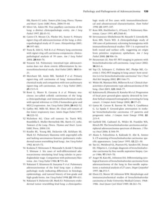
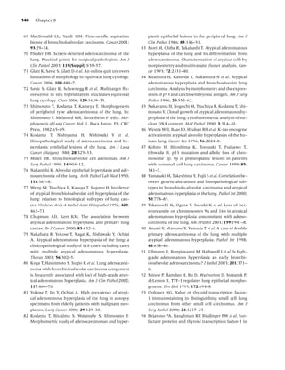
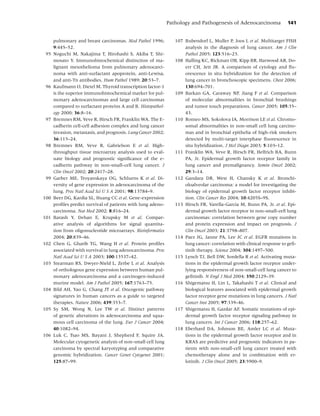
![CHAPTER 9
Treatment of Bronchioloalveolar
Carcinoma
Ji-Youn Han, Dae Ho Lee, and Jin Soo Lee
Introduction scar of previous inflammation, such as tuberculosis,
could be the bed of BAC [7].
The latest World Health Organization (WHO) clas- It is generally observed that the prognosis for BAC
sification divides lung adenocarcinoma mainly into is better than those for other types of lung cancer
adenocarcinoma mixed subtype, acinar adenocarci- [6,8–12]. Previously, Noguchi et al. pointed out that
noma, papillary adenocarcinoma, bronchioloalve- small (3 cm or less) peripheral lung adenocarcinoma
olar carcinoma (BAC), and solid adenocarcinoma with a pure BAC pattern and no invasion had 5-year
with mucin production [1]. BAC is characterized by survival rate of 100%, and patients with mixed BAC
distinct clinical presentation, radiographic appear- and invasive components had a survival of 75% in
ance, tumor biology, response to therapy, and prog- contrast to those with a purely invasive growth pat-
nosis. It disproportionately affects women, never tern who had a survival of 52% [13]. These findings
smokers, and Asians and tends to be younger at greatly influenced the 1999 WHO/the International
diagnosis. These differences raise the question of Association for the Study of Lung Cancer (IASLC)
whether BAC represents a separate biologic entity panel, which strictly redefined BAC as a subtype
[2,3]. Although pure BAC accounts for approxi- of pulmonary adenocarcinoma with growth of neo-
mately 4% of lung cancers, tumors with histolog- plastic cells along pre-existing alveolar structures
ically mixed BAC and adenocarcinoma account for (lepidic growth) without evidence of stromal, vas-
>20% of all lung cancers, and the incidence of BAC cular, or pleural invasion [14]. BACs are formally
might be increasing [3–5]. In a recent report of 278 classified into three subtypes, nonmucinous, muci-
lung resections for adenocarcinoma between 1992 nous, and mixed mucinous and nonmucinous type
and 2001, the proportion of BAC rose from 6.9% of [1,14]. They have been recognized as a solitary pe-
adenocarcinoma in 1992 to 46.9% in 2001 [6]. The ripheral nodule, multiple nodules, and lobar consol-
increasing numbers of BAC cases suggest that there idation and characterized by a higher incidence of
might be a change in the etiologic factors of lung intrathoracic recurrence and second primary lung
cancer. Although smoking remains a very important cancers, and less frequent distant metastasis com-
factor of lung cancer, the higher proportion of BAC pared to other type of nonsmall cell lung cancer
in never smoker postulates that BAC can develop in (NSCLC) [9,11,14]. The mucinous BAC, which ac-
a previously scarred area of the lung parenchyma. A counts for 20% of BACs, has worse outcome than
the nonmucinous BACs. While nonmucinous BACs
most commonly present as small peripheral nod-
Lung Cancer, 3rd edition. Edited by Jack A. Roth, James D. Cox,
ules, mucinous BACs tend to spread aerogenously
and Waun Ki Hong. c 2008 Blackwell Publishing, and develop multifocal lesions and frequently mas-
ISBN: 978-1-4051-5112-2. querade as pneumonia often resulting in a delay
144](https://image.slidesharecdn.com/lungcancer3rded-120126215311-phpapp01/85/Lung-cancer-3rd-ed-155-320.jpg)
![Treatment of Bronchioloalveolar Carcinoma 145
in diagnosis [15]. As with other subsets of NSCLC, than those of other types of lung cancer [11–13].
surgical resection is the only potentially curative In stage I disease, both 5-year DFS and OS were
treatment. Patients with unresectable BAC are more significantly higher in patients with pure BAC than
likely to respond to the epidermal growth factor re- adenocarcinoma (DFS, 81% versus 51%; OS, 86%
ceptor (EGFR) tyrosine kinase inhibitors (TKIs) gefi- versus 71%; p = 0.005) [11]. In addition, 76–95%
tinib and erlotinib than patients with other subtypes of patients with recurrence initially recur locally,
of NSCLC [16,17]. which is higher than other types of lung cancer
[11–13]. Early stage, nonmucinous type, and the
absence of vascular or lymphatic invasion are as-
Surgery sociated with better survival after surgical resection
[6,11,20,22,26]. Apart from the fact that advanced
Surgery in a curative intent stage is generally associated with a worse progno-
BAC is most commonly found in small peripheral le- sis, the management of patients with multiple nod-
sions, and it is subjected to surgery in an early stage ules is controversial and the presence of multiple
[18]. While only 15–25% of all lung cancers are in or satellite nodule is not an adverse prognostic fac-
stage I, the proportion of stage I in BAC is 68%, sug- tor in BAC [20,27]. Thus, surgery should not be
gesting the growth of BAC is slower than other types denied for patients with multiple nodules who are
of NSCLC [6,12,18–21]. BAC usually spreads by the younger than 60 years of age and without lymph
aerogenous route, and lymph node metastasis is node involvement. In the absence of mediastinal in-
rare, reported in less than 10% [18,19]. After cura- volvement or distant metastasis, possible multifocal
tive resection, 92% of patients display intrathoracic BACs should be treated as separate primary tumors
recurrence, while 29% of resected cases display ex- [17,27].
trathoracic recurrence. For example, brain metasta-
sis is frequently occurred in non-BAC lung cancer, Palliative surgery for BAC
for about 20%, it is rarely seen in BAC, for only In a highly selected subgroup of patients who
about 8% [9,11]. Thus, surgical resection appears present with bilateral BAC, palliative pneumonec-
to have a pivotal role in the treatment of BAC [9]. tomy or lobectomy could be considered because of
Localized BAC is treated like other NSCLCs with the unequal involvement of the lungs and major
lobar lung resection and ipsilateral mediastinal lym- hypoxemia in relation to a severe intrapulmonary
phadenectomy [20,22]. Although lobectomy is the shunting. Both symptoms and respiratory function
most commonly performed surgical procedure, the could be quickly improved postoperatively with tol-
extent of resection has been somewhat controver- erable morbidity [28]. However, palliative surgery
sial. Some investigators have suggested that patients should be considered exclusively for very highly se-
treated with less than a lobectomy have higher re- lected patients and further discussion in a multidis-
currence rates and a worse prognosis [18,23]. Given ciplinary manner would be needed [22,29].
the propensity of the disease to occur in a multi-
focal fashion, others have advocated lung-sparing Transplantation
procedures (wedge or segmentectomy) [9,24,25]. Because of the theoretical risk of rapid cancer dis-
Sometimes bilobectomy and pneumonectomy are semination with posttransplant immunosuppres-
required for complete resection of the diffuse or sion, bronchogenic carcinoma has been consid-
multifocal BAC. However, the extent of resection ered a strict contraindication to lung transplantation
never remained significant in multivariate analy- [30]. BAC can present as a localized discrete lesion
sis maybe because the surgical procedure is closely or with a diffuse multifocal pattern involving one
linked to the tumor node metastasis (TNM) stage or both lungs [14]. Pulmonary recurrence without
[22]. systemic dissemination is often observed after lung
Completely resected BAC is associated with bet- resection for multifocal BAC, and survival beyond 2
ter disease-free (DFS) and overall survival (OS) rate years is uncommon [9,11]. Death usually occurs as](https://image.slidesharecdn.com/lungcancer3rded-120126215311-phpapp01/85/Lung-cancer-3rd-ed-156-320.jpg)
![146 Chapter 9
Table 9.1 Paclitaxel chemotherapy trials in advanced BAC or NSCLC.
Patients Response Median
Study Histology Phase no. Schedule rate survival
SWOG 9714 [35] BAC II 58 35 mg/m2 /24 h CI over 96 h 8/58 (14%) 12 M
2
EORTC 08956 [36] BAC II 19 200 mg/m IV for 3 h 2/18 (11%) 8.6 M
2
Ranson et al. [37] NSCLC III 79 200 mg/m IV for 3 h 12/76 (16%) 6.8 M
BAC, bronchioloalveolar carcinoma; NSCLC, nonsmall cell lung cancer; CI, continuous infusion; IV, intravenously; M, month;
BST, best supportive care.
a result of pulmonary failure secondary to tumor re- may be indolent enough to follow asymptomatic pa-
placement of the functioning lung [19]. Despite case tients without any systemic therapy if patients are
reports and small case series on lung transplantation comfortable with this approach. It is because the
for multifocal BAC, the role of lung transplantation rate of disease progression may be slow enough to
in the treatment of patients with bronchogenic car- warrant no therapy for many months or even years.
cinoma and end-stage lung disease remains unre- For patients with symptoms and/or clear evidence
solved [31,32]. of disease progression over a short interval, standard
A recent international survey determined the chemotherapy is appropriate [17].
outcome of patients who presented with bron- Advanced multifocal BAC has been considered
chogenic carcinoma in the explanted lung at the to be relatively resistant to chemotherapy based on
time of transplantation [33]. This survey demon- limited retrospective data [27]. However, all BAC
strates that the 5-year actuarial survival rate was chemotherapy studies are limited by their sample
51% in patients with stage I bronchogenic carci- size and the use of old regimens. In addition, analy-
nomas, which was significantly better than for pa- sis of the response of BAC to chemotherapy is com-
tients with stage II and III bronchogenic carcinomas plicated by the classification of most patients with
(survival of 14%) or with incidental multifocal BAC mixed BAC as adenocarcinoma, and also by mis-
(survival of 23%). Time from transplantation to re- classification of histologic type when the diagnosis
currence and from recurrence to death was signifi- is made by cytology and by the absence of indepen-
cantly longer in patients with multifocal BAC than dent pathologic review in most chemotherapy trials
in patients with other types of bronchogenic carci- [2]. Moreover, retrospective reviews have suggested
noma. In addition, the site of recurrence was limited that patients with BAC survive longer than those
to the transplanted lung in 88% of the patients with with other types of NSCLC after chemotherapy
multifocal BAC, whereas it was always widespread [8,34]. Thus, little can be said about the chemosen-
in patients with other types of bronchogenic car- sitivity of these tumors based on the limited retro-
cinoma. Thus, although rarely curative, lung trans- spective data.
plantation remains a valuable procedure for patients Recently, two clinical trials of chemotherapy in
with impending respiratory failure secondary to ad- advanced BAC have been reported (Table 9.1). In
vanced multifocal BAC [33]. the Southwest Oncology Group (SWOG) trial 9714
[35], 58 chemona¨ve patients with advanced BAC
ı
Chemotherapy received a 96-hour infusion of paclitaxel (35 mg/m2
Despite the slow growth kinetics and prolonged sur- for 24 h). The overall response rate was 14% with
vival after repeated surgical resection of multifocal the time to progression of 5 months, median sur-
lesions, advanced bilateral multifocal BAC remains vival of 12 months, and 3-year overall survival rate
incurable. There is no optimal established therapy of 10%. However, the toxicity of the infusional pa-
for unresectable multifocal BAC. Multifocal BAC clitaxel dissuaded the general use of this regimen.](https://image.slidesharecdn.com/lungcancer3rded-120126215311-phpapp01/85/Lung-cancer-3rd-ed-157-320.jpg)
![Treatment of Bronchioloalveolar Carcinoma 147
Table 9.2 Phase II trials of EGFR-TKIs in patients with previously treated NSCLC.
Patients Median 1-year
Study Agent Dose no.∗ Response rate survival survival
IDEAL 1 [39] Gefitinib 250 vs. 500 mg/day 216 12/102 (12%) vs. 7 M vs. 6 M 27% vs. 24%
10/114 (9%)
IDEAL 2 [40]† Gefitinib 250 vs. 500 mg/day 208 18/103 (17.5%) vs. 7.6 M vs. 8 M 35% vs. 29%
19/105 (18.1%)
Perez-Soler et al. [41]‡ Erlotinib 150 mg/day 55 7/55 (12.7%, 2CR, 5PR) 8.4 M 40%
∗
Evaluable patients.
†
The response rate was higher for Japanese patients than non-Japanese patients (27.5% vs. 10.4%; odds ratio = 3.27;
p = 0.0023).
‡
NSCLC with EGFR expression.
A similar study was conducted by the European Or- after both platinum- and docetaxel-containing reg-
ganisation for Research and Treatment of Cancer; imens [38]. Belonged to this class of agents are
EORTC trial 08956 [36]. In this study, a 3-hour gefitinib (Iressa®, AstraZeneca, Wilmington, DE)
infusion of paclitaxel (200 mg/m2 on day 1 every and erlotinib (Tarceva®, OSI Pharmaceuticals Inc,
3 weeks) was used to treat chemona¨ve patients
ı Melville, NY). Both agents are small molecules that
with advanced BAC. The overall response rate was belong to the quinazolinamine class and inhibit the
11%, time to progression was 2.2 months, median tyrosine kinase (TK) activity of the EGFR by com-
survival was 8.6 months, and 1-year overall sur- peting ATP for the ATP-binding site. Single agent
vival was 35%. These results are comparable to the trials of gefitinib or erlotinib in patients with previ-
single-agent paclitaxel activity in advanced NSCLC ously treated advanced NSCLC have reported re-
[37]. It is generally accepted that patients with BAC sponse rates in approximately 10% of Caucasian
should not be excluded from the clinical trials eval- patients and 28% of Japanese patients (Table 9.2)
uating efficacy of chemotherapy and new targeted [39–41].
agents. Particularly in the absence of an optimal Accumulating evidence from these trials of gefi-
standard therapy, clinical trials should remain as a tinib or erlotinib suggest that such patient char-
viable treatment option for these patients. acteristics as never smoker, female gender, East
Asian origin, adenocarcinoma histology, and bron-
chioloalveolar carcinoma subtype, are associated
Epidermal growth factor with a grater benefit from treatment with these TKIs
receptor-tyrosine kinase inhibitors [16,34,42,43]. In light of these observations, two
prospective phase II trials were undertaken in pa-
Although patients with advanced NSCLC are com- tients with advanced BAC or adenocarcinoma with
monly treated with chemotherapy, there is a gen- BAC features (Table 9.3). In SWOG S0126 trial [44],
eral consensus in that the benefits of traditional 136 (101 chemona¨ve, 35 previously treated) pa-
ı
chemotherapy have reached a plateau. In the tients with advanced BAC received gefitinib sin-
past few years, several agents that are more spe- gle treatment. The response rates were 17 and 9%
cific for cancer cell targets have shown signifi- for chemona¨ve and previously treated patients, re-
ı
cant antitumor activity in NSCLC. The EGFR-TKI spectively. Median survival was 13 months for both
is the first class of molecular-targeted agent ap- chemona¨ve and previously treated patients. These
ı
proved in the United States and other countries for results are similar to that achieved in SWOG S9714
the treatment of advanced NSCLC that progressed trial with 96-hour paclitaxel infusion. However,](https://image.slidesharecdn.com/lungcancer3rded-120126215311-phpapp01/85/Lung-cancer-3rd-ed-158-320.jpg)
![148 Chapter 9
Table 9.3 Clinical trials of EGFR-TKIs in BAC.
Median 1-year
Study Agent Dose Patients no.∗ Response rate survival survival
SWOG S0126 [44] Gefitinib 500 mg/day Previously untreated-69 17% (12/69, 4CR, 8PR) 13 M 51%
Previously treated-22 9% (2/22, 2PR) 12 M 51%
Kris et al. [45] Erlotinib 150 mg/day 59 25% (15/59) NR 58%
∗
Evaluable patients.
CR, completer response; PR, partial response.
survival at 2 and 3 years in S0126 (chemona¨ve ı nomas investigated. A total of 39 mutations were
patents) is 39% and 23% versus 29% and 13% found in a series of 375 adenocarcinomas. Among
in S9714, respectively. Kris et al. [45] also con- them, 22 (26%) were found in 86 BACs, which is
ducted a similar study of erlotinib in 69 patients very similar to the response rates of BAC to EGFR-
with adenocarcinoma with any BAC features. The TKIs (Table 9.3). These findings suggest that about
preliminary data also showed encouraging results one fourth of BACs may respond to EGFR-TKIs be-
of erlotinib single treatment in these patients. These cause mutations affecting the gene.
long-term survival results observed in EGFR-TKI tri- Nevertheless, it is unclear whether all BACs re-
als in advanced BAC support the preferred treat- spond better than conventional adenocarcinoma to
ment of these agents as the first-line or second-line EGFR-TKIs. Although it is preliminary, Kris et al.
for patients with BAC feature. However, whether [45] reported that patients with adenocarcinoma
the results of EGFR-TKIs in BAC are superior to with BAC features showed higher response to er-
those achieved with platinum-based chemotherapy lotinib compared to pure BAC (30% versus 7%,
remains unclear. respectively). Marchetti et al. [47] reported that
The discovery of EGFR gene mutations in the re- all the EGFR mutations found in BAC were seen
ceptor TK domain and their association with high in the nonmucinous type. Eighteen (35%) of 52
response rate to EGFR-TKIs has had a profound im- nonmucinous BACs had EGFR mutations. Con-
pact on understanding the role of these agents in versely, mucinous BACs were always negative for
NSCLC. A recent comprehensive review on EGFR EGFR mutations. They also investigated for K-ras
somatic mutations provided detailed information on mutations at codon 12 and all of the tumors af-
clinicopathologic features in lung cancer that are fected by EGFR were found to be negative for K-ras
associated with these mutations. A total of over mutations, whereas, tumors negative for EGFR mu-
2000 NSCLC samples have been analyzed, and a tations showed a K-ras mutation in 32% of cases
total of 477 mutations were detected. A signifi- ( p = 0.000001). The prevalence of K-ras mutations
cantly higher frequency of EGFR kinase domain was 29% in conventional lung adenocarcinoma and
mutations found in patients with adenocarcinoma 27% in BAC. Fourteen percent of nonmucinous
(30% versus 2%; p < 0.001), never smoker (45% BACs carried K-ras mutations, while 76% of muci-
versus 7%; p < 0.001), female (38% versus 10%; nous BACs showed K-ras mutations ( p = 0.00002)
p < 0.001), and East Asian individuals (33% ver- [47]. K-ras mutations are strongly associated with
sus 6%; p < 0.001), which matches the profile of smoking status and more common in nonrespon-
NSCLC patients who likely to respond to EGFR-TKIs ders to EGFR-TKIs [48]. These findings suggest that
[46]. In a study of EGFR tyrosine kinase domain EGFR and K-ras genes seem to be related to the
mutations in 860 lung cancers, Marchetti et al. [47] development of different BAC types and result-
found that there were no EGFR mutations in 454 ing different effects on responding to EGFR-TKIs.
squamous cell carcinomas and 31 large cell carci- However, due to the limited number of BAC cases,](https://image.slidesharecdn.com/lungcancer3rded-120126215311-phpapp01/85/Lung-cancer-3rd-ed-159-320.jpg)
![Treatment of Bronchioloalveolar Carcinoma 149
additional studies are required to confirm these of 17.8 months. Notably, two of these responders
data. Prospective trials evaluating response to had BAC. Responses in the two BAC patients were
EGFR-TKIs including EGFR and K-ras sequencing durable and complete and the response duration
are under way to help shed more light on this in- measured 18 months in one patient and exceeded
triguing finding. 22 months in the other patient. The median survival
of all 33 patients was 11.6 months, which com-
pares favorably to the approved second-line doc-
Other molecular target agents etaxel chemotherapy for such patients [54]. An on-
in BAC going phase II CG8123 (GVAX) trial (SWOG 0310)
will treat nearly 100 patients with advanced BAC
Eastern Cooperative Oncology Group (ECOG) has both untreated and previously treated [34].
conducted a study evaluating the role of adenovirus Another agent of interest is a proteasome in-
p53 administered by bronchoalveolar lavage to pa- hibitor. The proteasome plays a critical role in
tients with BAC (ECOG 6597). The rationale for this the degradation of proteins involved in the reg-
study included the fact that NSCLCs have a high ulation of cell cycle, apoptosis, and angiogenesis
frequency of p53 mutations and that BAC tends to [55]. Bortezomib (VELCADE, Vc, Millennium Phar-
spread as a thin layer of cells along the airways maceuticals) is a reversible novel proteasome in-
rather than developing into a solid tumor mass. hibitor approved for the treatment of relapsed mul-
Some previous gene therapy studies have used in- tiple myeloma [56]. In the initial phase I study of
jections into the tumor mass, but success is often bortezomib in advanced solid tumors, one of 43
limited because the gene-carrying viruses cannot patients, a patient with heavily pretreated BAC,
reach most of the cancer cells in the solid tumor. In achieved a PR [57]. Recent studies in patients with
BAC, on the other hand, it may be possible to de- advanced NSCLC as a single agent or in combina-
liver gene therapy directly through the air passages. tion with other chemotherapy have demonstrated
Thus, adequate dissemination of adenovirus p53 encouraging results (Table 9.4) [58–60]. In addition,
might be obtained in this locoregional setting. Of there are anecdotal reports of objective responses of
27 patients enrolled, 25 were treated and one par- bortezomib in BAC [61]. Currently, the California–
tial response and 17 stable diseases were observed of Pittsburgh Cancer Consortium is conducting a phase
24 evaluable patients. Three patients had more than II study to evaluate the activity and safety of borte-
20% improvement in the diffusion capacity of car- zomib in patients with advanced BAC or adenocar-
bon monoxide, and subjectively improved breath- cinoma with BAC features.
ing was noted in many patients [49].
Therapeutic cancer vaccines derived from whole
tumor cells have also been tested in patients with re- Summary
sected early-stage NSCLC, which demonstrated im-
munologic activity and the suggestion of survival BAC is previously considered an uncommon sub-
advantage [50,51]. G8123 (GVAX) is a genetically set of NSCLC with unique epidemiology, pathol-
engineered autologous anticancer vaccine to secrete ogy, clinical features, radiographic presentation, and
granulocyte-macrophage colony-stimulating factor natural history compared with other subtypes of
(GM-CSF). In head-to-head comparison, geneti- NSCLC. However, recent data suggest that the in-
cally engineered anticancer vaccines secreting GM- cidence of BAC is increasing. Despite reports of
CSF have shown greater activity than vaccines using prolonged survival after repeated surgical resection
other cytokines in murine tumor models including of multifocal lesions and slow growth kinetics, ad-
the Lewis lung carcinoma [52,53]. In a phase I/II vanced bilateral or recurrent diffuse BAC remains
trial of this vaccine, complete responses were seen incurable, with the vast majority of patients dy-
in three of 33 previously treated patients with ad- ing of respiratory failure or intercurrent pneumo-
vanced NSCLC with a median duration of response nia within 5 years. Most of the clinical information](https://image.slidesharecdn.com/lungcancer3rded-120126215311-phpapp01/85/Lung-cancer-3rd-ed-160-320.jpg)
![150 Chapter 9
Table 9.4 Clinical trials of Bortezomib in NSCLC.
Patients Prior Median
Author Phase no.∗ chemotherapy Regimen Efficacy TTP
Davies et al. [58] I 10 0 Bortezomib + gemcitabine + PR 40% (4/10) NR
carboplatin SD 50% (5/10)
Stevenson et al. [59] II 22 ≤1 Bortezomib PR 5% (1/22) NR
SD 41% (9/22)
Fanucchi et al. [60] II 155 1 Bortezomib vs. PR 8% (6/75) vs. 1.5 mo vs.
Bortezomib + docetaxel PR 9% (7/80) 4 mo
∗
Evaluable patients.
PR, partial response; SD, stable disease; TTP, time to progression; NR, not reported.
on BAC comes from retrospective institutional re- 5 Auerbach O, Garfinkel L. The changing pattern of
views. Recent studies of molecular-targeted ther- lung carcinoma. Cancer 1991; 68:1973–7.
apies, however, have focused more specifically on 6 Furak J, Trojan I, Szoke T et al. Bronchioloalveolar
BAC. In particular, clinical trials of EGFR-TKIs have lung cancer: occurrence, surgical treatment and sur-
led to a deeper understanding of the distinct features vival. Eur J Cardiothorac Surg 2003; 23:818–23.
7 Barkley JE, Green MR. Bronchioloalveolar carci-
of this cancer and suggest that BAC may require
noma. J Clin Oncol 1996; 14:2377–86.
a new therapeutic paradigm different from that of
8 Breathnach OS, Ishibe N, Williams J, Linnoila RI, Ca-
other NSCLCs. The limited utility of chemotherapy poraso N, Johnson BE. Clinical features of patients
necessitates the investigation of novel agents and with stage IIIB and IV bronchioloalveolar carcinoma
combinations for the treatment of BAC. Identifica- of the lung. Cancer 1999; 86:1165–73.
tion of molecular pathways unique to BAC would 9 Breathnach OS, Kwiatkowski DJ, Finkelstein DM
enable faster the development of more efficacious et al. Bronchioloalveolar carcinoma of the lung: recur-
therapy for this unique subset of NSCLC. rences and survival in patients with stage I disease. J
Thorac Cardiovasc Surg 2001; 121:42–7.
10 Grover FL, Piantadosi S. Recurrence and survival fol-
lowing resection of bronchioloalveolar carcinoma of
References the lung—The Lung Cancer Study Group experience.
Ann Surg 1989; 209:779–90.
1 Travis WD, Brambilla E, Muller-Hermelink HK, Har- 11 Rena O, Papalia E, Ruffini E et al. Stage I pure bron-
ris CC. World Health Organization classification. In: chioloalveolar carcinoma: recurrences, survival and
Tumors of the Lung, Pleura, Thymus, and Heart. Lyon, comparison with adenocarcinoma of the lung. Eur J
France: IARC Press, 2004. Cardiothorac Surg 2003; 23:409–14.
2 Laskin JJ, Sandler AB, Johnson DH. Redefining bron- 12 Sakurai H, Dobashi Y, Mizutani E et al. Bronchi-
chioloalveolar carcinoma. Semin Oncol 2005; 32:329– oloalveolar carcinoma of the lung 3 centimeters or
35. less in diameter: a prognostic assessment. Ann Thorac
3 Zell JA, Ou SH, Ziogas A, Anton-Culver H. Epidemi- Surg 2004; 78:1728–33.
ology of bronchioloalveolar carcinoma: improvement 13 Noguchi M, Morikawa A, Kawasaki M et al. Small
in survival after release of the 1999 WHO classifica- adenocarcinoma of the lung. Histologic characteristics
tion of lung tumors. J Clin Oncol 2005; 23:8396–405. and prognosis. Cancer 1995; 75:2844–52.
4 Barsky SH, Cameron R, Osann KE, Tomita D, Holmes 14 Travis WD, Garg K, Franklin WA et al. Evolving con-
EC. Rising incidence of bronchioloalveolar lung carci- cepts in the pathology and computed tomography
noma and its unique clinicopathologic features. Can- imaging of lung adenocarcinoma and bronchioloalve-
cer 1994; 73:1163–70. olar carcinoma. J Clin Oncol 2005; 23:3279–87.](https://image.slidesharecdn.com/lungcancer3rded-120126215311-phpapp01/85/Lung-cancer-3rd-ed-161-320.jpg)
![152 Chapter 9
previously treated patients with advanced non-small- 52 Dranoff G, Jaffee E, Lazenby A et al. Vaccination
cell lung cancer. J Clin Oncol 2003; 21:2237–46. with irradiated tumor cells engineered to secrete
41 Perez-Soler R, Chachoua A, Hammond LA et al. De- murine granulocyte-macrophage colony-stimulating
terminants of tumor response and survival with er- factor stimulates potent, specific, and long-lasting
lotinib in patients with non-small-cell lung cancer. anti-tumor immunity. Proc Natl Acad Sci U S A 1993;
J Clin Oncol 2004; 22:3238–47. 90:3539–43.
42 Blackhall F, Ranson M, Thatcher N. Where next for 53 Lee CT, Wu S, Ciernik IF et al. Genetic immunother-
gefitinib in patients with lung cancer? Lancet Oncol apy of established tumors with adenovirus-murine
2006; 7:499–507. granulocyte-macrophage colony-stimulating factor.
43 Calvo E, Baselga J. Ethnic differences in response to Hum Gene Ther 1997; 8:187–93.
epidermal growth factor receptor tyrosine kinase in- 54 Nemunaitis J, Sterman D, Jablons D et al.
hibitors. J Clin Oncol 2006; 24:2158–63. Granulocyte-macrophage colony-stimulating fac-
44 West HL, Franklin WA, McCoy J et al. Gefitinib tor gene-modified autologous tumor vaccines in
therapy in advanced bronchioloalveolar carcinoma: non-small-cell lung cancer. J Natl Cancer Inst 2004;
Southwest Oncology Group Study S0126. J Clin Oncol 96:326–31.
2006; 24:1807–13. 55 Mitchell BS. The proteasome—an emerging thera-
45 Kris MG, Sandler A, Miller V et al. Cigarette smok- peutic target in cancer. N Engl J Med 2003; 348:2597–
ing history predicts sensitivity to erlotinib: results of 8.
a phase II trial in patients with bronchioloalveolar car- 56 San Miguel J, Blade J, Boccadoro M et al. A practical
cinoma (BAC) [Abstract 7062]. Proc Am Soc Clin Oncol update on the use of bortezomib in the management
2004; 22:14S. of multiple myeloma. Oncologist 2006; 11:51–61.
46 Shigematsu H, Gazdar AF. Somatic mutations of epi- 57 Aghajanian C, Soignet S, Dizon DS et al. A phase
dermal growth factor receptor signaling pathway in I trial of the novel proteasome inhibitor PS341 in
lung cancers. Int J Cancer 2006; 118:257–62. advanced solid tumor malignancies. Clin Cancer Res
47 Marchetti A, Martella C, Felicioni L et al. EGFR muta- 2002; 8:2505–11.
tions in non-small-cell lung cancer: analysis of a large 58 Davies M, Lara PN, Lau DH et al. The proteasome
series of cases and development of a rapid and sen- inhibitor, bortezomib, in combination with gemc-
sitive method for diagnostic screening with potential itabine (Gem) and carboplatin (Carbo) in advanced
implications on pharmacologic treatment. J Clin Oncol non-small cell lung cancer (NSCLC): final results of a
2005; 23:857–65. phase I California Cancer Consortium study [Abstract
48 Janne PA, Engelman JA, Johnson BE. Epidermal 7106]. Proc Am Soc Clin Oncol 2004; 22:14S.
growth factor receptor mutations in non-small-cell 59 Stevenson JP, Nho CW, Johnson SW et al. Effects of
lung cancer: implications for treatment and tumor bi- bortezomib (PS-341) on NF-κB activation in periph-
ology. J Clin Oncol 2005; 23:3227–34. eral blood mononuclear cells (PBMCs) of advanced
49 Carbone DP, Adak S, Schiller J et al. Adenovirus p53 non-small lung cancer (NSCLC) patients: a phase
administered by bronchoalveolar lavage in patients II/pharmacodynamic trial [Abstract 7145]. Proc Am
with bronchioalveolar cell lung carcinoma (BAC) Soc Clin Oncol 2004; 22:14S.
[Abstract 2492]. Proc Am Soc Clin Oncol 2003; 22: 60 Fanucchi MP, Fossella FV, Belt R et al. Randomized
620. phase II study of bortezomib alone and bortezomib in
50 Schulof R, Mai D, Nelson M et al. Active specific im- combination with docetaxel in previously treated ad-
munotherapy with an autologous tumor cell vaccine vanced non-small-cell lung cancer. J Clin Oncol 2006;
in patients with resected non-small cell lung cancer. 24:5025–33.
Mol Biother 1988; 1:30–6. 61 Subramanian J, Pillot G, Narra V, Govindan R. Re-
51 Stack BH, McSwan N, Stirling JM et al. Autologous sponse to bortezomib (velcade) in a case of advanced
x-irradiated tumour cells and percutaneous BCG in bronchiolo-alveolar carcinoma (BAC). A case report.
operable lung cancer. Thorax 1982; 37:588–93. Lung Cancer 2006; 51:257–9.](https://image.slidesharecdn.com/lungcancer3rded-120126215311-phpapp01/85/Lung-cancer-3rd-ed-163-320.jpg)
![CHAPTER 10
Molecular Profiling for Early
Detection and Prediction of
Response in Lung Cancer
Jacob M. Kaufman and David P. Carbone
Introduction will provide a more comprehensive understanding
of the biology and clinical behavior of cancer in gen-
Patterns of gene and protein expression determine eral, and, in particular for the purposes of this re-
the biologic behavior of all cells, normal and ma- view, lung cancer.
lignant, and it is now well accepted that aberrant Investigations of the expression of single genes
gene expression underlies all aspects of the malig- and proteins, particularly oncogenes and tumor
nant phenotype, including uncontrolled cell prolif- suppressor genes—e.g., membrane tyrosine kinases,
eration, faulty apoptosis, local invasion, and metas- ras family signaling proteins, p53, and bcl-2 [1–
tasis. Three decades of molecular and cellular re- 4]—have identified important correlations with cer-
search have led to a complex picture of the cancer tain clinical variables, such as survival. Unfortu-
cell, with disruptions of normal controls of cellular nately, these correlations generally lack the pre-
homeostasis at every level, from the outer and inner dictive power to affect clinical decision-making.
cell membranes, to cytoplasmic signaling cascades, Molecular profiling, on the other hand, because of
to nuclear transcription factors; these processes re- the very large numbers of genes simultaneously an-
sult from as well as lead to abnormalities in regu- alyzed, may yield more statistically powerful cor-
lation of transcription, DNA replication and cell di- relations and, thus, clinically important diagnostics
vision, DNA repair, energy utilization, and cellular and treatment planning.
waste management. It is clear that abnormal ex- Molecular profiling techniques of either mRNA
pression of not just a few, but potentially dozens or or proteins have potential use for earlier and more
hundreds of genes and proteins may be detectable in accurate detection and diagnosis of cancer, as an ad-
biopsy specimens or in serum or other body fluids. junct to traditional pathological diagnosis and sub-
With the sequencing of the entire human genome classification, for assigning prognostic categories,
and the development of technologies for simulta- and for guiding the inclusion of chemotherapy, ra-
neously assaying the expression of thousands of diation or surgery or for selecting agents for use
mRNA species or proteins from a single biological with conventional or targeted chemotherapy. Ad-
sample, the emerging field of molecular profiling ditionally, molecular profiling can enrich the infor-
mation obtained from molecular biology to give a
greater understanding of lung cancer and to gener-
Lung Cancer, 3rd edition. Edited by Jack A. Roth, James D. Cox, ate hypotheses about lung cancer that can guide fur-
and Waun Ki Hong. c 2008 Blackwell Publishing, ther in depth basic laboratory investigations of lung
ISBN: 978-1-4051-5112-2. cancer.
153](https://image.slidesharecdn.com/lungcancer3rded-120126215311-phpapp01/85/Lung-cancer-3rd-ed-164-320.jpg)
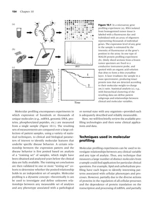
![Molecular Profiling for Early Detection 155
due to key enabling technological developments, the genes of interest, up to all possible expressed
the profiling of proteins (proteomics), and of mRNA genes, attached to an inert platform. A microar-
and DNA (genomics) are best developed, and per- ray chip can then simultaneously assay thousands
haps hold the most potential for the understanding of expressed genes by measuring hybridization of
of human disease. Hence, this chapter will focus on sample mRNA to RNA oligomers fixed to an inert
the reported and potential future applications of ge- substrate in a two-dimensional array. Hybridization
nomics and proteomics in lung cancer. of complementary strands of RNA or DNA occurs
Genomics and proteomics are complementary with extremely high affinity allowing very sensitive
modes of investigation. Genomics is currently more and specific detection of low-level species with the
widely used and more developmentally mature ability to distinguish between sequences differing by
than proteomics. The physical properties of DNA only a single nucleotide.
and RNA that allow complementary base pairing Messenger RNA is isolated from a homogenized
simplify their analysis. Proteins are, by virtue of biological sample and a fluorescent dye is bound to
their heterogeneity, more difficult to characterize. the mRNA. The labeled RNA is then applied to the
However, proteins have more direct impact on gene chip and allowed to hybridize with the pre-
pathological behavior, and many complexities of fixed oligomers, each of which represents a short
their regulation and function are not captured by sequence usually selected from the exon region of a
studies of gene expression levels. Studies that com- particular gene. After hybridization and washing to
pare genomic and proteomic data show that the cor- remove unhybridized species, a scanner measures at
relation between gene and protein levels is quite each position in the array the level of fluorescence
low, and changes in expression level often occur dis- that corresponds to the measure of expression for
cordantly [5–7]. Thus, although it is easier to assay each gene represented on the chip. The fluorescence
mRNA levels, they may provide a rougher surro- measurements for each of the thousands of genes
gate of biologic activity than knowledge of protein across each of the samples are stored in a database.
expression and modifications. Quackenbush offers an excellent review of the use
of microarrays for tumor analysis [8].
Genomics Early microarray studies used fewer than 10,000
Genomics can be defined as the study of the genes oligomer probes mounted on in-house produced
and their expression in normal or diseased cells chips. The potential power of microarray technol-
and tissues, to determine correlations with growth, ogy was readily apparent, and the technique was
development and disease states. Technologies that featured on the cover of the journal Science in 1991
have enabled the efficient detection of patterns of 3 years after the first prototype was manufactured.
altered expression of many genes in a single exper- As interest and use increased dramatically, chips
iment, coupled with powerful statistical techniques are now commercially manufactured and the num-
correlating the expression pattern with the biologic ber of oligomer probes represented on a chip has
phenotype, afford a deeper understanding of the also greatly increased, recently culminating in the
phenotype and offer the possibility to predict biolog- Affymetrix Human Exon chip, which contains ∼1.4
ical or clinical behavior in subsequent unknown an- million probes that cover each exon in the human
alytes. Genomics has already yielded clinically use- genome, allowing alternate splicing to be investi-
ful results in oncology, particularly breast cancer, gated over the entire genome. In addition to char-
lymphomas, and leukemias. acterizing exon regions of genes, probes represent-
The predominant technology currently employed ing mutated sequences have been utilized to rapidly
is the DNA microarray chip, which has rapidly characterize mutations occurring at known sites,
evolved into a highly versatile, standardized re- while other probes can be designed to profile micro-
search tool and is poised to have an important im- RNA or other genetic elements of interest [9,10].
pact clinically. A chip is composed of short syn- Other profiling techniques can give information
thetic oligomer probes, corresponding to each of about the genome that may be complementary to](https://image.slidesharecdn.com/lungcancer3rded-120126215311-phpapp01/85/Lung-cancer-3rd-ed-166-320.jpg)
![156 Chapter 10
mRNA expression levels [5,6,11–13]. Comparative proteins in low concentrations. MS can be coupled
genomic hybridization allows a rapid means of de- with a variety of protein separation techniques in-
termining changes in gene copy number across the cluding gel and column separations, and can also be
genome, relative to that in normal tissues. This used directly to assay protein profiles in tissues or
technique can be performed with gene arrays sim- blood without a separation step.
ilar to those used for profiling mRNA, or can be A mass spectrometer is an instrument that ionizes
used without arrays to analyze metaphase chro- molecules from a sample and measures the abun-
mosomal preparations. For example, specific am- dance and the ratio of molecular mass to unit charge
plification on chromosome 3q was identified to be (m/z) for all ions produced above a certain sensi-
associated with squamous cell lung cancer [12]. tivity threshold. A sample is ionized in a vacuum
Profiling of large numbers of single nucleotide poly- and the m/z ratios of each ionized species are dis-
morphisms (SNPs) allow linkage of phenotype in- tinguished by different physical properties depend-
heritance with specific chromosomal regions in pop- ing on the type of detector: time-of-flight (TOF),
ulations as well as uncovering losses of heterozy- Fourier-transform (FT), quadrupole etc. Two meth-
gosity in tumor samples that may be related to ods of ionization are generally employed: matrix-
malignant transformation and disease phenotype. assisted laser desorption/ionization (MALDI), and
However, these techniques that measure gene copy electrospray ionization (ESI). The important distinc-
number lack the capacity to detect changes in gene tion between MALDI and ESI is that MALDI pro-
expression produced by any of a myriad of other duces ions by laser irradiation of samples fixed to
factors regulating gene expression, whereas cDNA a solid conductive plate coated with a crystalline
microarray experiments assessing mRNA levels can, layer of organic acid; whereas, ESI produces ions
at least theoretically, detect perturbations reflecting from a sample in solution as it passes through a
underlying epigenetic mechanisms as well as muta- small-bore highly charge capillary. MALDI, and the
tions or chromosomal aberrations. These genomic closely related technique, surface enhanced laser
techniques thus capture less of the complexity re- desorption/ionization (SELDI), can be used to pro-
sponsible for phenotypic variability in lung cancer duce protein profiles from frozen tissue sections or
and, while of demonstrated utility in specific cases whole cell cytological preparations; unfortunately,
such as associating epidermal growth factor recep- with limited exceptions, paraffin-embedded tissue
tor (EGFR) amplification with clinical benefit from cannot be used. Multiple protein mass spectra can
EGFR tyrosine kinase inhibitors, may be less useful be obtained from different positions on a frozen
for developing complex predictive models of clinical tissue section, allowing analysis of a protein’s spa-
utility. However, specific genomic alterations may tial distribution and production of two-dimensional
be important in offering insight into the biological images of protein location and abundance. Thus,
relationships discovered by profiling mRNA levels, MALDI is useful for discerning field effects or dis-
especially when a single or limited number of ge- tinguishing protein expression in tumor from pro-
nomic alterations drive the biology. teins found in surrounding stroma or lymphocytic
infiltrate. MALDI can also be used to analyze dried
Proteomics droplets of biologic samples such as blood, serum,
Proteomics encompasses the study of protein ex- or plasma, pleural or ascitic fluid, or fractions col-
pression, including spatial and temporal profiles, lected from column separations or extracted pro-
posttranslational modifications, and interactions teins from one-dimensional or two-dimensional gel
with other molecules. Several important analytical separations. ESI requires samples to be in solution
tools and methodologies have been used extensively and has been particularly useful for online analy-
in proteomic research. sis of proteins and peptides in the eluate of column
Mass spectrometry (MS) features prominently as separations.
the most accurate means of determining the molec- MALDI or SELDI experiments have both been
ular weight of proteins, and is sensitive in detecting used in profiling studies of resected lung tumors](https://image.slidesharecdn.com/lungcancer3rded-120126215311-phpapp01/85/Lung-cancer-3rd-ed-167-320.jpg)
![Molecular Profiling for Early Detection 159
stringent similarity cutoff can define a larger num- the limited size of many studies necessitates the
ber of subgroups, each composed of samples that most efficient possible use of samples, and setting
have more uniform gene expression profiles; how- aside a large number of samples for adequate valida-
ever, this makes it more difficult for observed dif- tion leaves fewer samples for training and less robust
ferences (e.g., in survival) to reach statistical signif- predictive models. Furthermore, the genes picked
icance, due to the shrinking number of patients in as predictive are very dependent on the patients in-
each group and increase the likelihood of arriving cluded in the training set [16]. Cross-validation is
at false conclusions due to multiple subgroup anal- an approach that maximizes the size of the training
ysis. On the other hand, if newly discovered clinical set while still allowing validation of results on a suf-
correlations are statistically robust, the information ficiently large testing set [17–19]. In this method, a
may be further validated in prospective studies and single sample or a small number of samples is re-
be applied in individualizing treatment decisions. moved from a set and the remaining samples are
Supervised classification schemes start with two used to construct a predictive model. The model is
or more clinically or biologically defined groups— applied to the sample or samples that were left out
for instance, node negative versus node positive, of the model building process, and its accuracy in
responders versus nonresponders, or tumors that predicting the feature of interest is recorded. This
express a mutation versus those that do not—and is then repeated a large number of times and the
attempt to determine genetic profiles capable of pre- combined accuracy over all the left-out samples is
dicting to which group an unknown sample be- reported as an estimate of the classification accuracy
longs. One can perform a clustering approach simi- rate of the model.
lar to that described earlier but with the constraint Another method to minimize the necessary num-
that the two dominant clusters must differ by the ber of precious samples required for validating re-
clinical variable of interest. Alternatively, one can sults is to use online databases of previously pub-
use statistical tests to determine individual genes or lished gene expression profiles as independent test
proteins that may correlate with the clinical vari- sets. This is an attractive method made possible by
able of interest and then construct a model using the increasing standardization of some of the tech-
weighted linear combinations of these features to nologies [20–26], but there are challenges in inte-
make predictions on unknown samples. grating data sets from different experiments. There
Both supervised and unsupervised techniques may be substantial differences in the methods used
have the potential to “overtrain” the data, yielding to obtain the expression profiles; for instance, stud-
models that perform well on the sample set from ies use microarray chips from different manufac-
which they were derived, but with poor accuracy turers and even arrays produced at in-house mi-
on independent testing cohorts [14,15]. Validation croarray facilities. Even data obtained for the same
of results is thus the most important step in a molec- genes from newer versions of chips from a given
ular profiling experiment; studies without effective manufacturer may not be directly comparable to
validation deserve little credibility, since even rela- earlier versions. Also, newer microarrays typically
tionships that appear convincing may fail to have have more probes spanning more genetic elements
significance when applied to an independent set of than older versions. Thus, many genes may not be
samples. Some studies have approached this prob- represented in both data sets, and it may be dif-
lem by arbitrarily splitting sample sets into separate ficult to translate expression data from one set to
training and testing groups. However, to be mean- another. With the advent of exon arrays, it has be-
ingful, the testing set should be used as such only come clear that the location of the oligo within the
once, without recursive retraining. putative transcript yields different measurement re-
It is often underappreciated that high-quality sults. Furthermore, idiosyncratic differences in sam-
clinical samples are difficult to obtain and costly ple handling and experimental protocol can lead
to collect and store, and maintaining accurate and to other systematic differences between gene ex-
complete clinical follow-up is labor intensive. Thus, pression sets. Despite these challenges many studies](https://image.slidesharecdn.com/lungcancer3rded-120126215311-phpapp01/85/Lung-cancer-3rd-ed-170-320.jpg)
![160 Chapter 10
have shown that relationships observed in one data lung cancer in patients at high risk, including yearly
set can be successfully and convincingly demon- chest X-rays, sputum cytologies, even when cou-
strated to hold true in large independent sets. When pled with immunofluorescence or PCR, and bron-
efforts are taken to standardize procedures, interlab- choscopic screening, have had a discouragingly neg-
oratory reproducibility has been shown to be quite ligible impact on survival. A recent report of a non-
good [20–26]. Current microarray-based profiling randomized trial of spiral CT screening of smokers
will likely benefit from such increased standardiza- suggests the possibility that very small tumors can
tion across laboratories, as well as the use of a sin- be detected with encouraging survival results [33].
gle microarray platform and the ongoing develop- However, these results must be validated prospec-
ment of bioinformatics tools to permit sharing of tively, since lead-time bias is a possible explana-
large data sets and integrating data optimally de- tion for the apparently salutary effect. Moreover,
spite lab-to-lab differences. These advances will lead screening detected a number of small, indetermi-
not only to more effective validation of models, but nate nodules, of which a significant minority proved
will also allow large-scale meta-analyses to look for to be benign. The cost of CT screening is not in-
important molecular–clinical relationships in much significant and a large additional cost—including
larger sample sets, with better statistical power for increased morbidity and potentially mortality—is
developing robust molecular classifiers [27–32]. Of incurred by the necessity of additional diagnos-
course, regardless of its reproducibility in training tic testing of suspicious nodules that subsequently
and validation sets, the real rubric of success for any prove to be benign. A noninvasive test for cancer de-
molecular classification scheme will be its perfor- tection potentially could be integrated with imaging
mance in prospective clinical trials that determine modalities to help select patients with higher risk
whether its clinical use improves patient outcomes. nodules for more intensive efforts at diagnosis and
follow-up. A very high positive and negative pre-
dictive value for the targeted patient population is
Clinical applications mandatory, but most important, the screening test
or combination of tests must result in improved sur-
Early detection and diagnosis vival.
The peripheral blood is clearly the most readily MALDI and SELDI have been used to profile
available and clinically practical biospecimen source blood, serum, and plasma, as have antibody mi-
for early detection and diagnosis. Pathological states croarrays [34–37]. Other studies have attempted
can cause disease-related changes in molecules cir- to discover patient antibodies directed against tu-
culating in the blood, due to altered cellular ex- mor antigens that could have diagnostic utility
pression of secreted proteins, proteins being directly [37–40]. To date, these studies have been small
released into extracellular fluid after cell death, and incompletely validated and have yielded sen-
posttranslational modification by cleavage, glyco- sitivities and specificities much lower than what
sylation, or other processes, or as a result of the would be required for clinical use. However, as
host’s response to the disease. If a change in blood proteomic technology improves, a wider range of
protein composition occurs reproducibly in the low-concentration protein species may be detected,
presence of a disease, then such a change may be some of which may be useful in detecting early lung
useful for disease detection and diagnosis. For ex- cancer.
ample, the demonstration of proteins leaked from Other possible avenues for research include pro-
damaged cardiac myocytes has become the gold teomic profiling bronchial washings or pleural
standard for detection of myocardial infarction, effusion [41] to determine diagnostic or prognostic
even though they are not themselves causal. indicators. Gene profiles might also be helpful in dis-
One reason for the abysmal survival rates in lung tinguishing poorly differentiated lung cancers from
cancer may be the absence of effective screening other malignancies with similar histological appear-
for early detection. A variety of screening tests for ance [42].](https://image.slidesharecdn.com/lungcancer3rded-120126215311-phpapp01/85/Lung-cancer-3rd-ed-171-320.jpg)
![Molecular Profiling for Early Detection 161
Subclassification and staging filing, with corresponding differences in prognosis
Treatment decisions in lung cancer are based largely and response to therapy, including a previously un-
on the extent of disease as determined by staging recognized subtype, the basal-like, or “triple neg-
criteria and generalizations on disease behavior as- ative” subtype with a distinctly worse prognosis
sociated with histological distinctions. Stage has a [48,51–53]. Other expression studies have uncov-
strong impact on determining which patients are ered independent sets of genes capable of reliably
most likely to benefit from different types of treat- distinguishing good prognosis from poor prognosis
ment: surgery does not improve survival of stage patients among women with node-negative, estro-
IV patients since it has no effect on metastatic foci; gen receptor-positive breast cancer treated with ta-
conversely, patients with stage I lung cancer receive moxifen. The Oncotype DX assay (Genomic Health,
little or no survival advantage as a group from cur- Redwood City, CA) utilizes a 21-gene marker set,
rently available adjuvant chemotherapy because a using RT-PCR to profile expression using paraffin-
significant percentage will have no residual disease fixed biopsy specimens; from these a 10-year
following surgery and chemotherapy has limited ef- Recurrence Score predicting the rate of distant re-
fectiveness for those with micrometastases. More- currence among women treated with tamoxifen is
over, there is significant variability in the clinical generated [49]. Survival benefit with chemotherapy
outcome of individual patients within each stage appears to be limited to the poor prognosis group,
classification; there are differences in metastatic po- indicating that this risk stratification may be useful
tential, aggressiveness of metastatic disease, and re- in guiding treatment [50]. These risk predictors, as
sponse to chemotherapy. The ability to elucidate fac- well as the Affymetrix 70-gene assay [54,55], and
tors predicting individual prognosis and response to the wound response model [44,45], are based on
therapy will be invaluable clinically. different sets of genes with relatively little overlap,
The most important pathologic distinction de- yet they have demonstrated similar ability to distin-
scribed so far is that between small cell and nonsmall guish high risk from low risk node-negative, estro-
cell lung cancer (NSCLC). These are readily appar- gen receptor-positive patients treated with tamox-
ent by histological and immunohistochemical eval- ifen [46]. Further prospective studies are underway
uation and have fundamental differences in prob- to determine whether adding chemotherapy will
able cell of origin, natural history, and response to benefit those patients predicted to have worse prog-
chemotherapy. Subtype classification of NSCLC has nosis when treated with tamoxifen alone.
minimal impact on patient care, with a few notable Efforts to uncover useful predictive classifiers in
exceptions: e.g., recent studies suggest that EGF-R- lung cancer have also been fruitful. Many stud-
targeted therapy may show overall benefit in treat- ies [6,11,14,15,28–30,56–78] have tried to identify
ing certain patients with adenocarcinoma and es- subtypes of lung cancer based on overall similarities
pecially the subset with receptor mutations. Unfor- in gene and protein expression profiles using similar
tunately, the behavioral features that most likely approaches to those successfully employed in breast
affect clinical outcome—aggressiveness, likelihood and other cancers. The number of patients enrolled
of metastasis, resistance to apoptosis, and re- in these studies range from approximately 50 to 200,
sponse to therapy, inter alia—are indistinguishable and the number of RNA probes has increased in re-
with conventional microscopic evaluation. How- cent studies as commercial chip technology has ad-
ever, these features clearly have underlying genetic vanced. Early studies demonstrated that many clini-
determinants, and uncovering these with molecu- cal or histological groups clustered together by their
lar profiling may lead to dramatic changes in the natural genetic similarity. Normal lung could be
management of lung cancer. distinguished from tumor, NSCLC from SCLC, and
Gene profiling experiments in other cancer disci- adenocarcinomas, squamous cell carcinomas, and
plines have uncovered such determinants [43–55]. large cell carcinomas largely segregated into their re-
Five reproducible genetic clusters within breast can- spective groups. The ability to differentiate samples
cer have been disclosed by mRNA expression pro- with obvious histological differences was not a step](https://image.slidesharecdn.com/lungcancer3rded-120126215311-phpapp01/85/Lung-cancer-3rd-ed-172-320.jpg)
![162 Chapter 10
forward in itself, but proved the concept that Gene expression and annotated clinical data from
gene profiling experiments could make meaningful these early studies and several others, e.g. .[62],
classifications. Several studies have demonstrated were made publicly available, permitting the data
clustering of samples within one or more histo- to be further investigated through several meta-
logical subtypes [6,14,15,56–58,61–65,71–74,77]. analyses [27–32]. These studies showed that many
These results suggest that there may be genetic sub- underlying genetic relationships were observed in
groups within the main histologic subtypes, with multiple data sets, and that data sets could be com-
possible different biologic implications, although the bined to make larger training sets or for use as
reproducibility of observed subgroups has not been independent testing sets, despite significant differ-
conclusively verified across studies. ences between sample sets. Furthermore, meta-
Bhattacharjee et al. assayed gene profiles of 186 analyses combining lung cancer microarray studies
patients and described four distinct clusters of tu- with studies from other cancer disciplines have dis-
mors, one of which was said to have worse survival covered profiles of chemotherapy response, metas-
than the others [57]. However, a majority of samples tasis, and oncogenesis that are predictive in multiple
did not fit into distinct clusters, and the prognostic types of cancer [27,31,32]. Efforts to standard-
differences have not been validated in subsequent ize microarray platforms and sample handling
studies. Nevertheless, these were interesting obser- technique will also likely improve reproducibility
vations about the general appearance of clustering across studies and improve the power of future
within lung cancer, and the gene data and annotated studies.
clinical information has been a valuable resource for Potti et al. have also recently described a prog-
deriving and testing new predictive models. nostic indicator developed from 89 early-stage lung
Beer et al. studied 86 patients with adenocarci- cancer patients and tested in two independent test
noma, and described three dominant clusters within sets from cooperative group studies [70]. The results
this cohort, one cluster differing significantly from from this work are being used to design a prospec-
the others prognostically [56]. A linear model based tive clinical trial to determine whether this classi-
on 50 genes that could predict prognostic class in fier can guide recommendations for adjuvant treat-
unknown patients was derived using a split sample ment. In one potential design, tumors resected from
training–testing approach in “leave-one-out” cross- early-stage patients will be tested to predict good
validation. The conclusions were also tested in the or bad prognostic category. All patients will be ran-
Bhattacharjee et al.’s data set and found to be valid. domized to either receive adjuvant chemotherapy
The model was predictive for stage I patients in all or not, in order to test the hypothesis that adjuvant
testing sets and represents the first well-validated therapy will improve the survival of patients with
discovery of a prognostic indicator in lung cancer worse prognosis, regardless of stage, whereas, good
using molecular profiling. Chen et al. performed a prognosis patients will derive little or no benefit
proteomic analysis of the same patient cohort using from chemotherapy. This would then test whether
two-dimensional PAGE and defined a protein-based treatment strategies incorporating molecular pre-
risk index predictive of prognosis [6]. The proteins dictors to guide treatment are justified.
selected did not overlap with the previously devel- The above-mentioned and other similar clinical
oped 50-gene classifier, although the mRNA levels trials may demonstrate that patients have better
of several proteins were significantly associated with overall survival when treatment decisions take into
survival. Raponi et al. further validated the 50-gene account prognostic gene profiles. If this proves to
prognostic profile and developed a separate 50-gene be the case, then molecular profiling may be as-
classifier through analysis of 129 patients with squa- similated into clinical practice as a staging adjunct,
mous cell carcinoma [71]. The two classifiers were particularly to predict which patients may not need
combined to form a single 100-gene classifier appli- chemotherapy. However, it is not at all clear that
cable to both histologic types, which was shown to likelihood of relapse predicts likelihood of benefit
be a significant prognosticator. from treatment.](https://image.slidesharecdn.com/lungcancer3rded-120126215311-phpapp01/85/Lung-cancer-3rd-ed-173-320.jpg)
![Molecular Profiling for Early Detection 163
Selection of therapy and additionally, a small sample in a heterogeneous
Although there are numerous chemotherapy single tumor may not be representative of the entire tu-
agents and combination regimens with clear clini- mor. Furthermore, research studies attempting to
cal activity in the treatment of lung cancer, there analyze these samples are often hampered by the
is wide variability in their observed effectiveness frequent opinion that nonclinically indicated biop-
in individual patients. The ability to select an ac- sies from metastatic lesions are not warranted.
tive regimen de novo in individual patients would Many groups have applied molecular profiling to
be particularly valuable, especially considering the panels of cell lines, in which sensitivity to differ-
substantial treatment-related morbidity and the tu- ent agents is measured with in vitro techniques,
mor morbidity and death associated with ineffective and it is possible that the correlations between drug
therapy. sensitivity and molecular profile could be trans-
In vitro drug sensitivity assays have been touted lated to patient specimens [79–87]. Studies involv-
as a potential means to individualize chemother- ing cell lines would allow for greater control of ex-
apy selection, but so far have had limited clinical perimental factors affecting microarray assays and a
utility. If molecular profiling can effectively predict straightforward determination of in vitro drug effi-
the clinical efficacy of different therapies against a cacy. Furthermore, these experiments offer the ad-
particular tumor, it will profoundly impact patient vantage that large numbers of chemotherapeutics
care, particularly in the age of highly selective tar- can be economically investigated in parallel, includ-
geted therapies [27,69,72,79–87]. It is likely that ing first-line and second-line agents in lung cancer,
such selective therapies will be useful only in small drugs used primarily for other types of tumors, and
subsets of lung cancer patients, and clinical benefit experimental therapies.
can only be detected if these subsets can be molec- However, as for any model, cell lines only par-
ularly identified. A number of factors make it more tially represent clinical lung cancer and are only
challenging to discover predictors of chemotherapy hypothesis-generating. Human tumors may differ
response using actual patient samples. Surgical re- profoundly from their cell line derivates, and gene
section of NSCLC is typically performed only if it expression profiles of cells grown on culture dishes
can remove all known disease burden, and this is in growth media supplemented with various growth
only possible in about a quarter of NSCLC patients factors are likely quite distinct from those of tumors
and is rarely an option in SCLC. Thus, while re- in vivo. Thus, it may be difficult to translate these
sected primary tumors are well suited for molecular molecular assays into a clinically applicable form.
profiling, the requirement for fresh resected tumor If this proves to be the case, it may be possible to
tissue poses problems for prediction of chemosensi- develop predictors on other models that better rep-
tivity. Chemotherapy is most commonly used in dis- resent human disease, such as implanting cell lines
ease diagnosed in advanced stages where fresh tis- or cells from fresh human tumors into mice.
sue is rarely available, in the adjuvant setting, where Molecular profiling may be particularly useful
there is no followable disease, or after local or dis- in the selection of targeted therapies. This class
tant recurrence, in which case the disease may be of anticancer drugs comprises a growing number
genetically distinct from the original primary tumor. of molecular agents that inhibit a particular sig-
With effort, tissue specimens can be obtained from naling pathway or other crucial cellular process.
metastatic sites to be analyzed for molecular profil- Currently available examples of targeted agents in-
ing in order to determine correlates with sensitivity; clude the EGFR inhibitors gefitinib, erlotinib, and
however, this poses new problems. The quality of cetuximab, the inhibitor of vascular endothelial
the specimens may vary, the entirety of the spec- growth factor (VEGF) receptors, bevacizumab, and
imen may be required for pathological evaluation, the multitargeted agents, sorafenib and sunitinib.
the number of tumor cells observed can be quite These agents can induce dramatic responses in some
small, often too few to perform microarray analysis, patients whose tumors are particularly reliant on
highly contaminated with normal tissue elements, the targeted pathway, but they often show little](https://image.slidesharecdn.com/lungcancer3rded-120126215311-phpapp01/85/Lung-cancer-3rd-ed-174-320.jpg)
![164 Chapter 10
benefit in an unselected patient population. Molec- aided by efforts to accommodate smaller samples
ular profiles can potentially be correlated with drug and to get accurate results from potentially lower
response, using methods similar to those described quality real-world clinical samples. MALDI has been
above for conventional agents, or they can be used shown to be effective in analyzing very small col-
to determine which tumors have profiles associated lections of cells, thus allowing analysis of biopsy
with the target gene or protein [80,81,83]. samples and fine needle aspirates [88]. For microar-
Bild et al. observed changes in gene expression rays, however, increases in sensitivity are required
after transfection of lung cancer cell lines with to expand their clinical utility to these types of spec-
different oncogenes, and then used the resulting imens. Quantitative real-time PCR can provide such
gene profiles to predict whether patient samples sensitivity and can be clinically used to measure
had overexpression or mutations of those genes expression levels of each gene used in a profile,
[80]. Conversely, another approach that has re- and also can be applied to fixed tissue embedded
cently been proposed is the compilation of gene ex- in paraffin, rather than the fresh-frozen samples re-
pression changes after perturbation of a set of cell quired for full microarray analysis.
lines with a panel of pharmaceutical compounds Another challenge is the interpretation of results
[83]. These approaches can yield signatures of key in a meaningful biological context. Significant clin-
molecular pathways and drug effects that can poten- ical predictions and classifications can be made us-
tially be used to select patients for targeted therapy ing a gene or protein profile as a “fingerprint” or
and may help direct basic research by implicating “signature” without identification of its molecular
novel targets for drug design. constituents or knowledge of the biological rela-
tionships it represents. The theoretical and statistical
framework of molecular profiling does not require
Future directions and conclusions that the underlying biology be understood, but such
knowledge would undoubtedly aid scientists’ ability
There are many challenges that must be met be- to form and test hypotheses and identify new targets
fore profiling techniques can reach their full poten- for therapy. Understanding the biology that under-
tial. A major limitation of many studies has been lies clinically significant molecular profiles will re-
that the number of patient samples investigated has quire the coordinated efforts of proteomics and ge-
been too small to draw meaningful conclusions. nomics researchers, molecular biologists and clinical
One attractive means of increasing sample sizes is to scientists, as well as statisticians and bioinformatics
combine data from multiple laboratories into shared experts to explore the actions of individual proteins
databases. Improved standardization of sample han- and pathways responsible for various phenotypes
dling, microarray or other analytical platforms, and and behaviors of lung cancer.
quality control metrics will be essential for the suc- Although many challenges remain, molecular
cess of such data sharing [20–26]. Advancements in profiling experiments have produced encouraging
bioinformatics and computational tools will also be results in the study of breast, lung, and a vari-
required to efficiently utilize the large quantities of ety of other cancers. In breast cancer, gene pro-
data, to adjust for variability between labs, and to in- files are already being utilized clinically to predict
tegrate experiments that profile different analytes, recurrence risk of certain patients and better in-
e.g., proteomic and genomic studies. form the decision to include adjuvant chemother-
Once a predictive profile has been described and apy. Similar prognostic assays have been developed
validated, an additional challenge is posed in de- from microarray analyses of lung cancer, and a
veloping a suitable clinical assay. As already men- prospective clinical trial has been proposed to as-
tioned, this may pose a particular challenge for pro- sess the efficacy of one test in selecting patients
files derived from cell lines or animal models, or for adjuvant treatment of early-stage NSCLC. These
from highly technical, limited availability platforms. early successes attest to the potential of profiling
Once assays are developed, clinical utility will be experiments to improve patient outcome through](https://image.slidesharecdn.com/lungcancer3rded-120126215311-phpapp01/85/Lung-cancer-3rd-ed-175-320.jpg)
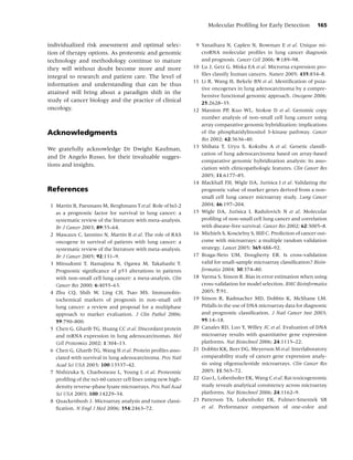
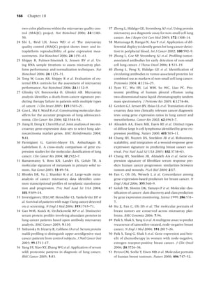

![CHAPTER 11
The Role for Mediastinoscopy in the
Staging of Nonsmall Cell Lung Cancer
Carolyn E. Reed
Introduction as multimodality therapy has become more com-
mon. Therapeutic options will vary for subsets of
The staging of nonsmall cell lung cancer (NSCLC) the N2 patient population, and comparison of study
aids the physician in selecting appropriate treat- results will require appropriate stratification.
ment, communicating prognosis, and studying The methods of staging the mediastinum in-
treatment results in equivalent patient populations. clude radiographic modalities, minimally invasive
Once distant metastatic disease has been ruled out, techniques, and invasive procedures as listed in
focus shifts to the assessment of lymph node in- Table 11.1 [4]. This chapter will focus on the role
volvement. It is estimated that 26–44% of patients of mediastinoscopy in staging NSCLC, which has
diagnosed with NSCLC will have mediastinal lymph been considered the “gold standard.” However, dis-
node disease [1,2]. cussion of alternate techniques will emphasize that
Detection of metastatic disease in contralateral radiographic and endoscopic sophistication in stag-
mediastinal or hilar lymph nodes or supraclavicu- ing has changed the present and probably the future
lar lymph nodes (stage IIIB) generally renders the utilization of mediastinoscopy.
patient unresectable. Patients with ipsilateral medi-
astinal lymph node involvement (stage IIIA) consti-
tute a heterogeneous population with varying treat- Radiographic staging of the
ment options and outcomes. This heterogeneity is mediastinum
emphasized by a study of 702 consecutive patients
with N2 disease undergoing surgical resection. Five- The initial staging of the mediastinum usually
year survival varied from 34% when one level was begins with noninvasive radiographic techniques.
involved with microscopic disease (n = 244), 11% These techniques may direct who undergoes inva-
when multiple levels were microscopically positive sive staging and/or the most likely location of N2
(n = 78), 8% when clinical N2 disease in one level positivity and therefore modality chosen for staging.
was detected (n = 118), and only 3% when multiple There are presently three commonly used meth-
levels of clinical N2 disease were apparent (n = 122) ods of radiographic mediastinal assessment: com-
[3]. The need to define the presence and extent of puterized tomography (CT), positron emission to-
mediastinal lymph node involvement is important mography (PET), and integrated CT-PET. Table 11.2
illustrates the sensitivity, specificity, positive pre-
dictive value (PPV), and negative predictive value
Lung Cancer, 3rd edition. Edited by Jack A. Roth, James D. Cox,
(NPV) of each modality [2,5]. CT scanning of the
and Waun Ki Hong. c 2008 Blackwell Publishing, mediastinum has poor accuracy, and denying a
ISBN: 978-1-4051-5112-2. patient surgery based on CT-enlarged (>1 cm in
169](https://image.slidesharecdn.com/lungcancer3rded-120126215311-phpapp01/85/Lung-cancer-3rd-ed-180-320.jpg)
![170 Chapter 11
Table 11.1 Options for staging the mediastinum. ative PET and CT scanning [13]. However, other sur-
geons have adopted a more selective approach to
Noninvasive radiographic imaging
invasive staging if the mediastinum is negative by
Chest X-ray
radiographic assessment, especially if the tumor is
CT scan
MRI small (<2 cm in diameter) and peripheral. Selective
PET scan use of mediastinoscopy is further discussed later in
Minimally invasive staging the chapter. The additional effectiveness (i.e., per-
Transbronchial needle aspiration (TBNA) centage of positive lymph nodes) of endoscopic min-
Esophageal endoscopic ultrasonography with imally invasive staging in radiographically negative
fine-needle aspiration (EUS-FNA) patients awaits further clarification.
Endobronchial ultrasonography with fine-needle
aspiration (EBUS-FNA)
Invasive staging
Mediastinoscopy Mediastinoscopy
Mediastinotomy
Extended cervical mediastinoscopy History
Thoracoscopy Cervical mediastinal exploration using the Jackson
Thoracotomy
laryngoscope was originally described by Harken
and associates in 1954 [14]. Carlens, using a spe-
Adapted from Pass [4].
cially designed mediastinoscope and a suprasternal
notch incision, advanced the technique [15], and
Pearson and the Toronto surgical group further
diameter) mediastinal lymph nodes should not be
developed the indications and popularized the
accepted without histologic confirmation. Although
procedure as currently used [16]. Mediastinoscopy
PET scanning has improved accuracy of mediasti-
subsequently became the “gold standard” by which
nal staging [6], initial hope from early studies that
other methods of evaluating the mediastinum are
PET would almost eliminate the need for invasive
compared.
staging has dimmed as more recent reports do not
reach the level of accuracy desired [7,8]. PET fails
to identify microscopic metastatic disease and cer- Indications for mediastinoscopy
tain clinical factors, such as size and location of tu- The main indication for mediastinoscopy is en-
mor, need to be considered in the decision to pro- larged mediastinal lymph nodes detected by CT or
ceed with invasive staging. The integrated CT-PET PET positivity in any mediastinal lymph node level.
has the potential to improve accuracy and further There are certain instances where despite a nor-
refinements are expected [5,9]. mal CT scan, the incidence of N2 disease is in-
Controversy exists over whether invasive medi- creased and the application of mediastinoscopy is
astinal staging is still needed if radiographic assess- suggested. This includes large lesions and tumors
ment is negative. In a series of 271 clinical stage I CT- occupying the inner third of the lung field, espe-
negative NSCLC patients undergoing resection after cially when the histology is adenocarcinoma or large
negative mediastinoscopy, N2 disease was found in cell carcinoma [17]. As noted previously, the use
9.2% [10]. In a similar study, the negative predic- of mediastinoscopy will vary with surgeon prefer-
tive value of mediastinoscopy was 93.4% [11]. In a ence and tolerance for an acceptable limit of false
small study where both PET and CT scanning did not negativity. Other techniques are now replacing or
identify mediastinal lymph node involvement, 8% being used in a complementary fashion to medi-
of patients had pathologically positive nodes [12]. astinoscopy and will be discussed later in this chap-
This finding was confirmed in a subsequent report ter. One of the advantages of mediastinoscopy is
that concluded routine mediastinoscopy is still the the ability to obtain a large fragment of tissue for
most economically reasonable strategy despite neg- histopathology or even entire small lymph nodes as](https://image.slidesharecdn.com/lungcancer3rded-120126215311-phpapp01/85/Lung-cancer-3rd-ed-181-320.jpg)
![Role of Mediastinoscopy in Staging NSCLC 171
Table 11.2 Radiographic staging of the
mediastinum. Positive Negative
Number of predictive predictive
Modality patients Sensitivity Specificity value value
CT 3438 0.75 0.82 0.56 0.83
PET 1045 0.84 0.89 0.79 0.93
PET-CT 728 0.69 0.94 0.49 0.99
Modified from Toloza et al. [2] and Cerfolio et al. [5].
compared to clusters of aspirated cells for cytologic Procedure
examination. The technique of cervical mediastinoscopy is de-
Selective indications for mediastinoscopy in clin- scribed in textbooks of general thoracic surgery.
ical N0 patients include tumor factors (size and lo- Right and left, high and low paratracheal nodes (lev-
cation) and histology. In a study of 164 patients els 2R, 2L, 4R, 4L), pretracheal nodes, and anterior
with T1 adenocarcinomas or T1 squamous cell carci- subcarinal nodes (level 7) are accessible via this ap-
nomas undergoing mediastinoscopy, true negative proach. Correct positioning of the patient is impor-
(TN) and true positive (TP) rates for tumors 2 cm tant to facilitate insertion of the mediastinoscope
or less were 96% and 4% compared to large tu- into the pretracheal space. A metal suction catheter
mors (2–3 cm) with 84% TN and 14% TP rates can be used to dissect fascial planes. The habit of al-
[18]. In a study of T1N0 patients (N0 defined by CT ways aspirating the structure to be biopsied with a
scan with diameter <1.5 cm) with adenocarcinomas spinal needle avoids vascular mishaps.
and squamous cell carcinomas undergoing medi- Ideally, the surgeon should routinely examine
astinoscopy, positivity was 9.5% in the adenocarci- and sample five nodal levels (2R, 2L, 4R, 4L, 7). The
noma group. However, none of the T1N0 squamous false-negative rate of mediastinoscopy is undoubt-
cell patients had a positive mediastinoscopy [19]. In edly affected by the skill and diligence of dissection.
a meta-analysis of 14 studies involving the size of A survey of United States cancer practice showed
lymph nodes detected on CT scan, if PET was nega- how infrequently mediastinoscopy is done properly
tive and nodes were 10–15 mm in CT, probability of and that in over half of cases no tissue is submitted
N2 disease was only 5% [20]. In a cost-effectiveness for pathology [23].
study of NSCLC patients who were stage I by CT
and PET screening, routine mediastinoscopy was Results
found to be of questionable value given the high In a review of over 5687 patients undergoing cer-
cost (>$250,000) per life year (0.008) gained [21]. vical mediastinoscopy between 1983 and 1999, the
However, the prevalence of N2 disease was low sensitivity was 81% and negative predictive value
(3%) in this study. As prevalence exceeded 10% in 91% [1]. In several large series, it has been empha-
the decision analysis model, the cost-effectiveness sized that the majority of patients found to have
increased. In a somewhat similar study comparing N2 disease at thoracotomy after a negative medi-
PET with selected mediastinoscopy to routine medi- astinoscopy have had abnormal lymph nodes lo-
astinoscopy, prevalence of mediastinal involvement cated in levels that are inaccessible by standard me-
in potentially resectable NSCLC patients was as- diastinoscopy [24,25].
sumed to be 20% and sensitivities and specificities
of modalities were based on literature review [22].
Complications
The selective approach of using mediastinoscopy
The reported mortality rate of cervical medi-
only when PET was positive for N2/N3 disease was
astinoscopy ranges from 0 to 0.2%, and morbid-
shown to offer cost benefit.
ity rates vary from 0.6 to 3.7% [1]. Ginsberg](https://image.slidesharecdn.com/lungcancer3rded-120126215311-phpapp01/85/Lung-cancer-3rd-ed-182-320.jpg)
![172 Chapter 11
reported no deaths in 2259 mediastinoscopies [26]. Although unusual, the left recurrent laryngeal
Of the 2.0% of patients having a complication, nerve can be traumatized if there is excessive dis-
only 0.3% required surgical treatment (thoraco- section, sampling, or cautery at the left tracheo-
tomy or sternotomy). Significant life-threatening bronchial angle occurs. Pneumothorax, wound in-
complications include hemorrhage (the most com- fections, and arrhythmias are rare minor complica-
mon), tracheobronchial injury, and esophageal tions.
injury.
The management of major hemorrhage during
mediastinoscopy was reviewed by Park and asso- Extended cervical
ciates [27]. During a 12-year period at Memo- mediastinoscopy and
rial Sloan Kettering Cancer Center, 3391 medi- anterior mediastinotomy
astinoscopies were performed, and 14 patients
(0.4%) experienced major hemorrhage, which was Standard mediastinoscopy cannot reach lymph
defined as any bleeding that required additional sur- nodes in levels 5 and 6, which are major drainage
gical exploration for surgical control. The most com- basins for left upper lobe tumors. Extended cervi-
mon biopsy site resulting in major hemorrhage was cal mediastinoscopy as described by Ginsberg and
the lower right paratracheal region (level 4R), and associates allows exploration of subaortic and an-
the most frequently injured vessels were the azy- terior mediastinal lymph nodes via the same inci-
gos vein and the pulmonary and innominate arter- sion [29]. Blunt dissection with an index finger is
ies. The azygos vein can easily be mistaken for an used to open the fascia between the innominate
anthracotic lymph node. Needle aspiration prior to and carotid arteries superior to the arch of the aorta.
biopsy and avoidance of excessive traction are rec- A tunnel is created along the anterolateral surface
ommended to avoid bleeding. Possible factors that of the aorta, and the mediastinoscope is inserted
could increase the risk of hemorrhage include prior into this tunnel and gently advanced over the top
radiotherapy, induction therapy, prior surgical pro- of the aorta to the aortopulmonary window [30,31]
cedures in the mediastinum and reoperative medi- (Figure 11.1). Electrocautery is not used to avoid
astinoscopy. injury to the vagus and phrenic nerves. Extended
The initial first step to control major hemorrhage cervical mediastinoscopy when added to standard
is gauze packing. The mediastinoscope should not mediastinoscopy can increase the negative predic-
be removed because direct tamponade is facilitated. tive value by 10–20% [1]. However, this technique
Damage to the main pulmonary artery or innomi- is not commonly used in the United States.
nate artery usually requires prompt exploration via An alternate approach to the aortopulmonary
median sternotomy and this approach should also window is anterior mediastinotomy described by
be used if packing is unsuccessful and the patient McNeil and Chamberlain in 1966 [32]. Although
remains unstable. Median sternotomy is the most in the original report a vertical parasternal inci-
versatile incision, allows identification and control sion was used with resection of the second and
of most injuries, and facilitates institution of car- third costal cartilages, more common modifications
diopulmonary bypass if needed. If packing stabi- include a short transverse incision over the second
lizes the patient but hemorrhage is still ongoing, the costal cartilage which is excised or a second inter-
surgical approach to repair the injury may include costal incision without cartilage incision. The me-
consideration of thoracotomy for possible definitive diastinal pleura can be bluntly dissected allowing
pulmonary resection. access to the aortopulmonary window without en-
Injury to the trachea or mainstem bronchi is very tering the pleural space. A mediastinoscope can be
rare. It usually responds to packing with absorbable inserted through the incision and regional lymph
cellulose gauze and closes when the patient is extu- nodes identified and biopsied (Figure 11.2). When
bated and no longer subjected to positive pressure performed alone as directed by CT scan, the sen-
ventilation [28]. sitivity of anterior mediastinotomy is 63–86% and](https://image.slidesharecdn.com/lungcancer3rded-120126215311-phpapp01/85/Lung-cancer-3rd-ed-183-320.jpg)
![Role of Mediastinoscopy in Staging NSCLC 173
Figure 11.1 Extended cervical mediastinoscopy. Reproduced from [31] by permission of Elsevier.
the NPV 89–100% [1]. Today, anterior mediastino- surgery, excisional biopsy of entire lymph nodes,
tomy is most commonly used to obtain large biop- and is an excellent teaching tool. It does increase the
sies of anterior mediastinal masses as other tech- cost when compare to standard mediastinoscopy.
niques have become available to assess aortopul- In a study of 240 consecutive patients undergoing
monary lymphadenopathy. video-assisted mediastinoscopy, the mean number
of biopsies was 6, mean number of lymph node lev-
els sampled was 2.3, and mean operating room time
Video-assisted mediastinoscopy was 36.6 minutes [34]. The sensitivity, specificity,
and accuracy for the 154 patients with NSCLC were
Video-assisted mediastinoscopy was introduced in 97.3, 100, and 98.0%, respectively. There were no
1994 by Sortini and associates [33]. The use of deaths, and there were two complications (0.83%),
the video mediastinoscope allows wide and magni- a pneumothorax and injury to the innominate
fied operative exposure, ability to perform bimanual artery requiring manubrial split and direct repair.](https://image.slidesharecdn.com/lungcancer3rded-120126215311-phpapp01/85/Lung-cancer-3rd-ed-184-320.jpg)
![174 Chapter 11
Figure 11.2 Anterior mediastinotomy. Reproduced from [31] by permission of Elsevier.
Redo mediastinoscopy [38]. It was possible to obtain a biopsy at the same
level that was positive at the initial mediastinoscopy.
Multiple studies utilizing induction chemotherapy There were no complications other than a wound
or chemoradiotherapy have documented increased infection. Of the 12 patients found to be N2 neg-
survival in the subset of N2 patients found to have ative, 5 had residual N2 disease after thoracotomy
N0 or N1 disease at the time of surgery [35–37]. and lymphadenectomy. The sensitivity for redo me-
Although the use of PET scanning to assess re- diastinoscopy was 0.70 and accuracy 0.80. It was
sponse to induction therapy is being investigated, admitted that the technique was more complex sec-
histopathologic confirmation of residual N2 disease ondary to peritracheal adhesions. The authors em-
is required if alternate therapies to surgery are be- phasized digital dissection and gradual access cre-
ing considered. Most surgeons are fearful of redo ated by using the electrocautery. It was noted that
mediastinoscopy secondary to expected fibrosis and the mediastinoscope was more easily inserted via
difficulty reaching the original biopsy site and the the left side of the trachea. Dissection to create a
danger of increased complications. left paratracheal tunnel until the posterior arch of
In a small study of patients undergoing neoad- the aorta is reached is also emphasized in a series
juvant chemotherapy for N2 disease, 24 patients reported by Van Schil and associates [39]. Dissec-
underwent redo mediastinoscopy to assess response tion using the suction tip is then carried back to the](https://image.slidesharecdn.com/lungcancer3rded-120126215311-phpapp01/85/Lung-cancer-3rd-ed-185-320.jpg)
![Role of Mediastinoscopy in Staging NSCLC 175
mid line and right side of the trachea. Bleeding con-
trolled by tamponade was encountered in two of 27 1
patients. The accuracy of redo mediastinoscopy in
this series was 85%. It was noted that subcarinal
nodes were the most difficult to reach.
2 2
Video-assisted mediastinoscopy has also been
successfully used in the postinduction setting [40]. Aortic arch
The anticipated increased difficulty when radiother-
apy is added requires further evaluation.
Although redo mediastinoscopy is feasible, the 4 4 5
use of alternate minimally invasive techniques to
evaluate the mediastinum offers a simpler and less 10 10
12
risky method to reassess the postinduction medi- 7
astinum. Although limited by fine-needle aspira-
11
tions, the level of initial positivity can always easily 11
be rebiopsied and multiple procedures are possible.
8
Esaphagus
Alternative methods to 12
mediastinoscopy 9 9
Pulmonary ligament
Both endobronchial ultrasonography (EBUS) and
esophageal endoscopic ultrasonography (EUS) al-
low excellent visualization of the mediastinum. By EBUS-TBNA
adding real-time fine-needle aspiration (FNA) to
both techniques, histologic confirmation of medi-
astinal disease can be obtained in an outpatient EUS-FNA
setting with conscious sedation, and even minor
complications are rare (i.e., these methods are min- Figure 11.3 The diagnostic reach of EUS-FNA and
imally invasive). As shown in Figure 11.3, the diag- EBUS-FNA (TBNA).(Reproduced with permission from
nostic reach of EBUS-FNA and EUS-FNA is com- Elsevier.)
plementary [41]. EUS-FNA allows access to the
retrotracheal space (level 3), the lower paratracheal
region on the left (4L), the radiographic subaortic PET in the posterior mediastinum, the sensitivity
space (5), and the posterior mediastinum (levels 7, ranged from 92.5 to 96% and accuracy has been
8, and 9). EBUS-FNA provides access to the superior 97–98% [43,44].
mediastinum (levels 2R, 2L, 4R, 4L), the subcarinal EUS-FNA can prevent up to 70% of invasive
space (7), as well as hilar (10), interlobar (11) and staging in patients with radiographic evidence of
lobar (12) lymph node levels. N2/N3 disease [45,46] and therefore can clearly im-
pact on patient management [47]. The utility of
EUS-FNA EUS-FNA in the setting of a radiographic negative
In a large series of 242 consecutive patients with mediastinum has yet to be clarified. In a study by
suspected or proven NSCLC and enlarged (>1 cm Wallace and associates [48], a small cohort of pa-
short-axis diameter) lymph nodes by CT, the sen- tients (n = 24) without enlarged mediastinal lymph
sitivity, specificity, and accuracy for EUS in medi- nodes underwent EUS-FNA and a surprising 42%
astinal analysis were 91, 100, and 93%, respectively had stage III or IV disease detected by EUS. In a study
[42]. In patients with suspicious nodes by CT and/or specifically assessing EUS-FNA in the CT-negative](https://image.slidesharecdn.com/lungcancer3rded-120126215311-phpapp01/85/Lung-cancer-3rd-ed-186-320.jpg)
![176 Chapter 11
mediastinum, surgery was precluded by the find- EUS-FNA was successful in 78%[57]. Combining
ing of stage IIIA or IIIB disease in 12% of patients both approaches produced successful biopsies in
with NSCLC [49]. Malignant mediastinal adenopa- 97% and diagnoses in 94%.
thy was detected significantly more frequently in The learning curve for thoracic surgeons is proba-
lower lobe and hilar cancers combined compared bly least with EBUS. If both EBUS and EUS are avail-
with upper lobe cancers. able, it is recommended that EBUS be performed
EUS also has the ability to detect T4 disease and first unless lower mediastinal lymph nodes other
allows biopsy of suspicious left adrenal lesions. The than level 7 are the only enlarged nodes. In view
use of EUS-FNA is not meant to supplant medi- of the minimal invasiveness of these techniques,
astinoscopy as the methods are complementary. the yield and cost-effectiveness of assessing a CT
When used as a first approach to accessible enlarged and PET-negative mediastinum should be further
mediastinal lymph nodes, EUS-FNA has been found defined.
by decision analysis to be cost-effective [50,51].
EBUS-FNA
Video-assisted thoracoscopy
Transbronchial needle aspiration has not been
widely used in the staging of NSCLC as the reported
Video-assisted thoracoscopy can be utilized to assess
yield has been variable, success is related to the size
all lymph node levels if performed on both right and
and location of the lesion, and the technique is es-
left sides. It has been most frequently used to assess
sentially “blind” and highly operator dependent. Us-
level 5 and 6 lymph nodes or when tumor invasion
ing EBUS as a guide, the diagnostic yield was in-
or pleural tumor seeding is suspected. It may be use-
creased (71%) and lymph node size and location
ful after induction therapy when both assessment of
did not influence success [52]. In a randomized trial
the N2 positive site and/or tumor status could alter
of conventional versus ultrasound-guided trans-
attempt at surgical resection.
bronchial needle aspiration to assess 200 patients,
EBUS guidance significantly increased the yield in
all stations except in the subcarinal region [53].
As technology evolved, linear array EBUS has Future recommendations
been coupled with fine-needle aspiration capabil-
ity via the biopsy channel. In an initial report, the The staging of the mediastinum will continue to
sensitivity, specificity, and accuracy of EBUS-FNA be critical for optimal management of NSCLC. The
in distinguishing benign from malignant lymph combined use of EBUS-FNA and EUS-FNA offers
nodes were 95.7, 100, and 97.1%, respectively [54]. the potential of more comprehensive access to me-
EBUS-FNA was recently evaluated in NSCLC pa- diastinal and hilar lymph nodes than is usual by
tients with no evidence of enlarged lymph nodes in standard mediastinoscopy. These minimally inva-
CT scan [55]. Of 100 patients evaluated, 119 lymph sive approaches avoid anesthesia, do not necessitate
nodes with a mean size of 8.1 mm were biopsied. clinical admission, and reduce risk. The same lymph
Unnecessary surgical exploration was avoided in 19 node level can be repeatedly biopsied and this abil-
patients, and malignancy was missed in two pa- ity becomes increasingly important if downstaging
tients. Sensitivity was therefore 92.3%, specificity is essential to surgical intervention, chemothera-
was 100%, and the negative predictive value was peutic regimens could be added or changed, and
96.3%. correlation with PET findings need to be corrob-
The accuracy of the combination of EBUS-FNA orated. There will continue to be instances when
and EUS-FNA has been reported to be 100% [56]. despite negative evaluation of CT-enlarged or PET-
In a study of patients with enlarged lymph nodes in positive mediastinal nodes by EBUS and/or EUS,
one of eight lymph node stations with a crossover mediastinoscopy is still warranted by tumor charac-
design, EBUS-FNA was successful in 85% and teristics, surgeon judgment, or patient risk profile.](https://image.slidesharecdn.com/lungcancer3rded-120126215311-phpapp01/85/Lung-cancer-3rd-ed-187-320.jpg)
![Role of Mediastinoscopy in Staging NSCLC 177
References 13 Kelly RF, Tran T, Holmstrom A, Murar J, Segurola
RJ. Accuracy and cost-effectiveness of [18F]-2-Fluoro-
1 Toloza EM, Harpole L, Detterbeck F, McCrory DC. In- Deoxy-D-Glucose-positron emission tomography scan
vasive staging of non-small cell lung cancer: a re- in potentially resectable non-small cell lung cancer.
view of the current evidence. Chest 2003; 123:157S– Chest 2004; 125:1413–23.
66S. 14 Harken DE, Black H, Clauss R, Farrand RE. A sim-
2 Toloza EM, Harpole L, McCrory DC. Noninvasive stag- ple cervicomediastinal exploration for tissue diagno-
ing of non-small cell lung cancer: a review of the cur- sis of intrathoracic disease. N Engl J Med 1954; 251:
rent evidence. Chest 2003; 123:137S–46S. 1041–4.
3 Andre F, Grunenwald D, Pignon J-P et al. Survival of 15 Carlens E. Mediastinoscopy: a method for inspection
patients with resected N2 non-small cell lung cancer: and tissue biopsy of the superior mediastinum. Dis Chest
evidence for a subclassification and implications. J Clin 1959; 36:343–52.
Oncol 2000; 18:2981–9. 16 Pearson FG. An evaluation of mediastinoscopy in the
4 Pass HI. Mediastinal staging 2005: pictures, scopes, and management of presumably operable bronchial carci-
scalpels. Semin Oncol 2005; 32:269–78. noma. J Thorac Cardiovasc Surg 1968; 55:617–25.
5 Cerfolio RJ, Ojha B, Bryant AS, Raghuveer V, Mountz 17 Daly BD, Mueller JD, Faling LJ et al. N2 lung cancer:
JM, Bartolucci AA. The accuracy of integrated PET-CT outcome in patients with false-negative computed to-
compared with dedicated PET alone for the staging of mographic scans of the chest. J Thorac Cardiovasc Surg
patients with non small cell lung cancer. Ann Thorac 1993; 105:904–10.
Surg 2004; 78:1017–23. 18 Funatsu T, Matsubara Y, Ikeda S, Hatakenaka R,
6 Birim O, Kappetein AP, Stijnen T, Bogers AJJC. Meta- Hanawa T, Ishida H. Preoperative mediastinoscopic as-
analysis of positron emission tomographic and com- sessment of N factors and the need for mediastinal
puted tomographic imaging in detecting mediastinal lymph node dissection in T1 lung cancer. J Thorac Car-
lymph node metastases in nonsmall cell lung cancer. diovasc Surg 1994; 108:321–8.
Ann Thorac Surg 2005; 79:375–81. 19 De Leyn P, Vansteenkiste J, Cuypers P et al. Role of
7 Reed CE, Harpole DH, Posther KE et al. Results of the cervical mediastinoscopy in staging of non-small cell
American College of Surgeons Oncology Group Z0050 lung cancer without enlarged mediastinal lymph nodes
trial: the utility of positron emission tomography in on CT scan. Eur J Cardiothorac Surg 1997; 12:706–
staging potentially operable non-small cell lung cancer. 12.
J Thorac Cardiovasc Surg 2003; 126:1943–51. 20 de Langen AJ, Raijmakers P, Riphagen I, Paul MA,
8 Gonzalez-Stawinski GV, Lemaire A, Merchant F et al. Hoekstra OS. The size of mediastinal lymph nodes
A comparative analysis of positron emission tomog- and its relation with metastatic involvement: a meta-
raphy and mediastinoscopy in staging non-small cell analysis. Eur J Cardiothorac Surg 2006; 29:26–9.
lung cancer. J Thorac Cardiovasc Surg 2003; 126:1900– 21 Meyers BF, Haddad F, Siegel BA et al. Cost-
5. effectiveness of routine mediastinoscopy in computed
9 Lardinois D, Weder W, Hany TF et al. Staging of tomography—and positron emission tomography—
non-small cell lung cancer with integrated positron- screened patients with stage I lung cancer. J Thorac
emission tomography and computed tomography. N Cardiovasc Surg 2006; 131:822–9.
Engl J Med 2003; 348:2500–7. 22 Yap KK, Yap KS, Byrne AJ et al. Positron emission to-
10 Choi YS, Shim YM, Kim J, Kim K. Mediastinoscopy in mography with selected mediastinoscopy compared to
patients with clinical stage I non-small cell lung cancer. routine mediastinoscopy offers cost and clinical out-
Ann Thorac Surg 2003; 75:364–6. come benefits for pre-operative staging of non-small
11 Gurses A, Turna A, Bedirhan MA et al. The value of
¨ cell lung cancer. Eur J Nucl Med Mol Imaging 2005;
mediastinoscopy in preoperative evaluation of medi- 32:1033–40.
astinal involvement in non-small cell lung cancer pa- 23 Little AG, Rusch VW, Bonner JA et al. Patterns of surgi-
tients with clinical N0 disease. Thorac Cardiovasc Surg cal care of lung cancer patients. Ann Thorac Surg 2005;
2002; 50:174–7. 80:2051–6.
12 Kempstine KH, Stanford W, Mullan BF et al. PET, CT, 24 Luke WP, Pearson FG, Todd TR, Patterson GA, Cooper
and MRI with Combidex for mediastinal staging in JD. Prospective evaluation of mediastinoscopy for as-
non-small cell lung carcinoma. Ann Thorac Surg 1999; sessment of carcinoma of the lung. J Thorac Cardiovasc
68:1022–8. Surg 1986; 91:53–6.](https://image.slidesharecdn.com/lungcancer3rded-120126215311-phpapp01/85/Lung-cancer-3rd-ed-188-320.jpg)
![CHAPTER 12
Minimally Invasive Surgery for
Lung Cancer
Michael Kent, Miguel Alvelo-Rivera, and James Luketich
Introduction demonstrating decreased morbidity and equivalent
long-term survival is lacking.
Minimally invasive surgery has revolutionized the The other issue that has limited widespread adop-
management of many benign diseases. As an exam- tion of the procedure is the potential for uncon-
ple, laparoscopy has largely supplanted open tech- trolled bleeding. Injury to the pulmonary artery or
niques for the treatment of morbid obesity and vein may occur during lobectomy, particularly for
gastroesophageal reflux disease. However, this has more central tumors or those with nodal involve-
not been the case for patients with malignant dis- ment. Injury to these vessels is not more common
ease. For lung cancer in specific, only a few cen- during thoracoscopic lobectomy, but the concern is
ters have developed significant experience with tho- that valuable time will be spent while the procedure
racoscopic lobectomy. Proponents of this operation is converted to an open thoracotomy. Large series
note the decreased morbidity and equivalent long- of thoracoscopic lobectomy have shown that this
term survival that can be achieved with the mini- complication is extremely rare. However, this will
mally invasive approach. Despite these claims, the remain an issue until the procedure becomes com-
procedure has not found widespread acceptance. In monplace at training programs for thoracic surgery,
fact, among the 40,000 lobectomies performed an- regardless of the level of evidence that supports tho-
nually in the United States, only 5% are performed racoscopic lobectomy.
thoracoscopically [1]. In this chapter the technique of thoracoscopic
There are several reasons for this. The most im- lobectomy, and the evidence that it leads to lower
portant is that nearly all the data supporting tho- morbidity and equivalent long-term survival will
racoscopic lobectomy are retrospective. Although be critically evaluated. In addition, the controversy
some centers have extensive experience with the surrounding the role of thoracoscopic wedge
operation and have reported outstanding results resection for early-stage lung cancer will be briefly
[1,2], no large, multi-institutional trials have been discussed.
conducted. Furthermore, the few randomized trials
comparing thoracoscopic lobectomy to open resec-
tion are either small [3] or were performed when Technique of thoracoscopic
the procedure was still in its infancy [4]. Thus, crit- lobectomy
ics of the operation emphasize that strong evidence
Unfortunately there is no universally accepted
Lung Cancer, 3rd edition. Edited by Jack A. Roth, James D. Cox,
definition for what distinguishes a thoracoscopic
and Waun Ki Hong. c 2008 Blackwell Publishing, from an open lobectomy. In either procedure a
ISBN: 978-1-4051-5112-2. thoracotomy is required for removal of the
180](https://image.slidesharecdn.com/lungcancer3rded-120126215311-phpapp01/85/Lung-cancer-3rd-ed-191-320.jpg)
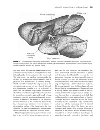
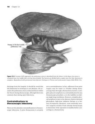
![Minimally Invasive Surgery for Lung Cancer 183
Another contraindication is involvement of the open and minimally invasive groups was equiva-
chest wall by tumor. Although the pulmonary re- lent. It should be noted that these results come from
section may be performed thoracoscopically, the a highly experienced center, and represent a care-
requirement for a chest wall resection would ob- fully selected group of patients. For most surgeons
viate any benefit gained from the thoracoscopic thoracoscopic lobectomy is reserved for peripheral,
approach. Thoracoscopy is certainly useful to de- early-stage tumors, although this may change as ex-
termine if the chest wall is involved, but if so the perience with the technique increases.
procedure should be converted to a thoracotomy.
There are also relative contraindications to thora-
coscopic lobectomy, although these are being chal- Benefits of thoracoscopic
lenged in experienced centers. The first is the pres- lobectomy
ence of a central tumor, in which involvement of the
hilar structures is suspected. Most surgeons would Proponents of thoracoscopic lobectomy claim that
consider these cases inappropriate for VATS for the there are several advantages to the minimally inva-
following reasons: (1) injury to a hilar vessel is more sive approach. Specifically, these are:
likely in this setting, with the possibility of signifi- – equivalent nodal clearance
cant blood loss, and (2) some patients may be spared – decreased pain
a pneumonectomy if a sleeve resection can be per- – lower perioperative morbidity
formed, and this is difficult to perform thoracoscop- – earlier discharge and return to work
ically. – equivalent long-term survival
However, minimally invasive pneumonectomies A detailed review of the literature that supports or
for central tumors have been performed [5]. Ex- challenges these claims follows.
perience with this operation is very limited, in fact
the largest series has only reported seven patients Adequacy of nodal clearance
[6]. In this report, a total of 25 pneumonectomies The benefit of a complete lymphadenectomy versus
were performed over the study period. Only seven nodal sampling for nonsmall cell lung cancer has not
were attempted thoracoscopically, and one was con- been resolved. Hopefully, a multi-institutional trial
verted to a thoracotomy. As such one can only con- sponsored by the American College of Surgeons On-
clude that the operation is technically feasible in a cology Group (ACOSOG protocol Z0030) will pro-
highly selected group of patients. However for most vide definitive data on this issue. What is clear at the
surgeons, even those with significant experience in present time is that mediastinal lymph nodes should
thoracoscopy, a central tumor or bulky adenopathy be adequately sampled during pulmonary resection.
would mandate a thoracotomy. This is important for two reasons: (1) it is critical
In most centers induction therapy is also consid- for prognosis, and (2) decisions regarding adjuvant
ered a contraindication to VATS lobectomy. Reasons therapy are driven by accurate staging. The issue of
for this are twofold: (1) preoperative therapy may adequate sampling is so important that some have
fuse the normal anatomic planes, rendering hilar suggested it be used to measure quality of care in
dissection more hazardous, and (2) many patients thoracic surgery [8].
will have residual nodal disease within the medi- Three prospective trials, all from Japan, have in-
astinum, and this may be difficult to completely re- vestigated the adequacy of nodal sampling during
sect during thoracoscopy. However, one report has thoracoscopic lobectomy. The first is a small trial
documented that thoracoscopic lobectomy can be of 29 patients who underwent thoracoscopic lobec-
safely performed in the setting of induction therapy tomy with mediastinal nodal dissection [9]. What is
[7]. In this series 97 consecutive patients were re- remarkable about this study is that following thora-
viewed, 12 of whom had a thoracoscopic lobectomy. coscopic dissection, a thoracotomy was carried out
There were no significant complications in the tho- by another surgeon and any remaining mediastinal
racoscopic group, and median survival between the lymph nodes were removed. For right-sided tumors,](https://image.slidesharecdn.com/lungcancer3rded-120126215311-phpapp01/85/Lung-cancer-3rd-ed-194-320.jpg)
![184 Chapter 12
40 nodes on average were removed during thora- Decreased pain
coscopy. During open thoracotomy, only one addi- Pain after thoracotomy can be considerable. For sev-
tional lymph node was removed on average. Simi- eral reasons, minimizing this pain may have a sig-
lar results were found for left-sided tumors. Based nificant impact on both morbidity and long-term
on weight and number of nodes, the authors con- quality of life. First, pain related to thoracic inci-
cluded that only 2–3% of nodal tissue was “missed” sions has a significant impact on chest wall me-
with thoracoscopic techniques. Unfortunately, de- chanics [11]. Patients with significant pain have a
spite the excellent design of this study the results reduced functional residual volume, and are more
are not clearly interpretable. This is because it does prone to develop atelectasis, sputum retention, and
not appear that a true “VATS lobectomy” was per- pneumonia. Secondly, regimens to treat postopera-
formed. The authors describe using an 8-cm tho- tive pain are themselves associated with morbidity.
racotomy with a “retractor.” Furthermore, approxi- Opiates, for example, are associated with decreased
mately 50% of the operation was performed under ventilatory drive, hypotension, nausea, and urinary
direct vision, the remainder using the thoracoscope. retention. These side effects persist even when pain
This does not meet the currently accepted definition control is supplemented with an epidural catheter
of a VATS lobectomy, and it is not clear if the same [12]. Perhaps most important, pain in the perioper-
degree of nodal clearance could be achieved using a ative period is a significant risk-factor for the devel-
true minimally invasive approach. opment of chronic postthoracotomy pain syndrome
Two randomized studies have also documented [13]. This syndrome is characterized by a burning
the degree of lymph node clearance that can be pain in the distribution of the thoracotomy incision
achieved during thoracoscopic lobectomy. In the that responds poorly to medication and may have a
first study, 100 patients with clinical stage IA lung significant impact on quality of life.
cancer were randomized to either a conventional Postthoracotomy pain is primarily related to two
or VATS lobectomy [3]. The mean number of hi- issues. The first is trauma to the intercostal nerve
lar and mediastinal nodes removed during open that occurs during rib-spreading [14]. The second is
lobectomy were 8 and 13 respectively, exactly the shoulder dysfunction caused by division of the latis-
same as in the thoracoscopy group. Furthermore, simus dorsi muscle. Thoracoscopy is likely to lead to
an equal number of patients were upstaged to N1 less pain, as the ribs are not spread and the latissimus
or N2 disease in each group. Unfortunately, this muscle is largely preserved.
study has been criticized for not describing the tho- However, the evidence to support these claims is
racoscopic technique in sufficient detail. The tho- largely retrospective. Only three randomized stud-
racotomy incision was 8 cm in size, and the au- ies have reported pain control as an outcome mea-
thors do not state if a rib-spreading retractor was sure. The first study randomized patients to either a
used. thoracoscopic lobectomy without rib-spreading or a
In the second trial, 39 patients were randomized “VATS-assisted lobectomy” with rib-spreading and
to undergo either a “complete VATS lobectomy” (in a large access incision. The design of this trial was
which the access incision was 4 cm in length and discussed previously [10]. In this trial analgesic re-
rib-spreading was not used) or an “assisted VATS quirements were less in the completely thoraco-
lobectomy” (in which the thoracotomy was 10 cm scopic group, although this did not reach statistical
in length and rib-spreading was used) [10]. Given significance ( p = 0.07). Unfortunately, this was the
the length of the thoracotomy and the use of a only measure of pain control that was reported. Nei-
rib-spreading retractor in the “assisted” group, we ther pain measured by a visual analogue scale (VAS)
would argue that this trial really compares the true nor the duration of narcotic use after discharge was
VATS procedure to an open lobectomy technique. In reported. In an earlier trail from the United States,
the complete VATS group 32 nodes were submitted 55 patients were randomized to either a conven-
for pathologic review, compared to 29 in the control tional muscle-sparing thoracotomy or a thoraco-
group ( p = 0.12). scopic lobectomy [15]. Acute pain was not reported](https://image.slidesharecdn.com/lungcancer3rded-120126215311-phpapp01/85/Lung-cancer-3rd-ed-195-320.jpg)
![Minimally Invasive Surgery for Lung Cancer 185
in this study, although the authors did note that pain [23]. By 2 years, only 4% of patients had any
there was no difference in disabling postthoraco- residual discomfort.
tomy pain between the two groups. The final study Shoulder dysfunction is another potential com-
randomized 47 patients with an undiagnosed pul- plication of thoracotomy. Preservation of shoulder
monary nodule to undergo either thoracoscopic or function is necessary for many activities of daily liv-
open surgery [16]. The majority of patients had a ing. The restriction of shoulder function after tho-
wedge resection; only seven had a lobectomy. How- racotomy may delay resumption of full activity, and
ever, we believe that this study is relevant given that could lead to significant long-term disability. To date
rib-spreading was not used in the VATS group and two retrospective and one prospective study have
the incisions were similar in size to those used dur- documented that shoulder dysfunction is less af-
ing VATS lobectomy. Pain as measured by a VAS ter VATS lobectomy. In the first retrospective study,
was significantly lower in the thoracoscopic group shoulder dysfunction was the same in both the VATS
up to 72 hours after surgery. Similarly, narcotic re- and muscle-sparing thoracotomy groups in the first
quirements were less in the VATS group at all time 3 days after surgery. However, shoulder function re-
points. turned to normal within 3 weeks in the VATS group,
Several retrospective studies have also shown whereas it remained significantly impaired in the
that the VATS approach is associated with less pain thoracotomy group [24]. A follow-up study com-
in the immediate postoperative period [17–19]. pared shoulder function in 178 patients who had
While this is important, evidence that VATS could a VATS resection (wedge or lobectomy) to 165 pa-
lessen the incidence of chronic postoperative pain tients who had a thoracotomy [22]. Within the first
would be more significant. Chronic postthoraco- year following surgery 25% of patients in the open
tomy pain is surprisingly common and can be quite group had shoulder dysfunction versus 10% in the
disabling. For example, in a prospective study from VATS group ( p < 0.001).
Finland 61% of patients had pain 1 year after thora- One prospective study has validated these ear-
cotomy [20]. Pain was considered severe in only 5% lier reports. In this report, 29 consecutive patients
of patients, however more than half of the patients who underwent lobectomy through a thoracotomy
said that pain interfered with their daily activities. (n = 11) or VATS (n = 18) were followed [25].
Unfortunately, there is no large study that docu- Shoulder function was measured preoperatively
ments the prevalence of chronic pain following tho- and at 1 week, 1 month, and 3 months following
racoscopic lobectomy. One small study compared surgery. Strength and function were determined by
22 patients who underwent a thoracoscopic lobec- a physiotherapist using the American Shoulder and
tomy versus an equal number who underwent open Elbow Surgeons Standardized Assessment forms.
lobectomy [21]. Patients were surveyed by ques- Shoulder strength and range of motion were found
tionnaire at a mean of 13 months in the VATS group to be significantly improved in the VATS group
and 34 months in the open group. Although the throughout the study period. Importantly, the anal-
follow-up was longer in the open group, four of gesic requirement was also lower in the VATS group,
these patients (18%) still required narcotics for pain and this persisted for up to 1 month after surgery.
control, compared to none in the VATS group. A
similar study from our center concluded that tho- Lower morbidity
racoscopy patients had less pain and narcotic re- The prevalence of complications following open
quirements than those who underwent a thora- lobectomy has been prospectively measured by
cotomy [22]. However, these differences did not the Z0030 trial, a randomized study sponsored by
persist beyond the first postoperative year. Other the American College of Surgeons [26]. In this
studies without a control group have drawn similar study, 1111 patients undergoing thoracotomy and
conclusions. In a large series of 173 VATS patients anatomic resection for lung cancer were random-
(16 had a lobectomy), 75% had no complaints at ized to either lymph node sampling or a com-
6 months after surgery, and no patient had severe plete nodal dissection. Overall, 38% of patients had](https://image.slidesharecdn.com/lungcancer3rded-120126215311-phpapp01/85/Lung-cancer-3rd-ed-196-320.jpg)
![186 Chapter 12
Table 12.1 Morbidity following open and thoracoscopic lobotomy.
Air Length
Author Procedure Evidence Number Mortality leak Arrhythmia MI Pneumonia of stay
Allen Open Prospective, 1111 1.4% 11.5% 14% 1% 2.5% Not Stated
(Z0030 randomized
trial)
McKenna VATS Retrospective 1100 0.8% 5% 3% 1% 1% 3 days
Onaitis VATS Retrospective 500 1.2% 4% 10% 0.4% 5% 3 days
one or more complications following surgery. How- these complications are not life-threatening, they
ever, the majority of these complications were mi- may have a significant impact on length of stay.
nor: atrial arrhythmias (14%) and prolonged air Serious complications such as pneumonia and
leak (11.5%) were the most common. More sig- myocardial infarction are rare after thoracoscopic
nificant complications such as pneumonia (2.5%) lobectomy, although comparable to open series.
and myocardial infarction (1%) were rare. The op- However, outstanding results from dedicated cen-
erative mortality was 1.4%. This multi-institutional ters need to be interpreted with caution. The most
series represents a benchmark against which se- important issue is whether such a low complication
ries of thoracoscopic lobectomy can be compared rate could be achieved in smaller centers without
(Table 12.1). the same degree of experience. The second issue is
The largest VATS lobectomy series was reported whether results from a single center with a few ded-
by McKenna [27]. Among 1100 cases, the opera- icated surgeons can be fairly compared to outcomes
tive mortality was 0.8%. There were no intraoper- from a large randomized trial that enrolled patients
ative deaths and only six patients required conver- from 63 institutions.
sion to a thoracotomy for bleeding. Overall 85% of There is no question that there is a learning curve
patients had no complications. Prolonged air leak associated with thoracoscopic lobectomy. The ap-
(5%) and atrial fibrillation (3%) were the most proach to the hilar structures is different, and the
common complications. Pneumonia and myocar- thoracoscopic view can be disorienting for those
dial infarction occurred in 1% of patients. used to open surgery. In our opinion, at least 30
Similar results were reported from Duke Uni- cases are required before a surgeon experienced in
versity [28]. Among 500 consecutive patients, the open pulmonary resection will be comfortable per-
mortality was 1.2%, and conversion to a thoraco- forming a VATS lobectomy. Experience with other
tomy was required in 1.6% of cases. Atrial fibrilla- minimally invasive procedures, such as thoraco-
tion and pneumonia were the most common com- scopic wedge resection or laparoscopic surgery will
plications, occurring in 10% and 5% of cases, re- shorten this learning curve.
spectively. Prolonged air leak occurred in 4% of The issue of whether outstanding results can
patients. be replicated in smaller centers will only be an-
What is clear from these series is that outstand- swered by a multi-institutional trial. Although the
ing results with very low mortality can be achieved results have not yet been published, such a trial
in dedicated centers. Massive intraoperative bleed- has been concluded by the Cancer and Leukemia
ing is extremely rare, and the conversion rate to Group B (CALGB protocol 93802). This prospective
thoracotomy is low. The incidence of minor com- trial was designed to determine the perioperative
plications such as atrial fibrillation and prolonged morbidity and mortality of patients undergoing
air leak seem to be lower after thoracoscopic lobec- thoracoscopic lobectomy, without an open control
tomy when compared to the Z0030 trial. Although group. A follow-up registry study is planned, in](https://image.slidesharecdn.com/lungcancer3rded-120126215311-phpapp01/85/Lung-cancer-3rd-ed-197-320.jpg)
![Minimally Invasive Surgery for Lung Cancer 187
which surgeons can prospectively enter patients and length of stay: 11.8 days in the completely tho-
who have undergone either an open or thoraco- racoscopic group, 15.3 days in the assisted group,
scopic lobectomy. This study was designed in lieu of and 17.9 days in the open group.
a randomized trial comparing the two procedures. Within the United States the largest series to re-
Unfortunately, it has been estimated that an ad- port length of stay is from McKenna [1]. Among
equately powered randomized study would re- 1100 patients who underwent VATS anatomic
quire approximately 800 patients. Accrual to such resection, the median length of stay was 3 days
a study would be limited by an unwillingness of (mean 4.8 days). Median length of stay was also
centers with experience in thoracoscopic lobectomy 3 days in the series from Duke University [28]. Al-
to randomize patients to an open procedure. A though there is no comparison group, these series
registry study may provide some data to support clearly demonstrate that patients can anticipate a
claims of decreased morbidity following thoraco- short hospital stay when thoracoscopic lobectomy
scopic lobectomy, although issues regarding out- is performed in a dedicated center. It may be argued
comes in smaller centers may remain unanswered. that these short stays reflect efficient postoperative
care and discharge planning as much as the opera-
Earlier discharge and return to work tive approach. However, we would note that a me-
Outside of a randomized trial it is difficult to sub- dian length of stay of 3 days would be most unusual
stantiate claims that length of stay is shorter after in series of open lobectomies.
thoracoscopic lobectomy. This is because length of
stay is not only related to pain control and the inci- Equivalent long-term survival
dence of complications, but also to the practices of Critics of thoracoscopic lobectomy suggest that in-
the institution and country where the procedure is adequate nodal sampling and the potential for port
performed. It is therefore difficult to compare length site contamination by tumor will lead to inferior sur-
of stay from different institutions or from separate vival compared to open lobectomy. However, sev-
time periods within a single institution. However, eral retrospective series and a single randomized
a trend toward earlier discharge has been demon- trial have minimized the significance of these con-
strated by several retrospective series. cerns (Table 12.2).
For example, a comparative study from the Uni- The single randomized trial to report survival
versity of Missouri evaluated 19 patients who had data was published in 2000 from Japan [3]. In this
undergone VATS lobectomy [29]. These patients trial 100 consecutive patients with clinical stage IA
were matched to a cohort of open lobectomy pa- lung cancer were randomized to either a thoraco-
tients on the basis of age, gender, and pulmonary scopic or open lobectomy. The median follow-up
function. In this study length of stay was markedly was 4.9 years. Within this period, 6% of patients
reduced in the VATS group (5.2 versus 12.2 days). in both the thoracoscopic and open group devel-
The median time required to return to full activity oped a local recurrence. The 5-year survival was
was 2.2 months in the VATS group compared to 3.6 85% in the open group and 90% in the thoraco-
months in the open group ( p < 0.01). scopic group ( p = 0.91). Although this study stands
Similar results have been reported from Japan. In as the only randomized study on the topic, it is
the largest study, the outcomes of 90 patients who unfortunately underpowered to document equiv-
underwent VATS lobectomy in three institutions alency between the two procedures. For compari-
were compared with 55 patients treated with an son, the Clinical Outcomes of Surgery Study Group
open lobectomy [10]. The thoracoscopic lobectomy (COST) trial was an adequately powered study to
group was subdivided into those who had a com- evaluate laparoscopic colectomy for colon cancer
pletely thoracoscopic procedure (no rib-spreading) [31]. The primary endpoint was time to recurrence.
and an “assisted” VATS procedure (a small thora- To document equivalency between the open and
cotomy with rib-spreading). There was a clear rela- laparoscopic groups, a sample size of 1200 patients
tionship between the invasiveness of the procedure was deemed necessary.](https://image.slidesharecdn.com/lungcancer3rded-120126215311-phpapp01/85/Lung-cancer-3rd-ed-198-320.jpg)
![188 Chapter 12
Table 12.2 Survival following thoracoscopic lobectomy for lung cancer.
Author Year Evidence Number Mean follow-up Stage 5-yr survival
Shiraishi [36] 2006 Retrospective 81 VATS, 79 open 46 mo IA 89% VATS, 78% open
Shigemura [10] 2006 Retrospective 145 VATS 39 mo IA 96%
McKenna [1] 2006 Retrospective 1015 VATS NR IA-IIIB Stage IA: 80%
Onaitis [2] 2006 Retrospective 416 VATS NR IA-IIIB Stage I: 85% at 2 yr
Iwasaki [35] 2004 Retrospective 140 VATS NR I/II Stage I: 81%, stage II: 70%
Walker [30] 2002 Prospective 158 VATS 38 mo I/II Stage I: 78%, stage II: 51%
Sugi [3] 2000 Prospective, 48 VATS, 52 open NR IA 90% VATS, 85% open
randomized
NR, not reported.
However, two large single-institution series have VATS lobectomy summary
shown that stage-specific survival is comparable
to historical series of open lobectomy. In the No appropriately powered study has documented
McKenna series, 1015 patients underwent thoraco- decreased morbidity and equivalent survival with
scopic lobectomy for nonsmall cell lung cancer. For VATS lobectomy compared to the open approach.
those with pathologic stage IA disease, the 5-year However, smaller studies have documented that
survival was 78%. Similar data was reported in the nodal clearance is equivalent and that periopera-
Duke series (2-year survival for stage I cancer was tive morbidity may be less with the minimally in-
85%). By comparison, Mountain reported survival vasive approach. Certainly, large single-institution
data of 1524 patients treated by open lobectomy in series have shown that the risk of uncontrolled
1997; this data was used to validate the current TNM bleeding during the procedure is extremely rare.
staging system [32]. In that series, the 5-year sur- It is likely that a well-designed randomized study
vival of stage IA patients was only 67%. to substantiate these findings will never be per-
The local recurrence rate was not reported in ei- formed, given the reluctance of surgeons skilled in
ther the McKenna or Duke series. However, this thoracoscopic lobectomy to randomize patients to a
data was provided in some smaller reports. For thoracotomy. A prospective study evaluating VATS
example, in an earlier paper from McKenna of lobectomy in a multi-institutional setting has been
298 patients, recurrence in the incision occurred completed, and a comparative registry study is be-
in one patient (0.3%) [33]. Comparable results ing planned. Currently, only a small percentage of
were reported in a large series from France [34]. lobectomies are performed thoracoscopically in the
In that report, outcomes of 110 patients with stage United States. Positive findings from these studies
I lung cancer treated thoracoscopically were com- may increase this percentage, despite the lack of a
pared with 405 patients resected through a tho- randomized trial.
racotomy. The recurrence rates (distant and local)
were 25% for the VATS group and 23% for the open
group. No patients in either group developed a re- Limited resection for lung cancer
currence at a port site or the thoracotomy incision.
Several other retrospective series have documented Lobectomy with mediastinal node dissection is
similar findings—that overall and recurrence-free considered the standard of care for patients with
survival are equivalent among patients treated by early-stage nonsmall cell lung cancer. Resection of
open and VATS lobectomy [9,35,36]. the primary tumor by less than a lobectomy (i.e.,](https://image.slidesharecdn.com/lungcancer3rded-120126215311-phpapp01/85/Lung-cancer-3rd-ed-199-320.jpg)
![Minimally Invasive Surgery for Lung Cancer 189
anatomic segmentectomy or a wedge resection) is sided statistical test. If a more rigorous two-sided test
considered a “limited resection.” Whether such an was used, the reported survival advantage would
operation is appropriate for any subset of patients not be significant at a p-value of 0.05. Second, only
has been intensely debated for 25 years [37]. The 60% of patients were able to undergo pulmonary
clear benefit of a limited resection is that more pul- function testing at 6 months. It is stressed by critics
monary parenchyma is conserved. Consequently, that the poor follow-up in this study should pre-
it has been argued that a limited resection should clude any conclusions regarding pulmonary func-
be offered to those patients with pulmonary dis- tion from being made.
ease severe enough that a lobectomy is contraindi- Since the Lung Cancer Study Group report, sev-
cated. However, two additional issues have made eral retrospective series have confirmed a higher
this debate more relevant for a larger population local recurrence after limited resections [40–42].
of patients. The first is that screening for lung can- In some of these series overall survival was also
cer in high-risk patients has become more common decreased in patients who underwent limited re-
[38]. As such an increasing number of patients section [40,41]. Some studies have suggested that
with subcentimeter tumors are presenting for re- limited resection may be appropriate for smaller
section. It has been argued that removing the en- tumors. For instance, Warren [40] demonstrated
tire lobe may not offer any survival benefit in these that the survival advantage for lobectomy was only
patients. The other issue is that a thoracoscopic evident if the tumor was greater than 3 cm in size.
wedge resection is technically straightforward. Most However, in another study [42] that only included
thoracic surgeons are very comfortable with this tumors smaller than 1 cm, 5-year survival was still
procedure, although this is not the case for tho- higher after lobectomy than limited resection (71%
racoscopic lobectomy. Therefore, in many centers versus 33%).
the distinction between a wedge resection and a It should be noted that excellent survival fol-
lobectomy is that the former can be performed tho- lowing limited resection have been reported from
racoscopically whereas the latter will mandate a Japan [43–45]. These studies have consistently
thoracotomy. shown that 5-year survival rates above 80% can
The single randomized trial designed to resolve be achieved with limited resection. A uniform con-
this debate was conducted by the Lung Cancer clusion of these studies is that limited resection is
Study Group, and reported in 1995 [39]. In this an acceptable alternative to lobectomy for “selected
study, 276 patients with clinical stage I lung cancer patients.”
were randomized to receive either a lobectomy or Therefore, the critical issue is to determine the cri-
a limited resection (both wedge resection and seg- teria by which patients should be selected for limited
mentectomy were allowed). Although the trial pre- resection. Unfortunately, retrospective series con-
dated the era of high-resolution CT and PET scan- tain significant biases that make it difficult to es-
ning, the study required hilar and mediastinal nodes tablish these criteria. For example, wedge resection
to be sampled before resection to confirm stage I dis- is often reserved for marginal surgical candidates
ease. The authors found no difference in periopera- with multiple comorbidities. Survival in the limited
tive morbidity or mortality between the two groups. resection group will be lower because of comorbid
Also, there was no difference in pulmonary function disease, regardless of the effectiveness of the oper-
at 6 months. Most importantly, the local recurrence ation. Another issue is the potential understaging
rate was three times higher in the limited resection of patients who undergo limited resection. The rate
group, and this was associated with a 50% increase of lymph node metastases in tumors less than 2 cm
in disease-specific mortality. in size is approximately 20% [46,47]. It is certainly
Although the conclusions of this trial would ap- possible that a significant number of patients who
pear definitive, the methodology of the study has undergo wedge resection with limited nodal sam-
been questioned. First, the survival advantage seen pling will be erroneously staged as N0, whereas they
with lobectomy was calculated on the basis of a one- may harbor occult nodal micrometastases. Thus](https://image.slidesharecdn.com/lungcancer3rded-120126215311-phpapp01/85/Lung-cancer-3rd-ed-200-320.jpg)
![190 Chapter 12
reports that claim to be restricted to stage IA pa- (a) 1
tients likely include some with stage II or III disease.
0.8
Cumulative survival
A recent paper from our institution supported this
Lobectomies
concern [48]. Over a 13-year period 784 patients 0.6
with stage I lung cancer were reviewed: 577 un-
derwent a lobectomy and 207 a sublobar resection. 0.4 Limited rsct
Those who underwent limited resection were much
0.2
more likely to have limited nodal sampling: 43% of p = 0.03
patients in the limited group had no nodes sampled 0
compared to 3% in the lobectomy group. Further- 0 10 20 30 40 50 60 70 80
more, a 2% decrease in mortality was observed for Time (mos)
every additional node resected, an observation that (b) 1
likely reflects more accurate staging with increased
nodal sampling. 0.8
Cumulative survival
Another issue that may impact on mortality after
0.6 Lobectomies
limited resection is age. Although limited resection
may have a survival disadvantage, this may not be 0.4
relevant for older patients who have a limited life Limited rsct
expectancy. This hypothesis was examined in a re- 0.2
p = 0.0009
view of 14,555 patients with lung cancer registered 0
in the Surveillance, Epidemiology and End Results 0 10 20 30 40 50 60 70 80
Database [49]. In this study age itself was a signif- Time (mos)
icant predictor of both overall and disease-specific
mortality. For younger patients (less than 65 yr), (c) 1
lobectomy conferred a significant survival advan-
0.8
Cumulative survival
tage over wedge resection. However, for older pa-
tients (75 yr or above) the choice of surgical proce- 0.6
dure had no impact on survival (Figure 12.3). This Lobectomies
0.4
paper suggests that age is a confounding factor in
series that compare limited resection to lobectomy. 0.2 Limited rsct
It also supports the practice of offering wedge re- p = 0.47
section to elderly patients, although this should be 0
individualized. For example, an otherwise healthy 0 10 20 30 40 50 60 70 80
Time (mos)
elderly patient would likely benefit from lobectomy.
On the other hand, an older patient with mul- Figure 12.3 Survival of patients undergoing lobectomy
tiple comorbidities and a limited life expectancy or limited resection based on SEER data. Panel (a):
Patients < 65 years of age; Panel (b): 65–74 years old;
may be appropriately managed with a wedge
Panel (c): > 75 years. As illustrated, lobectomy offers no
resection.
survival benefit for patients older than 75 years of age.
Adjuvant radiation therapy has been suggested (Reproduced with permission from Chest 2005;
as a means to reduce the high local recurrence rate 128:237–45.)
following limited resection. The largest study to in-
vestigate this was a prospective, multi-institutional
trial sponsored by the Cancer and Leukemia Group in thoracoscopy to participate in the trial, although
B (CALGB) [50]. This trial only included patients the manner by which this was established was not
with clinical T1 lesions considered to be “high-risk” specified. However, the results of surgical resection
for lobectomy on the basis of pulmonary function of these small tumors were poor: conversion to tho-
testing. Surgeons were required to show proficiency racotomy occurred in 17% of cases, and resection](https://image.slidesharecdn.com/lungcancer3rded-120126215311-phpapp01/85/Lung-cancer-3rd-ed-201-320.jpg)
![Minimally Invasive Surgery for Lung Cancer 191
margins were positive in 9% of patients. Further- thoracoscopic wedge resection. At the present,
more, a resection margin of more that 1 cm was data on these modalities is from small, single-
present in only 44% of pathologically staged T1 institution series [56]. However, a nonrandomized
lesions and 33% of those with T2 lesions. Fortu- clinical trial has begun accruing patients treated
nately, complications after surgery and radiother- with RFA. This prospective trial, sponsored by
apy were low in this high-risk population: the rates the American College of Surgeons, will document
of perioperative mortality, postoperative respiratory technical complications associated with the proce-
failure, and radiation pneumonitis were each 4%. dure and the rates of local recurrence and overall
The median survival after combined therapy was 27 survival.
months. The authors of this study concluded that In addition, two upcoming randomized trials will
thoracoscopic wedge resection had a high techni- provide more data on the role of limited resec-
cal failure rate, although radiation therapy could be tion and brachytherapy. The first is a trial spon-
administered safely. sored by the CALGB which will randomize patients
Another technique to decrease the risk of local with early-stage lung cancer to either lobectomy or
failure is to combine wedge resection with place- sublobar resection. This study will essentially re-
ment of brachytherapy mesh [51–53]. In contrast to visit the issues raised earlier by the Lung Cancer
external beam radiation, brachytherapy allows radi- Study Group trial. The second is a trial sponsored by
ation to be precisely delivered to the staple line, with the American College of Surgeons Oncology Group
100% patient compliance. At the moment the data (protocol Z4032). The study seeks to enroll patients
to support the use of brachytherapy is retrospec- with compromised pulmonary function and early-
tive. In one series from our center [52] the addition stage lung cancer (less than 3 cm). Patients are ran-
of brachytherapy to sublobar resection was demon- domized to receive sublobar resection with or with-
strated to lower the local recurrence rate from 17% out brachytherapy. The primary endpoint of the trial
to 3%. is local recurrence. Hopefully, the results of these
One issue with limited resections is whether func- trials will allow the treatment of early-stage lung
tional lung tissue is being preserved. In fact, pa- cancer to be guided by the highest levels of clinical
tients with severe emphysema have been shown to evidence.
improve their pulmonary function after lobectomy
[54,55]. In these patients lobectomy is thought to
improve pulmonary mechanics in much the same References
way as long-volume reduction surgery—by improv-
ing diaphragmatic excursion and restoring the elas- 1 McKenna R, Houck W, Fuller C. Video-assisted thoracic
tic recoil of the remaining lung. Although it may be surgery lobectomy: experience with 1,100 cases. Ann
difficult to predict which patients will benefit from Thorac Surg 2006; 81:421–6.
lobectomy, what is clear is that not all patients with 2 Onaitis M, Peterson R, Balderson S. Thoracoscopic
COPD should be consigned to limited resection or lobectomy is a safe and versatile procedure: experience
nonoperative therapy. with 500 consecutive patients. Ann Thorac Surg 2006;
244:420–5.
3 Sugi K, Kaneda Y, Esato K. Video-assisted thoraco-
scopic lobectomy achieves a satisfactory long-term
Future directions
prognosis in patients with clinical stage IA lung can-
cer. World J Surg 2000; 24(1):27–31.
Radiofrequency ablation and stereotactic radio-
4 Kirby T, Mack M, Landreneau R, Rice T. Initial experi-
surgery have both been explored as alternatives ence with video-assisted thoracoscopic lobectomy. Ann
to conventional radiation therapy for high-risk Thorac Surg 1993; 56:1248–52.
patients with early-stage lung cancer. These pa- 5 Conlan A, Sandor A. Total thoracoscopic pneumonec-
tients have such severe cardiopulmonary dysfunc- tomy: indications and technical considerations. J Tho-
tion that they are unlikely to tolerate even a rac Cardiovasc Surg 2003; 126:2083.](https://image.slidesharecdn.com/lungcancer3rded-120126215311-phpapp01/85/Lung-cancer-3rd-ed-202-320.jpg)
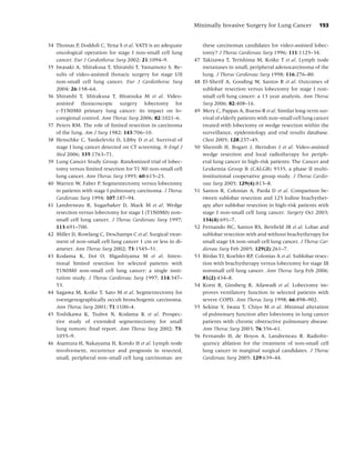
![CHAPTER 13
Extended Resections for Lung Cancer
Philippe G. Dartevelle, Bedrettin Yildizeli, and Sacha Mussot
Introduction surgery after induction therapy poses special peri-
operative risks and technical challenges because the
Lung cancer remains the most fatal cancer world- difference it is often difficult to distinguish between
wide with over 1,300,000 deaths estimated for the sclerotic tissue and tumor margins; moreover the
year 2000 [1,2]. Only 10% of patients with lung “down staging” phenomenon does not occur with a
cancers diagnosed in the European population are high frequency.
alive at 5 years, a figure that compares quite poorly Patients with T4 tumors with N0 or N1 disease
with the 50 and 70% 5-year survival rates for colon should benefit most from surgery because these tu-
and breast cancer, respectively [3]. Using the tu- mors are often more local-regionally than systemi-
mor node metastasis (TNM) classification to direct cally aggressive.
treatment can eliminate some patients from poten- This chapter will try to define the subsets of pa-
tially curative surgery. For example, Stage IIIb in- tients with locally advanced nonsmall cell lung can-
cludes T1N3 which is not curable by surgery as well cer (NSCLC) who are most likely to benefit from
as T4N0 which is sometimes amenable to surgical surgery.
resection and possible long-term survival.
Locally advanced lung cancer encompasses T3 tu-
mors with direct extension into the chest wall, di- Chest wall invasion
aphragm, mediastinal pleura, or within 2 cm of the
carina and nearly all T4 tumors invading the me- Five to eight percent of patients undergoing re-
diastinum, heart, great vessels, trachea, esophagus, section for NSCLC have involvement of the chest
vertebral body, or carina. wall [4]. Patients’ complaints are the most reliable
Extended resections for tumors that are locally indications of chest wall involvement, as infiltra-
advanced according to T status imply that surgical tion between the ribs may result in false-negative
resection is being performed under circumstances bone or computed tomography (CT) scans. It was
that are technically challenging and beyond the claimed that CT scan could be inaccurate in assess-
usual scope of an operation for lung cancer ing direct parietal pleura invasion of lung cancer [5].
(Table 13.1). However, Ratto et al. [6] and Rendina et al. [7] re-
Likewise, the development of preoperative ported that thickening of the pleura is not useful and
chemotherapy or chemoradiotherapy is said to obliteration of the extrapleural fat pad is the most
make curative treatment possible for tumors that sensitive and specific finding in CT scan. Further-
are locally advanced according to N status. However, more, visible rib destruction from direct invasion is
a very specific sign (and a bad prognosis) for chest
Lung Cancer, 3rd edition. Edited by Jack A. Roth, James D. Cox,
wall invasion. Although magnetic resonance imag-
and Waun Ki Hong. c 2008 Blackwell Publishing, ing (MRI) is not routinely used, it has the theoretic
ISBN: 978-1-4051-5112-2. advantage of being able to determine if the muscle
194](https://image.slidesharecdn.com/lungcancer3rded-120126215311-phpapp01/85/Lung-cancer-3rd-ed-205-320.jpg)
![Extended Resections for Lung Cancer 195
Table 13.1 New technical advances in T4 tumors are mandatory to confirm completeness of the
surgery. resection.
Majority of the chest wall resections do not re-
Vascular resection/reconstruction
quire prosthetic reconstruction. Resection of a por-
Tracheal/carinal resection
Anterior approach to thoracic inlet tumors tion of three or fewer ribs posteriorly rarely requires
Vertebrectomy prosthetic replacement, as the scapula lessens the
Safety of surgery after induction chemoradiation therapy cosmetic and functional impact of the chest wall re-
section. Resection of larger defect, especially when
located anterolateral aspects of the lower ribs, may
require prosthetic replacement, yet the risks of in-
layers are involved. Positron emission tomography fection should be balanced against the cosmetic and
(PET) is useful for detecting distant metastases but functional benefit of prosthetic replacement.
is not useful for determining local invasion because Reconstruction can be accomplished using non-
the resolution is not adequate to determine chest reinforced materials like Marlex mesh or Gore-
wall invasion. McCaughan et al. [8] reported an el- tex patch. The advantage of Marlex mesh (Bard
evated serum alkaline phosphatase level in 34% of Inc.), over Gore-tex patch (W. L. Gore and Assoc.,
the patients; however, this abnormality is not a spe- Flagstaff), is that it allows the ingrowth of the sur-
cific finding. Pulmonary function tests and quanti- rounding tissue and remains rigid over time [9]. For
tative perfusion lung scan are necessary to assess the small defects, the Marlex mesh is doubled cross-
patient’s ability to withstand operation and whether wise at a 90◦ angle for added strength, and tailored
the paradox motion of the residual wall requires sta- and sutured to the edges of the defect with non-
bilization. absorbable sutures. With larger and unsupported
The goals of surgery are to completely resect the defects, the chest wall rigidity can be obtained by
primary tumors with clear surgical margins and utilizing methylmethacrylate between two layers of
maintain a normal respiratory physiology by restor- Marlex mesh as described by Eschapasse et al. [9]
ing the rigidity of the chest wall and resected soft and McCormack et al. [10].
tissue. Knowledge of chest wall invasion preopera- In recent years, we adapted a more anatomical re-
tively is important because entering the chest at a construction, which is less prone to infection [11].
site remote from the chest wall invasion lessens the Following the chest wall resection, the Marlex mesh
risk of tumor spillage, allows the surgeon to assess is anchored to the surrounding tissues so that it re-
the extent of involvement, and avoids placement of mains beneath the ribs; 28-Fr silicone chest wall
the prosthetic material directly beneath the incision. tubes are then tailored so that they can be inter-
As a general rule, all tumors except those invad- posed between the healthy portion of the previously
ing the thoracic inlet or the anterior thoracic cage resected rib to cover the Marlex mesh. The methyl-
are approached through a standard posterolateral methacrylate is then spread into chest tubes, and
thoracotomy. Resection should include at least one while it becomes settled, the tubes are shaped ac-
segment of rib (with the related intercostal muscle) cording to the course of the resected ribs. Once hard
above and below the involved rib(s) and 3–5 cm and cool enough, the tube edges are telescoped and
laterally and medially. To prevent tumor spillage, fixed with nonabsorbable sutures to the edges of the
the entire tumor-bearing area should be resected en corresponding ribs. Presently, chest wall reconstruc-
bloc, and it is frequently easier to do the chest wall tion rarely requires the interposition of a myocuta-
resection initially (small involvements) and then neous flap. As with all other prosthetic substitutes,
proceed with the pulmonary resection. For large absolute sterility is required, and the amount of air
involvements, it is easier to do a wedge excision leaks should be minimal.
of the tumor-bearing area with a mechanical sta- All T3 tumors are resectable, but the prognosis
pler and to resect the remainder of the collapsed varies according to the involved site. A T3 tumor in-
lobe later. Frozen sections on the soft-tissue margins volving the chest wall provides the most favorable](https://image.slidesharecdn.com/lungcancer3rded-120126215311-phpapp01/85/Lung-cancer-3rd-ed-206-320.jpg)
![196 Chapter 13
Table 13.2 Results after complete resection of NSCLC invading the chest wall.
Operative
Survival rates, 5 yr (%)
Number of mortality
Author [ref.] Year patients (%) Overall NO N1 N2
Piehler et al. [12] 1982 66 15.2 32.9 54.0 7.4∗ 7.4∗
Patterson et al. [13] 1982 35 8.5 38.0 NS NS 0.0
McCaughan et al. [8] 1985 125 4.0 40.0 56.0 21.0∗ 21.0∗
Ratto et al. [6] 1991 112 1.7 NS 50.0 25.0 0.0
Allen et al. [14] 1991 52 3.8 26.3 29.0 11.0 NS
Shah and Goldstraw [15] 1995 58 3.4 37.2 45.0 38.0 0.0
Downey et al. [16] 1999 175 6.0 36.0 56.0 13.0 29.0
Facciolo et al. [17] 2001 104 0.0 61.4 67.0 100.0 17.0
Magdeleinat et al. [18] 2001 201 7.0 21.0 25.0 21.0 20.0
Burkhart et al. [19] 2002 94 6.3 38.7 44.0 26∗ 26.0∗
Chapelier et al. [20] 2000 100 1.8 18 22 9 0
Riquet et al. [21] 2002 125 7 22.5 30.7 0 11.5
Roviaro et al. [22] 2003 146 0.7 NS 78.5 7.2∗ 7.2∗
Matsuoka et al. [23] 2004 97 NS 34.2† 44.2 40.0 6.2
∗
N1 and N2 patients combined.
†
Complete resection.
NS, not stated.
prognosis among the resected T3 lesions. If com- with chest wall infiltration. Current trend for this
pletely excised, T3 (chest wall) N0 lung cancers pro- decision depends on the extent of the tumor. When
vide a 5-year survival in excess of 50% (Table 13.2). only flimsy adhesions are found, these can be safely
The strongest determinants of 5-year survival, by divided without a problem. When there is a ques-
far, are completeness of resection, depth of chest tion of whether the tumor invades into the chest
wall invasion, and nodal status. Patients with in- wall, however, an extrapleural resection is not ad-
complete resection may survive less than 2.5 years equate and only an en bloc chest wall resection
[6,8,12–23]. Likewise, the depth of chest wall inva- should be performed. We believe that when a lung
sion affects prognosis, as extension to the parietal cancer invades at least the parietal pleura, a wide re-
pleura only is associated with twofold increase of section of the chest wall with attached lung should
5-year survival (62% versus 35%) when compared be performed [20].
to deeper involvements [8]. Different opinions exist Most if not all series report no 5-year survivors
as to whether tumors confined to the parietal pleura with positive N2, compared to 5-year survival ex-
can be resected by simple extrapleural mobilization, ceeding 50% for N0 patients. In this sense, both PET
without resecting en bloc the adjacent soft and bony scan and mediastinoscopy is advocated for patients
tissues, as long as the resection margins are nega- with chest wall involvement and enlarged lymph
tive. While McCaughan et al. [8] showed that ex- nodes. If patients are found to have N2 disease be-
trapleural mobilization was sufficient for a signifi- fore thoracotomy, they should receive either preop-
cant number of patients whose tumors invaded the erative chemotherapy or chemoradiotherapy, either
parietal pleura only, Piehler et al. [12] reported a as induction or definitive treatment.
high incidence of local recurrence after extrapleural The final area of controversy is whether radiation
dissection for tumors invading the parietal pleura. therapy, administered either pre- or postoperatively,
Chapelier et al. [20] also found that patients with is indicated in patients who have lung cancer that
a tumor infiltration confined to the parietal pleura invade the chest wall. Potential benefits of preop-
had a significantly better 5-year survival than those erative therapy include the following: downstaging](https://image.slidesharecdn.com/lungcancer3rded-120126215311-phpapp01/85/Lung-cancer-3rd-ed-207-320.jpg)
![Extended Resections for Lung Cancer 197
the tumor, allowing potentially unresectable tumors anterior aspect; the subclavian artery with its pri-
to be resected, decreasing the rate of close margins, mary branches, except the posterior scapular artery;
and decreasing the risk of tumor spillage at the time and the trunks of the brachial plexus and middle
of resection [24]. However, recent reports showed scalene muscle (Figure 13.1). As the tumor involves
decreased survival rates in those patients who re- the brachial plexus, symptoms develop in the dis-
ceived radiation therapy [14,18]. Currently, radio- tribution of T1 (ulnar distribution of the arm and
therapy is proposed to reduce the incidence of local elbow) and C8 nerve roots (ulnar surface of the fore-
recurrence, and should be reserved for patients with arm and small and ring fingers).
close surgical margins, or those with hilar or medi- Tumors lying posterior to the middle scalene
astinal nodal involvement. Adjuvant chemotherapy muscles are usually located in the costovertebral
had no apparent effect on survival, but the num- groove and invade the nerve roots of T1, the pos-
ber of patients was too small to obtain statistically terior aspect of the subclavian and vertebral arter-
meaningful data. ies, paravertebral sympathetic chain, inferior cer-
vical (stellate) ganglion, and prevertebral muscles.
Some of these posterior tumors can invade trans-
Superior sulcus tumors verse process indeed the vertebral bodies (only
abutting the costovertebral angle or extending into
Superior sulcus lesions include a constellation of be- the intraspinal foramen without intraspinal exten-
nign or malignant tumors extending to the supe- sion may yet be resected). Because of the periph-
rior thoracic inlet. They cause steady, severe, and eral location of these lesions, pulmonary symptoms,
unrelenting shoulder and arm pain along the dis- such as cough, hemoptysis, and dyspnea, are un-
tribution of the eighth cervical nerve trunk and common in the initial stages of the disease. Abnor-
first and second thoracic nerve trunks. They also mal sensation and pain in the axilla and medial
cause Horner’s syndrome and weakness and atro- aspect of the upper arm in the distribution of the
phy of the intrinsic muscles of the hand, a clin- intercostobrachial (T2) nerve are more frequently
ical entity known as Pancoast–Tobias syndrome. observed in the early stage of the disease process.
Bronchial carcinoma represents the most frequent With further tumor growth, patients may present
cause of superior sulcus lesions. Superior sulcus le- with full-blown Pancoast’s syndrome.
sions of nonsmall cell histology account for less than Superior sulcus tumors are extremely difficult to
5% of all bronchial carcinomas. These tumors may diagnose at initial presentation. The time elapsed be-
arise from either upper lobe and tend to invade the tween the onset of the Pancoast–Tobias syndrome
parietal pleura, endothoracic fascia, subclavian ves- and diagnosis is still around 6 months. These pa-
sels, brachial plexus, vertebral bodies, and first ribs. tients usually present with small apical tumors that
However, their clinical features are influenced by are hidden behind the clavicle and the first rib on
their location. Tumors located anterior to the ante- routine chest radiographs. The diagnosis is estab-
rior scalene muscle may invade the platysma and lished by history and physical examination, bio-
sternocleidomastoid muscles, external and anterior chemical profile, chest radiographs, bronchoscopy
jugular veins, inferior belly of the omohyoid mus- and sputum cytology, fine-needle transthoracic or
cle, subclavian and internal jugular veins and their transcutaneous biopsy and aspiration, and CT of the
major branches, and the scalene fat pad. They in- chest. If there is evidence of mediastinal adenopa-
vade the first intercostal nerve and first rib more thy on chest radiographs, computed tomographic
frequently than the phrenic nerve or superior vena scanning or PET scan, histological proof is manda-
cava (SVC), and patients usually complain of pain tory because patients with clinical N2 disease are
distributed to the upper anterior chest wall. not suitable for operation. Neurologic examination,
Tumors located between the anterior and mid- MRI, and electromyography delineate the tumor’s
dle scalene muscles may invade the anterior sca- extension to the brachial plexus, phrenic nerve,
lene muscle with the phrenic nerve lying on its and epidural space. Vascular invasion is evaluated](https://image.slidesharecdn.com/lungcancer3rded-120126215311-phpapp01/85/Lung-cancer-3rd-ed-208-320.jpg)
![198 Chapter 13
Figure 13.1 A left superior sulcus
bronchial carcinoma invading the
middle thoracic inlet, including the
subclavian artery.
by venous angiography, subclavian arteriography, cure, optimal management for superior sulcus tu-
Doppler ultrasonography (cerebrovascular disor- mors continues to be a major challenge. The tra-
ders may contraindicate sacrifice of the vertebral ditional approach to superior sulcus tumors has
artery), and MRI. Magnetic resonance imaging has been preoperative radiotherapy followed by resec-
to be performed routinely when tumors approach tion, although this standard was established 45
the intervertebral foramina to rule out invasion of years ago solely on the basis of encouraging short-
the extradural space. term survival as compared with historical controls
The initial evaluation also includes all preopera- [25]. Then, high-dose curative primary radiother-
tive cardiopulmonary functional tests routinely per- apy [26], “sandwich” preoperative and postoper-
formed before any major lung resection and inves- ative radiotherapy [27], postoperative radiother-
tigative procedures to identify the presence of any apy alone [28], or intraoperative brachytherapy
metastatic disease. combined with preoperative radiation therapy and
Although it is now established that radical surgery operation [29] have been reported as the treat-
represents the only hope for long-term survival and ment modalities of superior sulcus tumors. In 2001,](https://image.slidesharecdn.com/lungcancer3rded-120126215311-phpapp01/85/Lung-cancer-3rd-ed-209-320.jpg)
![Extended Resections for Lung Cancer 199
SWOG 9416 (Intergroup 0160) [30], evaluated the rior transcervical approach as described by Dartev-
role of induction chemoradiotherapy and surgery elle and colleagues [28]. This operative procedure
for patients with superior sulcus tumors in multi- is increasingly accepted as a standard approach for
institutional setting and updated their results in all benign and malignant lesions of the thoracic in-
2003 [31] for the treatment of these tumors. And let structures, including nonbronchial cancers (e.g.,
then, preoperative concurrent chemotherapy and osteosarcomas of the first rib and tumors of the
radiotherapy has been explored by several other brachial plexus), and for exposing the anterolat-
groups [32,33]. The rate of complete resection was eral aspects of the upper thoracic vertebrae. Con-
92% as opposed to an average of 66% among his- traindications to this approach include extrathoracic
torical series of conventional treatment [30,34]. metastasis, invasion of the brachial plexus above the
The consistency of the data regarding preoperative T1 nerve root, invasion of the vertebral canal and
chemoradiotherapy and regarding preoperative ra- sheath of the medulla, massive invasion of the sca-
diotherapy alone is convincing that preoperative lene muscles and extrathoracic muscles, mediastinal
chemoradiotherapy represents a new standard of lymph node metastasis, and significant cardiopul-
care for patients with Pancoast tumors. Although monary disease.
no randomized data are available comparing these Our technique of anterior transcervical approach
approaches [35]. As to whether surgery should pro- has been reviewed in detail elsewhere [36]. Only
ceed or follow radiation therapy in newly diag- some specific points are presented herein. Follow-
nosed superior sulcus tumors, our strong opinion ing double-lumen endotracheal intubation, the pa-
is to first resect, because dissecting on a previously tient is in the supine position with the neck hy-
(chemo)irradiated thoracic inlet unquestionably in- perextended and the head turned away from the
creases the technical difficulties and postoperative involved side. An L-shaped cervicotomy incision
morbidity. Radiation therapy is to be discussed in is made, including a vertical presternocleidomas-
the postoperative course. toid incision carried horizontally below the clav-
Absolute surgical contraindications in the man- icle up to the deltopectoral groove (Figure 13.2).
agement of superior sulcus tumors are the presence
of extra-thoracic sites of metastasis, histologically
confirmed N2 disease, extensive invasion of the cer-
vical trachea, esophagus and the brachial plexus
above the T1 nerve root; this because it indicates
that the tumor is locally too extensive to achieve a
complete resection or that limb amputation is nec-
essary. Invasion of the subclavian vessels should
no longer be considered a surgical contraindica-
tion. Massive vertebral invasion, diagnosed preop-
eratively, is synonymous with unresectability. Inva-
sions limited to the intervertebral foramen without
extension into the spinal canal are resectable.
As a general rule, superior sulcus tumors not in-
vading the thoracic inlet are completely resectable
through the classic posterior approach of Shaw and
associates [25] alone. Because the posterior ap-
proach does not allow direct and safe visualiza-
tion, manipulation, and complete oncologic clear-
ance of all anatomic structures that compose the
thoracic inlet, superior sulcus lesions extending to
the thoracic inlet should be resected by the ante- Figure 13.2 Anterior transcervical approach.](https://image.slidesharecdn.com/lungcancer3rded-120126215311-phpapp01/85/Lung-cancer-3rd-ed-210-320.jpg)
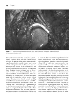
![Extended Resections for Lung Cancer 201
Figure 13.4 End-to-end anastomosis after freeing the carotid and subclavian arteries.
lobectomy, the chest wall resection is completed. It is costoclavicular ligaments rather than the sim-
through this cavity that an upper lobectomy can be ple sternoclavicular disarticulation. Clavicular os-
performed to complete the operation, although it is teosynthesis can then be accomplished by placing
technically demanding. Unlike our original descrip- metallic wires across the lateral clavicular edges and
tion [28], it has become evident that an additional across the divided manubrium.
posterior thoracotomy is usually not required. We also developed a technique for resecting pos-
There is increasing concern about the functional teriorly located superior sulcus tumors extending
and esthetic benefit of preserving the clavicle. We into the intervertebral foramen without intraspinal
believe that the indications for preserving and re- extension in collaboration with a spinal surgeon
constructing the clavicle are limited to the combined [37]. The underlying principle is that one can per-
resection of the serratus anterior muscle and the form a radical procedure by resecting the interver-
long thoracic nerve because this causes the scapula tebral foramen and dividing the nerve roots inside
to rotate and draw forward. This entity (scapula the spinal canal by a combined anterior transcervi-
alata), combined with the resection of the inter- cal and posterior midline approach. After division
nal half of the clavicle, pushes the shoulder ante- of the ipsilateral hemivertebral bodies, the speci-
riorly and medially and leads to severe cosmetic men is resected en bloc with the lung, ribs, and ves-
and functional discomfort. If this circumstance is sels through the posterior incision (Figure 13.5). On
anticipated, we recommend an oblique section of the side of the tumor, spinal fixation is performed
the manubrium that fully preserves the sternoclav- from the pedicle above to the pedicle below the re-
icular articulation, its intra-articular disc, and the sected hemivertebrae; on the contralateral side, a](https://image.slidesharecdn.com/lungcancer3rded-120126215311-phpapp01/85/Lung-cancer-3rd-ed-212-320.jpg)
![202 Chapter 13
Figure 13.5 Right-sided apical tumor
involving the costo-transverse space
and intervertebral foramen and part of
the ipsilateral vertebral body; this
tumor is first approached anteriorly
and then the operation is completed
through a hemivertebrectomy
performed through the posterior
midline approach. (Adapted from
Dartevelle et al. [28].)
screw is placed in each pedicle. However, the pres- ina with a 5-year and median survival rates of 20%
ence of an anterior spinal artery penetrating the and 27 months, respectively. Among the adverse
spinal canal through an invaded intervertebral fora- prognostic factors, the nodal status is the only pre-
men may contraindicates surgery. Tumors involving dictor of disease-free survival.
transverse processes should be resected with the an-
terior approach. The maneuver is similar to what
is used with the posterior approach but from the
front to the back, with a finger placed behind the Carinal resections
transverse process of T1 and T2 to give the correct
direction of the chisel. Refinement in techniques of tracheal surgery and
The reported surgical morbidity ranges from 7 bronchial sleeve lobectomy has made carinal re-
to 38% with surgical mortality generally around section and reconstruction possible. However, the
5–10% [38]. Surgical complications include spinal potential for complications remains high and few
fluid leakage, Horner’s syndrome and nerve deficits, centers only have cumulated sufficient expertise to
hematothorax, chylothorax, and prolonged ventila- safely perform the operation. Surgery is still infre-
tory support due to atelectasis because of the con- quently proposed because of its complexity and the
comitant extended chest wall resection and phrenic paucity of data demonstrating benefit in the long
nerve resection. term. However, results from recent series demon-
The overall 5-year survival rates after combined strate that carinal resection is safe in experienced
radiosurgical (posterior approach) treatment of su- centers with an operative mortality of less than 10%
perior sulcus tumors due to bronchial carcinoma and can be associated with good to excellent long-
range from 18 to 56% (Table 13.3). The best progno- term survival in selected patients. The current re-
sis is found in patients without nodal involvement sults are considerably better than those from earlier
who have had a complete resection. We reported a reported series and likely accounts for the improve-
complete resection rate of 100% with no postoper- ment in surgical and anesthetic techniques.
ative mortality or major complications. The 5-year Careful patient selection and detailed evaluation
and median survival rates were approximately 35% of the lesion is a key component to good surgi-
and 18 months, respectively. The local recurrence cal results in carinal resection. All patients should
rate was less than 1.8% using our approach. Fadel be evaluated to ascertain that they can tolerate the
et al. [37] reported 17 en bloc resections of NSCLCs operation and withstand the necessary removal of
invading the thoracic inlet and intervertebral foram- pulmonary parenchyma. The preoperative workup](https://image.slidesharecdn.com/lungcancer3rded-120126215311-phpapp01/85/Lung-cancer-3rd-ed-213-320.jpg)
![Extended Resections for Lung Cancer 203
Table 13.3 Results of patients treated
surgically for superior sulcus tumors. Number of 5-yr survival
Author (year) cases (%) Mortality (%)
Paulson [39] (1985) 79 35 3
Anderson et al. [40] (1986) 28 34 7
Devine et al. [41] (1986) 40 10 8
Miller et al. [42] (1979) 36 31 NS
Wright et al. [43] (1987) 21 27 —
Shahian et al. [27] (1987) 18 56 —
McKneally [44] (1987) 25 51 NS
Komaki et al. [26] (1990) 25 40 NS
Sartori et al. [45] (1992) 42 25 2.3
Maggi et al. [46] (1994) 60 17.4 5
Ginsberg et al. [47] (1994) 100 26 4
Okubo et al. [48] (1995) 18 38.5 5.6
Dartevelle [49] (1997) 70 34 —
Martinod et al. [50] (2002) 139 35 7.2
Alifano et al. [51] (2003) 67 36.2 8.9
Goldberg et al. [52] (2005) 39 47.9 5%
Total 807 34.5 ± 11.7 5.6 ± 2.2
Values are number ± standard deviation.
NS, not stated.
consists of chest radiography, chest CT scan, pul- cavography is performed if the SVC is potentially in-
monary function tests, arterial blood gas, ven- volved. Transesophageal echography is occasionally
tilation/perfusion scan, electrocardiography, and performed to evaluate tumor extension to the pos-
echocardiography. Stress thallium studies, maxi- terior mediastinum, especially the esophagus or the
mum oxygen uptake, and exercise testing are used left atrium.
when indicated. The operation is an elective pro-
cedure and efforts should be made to prepare Indications and contraindications
the patients for surgery with chest physiotherapy, The safe limit of resection between the lower tra-
deep breathing, and cessation of smoking. Airway chea and the contralateral main bronchus is usually
obstruction, bronchospasm, and intercurrent pul- considered to be 4 cm. This is particularly important
monary infection should be reversed. Steroids if a right carinal pneumonectomy is performed and
should be discontinued before surgery. the left mainstem bronchus is to be reanastomosed
Flexible or rigid bronchoscopy is crucial to evalu- end-to-end to the distal trachea. Upward mobiliza-
ate the overall length of the tumor, the adequacy tion of the left mainstem bronchus is limited because
of the remaining airway, and the feasibility of a of the aortic arch and can easily result in excessive
tension-free anastomosis. Besides routine investiga- anastomotic tension.
tion to rule out extrathoracic metastasis for patients In patients with bronchogenic carcinoma, cari-
with bronchogenic carcinoma, we also routinely nal resection should be considered for tumors in-
perform a mediastinoscopy at the time of surgery in vading the first centimeter of the ipsilateral main
patients presenting with bronchogenic carcinoma to bronchus, the lateral aspect of the lower trachea,
exclude N2 or N3 disease. the carina, or the contralateral main bronchus. This
Pulmonary angiography is performed for carinal applies usually for right-sided tumor, since left-sided
tumors arising from the anterior segment of the tumor rarely extend up to the carina without mas-
right upper lobe, because invasion of right upper sively invading structures situated in the subaortic
lobe (mediastinal) artery usually indirectly reveals space. The long-term results of carinal resection for
invasion of the posterior aspect of the SVC. Superior patients with bronchogenic carcinoma and N2 or N3](https://image.slidesharecdn.com/lungcancer3rded-120126215311-phpapp01/85/Lung-cancer-3rd-ed-214-320.jpg)
![204 Chapter 13
disease is poor, and therefore the findings of positive ing a left pneumonectomy, can provide access to a
mediastinal nodes at the time of mediastinoscopy is cervical collar incision for laryngeal or suprahyoid
usually considered a contraindication to surgery. In- release procedure, and affords access to both a right
duction therapy may be offered for these patients, and a left pulmonary hilar release if it is found to be
but we have found that this increases the techni- necessary intraoperatively.
cal difficulty of the operation and is associated with For carinal resection with sacrifice of pulmonary
greater operative mortality, particularly if carinal parenchyma approach depends on the lung con-
pneumonectomy is required. cerned by resection. On the right side, a right pos-
terolateral thoracotomy in the fifth intercostal space
Surgical technique
gives perfect exposure of the lower trachea and the
Our technique of carinal resection has been re-
origin of both main bronchi. On the left side, expo-
viewed in detail elsewhere [53,54]. Only some spe-
sure of the lower trachea and right main bronchus is
cific points are presented herein. Ventilation during
hindered by the aortic arch that is why the median
carinal resection has always been a major concern.
sternotomy is our preferred approach for left carinal
Our technique is similar to Grillo et al. [55]. The
pneumonectomy. It provides superb exposure to the
patient is initially intubated with an extra-long ar-
tracheobronchial bifurcation, causes less incisional
mored oral endotracheal tube that can be advanced
discomfort and results in less ventilatory restriction
into the opposite bronchus if one-lung ventilation
than a thoracotomy. The main disadvantages are
is desired. Once the carina has been resected, the
that freeing pleuroparietal adhesions can be diffi-
opposite main bronchus is intubated with a cross-
cult and mobilization of the left hilum requires car-
field sterile endotracheal tube connected to a ster-
diac retraction that may cause some hemodynamic
ile tubing system. The tube can be safely removed
instability.
intermittently to place the sutures precisely. Any
blood spillage into the contralateral lung should be
Type of carinal resection
carefully suctioned to preserve the lung. Once the
trachea and the bronchus are ready to be approxi-
Carinal resection without pulmonary
mated, the cross-field tube is withdrawn, and ven-
resection
tilation resumed with the original oral tube once
Carinal resection without pulmonary resection is
the anastomosis is completed. If a secondary end-
limited to the tumors located at the carina or at the
to-side anastomosis is required between the other
origin of the right or left main bronchus. Depend-
bronchus and the lateral trachea, the oral tube is
ing on the extent of invasion, different modes of re-
advanced across the first anastomosis and ventila-
construction exist. For very small tumors implanted
tion can proceed uninterrupted until the secondary
on the carina only, the medial wall of both main
anastomosis is completed.
bronchi can be approximate together to fashion a
Approaches new carina that is then anastomosed to the trachea
The incision varies according to the type of cari- (Figure 13.6). The main problem with this recon-
nal resection. Carinal resection without sacrifice of struction is that the “neocarina” has very limited
pulmonary parenchyma is approached through a mobility because of the aortic arch and therefore
median sternotomy. The pericardium is opened an- the trachea needs to be pulled down to the newly
teriorly and the tracheobronchial bifurcation is ex- created carina.
posed between the SVC and the ascending aorta. When the tumor is more extensive, requiring a
The exposure is facilitated by completely mobiliz- larger portion of the trachea to be resected, end-
ing the ascending aorta and both main pulmonary to-end plus end-to-side anastomosis is the method
arteries. The ligamentum arteriosum is systemati- of choice. Various methods of reconstruction have
cally sectioned. As previously reported by Pearson been described according to the length of resection
et al. [56], we find that this approach offers several of the trachea, right and left main bronchi. The tech-
advantages over a right posterolateral thoracotomy. nique described by Barclay et al. [57] involves end-
It allows any type of pulmonary resection, includ- to-end anastomosis between the trachea and right](https://image.slidesharecdn.com/lungcancer3rded-120126215311-phpapp01/85/Lung-cancer-3rd-ed-215-320.jpg)
![Extended Resections for Lung Cancer 205
(a) (b) (c)
Figure 13.6 Carinal resection with “neo-carina” recon- pleted, sutures for anastomosis between the trachea and
struction. (a) Carinal lesion involving little of the trachea. the bronchial circumference are placed. The joined main
Resection lines are indicated. (b) The medial walls of the bronchi are treated as a single unit. The anterior mattress
right and left main bronchi are approximated with inter- suture at the confluence of the trachea and both bronchi
rupted 4–0 PDS sutures to form a new carina. Note the is shown. (c) After all anastomotic sutures are placed, the
midlateral traction sutures in the lateral walls, one ring paired lateral traction sutures are tied on each side simulta-
distant from the cut edges. After the neocarina is com- neously. Note the mattress suture in the anterior midpoint.
main bronchus with end-to-side anastomosis of the anterior surface of the lower trachea while preserv-
left main bronchus across the mediastinum into ing the lateral blood supply as much as possible.
the bronchus intermedius (Figure 13.7). This recon- Umbilical tapes are passed around the distal trachea
struction is possible only if the right main bronchus and contralateral main bronchus. The hilum and
is left sufficiently long, but presents difficult access esophagus are then dissected and the esophagus
for the end-to-side anastomosis, often requiring hy- is retracted posteriorly. If there is no SVC involve-
poventilation of the right lung. Grillo et al. [55] de- ment, the pulmonary artery and veins are stapled
scribed anastomosing the left main bronchus into at their extrapericardial origin in order to have
the lateral wall of the trachea after an end-to-end the lung attached by the main bronchus only.
anastomosis between the trachea and the right main Subsequently, the cross-field intubation system
bronchus. This technique has rare indication and is is installed. The trachea and contralateral main
technically very demanding. In our experience, the bronchus are divided by sharp, straight transection
right main bronchus can usually be anastomosed to lines. The trachea is always sectioned first to provide
the lateral wall of the trachea after adequate release better exposure to section the left main bronchus.
maneuvers, regardless of the residual length of the In order to accomplish a tension-free anastomosis,
right main bronchus (Figure 13.8). it is crucial to limit the length of resection between
the distal trachea and left main bronchus to less
Right carinal pneumonectomy than 4 cm. Frozen section are obtained on the
Right carinal pneumonectomy is the most frequent tracheal and bronchial margins. The decision to
type of carinal resection for bronchogenic carci- resect further trachea or bronchus or leave residual
noma. No irrevocable step should be taken until tumor at the bronchial margin in case of positive
resection is certain. After division of the azygos vein, margins is balanced by the necessity to perform
the tracheobronchial bifurcation is gently mobilized a tension-free anastomosis. Enlarged subcarinal
and dissected. Dissection should be limited to the nodes can be resected, but otherwise the soft tissue](https://image.slidesharecdn.com/lungcancer3rded-120126215311-phpapp01/85/Lung-cancer-3rd-ed-216-320.jpg)
![206 Chapter 13
Figure 13.7 Barclay technique;
resection of the carina and a
significant length of the trachea. Right:
The length of trachea resected (dotted
lines) exceeds 4 cm. The trachea and
left main bronchus will not
approximate safely. Left: The elevated
right main bronchus is anastomosed to
the trachea after intrapericardial hilar
mobilization. The left main bronchus
is then anastomosed to the medial
wall of the bronchus intermedius.
around the carina should be preserved as much as intermedius is then transected below the take off
possible to ensure adequate vascularization of the of the right upper lobe bronchus. After completing
anastomosis and good lymphatic drainage for the the anastomosis between the trachea and the left
contralateral lung. After completing the anastomo- main bronchus, the bronchus intermedius is anas-
sis, the endotracheal tube is pulled back a sufficient tomosed 1 cm below the initial anastomosis to the
distance from the suture line to avoid any damage left main bronchus (Figure 13.9). We do not suggest
from the tip of the tube and the anastomosis is to perform an anastomosis of the bronchus inter-
checked for airtightness. The anastomosis is then medius above the initial anastomosis of the trachea
covered by the surrounding tissue. and left main bronchus (Figure 13.9b). Mobiliza-
If segmental resection of the SVC is planed, the tion of the pulmonary ligament and a right hilar
vascular procedure is usually performed before di- release is always required to limit the tension on
vision of the airway. The SVC is clamped proxi- the anastomosis. Occasionally, the bronchus inter-
mally at the confluence of the brachiocephalic veins medius can be anastomosed to the lateral wall of
and distally at the cavoatrial junction, and divided the trachea if the tension on this anastomosis is not
on each side of the tumor. Section of the SVC excessive.
facilitates exposure and stapling of the right pul-
monary artery in the interaorto-caval groove. The Left carinal pneumonectomy
SVC is reconstructed with a ring-less straight 18- or The aortic arch greatly hinders performance of
20-sized polytetrafluoroethylene (PTFE) graft. The the anastomosis in left carinal pneumonectomy
PTFE graft is protected with gauze soaked in beta- and renders the procedure technically challeng-
dine during reconstruction of the airway to prevent ing through a left thoracotomy. A one-step proce-
graft contamination. dure with mobilization of the aortic arch should,
however, be preferred to a two-stage approach in
Carinal resection with lobar resection which a left proximal pneumonectomy with posi-
Occasionally, the bronchogenic tumor can extend tive bronchial margin is followed 2–3 weeks later
from the right upper lobe to the carina and lower by the resection of the carina through a right tho-
trachea. The fissure is completed and the vessels for racotomy or a sternotomy. In our experience, we
the right upper lobe are ligated and sectioned before have favored a median sternotomy over a left tho-
dividing the lower trachea and left main bronchus racotomy in the past few years if a left carinal pneu-
as for a right carinal pneumonectomy. The bronchus monectomy is anticipated [58].](https://image.slidesharecdn.com/lungcancer3rded-120126215311-phpapp01/85/Lung-cancer-3rd-ed-217-320.jpg)
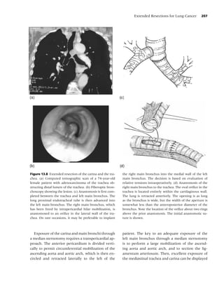
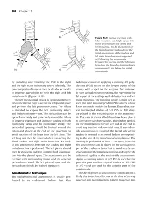
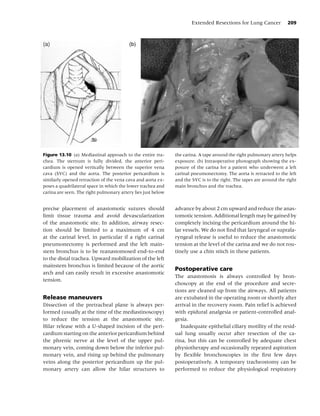
![210 Chapter 13
Table 13.4 Mortality and 5-year survival
Operative rates after carinal pneumonectomy.
Number of mortality 5-yr survival
Author (year) patients (%) (%)
Jensik et al. [59] 1982 34 29 15
Deslauriers et al. [60] 1989 38 29 13
Tsuchiya et al. [61] 1990 20 40 59 (2 yr)
Mathisen and Grillo [62] 1991 37 18.9 19
Roviaro et al. [63] 1994 28 4 20
Dartevelle et al. [64] 1995 60 6.6 43.3
Mitchell et al. [65] 1999 143 12.7 42
Roviaro et al. [66] 2001 49 8.2 24.5
Porhanov et al. [67] 2002 231 16 24.7
Regnard et al. [68] 2005 65 7.7 26.5
de Perrot et al. [58] 2006 119 7.6 44
Macchiarini et al. [69] 2006 50 4 51
Total 874 10.4 ± 11.6 25.6 ± 15.2
dead space and facilitate direct aspiration when- nodes can be sterilized prior to the lung resection.
ever the predicted residual ventilatory functional However, induction therapy could potentially be
reserve is borderline or the patient’s collaboration associated with increased operative morbidity and
reduced. The tracheostomy should be performed mortality in patients requiring right carinal pneu-
early in the postoperative period to prevent possible monectomy. Recently, we reported that operative
complications. mortality increased from 6.7 to 13% after induction
The results of carinal resection for bronchogenic therapy in patients undergoing right carinal pneu-
carcinoma have improved over time. Recent series monectomy [58]. Martin et al. [70] also reported an
have shown that carinal resection is relatively safe in operative mortality as high as 24% after right pneu-
experienced centers and can be associated with good monectomy following induction therapy.
long-term survival in selected patients [58–69]. The
median operative mortality is less than 7% and the
median 5-year survival is 43.3% in our experience SVC invasion
(Table 13.4).
Patients with positive mediastinal lymph node SVC syndrome is a distressing manifestation of be-
metastasis have a dismal prognosis; therefore, cari- nign and malignant disease obstructing venous re-
nal resection should be considered a potential turn through the SVC. Invasion of the SVC by the
contraindication. This also underscores the impor- right-sided bronchogenic carcinomas occurs in less
tance of performing routine preoperative medi- than 1% of operable patients [71] and is usually
astinoscopy in these patients. We recommend per- regarded as an absolute surgical contraindication
forming the mediastinoscopy at the time of the because of the dismal prognosis, absence of suit-
planed carinal resection in order to avoid the de- able graft material for reconstruction, and technical
velopment of scaring tissue along the trachea and fear concerning the effects of SVC clampage, graft
to take advantage of the tracheal mobilization to thrombosis, and infection. However, recent experi-
reduce tension at the anastomotic site. mental and clinical advances increased the popular-
Further studies should determine the role of in- ity of SVC replacement and expanded its therapeutic
duction therapy in patients presenting with bron- role in the management of patients with tho-
chogenic carcinoma and N2 disease. Induction ther- racic neoplasm [70–82]. Although, SVC resection
apy seems to improve survival if the mediastinal and revascularization is a technically demanding](https://image.slidesharecdn.com/lungcancer3rded-120126215311-phpapp01/85/Lung-cancer-3rd-ed-221-320.jpg)
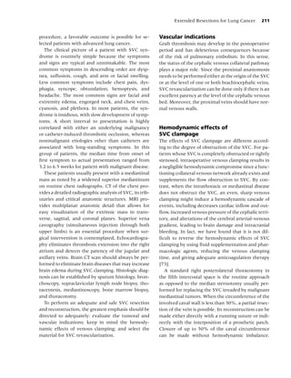
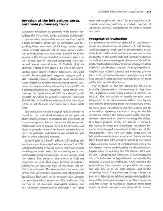
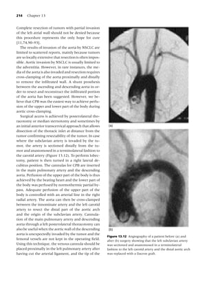
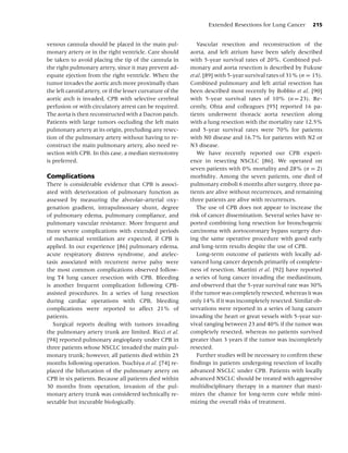


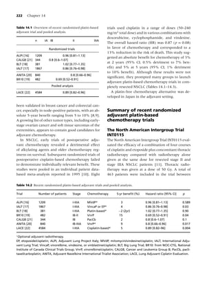
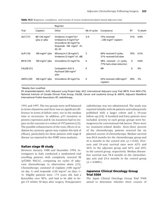
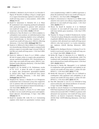
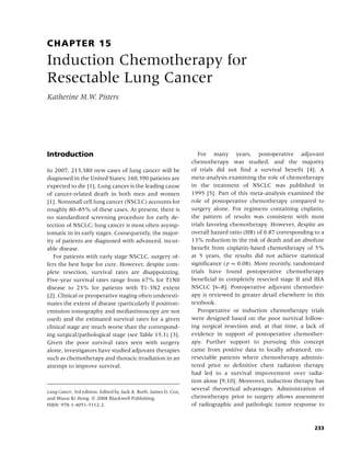
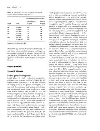
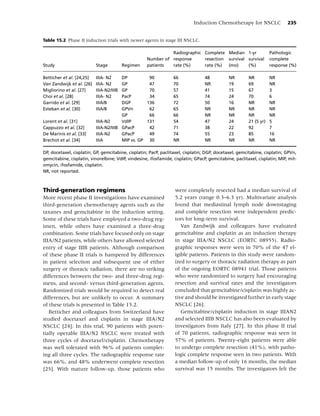
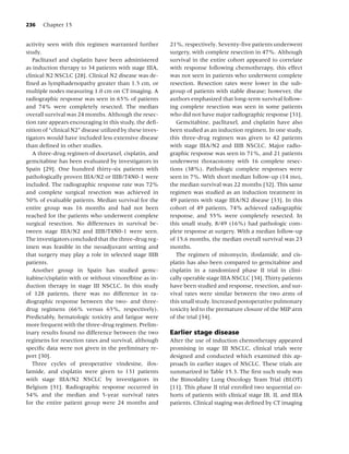
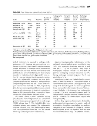
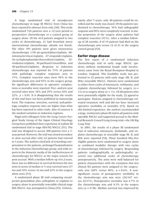
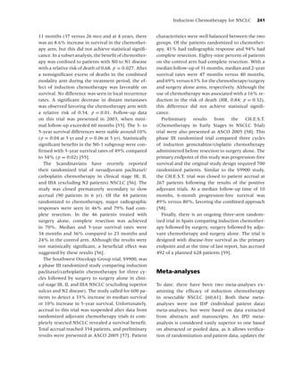
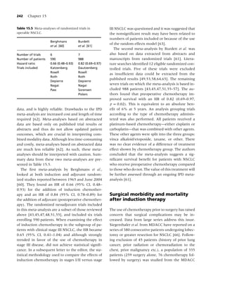
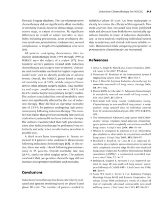
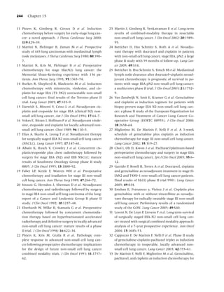
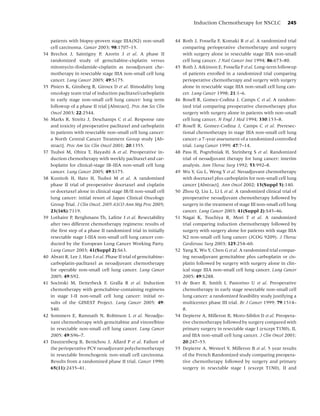
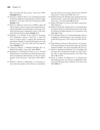
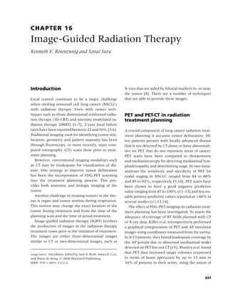

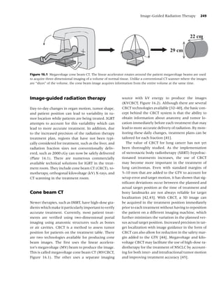
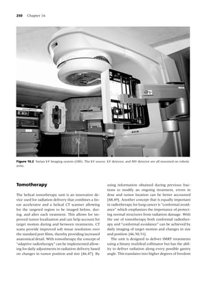

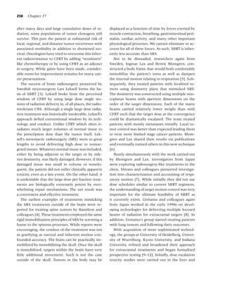
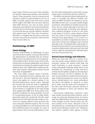
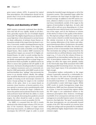
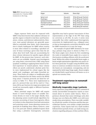
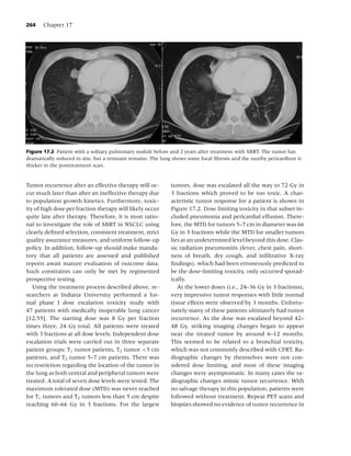
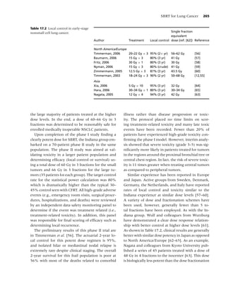
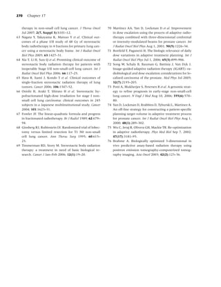
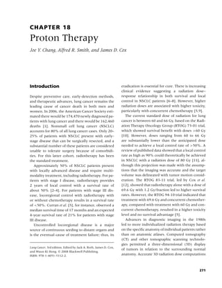
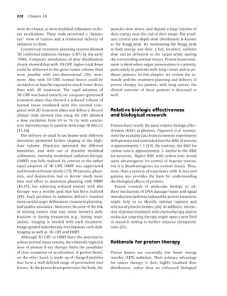
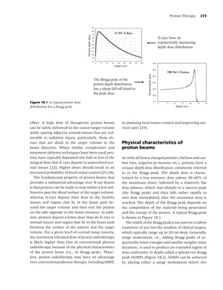
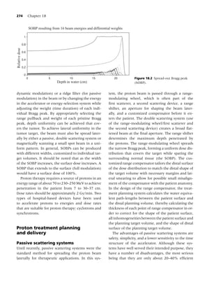
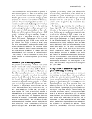
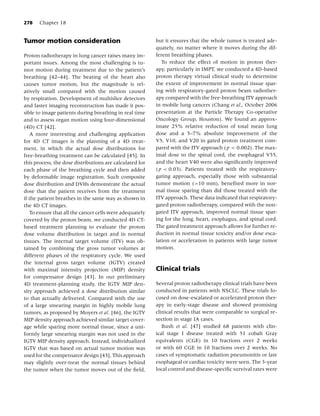
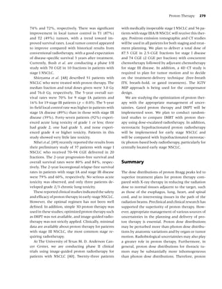

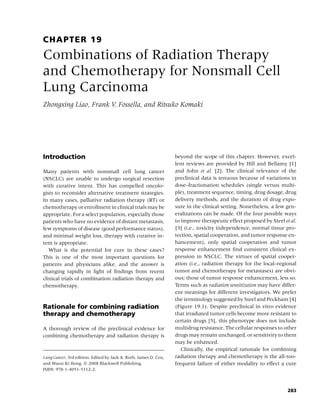
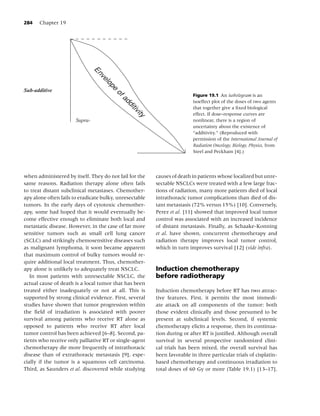
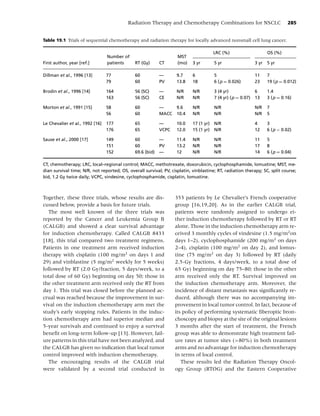
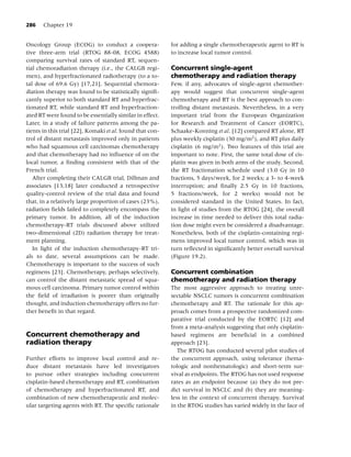
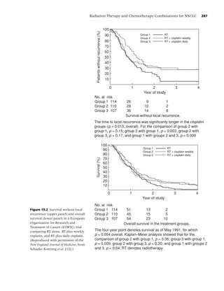
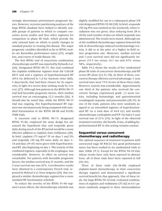
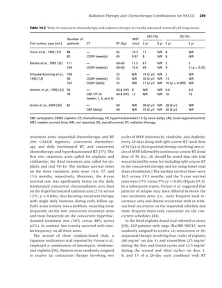
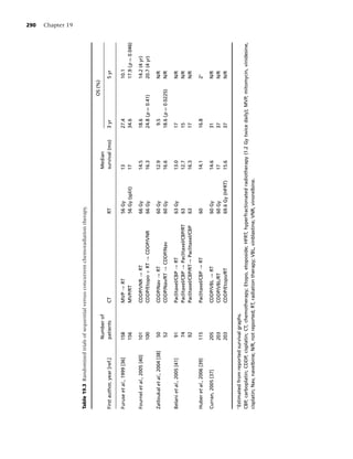
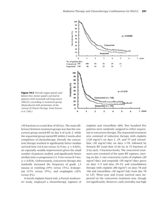
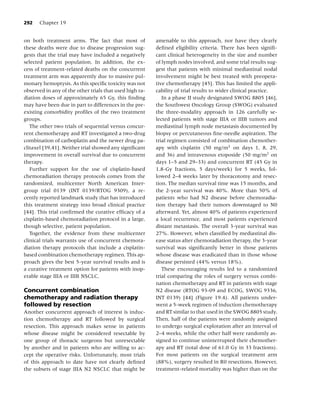
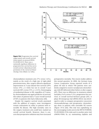
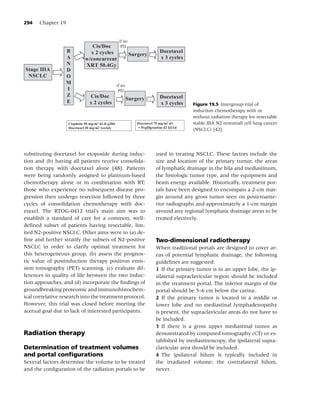
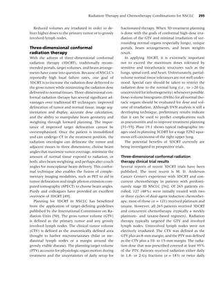
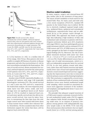
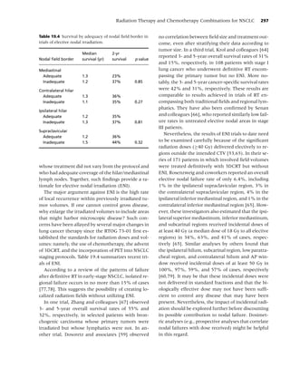
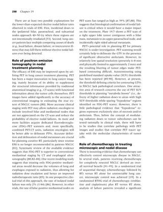
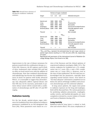
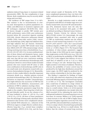
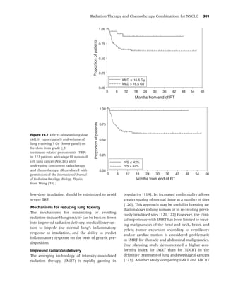
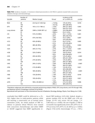
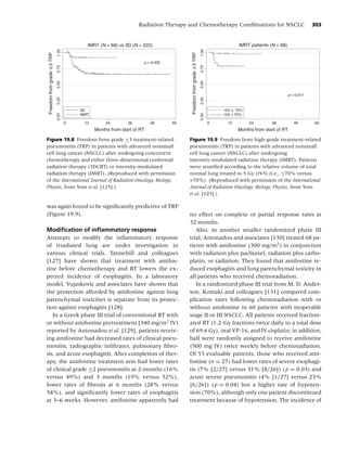
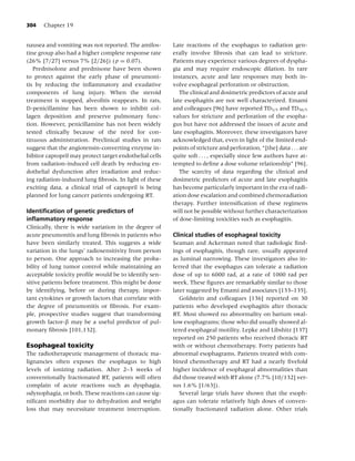
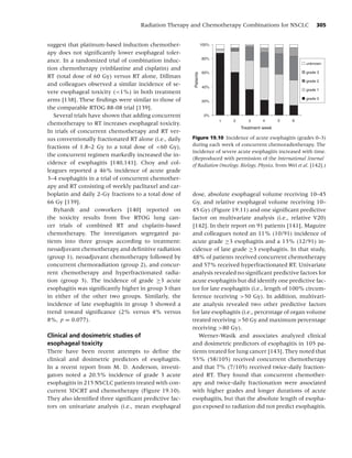
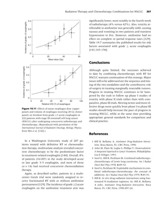
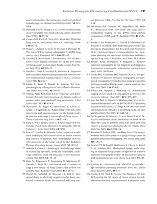
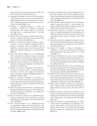
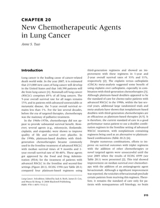
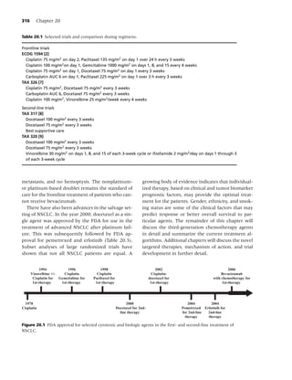
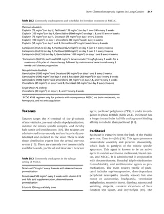
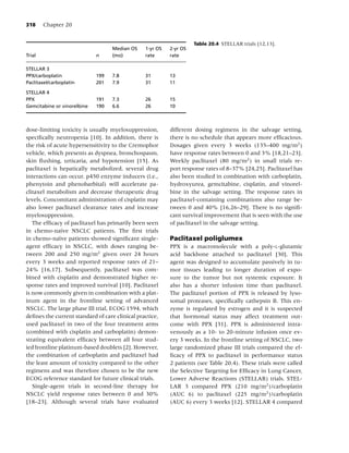
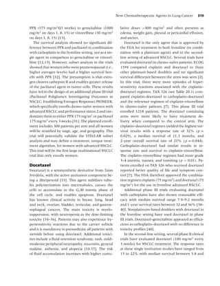
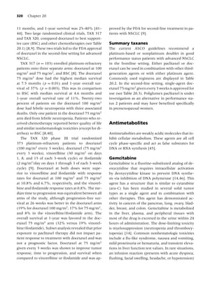
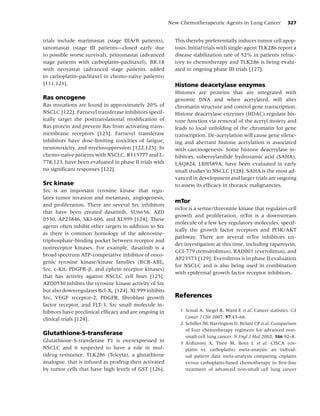
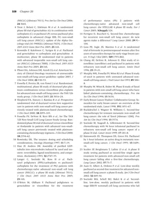
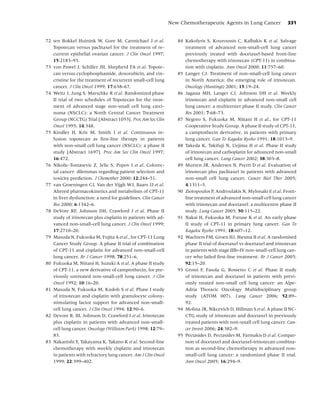
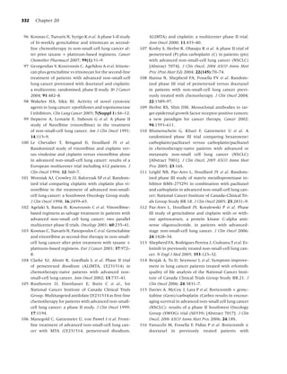
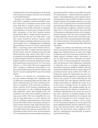
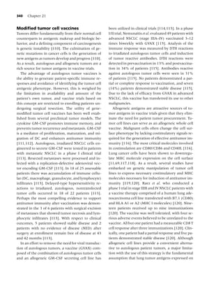
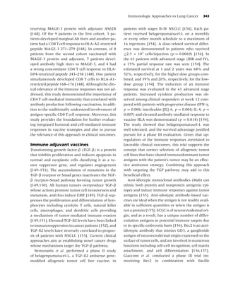
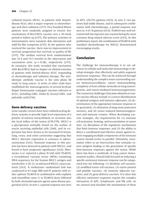
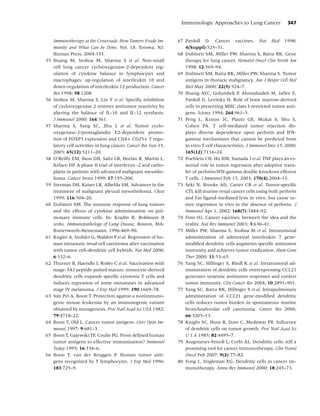
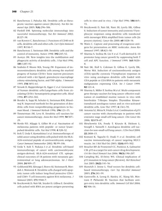
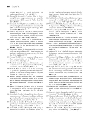

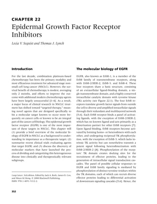
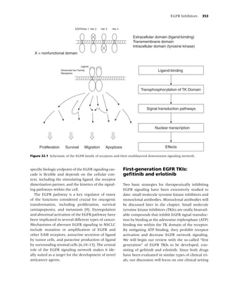
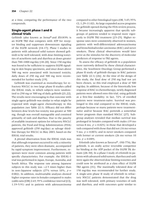
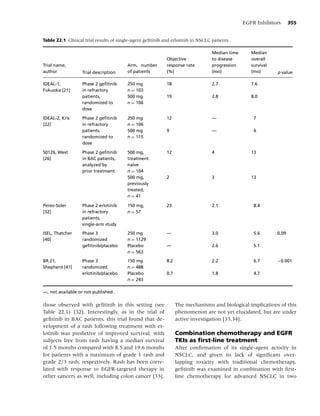
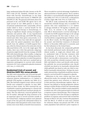
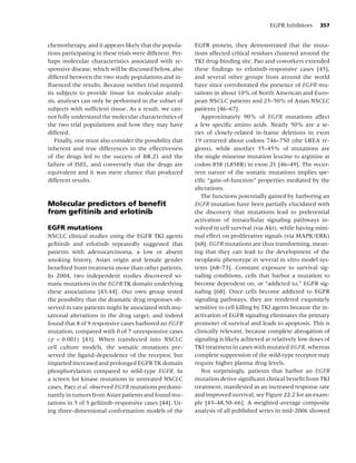
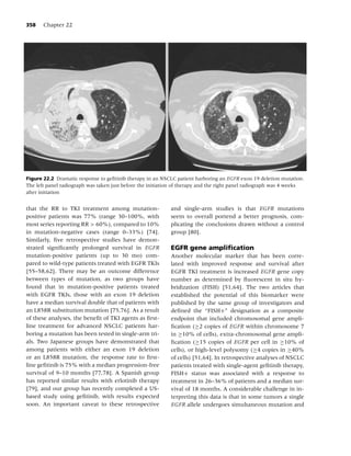
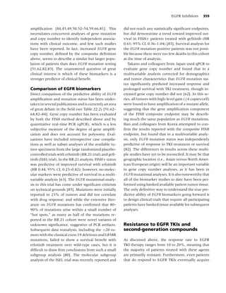
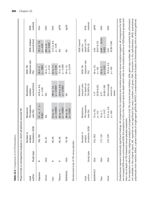
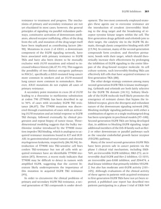
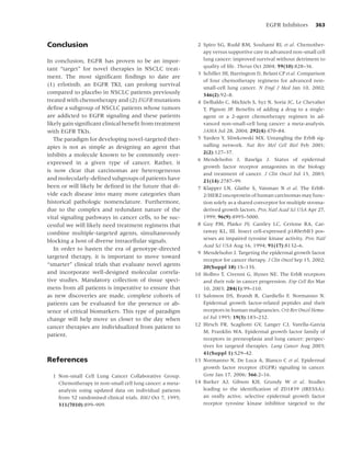
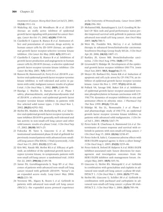
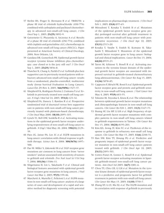
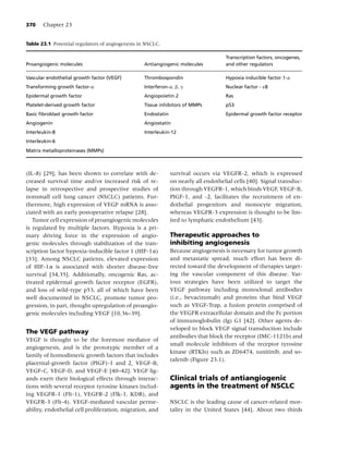
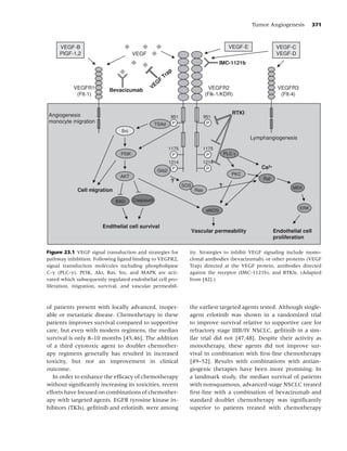
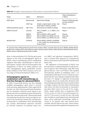
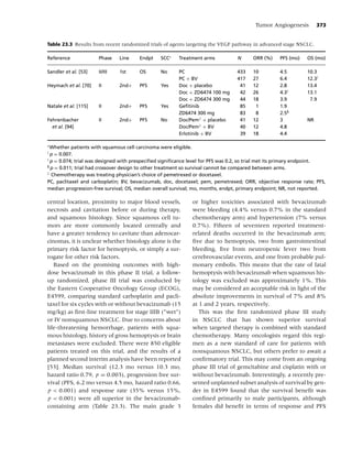
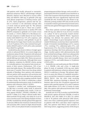
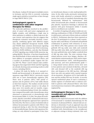
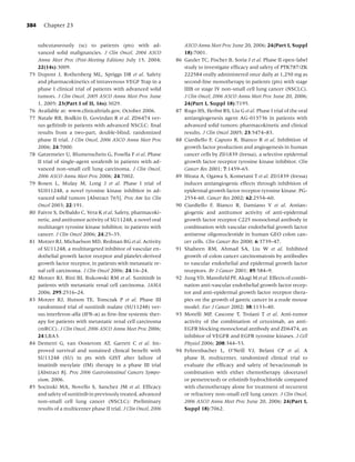
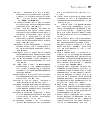
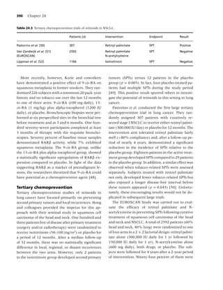
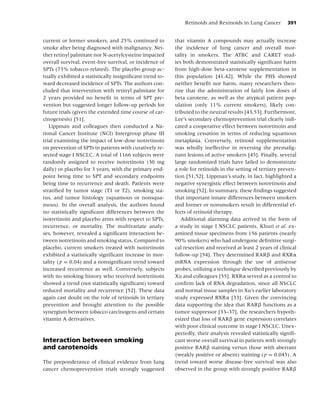
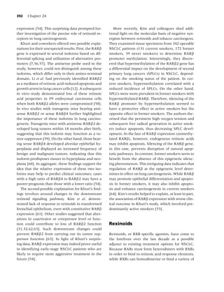
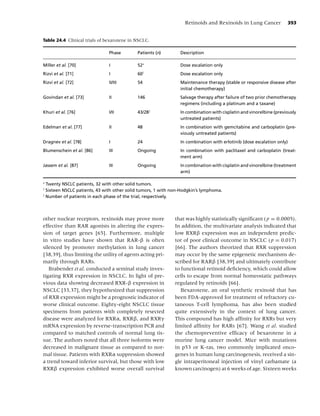
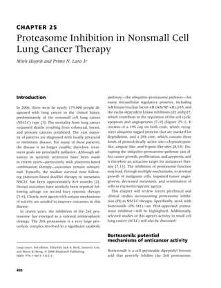
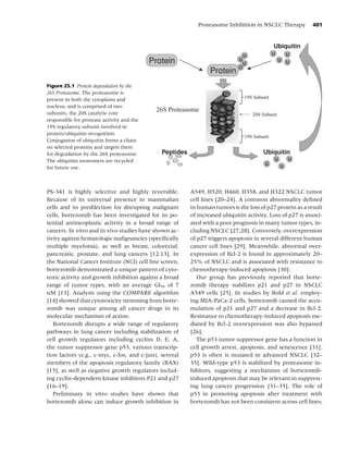
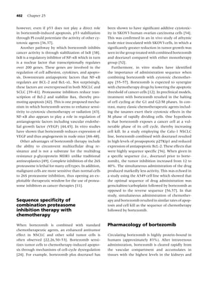
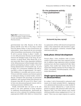
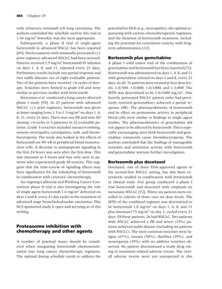
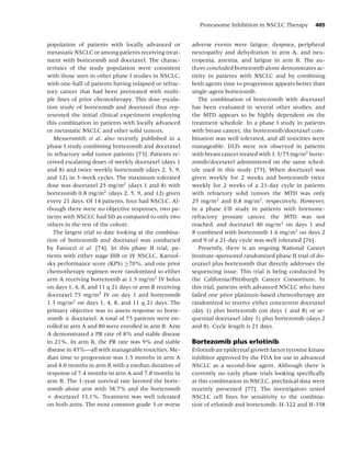
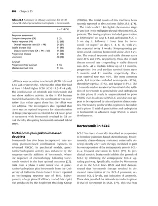
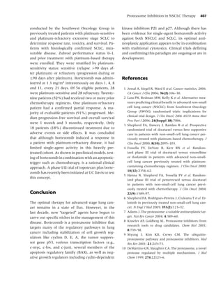
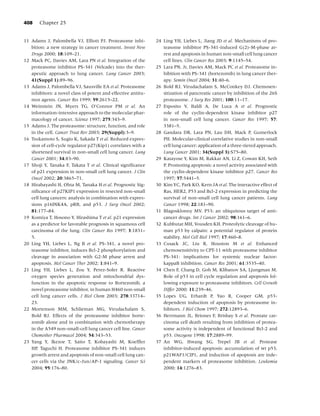
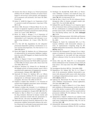
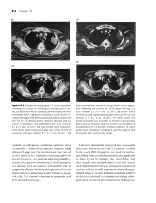
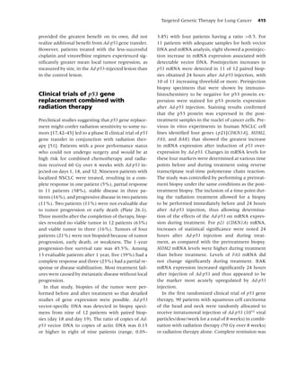
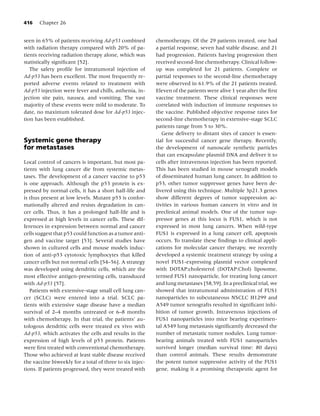
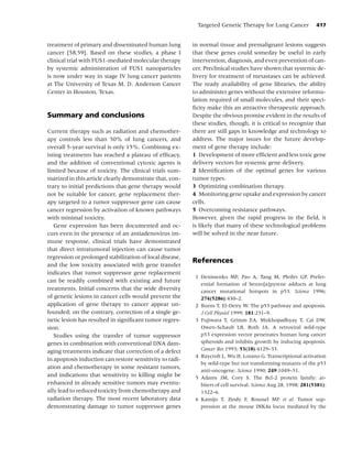

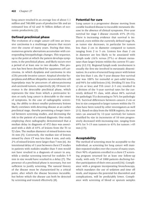
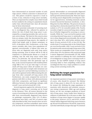
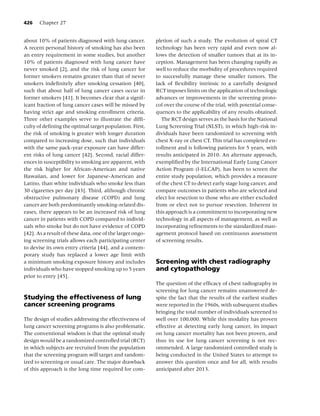
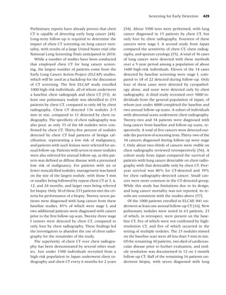
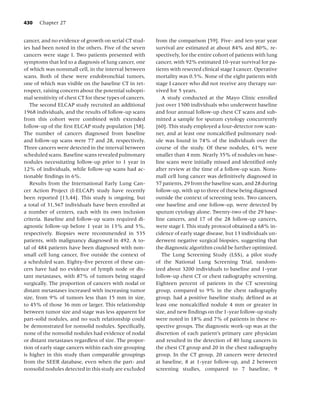
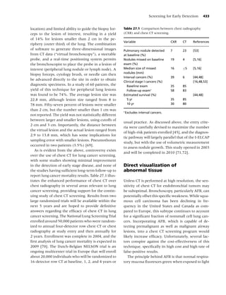
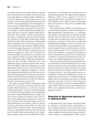
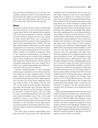
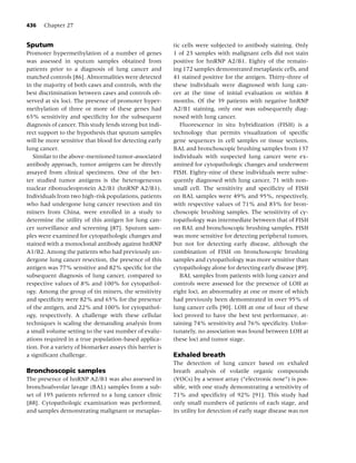
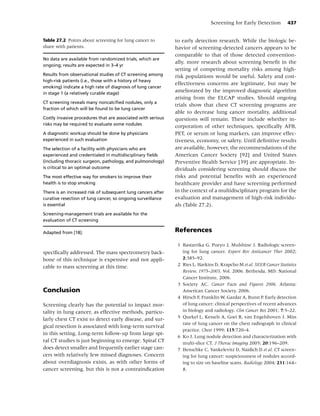
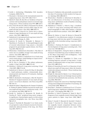
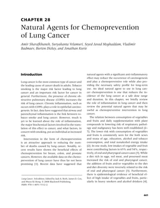
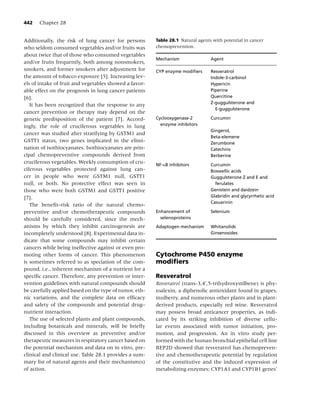
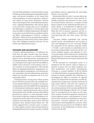
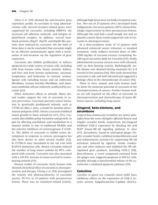
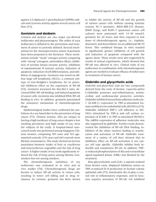
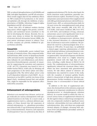
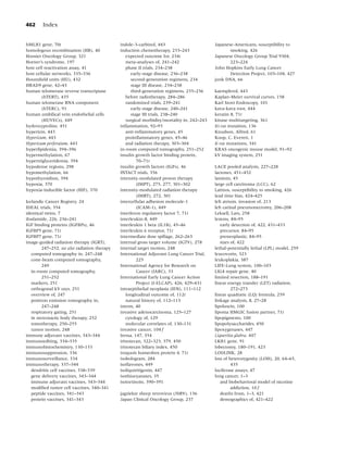
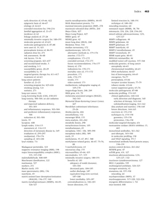
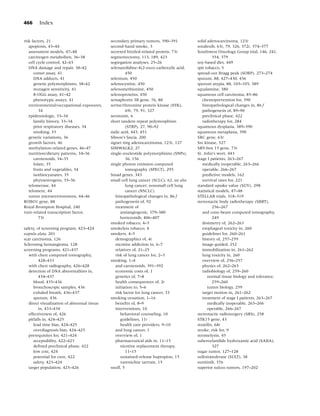
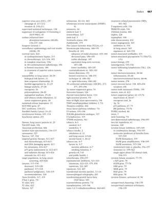

This document is the third edition of a book on lung cancer edited by Jack A. Roth, James D. Cox, and Waun Ki Hong. It contains chapters written by experts on various topics related to lung cancer, including smoking cessation, lung cancer genetics, detection and diagnosis of preneoplastic lung lesions, surgical and non-surgical treatments, targeted therapies, screening and prevention. The book provides a comprehensive overview of the current state of knowledge across the multidisciplinary field of lung cancer research and clinical management.


