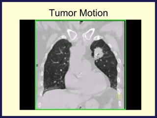
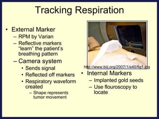
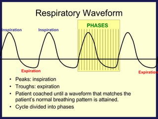
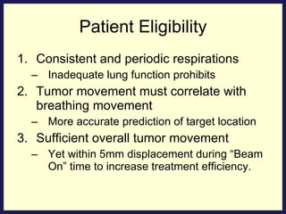
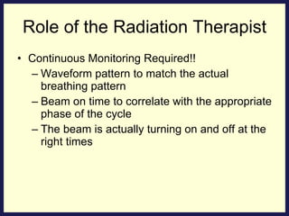
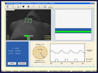
Respiratory Gating with IMRT Presented by: Katherine Adams
Respiratory Gating & IMRT Conforms to target Higher doses to target tissue Less side effects from normal tissue Sharp dose gradient between target tissue and normal tissue Target location important Misjudgment causes Underdose target Overdose normal tissue
Treatment Planning The prescription dose is targeted to a region that consist of the gross tumor volume, clinical target volume and the planning target volume. These margins need to be considered to make sure that the target volume remains in the field during treatment The resulting margins in conventional therapy can be as high as 3 to 5 cm depending on the anatomy treated.  GTV: visible or palpable tumor CTV: GTV + microscopic disease PTV: CTV + margin to account for Variations in treatment setup Patient motion Organ motion
Tumor Motion
Tumor Motion The previous slide showed a video clip of a 4DCT that shows the movement and location of a lung tumor during respiration. Internal structures can move a significant amount- The diaphragm and liver can move up to 3 cm and the pancreas, kidney and thorax can move up to 2 cm Respiratory gating is used to minimize the increased margins of the treatment volume that are directly related to respiratory movements
Tracking Respiration External Marker RPM by Varian Reflective markers “learn” the patient’s breathing pattern Camera system Sends signal  Reflected off markers Respiratory waveform created Shape represents tumor movement Internal Markers Implanted gold seeds Use flouroscopy to locate http://www.biij.org/2007/1/e40/fig1.jpg
Respiratory Waveform Peaks: inspiration Troughs: expiration Patient coached until a waveform that matches the patient’s normal breathing pattern is attained.  Cycle divided into phases Inspiration Expiration Inspiration Expiration PHASES
CT Images Spiral CT acquires its images with a moving table during many different phases of the breathing cycle The resulting image is a blurred snapshot of the structure and may contain motion artifacts
4D CT CT table stationary Info for complete cycle Divided into phases Table moves to next position Cycle repeats Information is gathered for each phase and combined to construct an image that represents the location of the anatomy at that precise moment.
Exhalation = Beam on Anatomy is relatively stationary More reproducible More phases included More efficient treatment Treatment Planning Beam Off Beam On Beam On
Cancer Treatment with Respiratory Gating
Irregular Respiration System automatically shuts down until normal breathing pattern is reestablished. Beam On Beam On Beam Off Normal Respiration Normal Respiration Beam Off Inspiration Expiration Inspiration Expiration Expiration Beam On Irregular Respiration
Cancers Treated Tumors subject to respiratory motion: Lung Breast Maximum inhalation Inflates lung- minimizing amount in field Separates breast from heart Pancreatic Stomach Liver Prostate Whole abdomen for ovarian cancer
Patient Eligibility Consistent and periodic respirations Inadequate lung function prohibits Tumor movement must correlate with breathing movement More accurate prediction of target location Sufficient overall tumor movement Yet within 5mm displacement during “Beam On” time to increase treatment efficiency.
Alternative Methods Require invasive surgical procedures submitting patient to infection: Intraoperative radiation therapy Interstitial brachytherapy Uncomfortable breath holds that many cannot withstand due to decreased pulmonary function: Deep inspiration breath hold (DIBH) Active breathing control (ABC)
Continuous Monitoring Required!! Waveform pattern to match the actual breathing pattern Beam on time to correlate with the appropriate phase of the cycle The beam is actually turning on and off at the right times Role of the Radiation Therapist
http://www.varian.com/us/oncology/radiation_oncology/clinac/rpm_respiratory_gating.html
THE END ANY QUESTIONS...???
References Brandner, E., Heron, D. E., Wu, A., Huq, M. S., Yue, N. J.,Chen, H. C., (2006). Localizing Moving Targets and organs using motion-managed CTs.  Medical Dosimetry , 31(2), 134-140. Bucsko, J. K., (2004, November). Managing Respiratory Motion.  Radiology Today , 5(23), 33. Retrieved February 27, 2008, from http://www.radiologytoday.net/archive/rt_110804p33.shtml Butler, L.E., Forster, K. M., Stevens, C. W., Bloch, C., Liu, H. H., Tucker, S. L., (2004). Dosimetric benefits of respiratory gating: a preminary study.  Journal of Applied Clinical Medical Physics , 5 (1), 16-24. Carlson, A. H., (2007, October). Breathing New Life into Respiratory Gating.  Medical Imaging , 42. Retrieved February 27, 2008, from http://www.medicalimagingmag.com/issues/articles/2007-10_04.asp Garsa, A.A., Andrade, R. S., Heron, D.E., Beriwal, S., Kim, H., Brandner, E., (2007). Four-dimensional computed tomography-based respiratory-gated whole abdominal intensity-modulated radiation therapy for ovarian cancer: a feasible study.  International Journal of Gynecological Cancer , 17, 55-60. Huntzinger, C., (n.d.). The Revolution in Radiation Therapy.  In Looking Forward- The Future is in Motion.  Varian Medical systems. Retrieved February 27, 2008, from varian.mediaroom.com/file.php/mr_varian/spinsite_docfiles/147/VRM395+IMRT+White+Paper+Final+Jan+2005.pdf -
References Jiang, S. B., (2006). Technical aspects of image guided respiration-gated radiation therapy.  Medical Dosimetry , 31(2), 141-151. Keall, P., Vedam, S., George, R., Bartee, C., Siebers, J., Lerma, F., (2006). The clinical implementation of respiratory-gated intensity-modulated radiotherapy.  Medical Dosimetry , 31(2), 152-162. Kornmehl, C., (2005). Every Move You Make. Image, 18(10). Retrieved February 25, 2008, from http://www.rt-image.com/030705FO Korreman, S., Mostafavi, H., Le, QT., Boyer, A., (2006).  Comparison of respiratory surrogates for gated lung radiotherapy without internal fiducials.  Acta Onclolgica , 45, 935-942. Rosenzweig, KE., Yorke, E., Amols, H., Mageras, GS., Giraud, P., Katz, MS., (2005). Tumor Motion Control in the Treatment of Non Small Cell Lung Cancer.  Cancer Investigation , 23, 129-133. Saw CB, Brandner E, Selvaraj R, Chen H, Saiful Huq M, Heron DE, (2007). A review on the clinical implementation of respiratory-gated radiation therapy,  Biomedical Imaging and Intervention Journal  3(1):e40. Retrieved February 25, 2008, from http://www.biij.org/2007/1/e40/> Underberg, R., van Sornsen de Koste, JR., Lagerwaard, FJ, Vincent, A., Slotman, BJ., Senan, S., (2006). A dosimetric analysis of respiration-gated radiotherapy in patients with stage II lung cancer.  Radiation Oncology . 1:8, Retrieved February 27, 2008, from http://www.ro-journal.com/content/pdf/1748-717X-1-8.pdf.

Respiratory Gating with IMRT

  • 1.
    Respiratory Gating withIMRT Presented by: Katherine Adams
  • 2.
    Respiratory Gating &IMRT Conforms to target Higher doses to target tissue Less side effects from normal tissue Sharp dose gradient between target tissue and normal tissue Target location important Misjudgment causes Underdose target Overdose normal tissue
  • 3.
    Treatment Planning Theprescription dose is targeted to a region that consist of the gross tumor volume, clinical target volume and the planning target volume. These margins need to be considered to make sure that the target volume remains in the field during treatment The resulting margins in conventional therapy can be as high as 3 to 5 cm depending on the anatomy treated. GTV: visible or palpable tumor CTV: GTV + microscopic disease PTV: CTV + margin to account for Variations in treatment setup Patient motion Organ motion
  • 4.
  • 5.
    Tumor Motion Theprevious slide showed a video clip of a 4DCT that shows the movement and location of a lung tumor during respiration. Internal structures can move a significant amount- The diaphragm and liver can move up to 3 cm and the pancreas, kidney and thorax can move up to 2 cm Respiratory gating is used to minimize the increased margins of the treatment volume that are directly related to respiratory movements
  • 6.
    Tracking Respiration ExternalMarker RPM by Varian Reflective markers “learn” the patient’s breathing pattern Camera system Sends signal Reflected off markers Respiratory waveform created Shape represents tumor movement Internal Markers Implanted gold seeds Use flouroscopy to locate http://www.biij.org/2007/1/e40/fig1.jpg
  • 7.
    Respiratory Waveform Peaks:inspiration Troughs: expiration Patient coached until a waveform that matches the patient’s normal breathing pattern is attained. Cycle divided into phases Inspiration Expiration Inspiration Expiration PHASES
  • 8.
    CT Images SpiralCT acquires its images with a moving table during many different phases of the breathing cycle The resulting image is a blurred snapshot of the structure and may contain motion artifacts
  • 9.
    4D CT CTtable stationary Info for complete cycle Divided into phases Table moves to next position Cycle repeats Information is gathered for each phase and combined to construct an image that represents the location of the anatomy at that precise moment.
  • 10.
    Exhalation = Beamon Anatomy is relatively stationary More reproducible More phases included More efficient treatment Treatment Planning Beam Off Beam On Beam On
  • 11.
    Cancer Treatment withRespiratory Gating
  • 12.
    Irregular Respiration Systemautomatically shuts down until normal breathing pattern is reestablished. Beam On Beam On Beam Off Normal Respiration Normal Respiration Beam Off Inspiration Expiration Inspiration Expiration Expiration Beam On Irregular Respiration
  • 13.
    Cancers Treated Tumorssubject to respiratory motion: Lung Breast Maximum inhalation Inflates lung- minimizing amount in field Separates breast from heart Pancreatic Stomach Liver Prostate Whole abdomen for ovarian cancer
  • 14.
    Patient Eligibility Consistentand periodic respirations Inadequate lung function prohibits Tumor movement must correlate with breathing movement More accurate prediction of target location Sufficient overall tumor movement Yet within 5mm displacement during “Beam On” time to increase treatment efficiency.
  • 15.
    Alternative Methods Requireinvasive surgical procedures submitting patient to infection: Intraoperative radiation therapy Interstitial brachytherapy Uncomfortable breath holds that many cannot withstand due to decreased pulmonary function: Deep inspiration breath hold (DIBH) Active breathing control (ABC)
  • 16.
    Continuous Monitoring Required!!Waveform pattern to match the actual breathing pattern Beam on time to correlate with the appropriate phase of the cycle The beam is actually turning on and off at the right times Role of the Radiation Therapist
  • 17.
  • 18.
    THE END ANYQUESTIONS...???
  • 19.
    References Brandner, E.,Heron, D. E., Wu, A., Huq, M. S., Yue, N. J.,Chen, H. C., (2006). Localizing Moving Targets and organs using motion-managed CTs. Medical Dosimetry , 31(2), 134-140. Bucsko, J. K., (2004, November). Managing Respiratory Motion. Radiology Today , 5(23), 33. Retrieved February 27, 2008, from http://www.radiologytoday.net/archive/rt_110804p33.shtml Butler, L.E., Forster, K. M., Stevens, C. W., Bloch, C., Liu, H. H., Tucker, S. L., (2004). Dosimetric benefits of respiratory gating: a preminary study. Journal of Applied Clinical Medical Physics , 5 (1), 16-24. Carlson, A. H., (2007, October). Breathing New Life into Respiratory Gating. Medical Imaging , 42. Retrieved February 27, 2008, from http://www.medicalimagingmag.com/issues/articles/2007-10_04.asp Garsa, A.A., Andrade, R. S., Heron, D.E., Beriwal, S., Kim, H., Brandner, E., (2007). Four-dimensional computed tomography-based respiratory-gated whole abdominal intensity-modulated radiation therapy for ovarian cancer: a feasible study. International Journal of Gynecological Cancer , 17, 55-60. Huntzinger, C., (n.d.). The Revolution in Radiation Therapy. In Looking Forward- The Future is in Motion. Varian Medical systems. Retrieved February 27, 2008, from varian.mediaroom.com/file.php/mr_varian/spinsite_docfiles/147/VRM395+IMRT+White+Paper+Final+Jan+2005.pdf -
  • 20.
    References Jiang, S.B., (2006). Technical aspects of image guided respiration-gated radiation therapy. Medical Dosimetry , 31(2), 141-151. Keall, P., Vedam, S., George, R., Bartee, C., Siebers, J., Lerma, F., (2006). The clinical implementation of respiratory-gated intensity-modulated radiotherapy. Medical Dosimetry , 31(2), 152-162. Kornmehl, C., (2005). Every Move You Make. Image, 18(10). Retrieved February 25, 2008, from http://www.rt-image.com/030705FO Korreman, S., Mostafavi, H., Le, QT., Boyer, A., (2006). Comparison of respiratory surrogates for gated lung radiotherapy without internal fiducials. Acta Onclolgica , 45, 935-942. Rosenzweig, KE., Yorke, E., Amols, H., Mageras, GS., Giraud, P., Katz, MS., (2005). Tumor Motion Control in the Treatment of Non Small Cell Lung Cancer. Cancer Investigation , 23, 129-133. Saw CB, Brandner E, Selvaraj R, Chen H, Saiful Huq M, Heron DE, (2007). A review on the clinical implementation of respiratory-gated radiation therapy, Biomedical Imaging and Intervention Journal 3(1):e40. Retrieved February 25, 2008, from http://www.biij.org/2007/1/e40/> Underberg, R., van Sornsen de Koste, JR., Lagerwaard, FJ, Vincent, A., Slotman, BJ., Senan, S., (2006). A dosimetric analysis of respiration-gated radiotherapy in patients with stage II lung cancer. Radiation Oncology . 1:8, Retrieved February 27, 2008, from http://www.ro-journal.com/content/pdf/1748-717X-1-8.pdf.