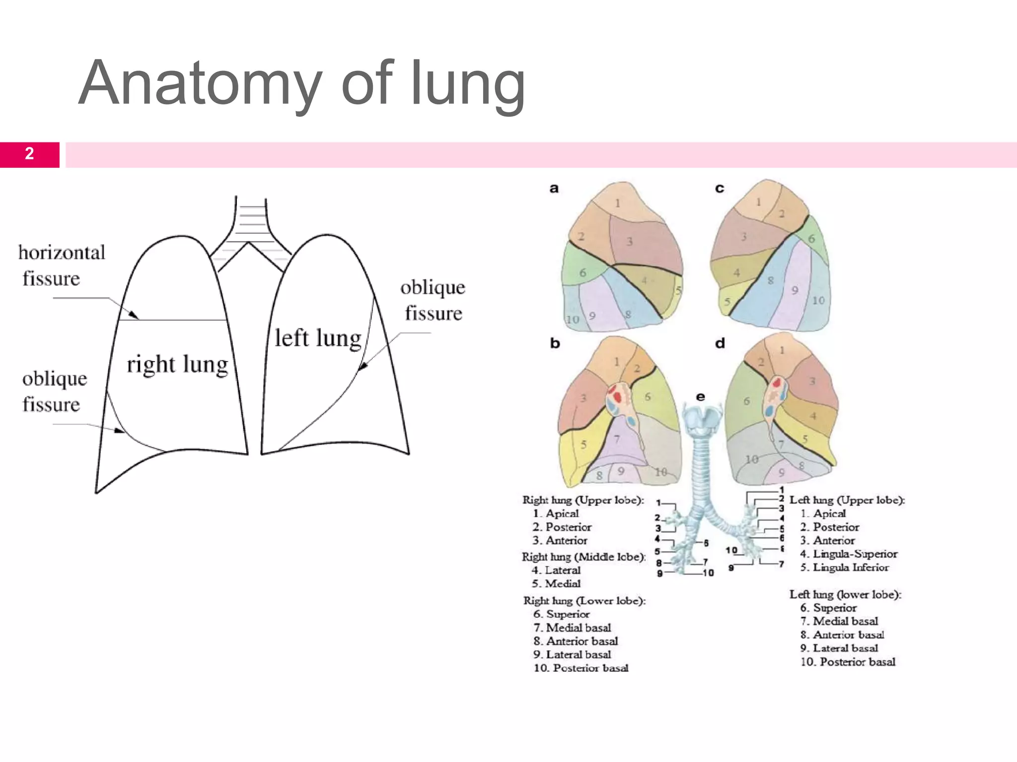
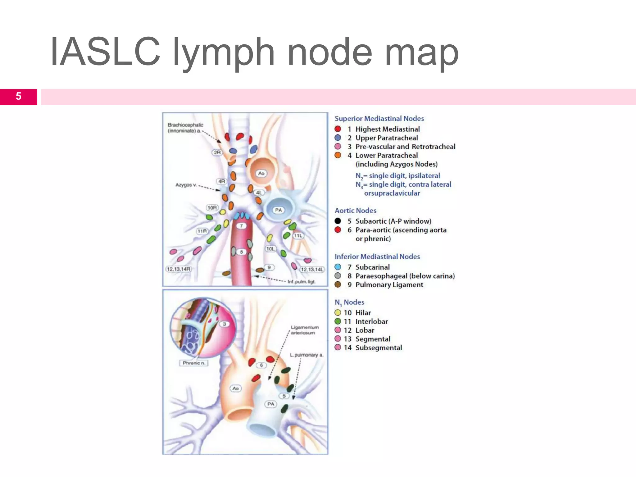
MANAGEMENT OF
NON SMALL CELL
LUNG CANCER
Dr. Ankita Pandey
JR 2
1
Anatomy of lung
2
Anatomy of lung
 Bronchopulmonary segment is the functional
unit of the lung and is defined by the
segmental bronchi
 Each bronchopulmonary segment consists of
1. Bronchus
2. Artery
3. Vein
4. lymphatics
3
 Lymphatics from each bronchopulmonary segment
drains into mediastinum
 Rich lymphatic network within thorax leads to
complex variability in drainage pattern
The international association for the study of lung
cancer developed lymph node map that provide
precise anatomic definitions for all lymph node
stations.
4
IASLC lymph node map
5
Clinical features
In NSCLC, half of the patients presents
with localised or locally advanced disease
while half presents with advanced stage
In SCLC, 20-30% cases presents with
localised or locally advanced disease while
70-80% presents with advanced stage
6
1. Cough
2. Dyspnea
3. Hemoptysis
4. Chest pain
5. Hoarsness of voice
6. SVC syndrome- SCLC
7. Pancoast syndrome- NSCLC
8. Bone pain
9. Headache, visual disturbances
7
Sites of distant mets
 Lymph node mets – most common
 Liver
 Adrenals
 Bones
 Brain
 Kidney
 Pancreas
 Contralateral lung
 Pleura
8
Paraneoplastic syndromes
 It is a disease or symptom that is the
consequence of cancer cells in the body but is
not attributable to the local presence of tumor.
 Treating the tumor usually resolves the
syndrome
Caused by-
1. Humoral factors secreted by
tumor
2. Immune response against tumor
9
1. Cushing syndrome
2. SIADH
3. Hypercalcemia
4. Lambert eaton myasthenic syndrome
5. Hypertrophic osteoarthropathy
10
Diagnostic staging and workup
 History and physical examination
 PFT
 Assess the overall health status
11
Investigations
 CT Scan thorax and upper abdomen
 PET CT
 Percutaneous fine needle aspiration
 Bronchoscopy
 Endoscopic ultrasound
 Thoracocentesis
 Mediastinoscopy
It should be considered in
patients with suspected lung
cancer even if chest x ray is
normal
Liver mets and adrenal
mets
12
Staging
13
14
Variants of NSCLC
 Adenocarcinoma (50%)
 Squamous cell carcinoma (35%)
 Large cell carcinoma (15%)
15
Histology is important
determinant in selection of
systemic therapy for
advanced NSCLC
Treatment algorithm
16
Management of stage I and II
NSCLC
 PRE OPERATIVE ASSESSMENT
1. Predicted postop FEV1 >60%
2. DLCO >60%
17
Management of stage I and II
NSCLC
 Standard care for stage I and II lung cancer is
surgical resection
Lobectomy with mediastinal LN dissection
 Standard of care for medically inoperable
early stage NSCLC is – SBRT
18
Types of surgeries
19
 What is the role of limited resection?
Limited resection is either wedge resection or
segmentectomy
 Trial by lung cancer study group- included T1-
2N0 NSCLC.
 Study confirmed lobectomy as standard of care
for early stage lung cancer
20
Limited resection Lobectomy
Risk of recurrence= 17% Risk of recurrence= 6%
What are the survival results after
surgery ?
 According to the recent IASLC lung cancer
staging project, following surgical resection for
lung cancer, the 5-year survival is -
 73% for stage IA
 58% for stage IB
 46% for stage IIA
 36% for stage IIB
21
 What is the role of mediastinal lymph node
dissection?
American college of surgeons oncology group
trial randomised N0 or N1 patients to
mediastinal LN sampling or dissection and
found no significant difference in DFS, LRR
and distant recurrence
Controversia
l
22
 Advantage of mediastinal LN dissection-
Complete mediastinal lymphadenectomy
provides surgical staging by removing all
lymph nodes.
This can help to identify patients who may
require postoperative adjuvant radiation
therapy
23
Indications of post op RT
 Positive surgical margin
 Pathologically N+ disease
24
Inoperable early stage NSCLC
Inoperable incurable
It simply means patient is medically not fit for
surgery due to old age/ cardiopulmonary
dysfunction/ chronic illness
25
Inoperable early stage NSCLC
 Radiotherapy with or without chemotherapy
remains the primary alternative to surgery for
inoperable NSCLC patients.
 higher radiation doses, greater than 70Gy,
enhanced local tumor control or survival.
 high doses increase the probability of normal
tissue toxicity
 Hence , it has poorer outcome compared to
surgery
26
Radiotherapy techniques
 2D conventional planning
 3D CRT/IMRT
 SBRT
 Brachytherapy
 Proton beam therapy
Respiratory
motion
management
27
Management of Stage III
NSCLC
 Stage IIIA= considered operable
 Stage IIIb= inoperable
28
 Primary surgery followed by
chemotherapy has been the
standard.
 Induction radiation therapy
or induction chemotherapy
followed by surgical
resection have been used to
downstage disease and are
considered options in
selected cases of non-bulky,
single-station N2 disease.
 Concurrent
chemotherapy and
radiation therapy
have been
considered the
standard treatment
Operable stage III Inoperable stage III
29
Role of Preoperative CCRT
 Results from phase III studies in patients with
stage IIIA non-small-cell lung cancer with
ipsilateral mediastinal nodal metastases (N2)
have shown the feasibility of resection after
concurrent chemotherapy and radiotherapy with
promising rates of survival
30
Role of preop chemotherapy
 There are multiple trials in which patients
undergoing surgical resection were
randomized to induction chemotherapy vs
surgery alone
31
Role of adjuvant chemotherapy in
stage IIIa NSCLC
 administration of adjuvant cisplatin-based
chemotherapy has become the standard of care
for patients with resected stage II and IIIA
disease
Lung Adjuvant Cisplatin Evaluation Meta-
analysis
32
Postoperative Radiation
Therapy
 Postoperative radiation therapy improves local
control and disease-free survival duration for
patients with pathologic N2 disease (Lung
Cancer Study Group 1986).
 a secondary analysis of the Adjuvant
Navelbine International Trialist Association
(ANITA) randomized study showed that PORT
improved overall survival in patients with N2
disease
33
Postoperative Radiation
Therapy34
Treatment of stage IIIB NSCLC
and inoperable stage IIIA NSCLC35
 Definitive radiotherapy
 Dose- 60-75Gy to gross disease
50Gy to microscopic disease
 RTOG 7301 trial- 60Gy as standard
 RTOG 8311 trial- 69.6Gy/1.2Gy per#/ twice daily
as standard regimen
 Hyperfractionation shows better result
Chemoradiotherapy
 A meta-analysis of six trials consisting of 1205
patients revealed that there was an absolute
benefit of 5.9% at 3 years in patients receiving
concomitant chemoradiation.
 improved locoregional progression, but did not
have an effect on distant progression.
 Severe acute esophagitis significantly increased
from 4% to 18%
36
Concurrent chemoRT regimens
 Cisplatin and etoposide
 Cisplatin and vinblastine
 Cisplatin and pemetrexed
 Carboplatin and pemetrexed
 Carboplatin and paclitaxel
37
Sequential vs concurrent
chemoRT38
Stage IV NSCLC
 Stage IV disease is incurable and treatment is
palliative.
 Treatment consists of palliative chemotherapy,
palliative radiation therapy, medications,
supportive care, and hospice care
39
Palliative RT
Mets Total dose Dose per fraction
Brain mets 30Gy 3Gy
Bone mets 20-30Gy 3-4Gy
Obstructive ds 30-45Gy 3Gy
40
Palliative chemotherapy
 Cisplatin
 Carboplatin
 Paclitaxel
 Docetaxel
 Venorelbine
 Gemcitabine
 Etoposide
 Bevacizumab
41
Targeted therapy
 Indicated in patients who have failed
platinum/taxane based chemotherapies
 Anti- EGFR agents-
1. Cetuximab
2. Panitumumab
3. Geftinib
4. Erlotinib
 VEGF monoclonal antibodies- bevacizumab
 ALK + patients - crizotinib
42
Radiotherapy techniques43
RT planning
 Lung tumor radiotherapy planning is complex
due to motion management.
 Respiratory motion management techniques
include-
1. Breath hold technique
2. Abdominal compression technique
3. Respiratory gating
4. Tumor tracking
5. ABC
44
Abdominal compression technique
 For forced shallow breathing
 A plate compresses the abdominal wall few
centimeters below the xiphoid
 This plate is fixed to the arch
 Arch is attached to the body frame
45
ABC ( active breathing
coordinator)
 Allows to pause a patient's breathing at a
precisely indicated tidal volume and coordinate
dose delivery
 Consist of Spirometer and balloon valve
 Breath-hold: 15-20 sec
 Self Gated technique
 Patients press switch when breath hold is
achieved
46
Respiratory gating
47
Real time tumor tracking
 Monitor tumor motion and synchronize the
radiation beam with tumor motion
 Real-time tracking using (i) dynamic MLC (ii)
Robotic couch
 Interrupt and restart the treatment when target
motion differs from the simulated pattern
48
CT simulation
Motion artifacts in free breathing
CT scans
Scan speed slower
than tumor motion
speed
Image of
tumor is
blurred
Scan speed faster
than tumor motion
speed
Tumor image
represents
arbitary phase
of respiration
Scan speed
similar to tumor
motion speed
Tumor
image is
distorted
49
4D CT
 4D CT provide multiple datasets for assessing
target motion during breathing
 Longer than conventional CT
 More dose
 Provides upto 10 different phases or sets of
CT images
50
51
4D CT
 PTV margin is reduced
 Less dose to normal tissue
 Dose escalation is possible
 Better tumor control
52
2D radiotherapy planning
53
54
55
3D planning
 GTV
 Macroscopic ds identified on imaging
 Lung window- for delineation of primary tumor
 Mediastinal window- for mediastinal
extensions
 LN >1cm is considered diseased
56
The Role of the PET Scan on
Target Delineation
 It distinguishes tumor from collapsed lung or
mediastinal structures.
 It detects mediastinal nodal involvement more
accurately, allowing an estimate of the ITV,
because the PET images are usually acquired
over an extended period (normally approximately
30 minutes).
 It decreases interobserver and intraobserver
variations in target delineation.
57
CTV
 Volumetric expansion of GTV to encompass
microscopic disease
6mm for squamous cell cancer
8mm for adenocarcinoma
9mm for others
 CTV of primary should not extend into chest wall or
mediastinum
 3mm margin around LN <2cm. Larger margins
required for LN >2cm
 In PORT, CTV includes the bronchial stump and high
risk draining LN stations
58
Internal Target Volume (ITV)
 tumour volume obtained using a 4DCT scan.
This is defined as tumour contoured using
either the
(i)maximum intensity projection scan,
(ii) maximum inspiratory and expiratory scans or
(iii) as contoured on all 10 phases of a 4DCT
scan.
59
PTV
 Volumetric expansion of CTV
 Decreased by immbilization, motion
management and IGRT techniques
60
SBRT
 SBRT also known as SABR uses short
courses of ablative , highly conformal and
dose intensive RT delivered to limited size
targets.
 Current standard of care for early stage non
operative NSCLC is SBRT
61
Patient selection criteria for
SBRT
1. Medically inoperable
2. Good PS
3. Able to lie on couch
4. Stage T1-3, N0
5. Not adjacent to great vessels/ esophagus/
heart
62
Types of lung lesion according to
location
a. Peripheral
lesionLesion located >2cm
from primary bronchi
or trachea
Lesion located within
2cm of primary bronchi
or trachea
Lesion abutting
central airways
63
Difference between fractionated
EBRT and SBRT64
SBRT dose
65
SBRT dose
 The dosing schemes used should achieve a
BED of >100 Gy, and dose fractionation
should be based on whether the tumor is
peripherally or centrally located.
 minimize normal tissue toxicities by using
multiple beams (>6, but typically 9–12)
66
 Two important principles of SBRT must be
obeyed:
(1) An ablative dose (BED>100 Gy) prescribed
to PTV is required to achieve >90% local
control and
(2) image-guided tumor volume delineation
and onboard image-guided radiation therapy
(IGRT) are required to account for daily
target/normal tissue motion so that the target
is not missed and to avoid normal tissue injury
67
68
Proton beam therapy
 proton therapy delivers its therapeutic dose to
a certain depth (as defined by the Bragg peak)
without any exit dose
 Advantage- tumor dose escalation is possible
without causing normal tissue toxicity
69
70
Brachytherapy
 Interstitial brachytherapy
 Endoluminal brachytherapy
71
Indications of endobronchial
brachytherapy
 Palliation of endobronchial disease recurring
after external beam radiotherapy
 Palliation of endobronchial disease from
metastatic disease
 Boost treatment after initial course of definitive
EBRT for primary cancer with endobronchial
component
72
Technique of endobronchial
brachytherapy
 100-cm-long 6-French catheter placed under
bronchoscopic guidance
 Catheter placed distal to area of obstruction with
visualization using dummy wires threaded through
catheter
 Localization films taken for treatment planning
(determination of length to be treated and initial
dwell positions)
 Dose prescription point 1 cm from source center
and 1–2 cm linear margin on the target
 Prescribed dose of 5 Gy in 3–4 fractions or 7 Gy
in 3 fractions with 1-week intervals between
treatments
73
Side effects of radiation
therapy
74
Acute Sequelae of therapy
 Esophagitis
 Skin reactions
 cough
75
Late sequelae
 Pneumonitis
 Pulmonary fibrosis
 Esophageal strictures
 Cardiomyopathy
 Spinal cord myelopathy
 Brachial plexopathy
76
Take home message
 Non–small cell lung cancers (NSCLC) account for over 85% of all cases;
the rates of small cell lung cancers (SCLC) fall with the reduction in
smoking rates.
 The most common presenting symptoms include dyspnea, cough, and
weight loss.
 Lobectomy is the standard treatment for early-stage NSCLC, although
stereotactic body radiation therapy is a good option for medically inoperable
patients.
 The optimal management of locally advanced NSCLC is controversial.
Treatment includes either definitive chemoradiation therapy or surgical
resection and lymph node dissection, with either induction or adjuvant
chemotherapy.
 Postoperative radiotherapy is indicated for positive-margin disease and
perhaps for patients with pathologic N2 disease.
 After surgical resection for stages IIA–IIIA NSCLC, adjuvant therapy with
platinum- based chemotherapy is now the standard of care.
77
Thank you
78

Non small cell lung cancer copy

  • 1.
    MANAGEMENT OF NON SMALLCELL LUNG CANCER Dr. Ankita Pandey JR 2 1
  • 2.
  • 3.
    Anatomy of lung Bronchopulmonary segment is the functional unit of the lung and is defined by the segmental bronchi  Each bronchopulmonary segment consists of 1. Bronchus 2. Artery 3. Vein 4. lymphatics 3
  • 4.
     Lymphatics fromeach bronchopulmonary segment drains into mediastinum  Rich lymphatic network within thorax leads to complex variability in drainage pattern The international association for the study of lung cancer developed lymph node map that provide precise anatomic definitions for all lymph node stations. 4
  • 5.
  • 6.
    Clinical features In NSCLC,half of the patients presents with localised or locally advanced disease while half presents with advanced stage In SCLC, 20-30% cases presents with localised or locally advanced disease while 70-80% presents with advanced stage 6
  • 7.
    1. Cough 2. Dyspnea 3.Hemoptysis 4. Chest pain 5. Hoarsness of voice 6. SVC syndrome- SCLC 7. Pancoast syndrome- NSCLC 8. Bone pain 9. Headache, visual disturbances 7
  • 8.
    Sites of distantmets  Lymph node mets – most common  Liver  Adrenals  Bones  Brain  Kidney  Pancreas  Contralateral lung  Pleura 8
  • 9.
    Paraneoplastic syndromes  Itis a disease or symptom that is the consequence of cancer cells in the body but is not attributable to the local presence of tumor.  Treating the tumor usually resolves the syndrome Caused by- 1. Humoral factors secreted by tumor 2. Immune response against tumor 9
  • 10.
    1. Cushing syndrome 2.SIADH 3. Hypercalcemia 4. Lambert eaton myasthenic syndrome 5. Hypertrophic osteoarthropathy 10
  • 11.
    Diagnostic staging andworkup  History and physical examination  PFT  Assess the overall health status 11
  • 12.
    Investigations  CT Scanthorax and upper abdomen  PET CT  Percutaneous fine needle aspiration  Bronchoscopy  Endoscopic ultrasound  Thoracocentesis  Mediastinoscopy It should be considered in patients with suspected lung cancer even if chest x ray is normal Liver mets and adrenal mets 12
  • 13.
  • 14.
  • 15.
    Variants of NSCLC Adenocarcinoma (50%)  Squamous cell carcinoma (35%)  Large cell carcinoma (15%) 15 Histology is important determinant in selection of systemic therapy for advanced NSCLC
  • 16.
  • 17.
    Management of stageI and II NSCLC  PRE OPERATIVE ASSESSMENT 1. Predicted postop FEV1 >60% 2. DLCO >60% 17
  • 18.
    Management of stageI and II NSCLC  Standard care for stage I and II lung cancer is surgical resection Lobectomy with mediastinal LN dissection  Standard of care for medically inoperable early stage NSCLC is – SBRT 18
  • 19.
  • 20.
     What isthe role of limited resection? Limited resection is either wedge resection or segmentectomy  Trial by lung cancer study group- included T1- 2N0 NSCLC.  Study confirmed lobectomy as standard of care for early stage lung cancer 20 Limited resection Lobectomy Risk of recurrence= 17% Risk of recurrence= 6%
  • 21.
    What are thesurvival results after surgery ?  According to the recent IASLC lung cancer staging project, following surgical resection for lung cancer, the 5-year survival is -  73% for stage IA  58% for stage IB  46% for stage IIA  36% for stage IIB 21
  • 22.
     What isthe role of mediastinal lymph node dissection? American college of surgeons oncology group trial randomised N0 or N1 patients to mediastinal LN sampling or dissection and found no significant difference in DFS, LRR and distant recurrence Controversia l 22
  • 23.
     Advantage ofmediastinal LN dissection- Complete mediastinal lymphadenectomy provides surgical staging by removing all lymph nodes. This can help to identify patients who may require postoperative adjuvant radiation therapy 23
  • 24.
    Indications of postop RT  Positive surgical margin  Pathologically N+ disease 24
  • 25.
    Inoperable early stageNSCLC Inoperable incurable It simply means patient is medically not fit for surgery due to old age/ cardiopulmonary dysfunction/ chronic illness 25
  • 26.
    Inoperable early stageNSCLC  Radiotherapy with or without chemotherapy remains the primary alternative to surgery for inoperable NSCLC patients.  higher radiation doses, greater than 70Gy, enhanced local tumor control or survival.  high doses increase the probability of normal tissue toxicity  Hence , it has poorer outcome compared to surgery 26
  • 27.
    Radiotherapy techniques  2Dconventional planning  3D CRT/IMRT  SBRT  Brachytherapy  Proton beam therapy Respiratory motion management 27
  • 28.
    Management of StageIII NSCLC  Stage IIIA= considered operable  Stage IIIb= inoperable 28
  • 29.
     Primary surgeryfollowed by chemotherapy has been the standard.  Induction radiation therapy or induction chemotherapy followed by surgical resection have been used to downstage disease and are considered options in selected cases of non-bulky, single-station N2 disease.  Concurrent chemotherapy and radiation therapy have been considered the standard treatment Operable stage III Inoperable stage III 29
  • 30.
    Role of PreoperativeCCRT  Results from phase III studies in patients with stage IIIA non-small-cell lung cancer with ipsilateral mediastinal nodal metastases (N2) have shown the feasibility of resection after concurrent chemotherapy and radiotherapy with promising rates of survival 30
  • 31.
    Role of preopchemotherapy  There are multiple trials in which patients undergoing surgical resection were randomized to induction chemotherapy vs surgery alone 31
  • 32.
    Role of adjuvantchemotherapy in stage IIIa NSCLC  administration of adjuvant cisplatin-based chemotherapy has become the standard of care for patients with resected stage II and IIIA disease Lung Adjuvant Cisplatin Evaluation Meta- analysis 32
  • 33.
    Postoperative Radiation Therapy  Postoperativeradiation therapy improves local control and disease-free survival duration for patients with pathologic N2 disease (Lung Cancer Study Group 1986).  a secondary analysis of the Adjuvant Navelbine International Trialist Association (ANITA) randomized study showed that PORT improved overall survival in patients with N2 disease 33
  • 34.
  • 35.
    Treatment of stageIIIB NSCLC and inoperable stage IIIA NSCLC35  Definitive radiotherapy  Dose- 60-75Gy to gross disease 50Gy to microscopic disease  RTOG 7301 trial- 60Gy as standard  RTOG 8311 trial- 69.6Gy/1.2Gy per#/ twice daily as standard regimen  Hyperfractionation shows better result
  • 36.
    Chemoradiotherapy  A meta-analysisof six trials consisting of 1205 patients revealed that there was an absolute benefit of 5.9% at 3 years in patients receiving concomitant chemoradiation.  improved locoregional progression, but did not have an effect on distant progression.  Severe acute esophagitis significantly increased from 4% to 18% 36
  • 37.
    Concurrent chemoRT regimens Cisplatin and etoposide  Cisplatin and vinblastine  Cisplatin and pemetrexed  Carboplatin and pemetrexed  Carboplatin and paclitaxel 37
  • 38.
  • 39.
    Stage IV NSCLC Stage IV disease is incurable and treatment is palliative.  Treatment consists of palliative chemotherapy, palliative radiation therapy, medications, supportive care, and hospice care 39
  • 40.
    Palliative RT Mets Totaldose Dose per fraction Brain mets 30Gy 3Gy Bone mets 20-30Gy 3-4Gy Obstructive ds 30-45Gy 3Gy 40
  • 41.
    Palliative chemotherapy  Cisplatin Carboplatin  Paclitaxel  Docetaxel  Venorelbine  Gemcitabine  Etoposide  Bevacizumab 41
  • 42.
    Targeted therapy  Indicatedin patients who have failed platinum/taxane based chemotherapies  Anti- EGFR agents- 1. Cetuximab 2. Panitumumab 3. Geftinib 4. Erlotinib  VEGF monoclonal antibodies- bevacizumab  ALK + patients - crizotinib 42
  • 43.
  • 44.
    RT planning  Lungtumor radiotherapy planning is complex due to motion management.  Respiratory motion management techniques include- 1. Breath hold technique 2. Abdominal compression technique 3. Respiratory gating 4. Tumor tracking 5. ABC 44
  • 45.
    Abdominal compression technique For forced shallow breathing  A plate compresses the abdominal wall few centimeters below the xiphoid  This plate is fixed to the arch  Arch is attached to the body frame 45
  • 46.
    ABC ( activebreathing coordinator)  Allows to pause a patient's breathing at a precisely indicated tidal volume and coordinate dose delivery  Consist of Spirometer and balloon valve  Breath-hold: 15-20 sec  Self Gated technique  Patients press switch when breath hold is achieved 46
  • 47.
  • 48.
    Real time tumortracking  Monitor tumor motion and synchronize the radiation beam with tumor motion  Real-time tracking using (i) dynamic MLC (ii) Robotic couch  Interrupt and restart the treatment when target motion differs from the simulated pattern 48
  • 49.
    CT simulation Motion artifactsin free breathing CT scans Scan speed slower than tumor motion speed Image of tumor is blurred Scan speed faster than tumor motion speed Tumor image represents arbitary phase of respiration Scan speed similar to tumor motion speed Tumor image is distorted 49
  • 50.
    4D CT  4DCT provide multiple datasets for assessing target motion during breathing  Longer than conventional CT  More dose  Provides upto 10 different phases or sets of CT images 50
  • 51.
  • 52.
    4D CT  PTVmargin is reduced  Less dose to normal tissue  Dose escalation is possible  Better tumor control 52
  • 53.
  • 54.
  • 55.
  • 56.
    3D planning  GTV Macroscopic ds identified on imaging  Lung window- for delineation of primary tumor  Mediastinal window- for mediastinal extensions  LN >1cm is considered diseased 56
  • 57.
    The Role ofthe PET Scan on Target Delineation  It distinguishes tumor from collapsed lung or mediastinal structures.  It detects mediastinal nodal involvement more accurately, allowing an estimate of the ITV, because the PET images are usually acquired over an extended period (normally approximately 30 minutes).  It decreases interobserver and intraobserver variations in target delineation. 57
  • 58.
    CTV  Volumetric expansionof GTV to encompass microscopic disease 6mm for squamous cell cancer 8mm for adenocarcinoma 9mm for others  CTV of primary should not extend into chest wall or mediastinum  3mm margin around LN <2cm. Larger margins required for LN >2cm  In PORT, CTV includes the bronchial stump and high risk draining LN stations 58
  • 59.
    Internal Target Volume(ITV)  tumour volume obtained using a 4DCT scan. This is defined as tumour contoured using either the (i)maximum intensity projection scan, (ii) maximum inspiratory and expiratory scans or (iii) as contoured on all 10 phases of a 4DCT scan. 59
  • 60.
    PTV  Volumetric expansionof CTV  Decreased by immbilization, motion management and IGRT techniques 60
  • 61.
    SBRT  SBRT alsoknown as SABR uses short courses of ablative , highly conformal and dose intensive RT delivered to limited size targets.  Current standard of care for early stage non operative NSCLC is SBRT 61
  • 62.
    Patient selection criteriafor SBRT 1. Medically inoperable 2. Good PS 3. Able to lie on couch 4. Stage T1-3, N0 5. Not adjacent to great vessels/ esophagus/ heart 62
  • 63.
    Types of lunglesion according to location a. Peripheral lesionLesion located >2cm from primary bronchi or trachea Lesion located within 2cm of primary bronchi or trachea Lesion abutting central airways 63
  • 64.
  • 65.
  • 66.
    SBRT dose  Thedosing schemes used should achieve a BED of >100 Gy, and dose fractionation should be based on whether the tumor is peripherally or centrally located.  minimize normal tissue toxicities by using multiple beams (>6, but typically 9–12) 66
  • 67.
     Two importantprinciples of SBRT must be obeyed: (1) An ablative dose (BED>100 Gy) prescribed to PTV is required to achieve >90% local control and (2) image-guided tumor volume delineation and onboard image-guided radiation therapy (IGRT) are required to account for daily target/normal tissue motion so that the target is not missed and to avoid normal tissue injury 67
  • 68.
  • 69.
    Proton beam therapy proton therapy delivers its therapeutic dose to a certain depth (as defined by the Bragg peak) without any exit dose  Advantage- tumor dose escalation is possible without causing normal tissue toxicity 69
  • 70.
  • 71.
  • 72.
    Indications of endobronchial brachytherapy Palliation of endobronchial disease recurring after external beam radiotherapy  Palliation of endobronchial disease from metastatic disease  Boost treatment after initial course of definitive EBRT for primary cancer with endobronchial component 72
  • 73.
    Technique of endobronchial brachytherapy 100-cm-long 6-French catheter placed under bronchoscopic guidance  Catheter placed distal to area of obstruction with visualization using dummy wires threaded through catheter  Localization films taken for treatment planning (determination of length to be treated and initial dwell positions)  Dose prescription point 1 cm from source center and 1–2 cm linear margin on the target  Prescribed dose of 5 Gy in 3–4 fractions or 7 Gy in 3 fractions with 1-week intervals between treatments 73
  • 74.
    Side effects ofradiation therapy 74
  • 75.
    Acute Sequelae oftherapy  Esophagitis  Skin reactions  cough 75
  • 76.
    Late sequelae  Pneumonitis Pulmonary fibrosis  Esophageal strictures  Cardiomyopathy  Spinal cord myelopathy  Brachial plexopathy 76
  • 77.
    Take home message Non–small cell lung cancers (NSCLC) account for over 85% of all cases; the rates of small cell lung cancers (SCLC) fall with the reduction in smoking rates.  The most common presenting symptoms include dyspnea, cough, and weight loss.  Lobectomy is the standard treatment for early-stage NSCLC, although stereotactic body radiation therapy is a good option for medically inoperable patients.  The optimal management of locally advanced NSCLC is controversial. Treatment includes either definitive chemoradiation therapy or surgical resection and lymph node dissection, with either induction or adjuvant chemotherapy.  Postoperative radiotherapy is indicated for positive-margin disease and perhaps for patients with pathologic N2 disease.  After surgical resection for stages IIA–IIIA NSCLC, adjuvant therapy with platinum- based chemotherapy is now the standard of care. 77
  • 78.