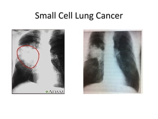
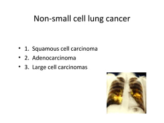
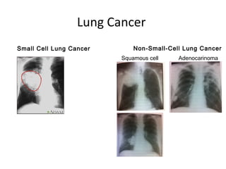
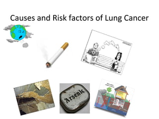
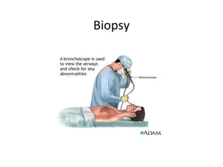
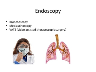
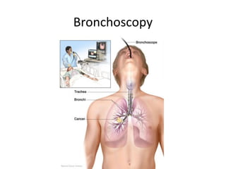
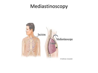
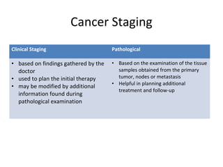
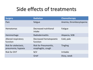
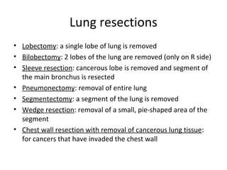
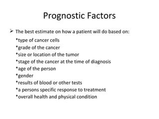
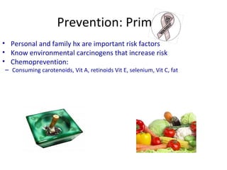
BRONCHOGENIC
CARCINOMA
Lung Cancer
Objectives
To provide a general overview of lung physiology
To explore the types and classifications of lung cancer
To provide causes and risk factors of lung cancer
To present the signs and symptoms of lung cancer in
throughout its progression
To explore assessment and diagnostic information of lung
cancer
To introduce diagnostic staging specific to lung cancer
To discuss treatments and side effects of lung cancer
General Overview of Lung Physiology:
Breathing
Healthy lung tissue
Diseased Lung Tissue
Types of Lung Cancer
Two main Types of Lung Cancer:
Small Cell Lung Cancer (20-25% of all lung cancers)
Non Small Cell Lung Cancer(most common ~80%)
Small Cell Lung Cancer
Non-small cell lung cancer
• 1. Squamous cell carcinoma
• 2. Adenocarcinoma
• 3. Large cell carcinomas
Squamous cell carcinoma
• Moderate to poor differentiation
• makes up 30-40% of all lung cancers
• more common in males
• most occur centrally in the large bronchi
• Uncommon metastasis that is slow effects the liver, adrenal
glands and lymph nodes.
• Associated with smoking
• Not easily visualized on xray (may delay dx)
• Most likely presents as a Pancoasts tumor
Adenocacinoma
• Increasing in frequency. Most common type of Lung cancer
(40-50% of all lung cancers).
• Clearly defined peripheral lesions (RLL lesion)
• Glandular appearance under a microscope
• Easily seen on a CXR
• Can occur in non-smokers
• Highly metastatic in nature
– Pts present with or develop brain, liver,
adrenal or bone metastasis
Large cell carcinomas
• makes up 15-20% of all lung cancers
• Poorly differentiated cells
• Tends to occur in the outer part (periphery) of lung, invading
sub-segmental bronchi or larger airways
• Metastasis is slow BUT
• Early metastasis occurs to the kidney, liver organs as well as
the adrenal glands
Lung Cancer
Small Cell Lung Cancer Non-Small-Cell Lung Cancer
Squamous cell Adenocarinoma
Causes and Risk factors of Lung Cancer
Signs and Symptoms of Lung Cancer
• Sometimes lung cancer does not cause any symptoms and is only found in
a routine x-ray.
• If a person with lung cancer does have symptoms, they will depend on the
location of the tumour in their lung.
• It is also imperative to note that the same symptoms can be caused by
other conditions, so may not necessarily mean cancer.
• Therefore it is important to consult a doctor when symptoms are present.
• Signs and symptoms also depend upon the location, size of the tumor,
degree of obstruction and existence of metastases
Signs and Symptoms of Lung Cancer
There are two types of signs and symptoms of
lung cancer:
1) Localized – involving the lung.
2) Generalized – involves other areas
throughout the body if the cancer has spread.
Localized Signs and Symptoms
Cough
Breathing Problems, SOB, stridor
Change in phlegm
Lung infection, hemoptysis
Hoarseness, Hiccups
Wt loss
Chest Pain and tightness
Pancasts Syndrome
Horner’s Syndrome
Pleural Effusion
Superior Vena Cava Syndrome
Fatigue
Generalized Signs and Symptoms
• Bone pain
• Headaches, mental status changes or neurologic
findings
• Abdominal pain, elevated liver function tests,
enlarged liver, gastrointestinal disturbances
(anorexia, cachexia), jaundice, hepatomegaly r/t liver
involvement
• Weight loss
Early/late Signs and Symptoms Of Lung
Cancer
Early Signs Late signs
Cough/chronic cough Bone pain, spinal cord compression
Dyspnea Chest pain/tightness
Hemoptysis Dysphagia
Chest/shoulder pain Head and neck edema
Recurring temperature Blurred vision, headaches
Recurring respiratory infections Weakness, anorexia, weight-loss, cachexia
Pleural effusion
Liver metastasis/regional spread
Diagnostic Tests
• CXR
• CT Scans
• MRI
• Sputum cytology
• Fibreoptic bronchoscopy
• Transthoracic fine needle aspiration
Laboratory Tests
 Blood Tests
*CBC-to check red/white blood cell & platelets
-to check bone marrow and organ function
*Blood Chemistry Test-to assess how organs
are functioning such as liver and kidney
 Biopsy-to determine if the tumor is cancer or not
-to determine the type of cancer
-to determine the grade of cancer (slow
or fast)
Biopsy
Endoscopy
• Bronchoscopy
• Mediastinoscopy
• VATS (video assisted thoracoscopic surgery)
Bronchoscopy
Mediastinoscopy
VATS (video assisted thoracoscopic
surgery)
Nursing Management for post endoscopic
procedures
Bronchoscopy Mediastinoscopy VATS
Monitor V/S; NPO status
maintained until return of
gag reflex.
Fever up to 101F can be
expected afterwards
Monitor VS; potential for
bleeding, infection and
dyspnea; NPO status until
return of gag reflex
Monitor V/S; potential for
bleeding, infection and
dyspnea; NPO status until
return of gag reflex
Post-op complications for those with
lung cancer
• Airway obstruction, dyspnea, hypoxemia, respiratory failure
• Anesthesia side effects (N/V)
• Bleeding (hypotension, cardiogenic shock)
• Cardiac dysthymias, CHF, fluid overload
• Fever, sepsis
• Pneumonia
• Pneumothorax
• Pulmonary embolus
• Wound dehiscence
• Prolonged hospitalization
• Death
Cancer Staging
Clinical Staging Pathological
• based on findings gathered by the
doctor
• used to plan the initial therapy
• may be modified by additional
information found during
pathological examination
• Based on the examination of the tissue
samples obtained from the primary
tumor, nodes or metastasis
• Helpful in planning additional
treatment and follow-up
Cancer Staging Systems
• The most common staging system for lung
cancer is the TNM System developed by the
International Union Against Cancer (UICC).
• Guides best course of treatment
• Estimates prognosis
• It is only useful in staging
NSCLC, when surgery is
considered.
TMN Staging system for Lung Cancer
T= Tumors : tumor size, (local
invasion)
N= Node : node involvement
(size and type)
M= Metastasis : general
involvement in organs and
tissues
Lung Cancer Staging Continued
• T: Tx, T0, Tis, T1-T4 (T3-
tumors greater than 7cm,
T4 is a tumor of any size)
• N: N0, N1, N2, N3
• M: M0, M1a, M1b
Medical Management
 The three main cancer treatments
are:
*surgery (lung resections)
*radiation therapy
*chemotherapy
 Other types of treatment that are
used to treat certain cancers are
hormonal therapy, biological therapy
or stem cell transplant.
Side effects of treatments
Surgery Radiation Chemotherapy
Pain fatigue Anemia, thrombocytopenia
Hemotomas Decreased nutritional
intake
Fatigue
Hemmorhage Radiodermatitis Alopecia, SOB
Altered respiratory
function
Decreased hematopoietic
function
Cold, pale
Risk for atelectasis,
pneumonia, hypoxia
Risk for Pneumonitis,
esophagitis, cough
Tingling
Risk for DVT N/V Irritable
Grief Dizzy, weak
Lung resections
• Lobectomy: a single lobe of lung is removed
• Bilobectomy: 2 lobes of the lung are removed (only on R side)
• Sleeve resection: cancerous lobe is removed and segment of
the main bronchus is resected
• Pneumonectomy: removal of entire lung
• Segmentectomy: a segment of the lung is removed
• Wedge resection: removal of a small, pie-shaped area of the
segment
• Chest wall resection with removal of cancerous lung tissue:
for cancers that have invaded the chest wall
Prognostic Factors
 The best estimate on how a patient will do based on:
*type of cancer cells
*grade of the cancer
*size or location of the tumor
*stage of the cancer at the time of diagnosis
*age of the person
*gender
*results of blood or other tests
*a persons specific response to treatment
*overall health and physical condition
Prevention: Primary
• Avoid the use of tobacco smoke
• Personal and family hx are important risk factors
• Know environmental carcinogens that increase risk
• Chemoprevention:
– Consuming carotenoids, Vit A, retinoids Vit E, selenium, Vit C, fat
Prevention: Secondary
• Aim is to early diagnose high risk populations
via screening
• CXR, MRI, CT scans, sputum cytology
Prevention: Tertiary
• Targeted at people who survived a cancer
disease
• Assists them to retain an optimal level of
functioning regardless of their potential
debilitating disease
Nurse’s Role with patients with lung cancer
• Coping with diagnosis
• Pre/post treatment education
• Education on managing breathlessness
• Referrals for ‘stop smoking advisor’
• Referring to services such as hospice care, dietitian, massage
therapy, counselors
• Advocate for appointments, scans etc.
• Educate client and family about disease process, treatment
options and S/E
• Provide resources for support groups and where information
and be sought
Nursing Interventions
• Management of N/V, weakness, fatigue, wt loss, appetite loss, altered
taste
• Pain management, education to avoid concern about addiction,
pharmacological and non-pharmacological
• Elevate HOB
• Splinting to aid in coughing
• Teach breathing exercises to ↑ diaphragmatic excursion and ↓ WOB
• DB&C
• Provide a vaporizer
• Relaxation techniques to ↓ anxiety r/t SOB
• Encourage energy conservation
• Encourage small amts of high-calorie and Pn foods freq.
Nursing Diagnoses
• Ineffective breathing pattern r/t loss of adequate ventilation
• Impaired gas exchange r/t excessive or thick secretions 2 to smoking;
r/t decreased passage of gases between alveoli of lungs and vascular
system
• Chronic pain related to Stage IV NSCLC diagnosis as evidenced by
client reporting “pain in right chest and lower ribs”.
• Risk of infection related to altered immune system secondary to
effects of cytotoxic drugs.
• Risk for disturbed self concept related to changes in lifestyle.
• Nausea related to effects of chemotherapy as evidenced by
client reporting feeling nauseated.
• Risk for deficient fluid volume related to gastrointestinal fluid loss
secondary to vomiting.
• Fatigue related to chemotherapy secondary to stage IV NSCLC as
evidenced by client reporting he “ no longer has the energy to play
with his grandchildren or visit his friends”.

Ca lung

  • 1.
  • 2.
  • 3.
    Objectives To provide ageneral overview of lung physiology To explore the types and classifications of lung cancer To provide causes and risk factors of lung cancer To present the signs and symptoms of lung cancer in throughout its progression To explore assessment and diagnostic information of lung cancer To introduce diagnostic staging specific to lung cancer To discuss treatments and side effects of lung cancer
  • 4.
    General Overview ofLung Physiology: Breathing
  • 5.
  • 6.
  • 7.
    Types of LungCancer Two main Types of Lung Cancer: Small Cell Lung Cancer (20-25% of all lung cancers) Non Small Cell Lung Cancer(most common ~80%)
  • 8.
  • 9.
    Non-small cell lungcancer • 1. Squamous cell carcinoma • 2. Adenocarcinoma • 3. Large cell carcinomas
  • 10.
    Squamous cell carcinoma •Moderate to poor differentiation • makes up 30-40% of all lung cancers • more common in males • most occur centrally in the large bronchi • Uncommon metastasis that is slow effects the liver, adrenal glands and lymph nodes. • Associated with smoking • Not easily visualized on xray (may delay dx) • Most likely presents as a Pancoasts tumor
  • 11.
    Adenocacinoma • Increasing infrequency. Most common type of Lung cancer (40-50% of all lung cancers). • Clearly defined peripheral lesions (RLL lesion) • Glandular appearance under a microscope • Easily seen on a CXR • Can occur in non-smokers • Highly metastatic in nature – Pts present with or develop brain, liver, adrenal or bone metastasis
  • 12.
    Large cell carcinomas •makes up 15-20% of all lung cancers • Poorly differentiated cells • Tends to occur in the outer part (periphery) of lung, invading sub-segmental bronchi or larger airways • Metastasis is slow BUT • Early metastasis occurs to the kidney, liver organs as well as the adrenal glands
  • 13.
    Lung Cancer Small CellLung Cancer Non-Small-Cell Lung Cancer Squamous cell Adenocarinoma
  • 14.
    Causes and Riskfactors of Lung Cancer
  • 15.
    Signs and Symptomsof Lung Cancer • Sometimes lung cancer does not cause any symptoms and is only found in a routine x-ray. • If a person with lung cancer does have symptoms, they will depend on the location of the tumour in their lung. • It is also imperative to note that the same symptoms can be caused by other conditions, so may not necessarily mean cancer. • Therefore it is important to consult a doctor when symptoms are present. • Signs and symptoms also depend upon the location, size of the tumor, degree of obstruction and existence of metastases
  • 16.
    Signs and Symptomsof Lung Cancer There are two types of signs and symptoms of lung cancer: 1) Localized – involving the lung. 2) Generalized – involves other areas throughout the body if the cancer has spread.
  • 17.
    Localized Signs andSymptoms Cough Breathing Problems, SOB, stridor Change in phlegm Lung infection, hemoptysis Hoarseness, Hiccups Wt loss Chest Pain and tightness Pancasts Syndrome Horner’s Syndrome Pleural Effusion Superior Vena Cava Syndrome Fatigue
  • 18.
    Generalized Signs andSymptoms • Bone pain • Headaches, mental status changes or neurologic findings • Abdominal pain, elevated liver function tests, enlarged liver, gastrointestinal disturbances (anorexia, cachexia), jaundice, hepatomegaly r/t liver involvement • Weight loss
  • 19.
    Early/late Signs andSymptoms Of Lung Cancer Early Signs Late signs Cough/chronic cough Bone pain, spinal cord compression Dyspnea Chest pain/tightness Hemoptysis Dysphagia Chest/shoulder pain Head and neck edema Recurring temperature Blurred vision, headaches Recurring respiratory infections Weakness, anorexia, weight-loss, cachexia Pleural effusion Liver metastasis/regional spread
  • 20.
    Diagnostic Tests • CXR •CT Scans • MRI • Sputum cytology • Fibreoptic bronchoscopy • Transthoracic fine needle aspiration
  • 21.
    Laboratory Tests  BloodTests *CBC-to check red/white blood cell & platelets -to check bone marrow and organ function *Blood Chemistry Test-to assess how organs are functioning such as liver and kidney  Biopsy-to determine if the tumor is cancer or not -to determine the type of cancer -to determine the grade of cancer (slow or fast)
  • 22.
  • 23.
    Endoscopy • Bronchoscopy • Mediastinoscopy •VATS (video assisted thoracoscopic surgery)
  • 24.
  • 25.
  • 26.
    VATS (video assistedthoracoscopic surgery)
  • 27.
    Nursing Management forpost endoscopic procedures Bronchoscopy Mediastinoscopy VATS Monitor V/S; NPO status maintained until return of gag reflex. Fever up to 101F can be expected afterwards Monitor VS; potential for bleeding, infection and dyspnea; NPO status until return of gag reflex Monitor V/S; potential for bleeding, infection and dyspnea; NPO status until return of gag reflex
  • 28.
    Post-op complications forthose with lung cancer • Airway obstruction, dyspnea, hypoxemia, respiratory failure • Anesthesia side effects (N/V) • Bleeding (hypotension, cardiogenic shock) • Cardiac dysthymias, CHF, fluid overload • Fever, sepsis • Pneumonia • Pneumothorax • Pulmonary embolus • Wound dehiscence • Prolonged hospitalization • Death
  • 29.
    Cancer Staging Clinical StagingPathological • based on findings gathered by the doctor • used to plan the initial therapy • may be modified by additional information found during pathological examination • Based on the examination of the tissue samples obtained from the primary tumor, nodes or metastasis • Helpful in planning additional treatment and follow-up
  • 30.
    Cancer Staging Systems •The most common staging system for lung cancer is the TNM System developed by the International Union Against Cancer (UICC). • Guides best course of treatment • Estimates prognosis • It is only useful in staging NSCLC, when surgery is considered.
  • 31.
    TMN Staging systemfor Lung Cancer T= Tumors : tumor size, (local invasion) N= Node : node involvement (size and type) M= Metastasis : general involvement in organs and tissues
  • 32.
    Lung Cancer StagingContinued • T: Tx, T0, Tis, T1-T4 (T3- tumors greater than 7cm, T4 is a tumor of any size) • N: N0, N1, N2, N3 • M: M0, M1a, M1b
  • 33.
    Medical Management  Thethree main cancer treatments are: *surgery (lung resections) *radiation therapy *chemotherapy  Other types of treatment that are used to treat certain cancers are hormonal therapy, biological therapy or stem cell transplant.
  • 34.
    Side effects oftreatments Surgery Radiation Chemotherapy Pain fatigue Anemia, thrombocytopenia Hemotomas Decreased nutritional intake Fatigue Hemmorhage Radiodermatitis Alopecia, SOB Altered respiratory function Decreased hematopoietic function Cold, pale Risk for atelectasis, pneumonia, hypoxia Risk for Pneumonitis, esophagitis, cough Tingling Risk for DVT N/V Irritable Grief Dizzy, weak
  • 35.
    Lung resections • Lobectomy:a single lobe of lung is removed • Bilobectomy: 2 lobes of the lung are removed (only on R side) • Sleeve resection: cancerous lobe is removed and segment of the main bronchus is resected • Pneumonectomy: removal of entire lung • Segmentectomy: a segment of the lung is removed • Wedge resection: removal of a small, pie-shaped area of the segment • Chest wall resection with removal of cancerous lung tissue: for cancers that have invaded the chest wall
  • 36.
    Prognostic Factors  Thebest estimate on how a patient will do based on: *type of cancer cells *grade of the cancer *size or location of the tumor *stage of the cancer at the time of diagnosis *age of the person *gender *results of blood or other tests *a persons specific response to treatment *overall health and physical condition
  • 37.
    Prevention: Primary • Avoidthe use of tobacco smoke • Personal and family hx are important risk factors • Know environmental carcinogens that increase risk • Chemoprevention: – Consuming carotenoids, Vit A, retinoids Vit E, selenium, Vit C, fat
  • 38.
    Prevention: Secondary • Aimis to early diagnose high risk populations via screening • CXR, MRI, CT scans, sputum cytology
  • 39.
    Prevention: Tertiary • Targetedat people who survived a cancer disease • Assists them to retain an optimal level of functioning regardless of their potential debilitating disease
  • 40.
    Nurse’s Role withpatients with lung cancer • Coping with diagnosis • Pre/post treatment education • Education on managing breathlessness • Referrals for ‘stop smoking advisor’ • Referring to services such as hospice care, dietitian, massage therapy, counselors • Advocate for appointments, scans etc. • Educate client and family about disease process, treatment options and S/E • Provide resources for support groups and where information and be sought
  • 41.
    Nursing Interventions • Managementof N/V, weakness, fatigue, wt loss, appetite loss, altered taste • Pain management, education to avoid concern about addiction, pharmacological and non-pharmacological • Elevate HOB • Splinting to aid in coughing • Teach breathing exercises to ↑ diaphragmatic excursion and ↓ WOB • DB&C • Provide a vaporizer • Relaxation techniques to ↓ anxiety r/t SOB • Encourage energy conservation • Encourage small amts of high-calorie and Pn foods freq.
  • 42.
    Nursing Diagnoses • Ineffectivebreathing pattern r/t loss of adequate ventilation • Impaired gas exchange r/t excessive or thick secretions 2 to smoking; r/t decreased passage of gases between alveoli of lungs and vascular system • Chronic pain related to Stage IV NSCLC diagnosis as evidenced by client reporting “pain in right chest and lower ribs”. • Risk of infection related to altered immune system secondary to effects of cytotoxic drugs. • Risk for disturbed self concept related to changes in lifestyle. • Nausea related to effects of chemotherapy as evidenced by client reporting feeling nauseated. • Risk for deficient fluid volume related to gastrointestinal fluid loss secondary to vomiting. • Fatigue related to chemotherapy secondary to stage IV NSCLC as evidenced by client reporting he “ no longer has the energy to play with his grandchildren or visit his friends”.

Editor's Notes

  • #3 Presently, lung cancer is the most commonly diagnosed cancer and the leading cause of cancer death in Canada for both men and women (Day et. a;l, 2010). It accounts “for 28.3% of cancer deaths in men and 26.3% in women. For men, the incidence of lung cancer has remained relatively constant, but in women it continues to rise.” (Day et. al, 2010, p. 630). In 2012, an estimated 25,600 Canadians will be diagnosed with lung cancer and 20,100 will die of it. Lung cancer remains the leading cause of cancer death for both men and women (Canadian lung association, 2011). Sadly, it is one of the most preventable of the cancers. In Nova Scotia this year, an estimated 2,700 people will die of cancer and 6,100 new cases will be diagnosed.Read more: http://www.cancer.ca/Nova%20Scotia/About%20cancer/Cancer%20statistics/Nova%20Scotia%20stats.aspx?sc_lang=en#ixzz23ed60ZZH This presentation will give you information regarding nursing a person with and at risk for lung cancer and the treatment options available.
  • #5 As we all know, normal respiration begins by inhaling air through the mouth and nose. This air flows down your trachea, which divides into the left and right bronchi, which carry air to each lung. Once inside the lung, the bronchi divide into smaller tubes called bronchioles and each bronchiole ends with alveoli. The alveoli are responsible for oxygenating the blood for circulation as well as removing carbon dioxide from the blood. As you can see, the right lobe has 3 sections or lobes and the left lung has 2 lobes. Read more: http://www.cancer.ca/Nova%20Scotia/About%20cancer/Types%20of%20cancer/What%20is%20lung%20cancer.aspx?sc_lang=en#ixzz23eYpoA8o
  • #8 95% of lung cancers are classified as either small cell lung cancer or non small cell lung cancer (Wallace, 2012).
  • #9 Small Cell Lung Cancer is the most aggressive form of lung cancer. It usually starts in the bronchi which is problematic because post-pneumonia and atelectasis often occur. These cancer cells are small and are considered to be quite aggressive in nature and they have a large growth factor. Because of these reasons, at the time of diagnosis, (60% of the time), these tumors have often metastasize to other parts of the body (brain, liver, and bone marrow) (Otto, 2001, p. 284). SCLC accounts for 20-25% of all lung cancers.
  • #10 NSCLC is any type of epithelial lung cancer other than small cell lung cancer. Non-small cell lung Ca usually grows and spreads more slowly than SCLC. **Add in 1 and 2 for a slide on each There are three sub-types of Non Small Cell Lung Cancer include: 1. Squamous cell carcinomas usually arise centrally in larger bronchi 2. Adenocarcinoma are often found in the periphery of the lungs 3. Large cell carcinomas can occur in any part of the lung and tend to grow and spread faster than the other two types
  • #11 “Squamous cell is the most likely lung cancer to present as a Pancoast’s tumor, which is high in the lung apex with extension to the chest wall, causing shoulder pain that radiates down the ulnar nerve” (Otto, 2001, p. 384) also known as epidermoid carcinoma. Squamous cell cancers are also known as epidermoid cancer that makes up 30-40% of all lung cancers This type of cancer is characterized by having cells that are moderate to poor in differentiation ( lacking in distinguishing features) This cancer is more common in males most originate in the central portion of the lungs such as in the large bronchi. It Is hard to detect by x-ray. For this reason, diagnoses will often be delayed. slow growing Uncommon metastasis that is slow, The cancer would eventually effects the liver, adrenal glands and lymph nodes. Associated with smoking
  • #12 most common type of lung cancer makes up 40-50% of all lung cancers most of these cancers originate at the peripheral areas of the lung, like the bronchial mucosa can also begin in scars caused by fibrosis, easily seen on x-rays can occur in non-smokers more common in women, strongly linked to smoking Slow metastasis can occur throughout the lung or other body organs
  • #13 http://www.youtube.com/watch?v=3wzjqbhbesI*** (NOT THAT GREAT) makes up 15-20%of all lung cancers and also has poorly differentiated cells tends to occur in the outer part (periphery) of lung, invading sub-segmental bronchi or larger airways Metastasis is slow BUT Early metastasis occurs to the kidney, liver organs as well as the adrenal glands
  • #15 Risk factors may increase a person’s chance of developing lung cancer. The factors that increase the risk of developing lung cancer include: smoking tobacco- is the predominant cause of Lung Ca and accounts for 80% of all new cases in women and 90% in men. Lung cancer is 10 times more likely to occur in smokers than non-smokers. second-hand smoke- studies have shown that people who are exposed to tobacco smoke in a closed environment (car, house, building) are at inc’d risk of developing lung Ca than those who are not exposed. Asbestos-Asbestos refers to a group of naturally occurring minerals that are used in some industries. Asbestos fibers have a tendency to easily shatter into small bits that can be suspended in the air and adhere to clothes. In the event that these asbestos particles are inhaled, they can enter into the lungs, damaging cells, escalating the risk for lung cancer development. Studies have revealed that workers exposed to great amount of asbestos are 3 to 4 times more at risk of developing lung cancer than those who work in asbestos free environment. Arsenic - Arsenic can be found in both surface water and groundwater sources, with levels generally higher in groundwater and is known to be a human carcinogenic (Health Canada, 2008, http://www.hc-sc.gc.ca/ewh-semt/pubs/water-eau/arsenic/rationale-justification-eng.php) Radon- Radon is gas that is undetectable, fragrance-free, and tasteless radioactive gas that occurs naturally in soil and rocks. It naturally occurs in can cause damage to the lungs that may lead to lung cancer. People who work in mines may be exposed to radon and, in some parts of the country, radon is found in houses. Smoking increases the risk of lung cancer even more for those already at risk because of exposure to radon. A kit available at most hardware stores allows homeowners to measure radon levels in their homes. The home radon test is relatively easy to use and inexpensive. Once a radon problem is corrected, the hazard is gone for good. There are also various carcinogens identified in the atmosphere from vehicle emissions and pollutants from refineries and manufacturing plants. Evidence suggests that the incidence of lung cancer is greater in urban areas as a result of the buildup of these pollutants (Day et. al, 2010, p. 630). Information from: http://info.cancer.ca/cce-ecc/default.aspx?Lang=E&toc=26 Some other risk factors are: Marijuana Pollution Industry work Lung Disease Personal History Diet Lung Diseases. Certain lung ailments, such as tuberculosis (TB), add to a person's likelihood of developing lung cancer. Lung cancer tends to grow in the regions of the lung that are scarred from TB. Other diseases such as tuberculosis (TB) and some types of pneumonia often leave scars on the lung. This scarring can increase the risk of developing lung cancer. People with diseases from breathing in certain minerals also have a higher risk of lung cancer. Personal History. A person who has a history of having lung cancer lung cancer is more prone to develop lung cancer again compared with someone who has never had lung cancer. Smoking cessation following a diagnosis of lung cancer may stop the development subsequent lung cancer. Additionally, People who have had prior experiences with radiation therapy on the chest at higher risk for lung cancer, especially if they smoke. Diet: Some reports propose that a diet low in fruits and vegetables may amplify the risk of lung cancer in people who are exposed to environmental tobacco smoke. It is believed that fruits and vegetables help protect against lung cancer. We already know greatest way to avoid developing lung cancer. Stop smoking or never start. The sooner you quit the better, it’s never too late to give up smoking.
  • #18 LOCALIZED The most common symptom is a persistent and non productive cough breathing problems:  shortness of breath         increased shortness of breath during physical activity wheezing due to the bronchus being partially obstructed high pitched breathing sounds (stridor) changes in phlegm (sputum): increased amount blood in the phlegm (hemoptysis) lung infection (pneumonia):  frequent lung infections may develop the lung infection might be found in the same area as the tumour Hoarseness: hiccups chest pain/tightness Pancoat’s syndrome is coined as a collective presentation of arm and shoulder pain, shrinking of the muscles of the hand and arm and Horner’s syndrome. It is likely to arise with the presence of lung Cancer. Horner’s syndrome: A condition that cause recessed eyeball, pupil constriction, droopy upper eyelid and decreased perspiration on the affected side of the face. This state is brought on my the paralysis of the trunk of cervical sympathetic nerve by such things as a lung tumor. Pleural effusion: build up of fluid in the space between the covering of the lung and the lining of the chest wall marked shortness of breath persistent cough pain in the chest that gets worse while breathing in (pleurisy) Superior vena cava syndrome: This condition occurs when the SVC is compressed or blocked by the tumor, resulting in little or no blood reaching the heart. Look for : -        shortness of breath -        sensation of fullness in the head -        facial swelling -        arm swelling -        chest pain -        difficulty swallowing Tracheal/esophageal obstruction Pericardial effusion which refers to fluid gathering in the pericardial space which houses the lungs. This excess fluid around the lungs may lead to cardiac tamponade. This is a life threatening event that weaken the body’s ability to fill up with blood during the diastolic portion of the heart beat. Hypoxia and dyspnea
  • #19 Bone pain r/t bone mets Headaches, mental status changes or neurologic findings resulting in brain mets Abdominal pain, elevated liver function tests, enlarged liver, gastrointestinal disturbances (anorexia, cachexia), jaundice, hepatomegaly r/t liver involvement Weight loss
  • #20 Often, lung cancer develops gradually and is asymptomatic until late in its course” (Day et. al, 2010, p. 631). The signs and symptoms depend on the location and size of the tumor, the degree of obstruction and the existence of metastases to regional or distant sites (Day et al., 2010). Early signs Chronic cough “The most frequent symptom of lung cancer is cough or a change in a chronic cough (a cough that changes character should alert us to be suspicious of lung cancer). Most people just ignore this and attribute it to smoking or a respiratory infection” (Day et al., 2010, pg. 632). Dyspnea occurs in 35-50% of patients Hemoptysis – blood expectorated Chest/shoulder pain- this may indicate chest wall or pleural involvement by a tumor. Recurring fever due to infection distal to tumor (suspect L Ca in those with repeated unresolved upper respiratory tract infections). Late signs If the tumor spreads to adjacent structure and regional lymph nodes, the patient may present with chest pain and tightness, hoarseness, dysphagia, head and neck edema and symptoms of pleural or pericardial effusion (Day et al., 2010, p. 632). The most common sites for metastases are lymph nodes, bone, brain, contralateral lung, adrenal glands and liver (Day et al., 2010). Other late sigs include: weakness, anorexia, weight-loss, cachexia (Otto, 2001).
  • #21 **As a nurse working with a client, what would be some things you could tell him or her about what to expect for each test? A chest x-ray is preformed to search for: pulmonary density, a solidary peripheral nodule (coin lesion) (a mass in the lung or airway) Atelectasis (collapsed lung) Infection fluid in the lung enlarged lymph nodes in the chest “A chest x ray is a painless, noninvasive test that creates pictures of the structures inside your chest, such as your heart, lungs, and blood vessels. "Noninvasive" means that no surgery is done and no instruments are inserted into your body Your ribs and spine are bony and absorb radiation well. They normally appear light on a chest x ray. Your lungs, which are filled with air, normally appear dark. A disease in the chest that changes how radiation is absorbed also will appear on a chest x ray”. (http://www.nhlbi.nih.gov/health/health-topics/topics/cxray/) Chest x rays have few risks. The amount of radiation used in a chest x ray is very small. A lead apron may be used to protect certain parts of your body from the radiation. CT scans shows the size, shape, and position of your lungs and other structures in your chest. Follow up on abnormal findings from standard chest x rays. Find the cause of lung symptoms, such as shortness of breath or chest pain. Find out whether you have a lung problem, such as a tumor, excess fluid around the lungs, or a pulmonary embolism. Most places will provide the patient with a gown. He/she will need to undress, usually down to their underwear, and put the gown on. If the place does not provide a gown the patient should wear loose-fitting clothes. Any woman who suspects she may be pregnant should tell her doctor beforehand. Doctors may ask the patient to fast (eat nothing) and even refrain from consuming liquids for a specific period before the scan. The patient will be asked to lie down on a motorized examination table, which then goes into the giant doughnut-like machine. The couch with the patient goes into the doughnut hole. MRI is like a CT only it uses magnetism instead of xrays, remove all metallic objects, fill out a screening form, asked to lie down on a comfortably padded table that gently glides you into the scanner. earplugs or headphones to protect your hearing because, when certain scanners operate, they may produce loud noises. These loud noises are normal and should not worry you. Nurse may inject a contrast agent called "gadolinium" in vein to help obtain a clearer picture of the area being examined. A saline solution will drip through IV to prevent clotting until the contrast material is injected at some point during the exam. The most important thing for the patient to do is to relax and lie still. Most MRI exams take between 15 to 45 minutes to complete depending on the body part imaged and how many images are needed.You will be asked to remain perfectly still during the time the imaging takes place, but between sequences some minor movement may be allowed. You will be guided.may breathe normally, however, for certain examinations it may be necessary for you to hold your breath for a short period of time. During your MRI examination, the MR system operator will be able to speak to you, hear you, and observe you at all times. Consult the scanner operator if you have any questions or feel anything unusual.When the MRI procedure is over, you may be asked to wait until the images are examined to determine if more images are needed. After the scan, you have no restrictions and can go about your normal activities. Sputum cytology is rarely used to make a dx of lung Ca; medical test in which a sample of sputum (mucus) is examined under a microscope to determine whether abnormal cells are present. A sample may be obtained either by the person coughing up mucus at home or in the doctor’s office or during a bronchoscopy. Remove dentures if you wear them. • Rinse your mouth with water. • Take about four deep breaths followed by a few short coughs, then inhale deeply and cough forcefully into the container. Make sure to get a sample from deep in your airway. (http://www.lung-cancer.com/sputum.html) however fibreoptic bronchoscopy is more commonly used and provides a detailed study of the tracheobronchial tree and allows for brushings, washings, and biopsies of suspicious areas. Test to see inside the airways of your lungs, or to get samples of mucus or tissue from the lungs. Bronchoscopy involves placing a thin tube-like instrument called a bronchoscope through the nose or mouth and down into the airways of the lungs. The tube has a mini-camera at its tip, and is able to carry pictures back to a video screen or camera. not to eat after midnight the night before (or about 8 hours before) the procedure. You will also receive instructions about taking your regular medicines, smoking and removing any dentures before the procedure. Before beginning the procedure, you will inhale an aerosol spray of a medicine like Novocain, which numbs the nose and throat area and helps to prevent coughing and gagging during the procedure. After that you will be given a sedative by vein. The sedative will help you to relax, and may make you feel sleepy. The sedative may also help you to forget any unpleasant sensations felt during the test. After the procedure, do not drink for 1⁄2 to 1 hour or until the numbness completely wears off. Do not drive home by yourself after the procedure; arrange for a family member or friend to take you home. Contact your doctor immediately if you have shortness of breath or chest pain, or you cough up more than a few tablespoons of blood at home. (http://patients.thoracic.org/information-series/en/resources/fiberoptic-bronchoscopy.pdf) A transthoracic fine needle aspiration A fine needle aspiration biopsy is a test done to see if a tumor is benign (non-cancerous) or malignant (cancerous.) Fine needle aspiration (FNA) is done by inserting a thin needle into a tumor and removing cells that can be evaluated under the microscope. A pathologist looks at the cells to see if the suspicious tumor is cancer, and if it is cancer, what type of cancer. With lung cancer, the needle is inserted into the chest through the skin. Doctors can make sure the needle goes to the right part of the lung by watching it through ultrasound or a CT scanner. Given cough suppressant, CT scan or help find target of biopsy, skin cleaned just above ribs, sedative and local anesthetic for area, <30mins, small incising in skin, hold breathe stay still, insert needle thru skin and chest wall, feel pressure and pain when reach surface of lung, pain when reach area for tissue extraction. CXR done to see no collapse, short recovery time and home the same day unless a complication. (http://www.youtube.com/watch?v=abvYaB2VcmI) http://lungcancer.about.com/od/glossary/g/FNA.htm
  • #23 A lung biopsy removes a small piece of lung tissue which can be analyzed at under a microscope to determine if the tumor is cancer or not to determine the type of cancer to determine the grade of cancer (slow or fast)
  • #24 There are three methods that I’ll discuss on how biopsies may be preformed and how surgeons can prove or disprove suspicions of lung cancer. These are Bronchoscopy, Mediastinoscopy and video assisted thoracoscopic surgery. After I explain each one I will talk about some nursing management that should be done post-op.
  • #25 This type of biopsy uses a lighted instrument (bronchoscope) inserted through the mouth or nose and into the airway to remove a lung tissue sample. (Web MD, 2012, http://www.webmd.com/lung/lung-biopsy). Bronchoscopies are used: To visually examine airways for tumors, obstructions, secretions or foreign bodies. To diagnose disease processes such as interstitial pulmonary diseases. To therapeutically remove foreign bodies, mucous plugs or excessive secretions. To locate the site and cause of hemoptysis. To treat malignant airway obstruction (http://www.endonurse.com/articles/2002/05/bronchoscopy-for-the-new-endoscopy-nurse.aspx)
  • #26 This procedure determines whether the cancer has spread the lymph nodes. Patients are usually given a general anesthesia. A small incision is made in the neck and a thin tube is inserted. Fluid samples and biopsies are then collected from the lymph nodes near the throat and lungs. The samples are tested for cancerous cells (http://www.lung.ca/diseases-maladies/cancer-cancer/signs-signes/diagnosis-diagnostique_e.php)
  • #27 Video-assisted thoracic surgery (VATS) is a recently developed type of surgery that enables doctors to view the inside of the chest cavity after making only very small incisions. It allows surgeons to take a biopsy close to the outside edges of the lung and to test them for cancer. It is also useful for diagnosing certain pneumonia infections, pneumothorax, infection, cysts and other thoracic disorders. (http://www.health.harvard.edu/diagnostic-tests/video-assisted-thoracic-surgery.htm) http://ca.video.search.yahoo.com/video/play;_ylt=A2KLqIqSYDZQuDsAakQWFQx.;_ylu=X3oDMTBrc3VyamVwBHNlYwNzcgRzbGsDdmlkBHZ0aWQD?p=lobectomy&vid=D81124D6FD06CA7C2ED6D81124D6FD06CA7C2ED6&l=2%3A00&turl=http%3A%2F%2Fts2.mm.bing.net%2Fvideos%2Fthumbnail.aspx%3Fq%3D4505773586186253%26id%3Daab7a3e98a6ab74f3a799026b5e8853d%26bid%3D1i58ygb91iQR2A%26bn%3DLargeThumb%26url%3Dhttp%253a%252f%252fwww.youtube.com%252fwatch%253fv%253detSe9915ZS4&rurl=http%3A%2F%2Fwww.youtube.com%2Fwatch%3Fv%3DetSe9915ZS4&tit=VATS+Lobectomy+for+Lung+Cancer&c=8&sigr=11auu2s71&fr=yfp-t-715
  • #28 Fowler's position. When oxygen saturations are greater than 92 percent, the patient is weaned off the oxygen therapy.
  • #30 Clinical staging is used to plan the preliminary therapy to treat the cancer. And it may be changed by additional data found during pathological examination. It is based on findings gathered by the doctor used to plan the initial therapy may be modified by additional information found during pathological examination.
  • #31 Clinical staging of cancer is founded on findings gathered from diagnostic tests preformed by the physician. This type of staging is used to plan the preliminary therapy to treat the cancer. And it may be changed by additional data found during pathological examination
  • #32 The TNM staging system is based on 3 parts – T for tumor, N for nodes, and M for Metastases.   T: indicates the size of the primary tumour and its degree of spread into nearby tissues (local invasion) N: indicates whether or not nearby lymph nodes are involved, the size of the involved nodes and/or how many contain cancer M: indicates whether or not cancer has spread (metastasized) to distant organs  Additional letters or numbers may be placed after "T", "N" and "M" to provide more specific details.
  • #33 Tx – The tumor size is unknown, or cancer cells are only found in sputum.T0 – There is no evidence of a primary tumor.Tis – Tumor in situ – The tumor is present only in the cells lining the airway and has not spread to nearby tissues.T1 – Tumors less than or equal to 3 cm (1 ½ inches). T3 – Tumors greater than 7 cmT4 – A tumor of any size. It is a tumor that invades structures in the chest such as the heart, major blood vessels near the heart, the trachea, the esophagus. N0 – No nodes are involved.N1 – The tumor has spread to nearby nodes on the same side of the body.N2 – The tumor has spread to nodes farther away, but on the same side of the chest.N3 – The tumor has spread to lymph nodes on the other side of the chest from the original tumor, or has spread to nodes near the collarbone or neck muscles. M – Metastasis (Spread) to Other Regions M0 - The tumor has not spread to distant regions.M1: M1a – The tumor has spread to the opposite lung, to the lung lining M1b – The tumor has spread to distant regions of the body, such as the brain or bones.
  • #35 Otto 2001 p. 408
  • #36 Lobectomy: Careful positioning of the patient is important, bed may elevated 30 to 45 degrees. Turning from back to operated side, but not completely to the un-operated side to prevent mediastinal shifting. **Deep breathing and coughing and ambulation.
  • #38 Beta carotene and vitamin A intake has been associated with decreased risk of lung cancer “The majority (at lease 85%) of lung cancer deaths are smoking related and therefore preventable” (Otto, 2001, p. 381). “As many as 1/3 of heavy smokers (25 cigs or more a day) who are 35 yrs will experience premature death from a smoking-related disease” (Otto, 2001, p. 382). Primary prevention mainly focuses on decreaseing the number of new smokers and helping present smokers quit (Otto, 2001). Smoking cessation programs and public education programs are crucial in primary prevention strategies against lung cancer (Otto, 2001). By 18, 75% of smokers have tried their first cigarette and 50% are regular smokers. Many teenage girls are recruited into smoking even before the age of 13 so education programs and peer leader presentations to be implemented at the junior high group or younger. With adults, there are many smoking cessation programs available but 95% of smokers who quit do no seek outside help Genetics: “There is some predisposition to lung cancer because the incidence of lung cancer appears to the 2-3 times that of the general population regardless of smoking status” (Day et al., 2010, p. 631). Arsenic, Radon and Asbestos are environmental agents known to be associated with the development of lung cancer. Much media coverage has been given to the risk of lung cancer resulting from exposure to these substances. Guidelines are now in place with occupational health and safety boards to control exposure, however, people should still have their homes tested to protect themselves (Otto, 2001). “Chemoprevention refers to the concept of reducing cancer risk in individuals who are highly susceptive to certain cancers by prescribing certain natural or chemical synthetic products or chemotherapy drugs that my reduce or supress the process of carcinogenesis. It seeks to stop the process of this process before the tumor develops” (Otto, 2001, p. 640). An example of this would be NSAIDS (cancer.net). “Smokers who eat a diet low in fruits and vegetables have an increased risk of developing lung cancer” (Day et al., 2010, p. 631). ”It has been hypothesized that carotenoids, particularly carotene or vit A, may be important. Vitamin E, selenium, vitamin C, fat, and retinoids are also being evaluated regarding their protective role against lung cancer” (Day et al., 2010, p. 631). “In order to greatly decrease the probability of getting lung cancer, people should consume a healthy diet, scheduled exercise and avoid tobacco products” (Otto, 2001, p. 640).
  • #39 “Secondary prevention is aimed at early diagnosis of lung cancer in populations at high risk (such as those over the age of 45 who have smoked heavily)” (Otto, 2001, p. 383). “In persons with symptoms, a history of lung disease, a family history of lung cancer, or a heavy smoking history, chest x-ray and sputum cytology are primary tools to screen for lung cancer” (Otto, 2001, p. 383). “Early findings showed that cells in sputum specimens that stained positive with anti-lung cancer antibodies were 91% predictive of the development of lung cancer within 2 years” (Otto, 2001, p. 383).
  • #40 “Tertiary prevention is aimed at individuals who have been diagnosed with cancer. The goal here is to assist them to an optimal level of functioning regardless of their potential disabling disease” (Otto, 2001, p. 640). Some strategies of tertiary prevention include surgery, radiation therapy and chemotherapy, as well as hospice and palliative care. Nurses also want to be cognizant of infection control for people receiving treatment to control their cancer because they are immunocompromised (N 405).
  • #41 Coping with diagnosis, pre/post treatment, advising on breathlessness and ways to improve it, referring to smoke stop advisor, living with lung cancer and referring to other services such as hospice care, dietician, massage, counsellors. Stop smoking advice is vitally important if patients are to have radical treatment as recovery can be complicated/delayed if smoking continues. Making sure patients aren't waiting for scans or appointments, service development and improvement. Educating patient and families about the disease process, treatment options and side-effects. Provide resources for support groups and where information and be sought (cancer.ca). Information taken from: http://news.bbc.co.uk/2/hi/health/7130216.stm
  • #42 Management of N/V, weakness, fatigue, wt loss, appetite loss, altered taste Pain management, education to avoid concern about addiction, pharmacological and non-pharmacological Elevate the head of the bed to ease the work of breathing and to prevent fluid collection in upper body (from superior vena cava syndrome). Teach breathing retraining exercises to increase diaphragmatic excursion and reduce work of breathing. Augment the patient’s ability to cough effectively by splinting the patient’s chest manually. Instruct the patient to inspire fully and cough two to three times in one breath. Provide humidifier or vaporizer to provide moisture to loosen secretions. Teach relaxation techniques to reduce anxiety associated with dyspnea. Allow the severely dyspneic patient to sleep in reclining chair. Encourage the patient to conserve energy by decreasing activities. Advise the patient to eat small amounts of high-calorie and high-protein foods frequently, rather than three daily meals. http://nursingcrib.com/nursing-notes-reviewer/lung-cancer/