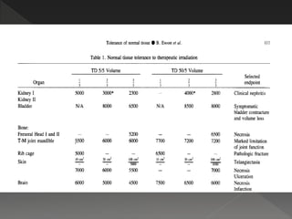
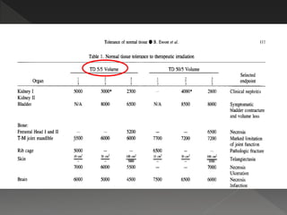
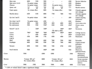
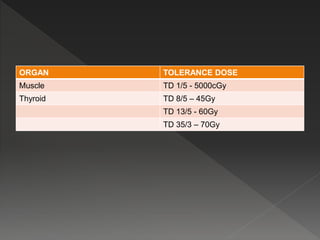
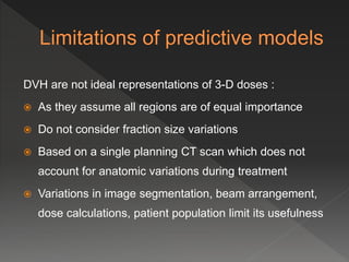
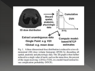
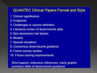
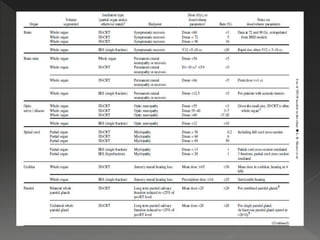
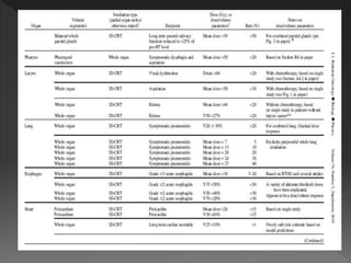
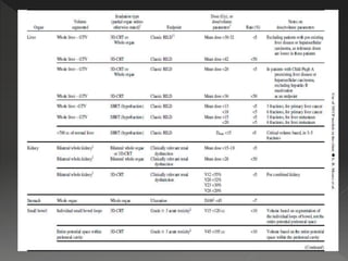
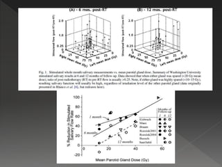
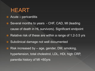
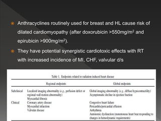
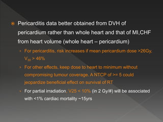
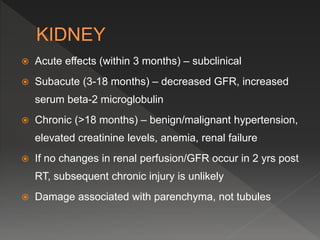
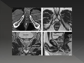
 Deals with the normal tissue complications with differing
doses of radiation
 The aim of a radiation oncologist is uncomplicated
locoregional control of cancer by radiation therapy.
 For this it is a must to have the precise knowledge of –
› Tumoricidal doses
› Normal tissue tolerances
 As part of the NCI contract a task force chaired by the
primary author was formed.
 Extensive collection and study of literature was carried
out to establish the protocols presented in the paper
with special emphasis to partial volume effects.
 However it was stated that it was stated that it is not a
comprehensive work
 The earliest step was the pulication of Rubin and
Cassarett of the tolerance doses in 1972.
 It summated the tolerance in terms of –
› TD 5/5 (the probability of 5% complication with in 5 years from
treatment)
› TD 50/5 (the probability of 50% complication with in 5 years
from treatment)
 28 critical sites were included
 Only conventional (1.8-2Gy/day, 5#/wk) was considered
 most severe end point was chosen
 normal organs were considered in terms of – 1/3, 2/3,
3/3 or whole volume (organs in which such division
could not be carried out – optic nerve, optic chiasma,
lens – whole organ was considered
 only adult tissue tolerance was considered
 The significant parameters considered were –
› Dose – time factors
› Partial volumes of normal tissues irradiated
 Only the linear component of the linear quadrantic
model was used.
 The expression of the NTCP model equation may be
written as :
NTCP (D,v) = exp [-N0 v-k exp {-aDG}]
 G = [1+d/(a/b)]
 a is the coefficient of lethal damage
 b/a is the reciprocal of a/b
 N0 (depicts proportion of stem cells) and k (depicts volume
dependence) are the tissue/organ specific, non-negative
adjustable parameters
 V is the uniformly irradiated partial volume of the tissue/organ
(=V/Vref where V is the uniformly irradiated volume of the
normal organ, Vref is the reference volume of the normal
organ.
 It was observed that the tolerance doses either showed
or did not show volume dependence.
 No volume dependence could be shown for – femoral
head and neck, rib cage, skin (telangeictasia), optic
nerve, optic chiasma, cauda equina, eye lens, retina,
ear (middle/external), parotid, larynx (edema), rectum,
thyroid.
ORGAN TOLERANCE DOSE
Muscle TD 1/5 - 5000cGy
Thyroid TD 8/5 – 45Gy
TD 13/5 - 60Gy
TD 35/3 – 70Gy
 All the planning and reporting was in terms of 2D
planning.
 With the advent and use of CT scan based 3D
conformal RT, a better understanding was needed and
was possible for the non-uniform irradiation of normal
organs.
 The steering committee of QUANTEC was formed and it
defined three aims :
› To provide a critical overview of the current state of knowledge
of dose-volume, dose-response relationships.
› To produce practical guidelines allowing the clinician to
reasonably categorize toxicity risk based on dose-volume
parameters.
› To identify future research avenues that would help improve
estimation or mitigation of side effects.
Reporting of the following is not proper :
 Side effects (grades)
 Different follow up duration
 Risk factors of patients
 Effect of concurrent chemotherapy
 Effect of altered fractionation schedules
 Altered beams (multiple beams, rotational delivery).
 Non-uniform dose distributions
 Uncertain alpha/beta ratio
 Need to confirm NTCP model by data in phase III trials
 Need to improve data analytic model
 Balancing the risks to different organs relating ‘‘whole
treatment’’ DVHs with acute toxicities
 Unknown applicability in children
DVH are not ideal representations of 3-D doses :
 As they assume all regions are of equal importance
 Do not consider fraction size variations
 Based on a single planning CT scan which does not
account for anatomic variations during treatment
 Variations in image segmentation, beam arrangement,
dose calculations, patient population limit its usefulness
 All the data available is for conventional 2Gy/#, 5#/week
schedule
 The data is for conventional parallel opposed fields with
shrinking field
 Now altered fractionation is widely used esp in trials
 The LQ model may not be of questionable benefit in
very novel fractions
 Chemotherapy exacerbates the severity of normal
tissue reactions
 Data regarding sequential/concurrent chemotherapy/RT
is lacking
 And the effects of different chemotherapy agent, doses,
schedules also differ
 Host factors – chronic liver disease, genetic, lifestyle –
may affect dose response relationships and are partly
responsible for inter-patient variations.
 Incorporating these data may produce better correlation
or analysis of toxicity.
 Earlier treatment was done keeping the normal organ
tolerance as the deciding factor
 Now the aim is to achieve a ‘tradeoff’ between the
benefit and risk from the planned treatment
 Aim is to have a good “quality of life adjusted tumour
control probability”
 Many times patients are lost to follow up
 Early death may result in a false low adverse event
reporting
 Relating acute toxicitiy during treatment with the DVH of
the whole treatment might be illogical
 Relating a late toxicity that occurs as a result of severe
acute toxicity with the DVH may give suboptimal results
of analysis
 In children the tissues develop at different rates and
temporal sequences
 In adults the same tissues are in a steady state with
relatively slow cell renewal kinetics.
 Same dose effect relationship does not exist with
children under radiation as with adults
 Generally, NTCP models attempt to reduce complicated
dosimetric and antomic information to a single risk
measure
 Most models fall into either of these three categories :
› DVH reduction models
› Tissue architecture models
› Multiple metric models
 Mechanism – damage to vasculature & myelin forming
oligodendriocytes diffuse cerebral edema & demyelination
 acute – nausea, vomiting, headache, seizures. Chronic –
radionecrosis, cognitive decline
 Endpoint – radionecrosis. Approx 1-2 yrs after RT
 Dose and volume dependent
 Toxicity increased by – age, chemotherapy, diabetes, spatial
factors, dose, dose/#, volume
 No evidence has shown that children are at increased risk of
radionecrosis
 For RT at <= 2.5Gy/#, incidence is 5% for 120 Gy (140-170
Gy) and 10% for 150 Gy (140-170Gy)
 Dmax < 60Gy – risk <3%
 For >2.5Gy/#, effect is unpredictable. Surprising toxicity with
>2Gy/# and 2#/day
 Risk increases if no of fractions/week >5
 For SRS, dose depends on volume. Maximum tolerated dose
for tumour volume –
› 31-40mm – 15Gy
› 21-30mm – 18Gy
› <20mm - >24Gy
 Neurocognitive decline is seen in children receiving >= 18Gy
within 4 yrs of RT.
 Enhanced by surgery, steroids, antiepileptics, opoids,
chemotherapy, female, NF-1 mutation, hydrocephalus
 13 point decreased IQ 5 yrs after RT. Poorer academic
achievement and self image and greater psychological
distress at 15 yrs
 In adults – many studies showed improved neurocognitive
effects due to antitumour effect of RT
 Mechanism – vascular damage radiation induced
optic neuropathy (RON).
 Other mechanisms – vascular insufficiency to optic
nerve/tracts, retina, occipital cortex
 Presents with rapid visual loss
 Endpoint – visual acuity 20/100
 Defined by the size and extent of the “visual field”
 Optic nerve – U/L, chiasm – B/L, optic tract spread out
near occipital cortes – small visual field defect
 Dmax is often the only data reported
 TD 5/5 – 50Gy, TD 50/5 – 65Gy (Emami et al)
 Researchers found incidence of RION is low for Dmax
<54Gy (<3%) with a steep increase in complications
>60Gy
 Complications for a given dose increase for dose >=
1.9Gy/#
 Increased risk in pituitary tumours
 Same tolerance for protons therapy (rather lower risk of
complications)
 For SRS, a dose of 12Gy for optic tolerance
 Risk increases with increasing age
 Inconsistent data on effect of diabetes, hypertension,
chemotherapy, previous irradiation
 Mechanism – vascular endothelial damage, glial cell
injury  pain, paresthesias, sensory deficits, paralysis,
bowel-bladder incontinence, Brown-Sequard syndrome
 Not included – Babinski’s sign, Lhermitte’s sign
 Endpoint – myelitis, MRI evidence of myelitis
 Presents after 6months to 2 years post RT
 Different tolerance for full thickness, partial thickness,
peripheral/central fibres may be defined in later years
 For 1.8-2Gy/#, estimated risk of myelopathy is 0.2%,
6% and 50% for doses of 50Gy, 60Gy and ~69Gy
 For Dmax SRS<=13Gy, hypofractionation <=20Gy
 For reirradiation, essentially no myelopathy if cumulative
dose <=60Gy
 Risk increased in immature spine, concurrent use of
intrathecal chemotherapy
 Cancer therapy evaluation programme (CTEP) grades
brain stem injury as –
› Grade 1 – mild/asymptomatic
› Grade 2 – moderate, not interfering with activities of daily living
(ADL)
› Grade 3 – severe, interfering with ADL
› Grade 4 – life threatening or disabling, intervention indicated
› Grade 5 - death
 End point – brain stem necrosis/MRI e/o injury
 Same mechanism as brain – brain stem has more white
matter
 Whole brainstem Dmax <54Gy, 1-10cc Dmax = 59Gy(2Gy/#) –
not different in pediatric age
 Risk increases > 64Gy
 Factors increasing risk – 2or more skull base surgeries,
diabetes, hypertension, V60> 0.9ml, hydrocephalus, tumour
close to brain stem
 SRS – cranial neuropathy >= 12.5Gy
 Does not refer to acute, chronic otitis media referred to
in Emami et al’s paper. It deals with damage to cochlea
and acoustic nerve
 Grading of hearing loss is done using Gardner-
Robertson hearing grade (GRGH scale)
 Sensorineural hearing loss (SNHL) is defined as an
increase in the bone conduction threshhold(BCT) at key
human speech frequencies (0.5-4kHz)
 Post RT hearing loss is more at higher frequencies
 For 2Gy/# are- mean dose to cochlea <=45Gy (<30%
risk of 4kHz frequency SNHL)
 For SRS dose <=12-14Gy
 Risk increased by – total dose, dose/#, FSRT better
hearing than SRS, adjuvant/concurrent cisplatin (not
with neoadjuvant cisplatin), older age, males>females,
better pre RT hearing, post RT otitis media, NF-2,
cerebral spinal fluid shunt
 Hearing assessment should be started 6 months post
RT and be done biannually
 Control is – pre RT hearing of same ear/C/L ear
hearing/age adjusted hearing data
 CAUTION –
› dose should be kept minimum with concurrent cisplatin
› Data presented may not be applicable for altered fractionation
› Data applies only to adult patients
› Data may not be representative in cases of vestibular
schwannoma with NF-2
 Stimulated saliva production is mainly (60-70%) from parotid
gland (balance from other glands). Unstimulated/resting from
submandibular sublingual and minor oral salivary glands
 Xerostomia leads to poor dental hygiene, oral pain, difficulty
in chewing and swallowing
 Endpoints –
› patient observation (loss of taste, dryness)
› objective – unstimulated and stimulated saliva production
› imaging (scintigraphy, dynamic MRI sialography
 Reduction upto 25% of pre RT saliva production
 “recovery overshoot” can occur with >100% saliva production
as compared to pre RT production
 Effect of doses –
› Minimum effect -10-15Gy (in 1 week of starting RT)
› Increases at 20-40Gy
› strong reduction (>75%) at >40Gy
 Severe xerostomia avoided if one parotid spared <20Gy or
both parotid <25Gy for 1.5-1.8Gy/#
 When oncologically safe, submandibular gland preservation
<35Gy reduces xerostomia
 Recording grade 4 xerostomia
 Recovery by 24 months
 Xerostomia risk is reduced by sparing atleast one
parotid or even one submandibular gland
 Under evaluation – hyperfractionation decreases effect,
sparing submandibular gland to achieve >25% pre RT
saliva production
 Amifostine increases tolerance of submandibular and
parotid by 9Gy
 80% present within 10 months of RT
 Approx 5-50% lung, 5-10% mediastinal LN, 1-5% lung
cases treated with RT develop RP
 Problems –
› Relevant grade of symptoms is controversial
› Dyspnea is non specific
› Different physicians have different assesment
› Lung tumour shrinkage makes symptoms better sometimes
 The gradual increase in dose response suggests that there is
no absolute MLD below which there is no pneumonitis
 Tolerance vary for different fraction sizes
 Tolerance depends on technique used
 Radiation associated dyspnea is seen more in lower
lung tumous than upper lobe
 Factors affecting risk of RP –
› Lesser in young age <60-70yrs
› Increased for mesothelioma treated with IMRT
› Effect of surgery not yet established
› INTERESTINGLY, CURRENT SMOKERS HAVE A LESSER
RISK
› Chemo may increase risk (docetaxel, gemcitabine) (not
cisplatin, carboplatin, paclitaxel, etoposide)
 No definite threshholds described
 Prudent to keep V20<= 30% and MLD <=20-23Gy
(conventional fractionation)
 Mesothelioma – V20<= 4-10% and MLD <= 8Gy
 Limit central airway dose to <= 80Gy to reduce risk of
bronchial stricture
 Acute esophagitis peaks 4-8 weeks after starting RT,
grade 2 or higher is considered
 Stricture develops ~3-8 months after RT. Later
esophagitis grade 1 or higher is considered
 Differentiate symptoms with that of candidiasis, herpes
simplex esophagitis, preexisting GERD, incidental
irradiation of stomach  gastritis, intero-observer
variation
 To assess dose, whole length of esophagus should be
included in the scan
 Parameters studied – absolute volume (a V dose),
absolute area (aAdose), percentage of a reference
volume (V dose), reference area (Adose)
 Intermediate toxicity develops at 30-50Gy
 DVH showing cumulative dose >50Gy has high
statistical significance of esophagitis
 Rates of acute grade 2 or greater esophagitis increasing
to >30% as V70 > 20%, V50>40%, V35>50%
 Risk increased by hyperfractionation, concurrent boost,
CCT, preexisting dysphagia, increasing nodal stage
 Ongoing Phase III Intergroup Trial (RTOG0617) has
recommended but not mandated – mean dose to
esophagus <34Gy and V60 be calculated for all patients
 Nausea & vomiting within hours after radiation
 Days to weeks later – dyspepsia, ulceration, bleeding (may
be life threatening)
 Long term effects – long-term dyspepsia, ulceration
 Emami et al referred to the TD5/5 (50Gy) and TD 50/5
(65Gy) for late effects and not for the acute effects
 Whole stomachD100 <45Gy  <5-7% risk of ulceration
 SBRT dose, limit Dmax <22.5Gy to <4% or 5cc with max
point dose of <30Gy for 3 fraction SBRT
 1-2 weeks after RT – cramping and diarrhea d/t
interference with nutrient absorption  weight loss
 Weeks to months post RT – late obstruction as a result
of adhesions that restrict intestinal mobility
 Long term effects – (usually within 3 yrs post RT)
ulceration, fistula, perforation, bleeding
 Grade 2-4 toxicity is usually considred life threatening
 Emami et al , whole organ TD5/5 – 40Gy, TD 50/5 –
55Gy. Partial organ TD5/5 - 50Gy, TD 50/5 – 60 Gy
 In modern series doses are concordant with the Emami
series
 Restrict V15 < 120cc – when delineating individual bowel
loops and V45 <195cc - when delineating entire
peritoneal cavity.
 For SBRT , volume receiving >12.5 Gy be kept < 30cc
and max point dose <30Gy
 Acute – pericarditis
 Several months to years - CHF, CAD, MI (leading
cause of death in HL survivors). Significant endpoint
 Relative risk of these are within a range of 1.2-3.5 yrs
 Subclinical damage not well documented
 Risk increased by – age, gender, DM, smoking,
hypertension, total cholestrol, LDL, HDl, high CRP,
parentla history of MI <60yrs
 Anthracyclines routinely used for breast and HL cause risk of
dilated cardiomyopathy (after doxorubicin >550mg/m2 and
epirubicin >900mg/m2).
 They have potential synergistic cardiotoxic effects with RT
with increased incidence of MI, CHF, valvular d/s
 Pericarditis data better obtained from DVH of
pericardium rather than whole heart and that of MI,CHF
from heart volume (whole heart – pericardium)
› For pericarditis, risk increases if mean pericardium dose >26Gy,
V30 > 46%
› For other effects, keep dose to heart to minimum without
compromising tumour coverage. A NTCP of >= 5 could
jeopardize beneficial effect on survival of RT
› For partial irradiation, V25 < 10% (in 2 Gy/#) will be associated
with <1% cardiac mortality ~15yrs
 Acute effects (within 3 months) – subclinical
 Subacute (3-18 months) – decreased GFR, increased
serum beta-2 microglobulin
 Chronic (>18 months) – benign/malignant hypertension,
elevated creatinine levels, anemia, renal failure
 If no changes in renal perfusion/GFR occur in 2 yrs post
RT, subsequent chronic injury is unlikely
 Damage associated with parenchyma, not tubules
 Whole kidney tolerance (as in TBI) is in accordance with
Emami et al – TD5/5 – 18-23Gy and TD 50/5 – 28Gy in
0.5-1.25 Gy/#. Creatinine clearence affected after 10-20
Gy. Mean dose <15-18Gy
 Other parameters for <5% clinically relevant renal
dysfunction –
› V12 <55%
› V20 <32%
› V23 < 30%
› V28 <20%
 These findings improved with time due to the reserve
capacity of the spared renal tissue though reparative
capacity blunted with RT
 Pediatric kidneys are very sensitive with 12-14 Gy at
1.25-1.5Gy/# causing decreased GFR etc. no kidney
failure after 10-12 Gy in 1.5-2Gy/#
 Age <5 yrs associated with increased risk of acute renal
dysfunction post TBI
 Risk increased with –
› Chemotherapy
› Diabetes
› Hypertension
› underlying renal
insufficiency
› liver disease
› heart disease
› smoking
 Acute – during/soon after RT. softer./diarrhea-like stools,
pain, sensation of rectal distention and cramping,
frequency, occasionally ulceration and bleeding
 Late – 3-4 years after RT. Stricture, diminished rectal
compliance, decreasing storage capacity  frequent
bowel movements, anal musculature damage  fecal
incontinence/stricture
 Endpoint – rectal bleeding/grade>=2 rectal toxicity
 Significant late rectal toxicity with >=60Gy. Vdose has not
been found to be significant for rectal doses <=45Gy
 Risk increased by DM, hemorrhoids, inflammatory
bowel disease, advanced disease, IBD, androgen
deprivation therapy, rectum size, prior abdominal
surgery, severe acute rectal toxicity
 Severe acute toxicity can result in high late rectal
proctopathy
 Dose limits described to limit grade 2 toxicity to <15%
and grade 3 <10% for upto 79.2Gy in 1.8-2Gy/# –
› V50 <50%
› V60 <35%
› V65 < 25%
› V70 < 20%
› V75 < 15%
 Acute – during or soon after RT. Dysuria, haematuria,
frequency, nocturia
 Late – underreported due to latency period of decades.
Dysuria, urgency, frequency, contacture, spasm, reduced
flow, incontinence, hematuria, fistula, obstruction, ulceration,
necrosis
 Symptoms may be urethral in origin and difficult to
differentiate
 Physician observation differences may be there
 Risk increased by – pre RT GU morbidity, increasing
age, hormonal therapy, surgery (TURP), prostatectomy,
hysterectomy, biopsy), chemotherapy
 Prescribed dose limits –
› Dmax <=65 (for <=6% grade 3 RTOG toxicity)
› V65 < 50%
› V70 <35%
› V75 < 25%
› V80 < 15%
 Grade 2,3,4,5 liver dysfunction ( jaundice, asterixis,
encephalopathy, coma, death respectively) rarely occur with
RT. Elevation of liver enzymes occur (grade 1 CTCAE) in 3-4
months
 Endpoint – Child Pugh score >=2, reactivation of hepatitis B
 Divided into classic (elevation of liver enzymes 2 fold,
pathologic evidence of injury) and non classic (elevation of
liver enzymes >5fold, child pugh score >=2) RILD (radiation
induced liver disease)
 Risk increased in – HCC> metastases, Child Pugh B/C
> A, cirrhosis, hepatitis B carrier, prior transcatheter
arterial chemoembolization, concurrent chemotherapy,
portal vein tumor thrombus, tumour stage, male,
increased dose/#
 Progressive edema & fibrosis after RT could defeat the
purpose of RT – organ & functional preservation
 Endpoints – laryngeal edema, vocal function
 Edema – RTOG grade 0 – no edema, 1- slight edema,
2- moderate, 3 – severe, 4 – necrosis
 Vocal function – assessed by patient focused
questionnaire, instruments (videostroboscopy,
aerodynamic measurement of phonation time, human
observation
 Risk increased by – initial advanced disease,
concurrent chemotherapy (doubles risk)
 Vocal dysfunction develops as a result of damage to
larynx, supralaryngeal structures, decreased saliva
production, damage to intrinsic musculature and soft
tissue
 Endpoints – instruments (modified barium swallow,
esophagography), subjective evaluation (CTCAE,
RTOG), patient reported QOL
 Risk of dysphagia  aspiration could be decreased by
keeping dose to supraglottic area, larynx, upper
esophageal sphincter to < 60Gy or using brachytherapy
to reduce pharyngeal dose
 Nasogastric tube decreases risk
 Prescribed dose limits
› Vocal dysfunction – Dmax <66Gy
› Aspiration – mean dose < 50Gy
› Edema – mean dose < 44Gy
- V50 < 27%
 Erectile dysfunction is a common complication following RT
for prostate cancer. Rates are
› 24% after brachytherapy alone
› 40% after brachy + EBRT
› 45% after EBRT alone
› 66% after nerve sparing –RP
› 75% after non-nerve sparing RP
› 87% for cryosurgery
 Time course – days to years, then evolves gradually
 Additive effect of age, diabetes, hypertension, smoking
 Assessment by self-administered questionnaires of IIEF
(international Index of Erectile Function).
 Tests - nocturnal penile tumescence, somatosensory
evoked potentials, bulbocavernous reflex latency, penile
electromyography, colour duplex doppler ultrasound,
dynamic infusion cavernosometry, pharmacotesting
 IIEF scoring below 21 is to be considered – none (25-
22), mild (21-17), mild to moderate (16-12), moderate
(11-8), severe (7-5)
 It is prudent to keep dose to
95% of penile bulb volume
<50Gy. D70 < 70Gy, D90 <
50Gy
 Its acknowledged that penile
bulb may not be the critical
component of the erectile
apparatus, but is used as a
surrogate.

Quantec dr. upasna saxena (2)

  • 2.
     Deals withthe normal tissue complications with differing doses of radiation  The aim of a radiation oncologist is uncomplicated locoregional control of cancer by radiation therapy.  For this it is a must to have the precise knowledge of – › Tumoricidal doses › Normal tissue tolerances
  • 4.
     As partof the NCI contract a task force chaired by the primary author was formed.  Extensive collection and study of literature was carried out to establish the protocols presented in the paper with special emphasis to partial volume effects.  However it was stated that it was stated that it is not a comprehensive work
  • 5.
     The earlieststep was the pulication of Rubin and Cassarett of the tolerance doses in 1972.  It summated the tolerance in terms of – › TD 5/5 (the probability of 5% complication with in 5 years from treatment) › TD 50/5 (the probability of 50% complication with in 5 years from treatment)
  • 6.
     28 criticalsites were included  Only conventional (1.8-2Gy/day, 5#/wk) was considered  most severe end point was chosen  normal organs were considered in terms of – 1/3, 2/3, 3/3 or whole volume (organs in which such division could not be carried out – optic nerve, optic chiasma, lens – whole organ was considered  only adult tissue tolerance was considered
  • 7.
     The significantparameters considered were – › Dose – time factors › Partial volumes of normal tissues irradiated  Only the linear component of the linear quadrantic model was used.  The expression of the NTCP model equation may be written as : NTCP (D,v) = exp [-N0 v-k exp {-aDG}]
  • 8.
     G =[1+d/(a/b)]  a is the coefficient of lethal damage  b/a is the reciprocal of a/b  N0 (depicts proportion of stem cells) and k (depicts volume dependence) are the tissue/organ specific, non-negative adjustable parameters  V is the uniformly irradiated partial volume of the tissue/organ (=V/Vref where V is the uniformly irradiated volume of the normal organ, Vref is the reference volume of the normal organ.
  • 9.
     It wasobserved that the tolerance doses either showed or did not show volume dependence.  No volume dependence could be shown for – femoral head and neck, rib cage, skin (telangeictasia), optic nerve, optic chiasma, cauda equina, eye lens, retina, ear (middle/external), parotid, larynx (edema), rectum, thyroid.
  • 13.
    ORGAN TOLERANCE DOSE MuscleTD 1/5 - 5000cGy Thyroid TD 8/5 – 45Gy TD 13/5 - 60Gy TD 35/3 – 70Gy
  • 15.
     All theplanning and reporting was in terms of 2D planning.  With the advent and use of CT scan based 3D conformal RT, a better understanding was needed and was possible for the non-uniform irradiation of normal organs.
  • 16.
     The steeringcommittee of QUANTEC was formed and it defined three aims : › To provide a critical overview of the current state of knowledge of dose-volume, dose-response relationships. › To produce practical guidelines allowing the clinician to reasonably categorize toxicity risk based on dose-volume parameters. › To identify future research avenues that would help improve estimation or mitigation of side effects.
  • 17.
    Reporting of thefollowing is not proper :  Side effects (grades)  Different follow up duration  Risk factors of patients  Effect of concurrent chemotherapy  Effect of altered fractionation schedules  Altered beams (multiple beams, rotational delivery).
  • 18.
     Non-uniform dosedistributions  Uncertain alpha/beta ratio  Need to confirm NTCP model by data in phase III trials  Need to improve data analytic model  Balancing the risks to different organs relating ‘‘whole treatment’’ DVHs with acute toxicities  Unknown applicability in children
  • 20.
    DVH are notideal representations of 3-D doses :  As they assume all regions are of equal importance  Do not consider fraction size variations  Based on a single planning CT scan which does not account for anatomic variations during treatment  Variations in image segmentation, beam arrangement, dose calculations, patient population limit its usefulness
  • 22.
     All thedata available is for conventional 2Gy/#, 5#/week schedule  The data is for conventional parallel opposed fields with shrinking field  Now altered fractionation is widely used esp in trials  The LQ model may not be of questionable benefit in very novel fractions
  • 23.
     Chemotherapy exacerbatesthe severity of normal tissue reactions  Data regarding sequential/concurrent chemotherapy/RT is lacking  And the effects of different chemotherapy agent, doses, schedules also differ
  • 24.
     Host factors– chronic liver disease, genetic, lifestyle – may affect dose response relationships and are partly responsible for inter-patient variations.  Incorporating these data may produce better correlation or analysis of toxicity.
  • 25.
     Earlier treatmentwas done keeping the normal organ tolerance as the deciding factor  Now the aim is to achieve a ‘tradeoff’ between the benefit and risk from the planned treatment  Aim is to have a good “quality of life adjusted tumour control probability”
  • 26.
     Many timespatients are lost to follow up  Early death may result in a false low adverse event reporting
  • 27.
     Relating acutetoxicitiy during treatment with the DVH of the whole treatment might be illogical  Relating a late toxicity that occurs as a result of severe acute toxicity with the DVH may give suboptimal results of analysis
  • 28.
     In childrenthe tissues develop at different rates and temporal sequences  In adults the same tissues are in a steady state with relatively slow cell renewal kinetics.  Same dose effect relationship does not exist with children under radiation as with adults
  • 30.
     Generally, NTCPmodels attempt to reduce complicated dosimetric and antomic information to a single risk measure  Most models fall into either of these three categories : › DVH reduction models › Tissue architecture models › Multiple metric models
  • 38.
     Mechanism –damage to vasculature & myelin forming oligodendriocytes diffuse cerebral edema & demyelination  acute – nausea, vomiting, headache, seizures. Chronic – radionecrosis, cognitive decline  Endpoint – radionecrosis. Approx 1-2 yrs after RT  Dose and volume dependent  Toxicity increased by – age, chemotherapy, diabetes, spatial factors, dose, dose/#, volume  No evidence has shown that children are at increased risk of radionecrosis
  • 39.
     For RTat <= 2.5Gy/#, incidence is 5% for 120 Gy (140-170 Gy) and 10% for 150 Gy (140-170Gy)  Dmax < 60Gy – risk <3%  For >2.5Gy/#, effect is unpredictable. Surprising toxicity with >2Gy/# and 2#/day  Risk increases if no of fractions/week >5  For SRS, dose depends on volume. Maximum tolerated dose for tumour volume – › 31-40mm – 15Gy › 21-30mm – 18Gy › <20mm - >24Gy
  • 41.
     Neurocognitive declineis seen in children receiving >= 18Gy within 4 yrs of RT.  Enhanced by surgery, steroids, antiepileptics, opoids, chemotherapy, female, NF-1 mutation, hydrocephalus  13 point decreased IQ 5 yrs after RT. Poorer academic achievement and self image and greater psychological distress at 15 yrs  In adults – many studies showed improved neurocognitive effects due to antitumour effect of RT
  • 42.
     Mechanism –vascular damage radiation induced optic neuropathy (RON).  Other mechanisms – vascular insufficiency to optic nerve/tracts, retina, occipital cortex  Presents with rapid visual loss  Endpoint – visual acuity 20/100  Defined by the size and extent of the “visual field”  Optic nerve – U/L, chiasm – B/L, optic tract spread out near occipital cortes – small visual field defect
  • 43.
     Dmax isoften the only data reported  TD 5/5 – 50Gy, TD 50/5 – 65Gy (Emami et al)  Researchers found incidence of RION is low for Dmax <54Gy (<3%) with a steep increase in complications >60Gy  Complications for a given dose increase for dose >= 1.9Gy/#
  • 45.
     Increased riskin pituitary tumours  Same tolerance for protons therapy (rather lower risk of complications)  For SRS, a dose of 12Gy for optic tolerance  Risk increases with increasing age  Inconsistent data on effect of diabetes, hypertension, chemotherapy, previous irradiation
  • 46.
     Mechanism –vascular endothelial damage, glial cell injury  pain, paresthesias, sensory deficits, paralysis, bowel-bladder incontinence, Brown-Sequard syndrome  Not included – Babinski’s sign, Lhermitte’s sign  Endpoint – myelitis, MRI evidence of myelitis  Presents after 6months to 2 years post RT  Different tolerance for full thickness, partial thickness, peripheral/central fibres may be defined in later years
  • 47.
     For 1.8-2Gy/#,estimated risk of myelopathy is 0.2%, 6% and 50% for doses of 50Gy, 60Gy and ~69Gy  For Dmax SRS<=13Gy, hypofractionation <=20Gy  For reirradiation, essentially no myelopathy if cumulative dose <=60Gy  Risk increased in immature spine, concurrent use of intrathecal chemotherapy
  • 48.
     Cancer therapyevaluation programme (CTEP) grades brain stem injury as – › Grade 1 – mild/asymptomatic › Grade 2 – moderate, not interfering with activities of daily living (ADL) › Grade 3 – severe, interfering with ADL › Grade 4 – life threatening or disabling, intervention indicated › Grade 5 - death
  • 49.
     End point– brain stem necrosis/MRI e/o injury  Same mechanism as brain – brain stem has more white matter  Whole brainstem Dmax <54Gy, 1-10cc Dmax = 59Gy(2Gy/#) – not different in pediatric age  Risk increases > 64Gy  Factors increasing risk – 2or more skull base surgeries, diabetes, hypertension, V60> 0.9ml, hydrocephalus, tumour close to brain stem  SRS – cranial neuropathy >= 12.5Gy
  • 50.
     Does notrefer to acute, chronic otitis media referred to in Emami et al’s paper. It deals with damage to cochlea and acoustic nerve  Grading of hearing loss is done using Gardner- Robertson hearing grade (GRGH scale)  Sensorineural hearing loss (SNHL) is defined as an increase in the bone conduction threshhold(BCT) at key human speech frequencies (0.5-4kHz)  Post RT hearing loss is more at higher frequencies
  • 53.
     For 2Gy/#are- mean dose to cochlea <=45Gy (<30% risk of 4kHz frequency SNHL)  For SRS dose <=12-14Gy  Risk increased by – total dose, dose/#, FSRT better hearing than SRS, adjuvant/concurrent cisplatin (not with neoadjuvant cisplatin), older age, males>females, better pre RT hearing, post RT otitis media, NF-2, cerebral spinal fluid shunt
  • 54.
     Hearing assessmentshould be started 6 months post RT and be done biannually  Control is – pre RT hearing of same ear/C/L ear hearing/age adjusted hearing data  CAUTION – › dose should be kept minimum with concurrent cisplatin › Data presented may not be applicable for altered fractionation › Data applies only to adult patients › Data may not be representative in cases of vestibular schwannoma with NF-2
  • 55.
     Stimulated salivaproduction is mainly (60-70%) from parotid gland (balance from other glands). Unstimulated/resting from submandibular sublingual and minor oral salivary glands  Xerostomia leads to poor dental hygiene, oral pain, difficulty in chewing and swallowing  Endpoints – › patient observation (loss of taste, dryness) › objective – unstimulated and stimulated saliva production › imaging (scintigraphy, dynamic MRI sialography
  • 56.
     Reduction upto25% of pre RT saliva production  “recovery overshoot” can occur with >100% saliva production as compared to pre RT production  Effect of doses – › Minimum effect -10-15Gy (in 1 week of starting RT) › Increases at 20-40Gy › strong reduction (>75%) at >40Gy  Severe xerostomia avoided if one parotid spared <20Gy or both parotid <25Gy for 1.5-1.8Gy/#  When oncologically safe, submandibular gland preservation <35Gy reduces xerostomia
  • 57.
     Recording grade4 xerostomia  Recovery by 24 months  Xerostomia risk is reduced by sparing atleast one parotid or even one submandibular gland  Under evaluation – hyperfractionation decreases effect, sparing submandibular gland to achieve >25% pre RT saliva production  Amifostine increases tolerance of submandibular and parotid by 9Gy
  • 59.
     80% presentwithin 10 months of RT  Approx 5-50% lung, 5-10% mediastinal LN, 1-5% lung cases treated with RT develop RP  Problems – › Relevant grade of symptoms is controversial › Dyspnea is non specific › Different physicians have different assesment › Lung tumour shrinkage makes symptoms better sometimes
  • 60.
     The gradualincrease in dose response suggests that there is no absolute MLD below which there is no pneumonitis  Tolerance vary for different fraction sizes
  • 61.
     Tolerance dependson technique used  Radiation associated dyspnea is seen more in lower lung tumous than upper lobe  Factors affecting risk of RP – › Lesser in young age <60-70yrs › Increased for mesothelioma treated with IMRT › Effect of surgery not yet established › INTERESTINGLY, CURRENT SMOKERS HAVE A LESSER RISK › Chemo may increase risk (docetaxel, gemcitabine) (not cisplatin, carboplatin, paclitaxel, etoposide)
  • 62.
     No definitethreshholds described  Prudent to keep V20<= 30% and MLD <=20-23Gy (conventional fractionation)  Mesothelioma – V20<= 4-10% and MLD <= 8Gy  Limit central airway dose to <= 80Gy to reduce risk of bronchial stricture
  • 63.
     Acute esophagitispeaks 4-8 weeks after starting RT, grade 2 or higher is considered  Stricture develops ~3-8 months after RT. Later esophagitis grade 1 or higher is considered  Differentiate symptoms with that of candidiasis, herpes simplex esophagitis, preexisting GERD, incidental irradiation of stomach  gastritis, intero-observer variation
  • 64.
     To assessdose, whole length of esophagus should be included in the scan  Parameters studied – absolute volume (a V dose), absolute area (aAdose), percentage of a reference volume (V dose), reference area (Adose)  Intermediate toxicity develops at 30-50Gy  DVH showing cumulative dose >50Gy has high statistical significance of esophagitis  Rates of acute grade 2 or greater esophagitis increasing to >30% as V70 > 20%, V50>40%, V35>50%
  • 65.
     Risk increasedby hyperfractionation, concurrent boost, CCT, preexisting dysphagia, increasing nodal stage  Ongoing Phase III Intergroup Trial (RTOG0617) has recommended but not mandated – mean dose to esophagus <34Gy and V60 be calculated for all patients
  • 66.
     Nausea &vomiting within hours after radiation  Days to weeks later – dyspepsia, ulceration, bleeding (may be life threatening)  Long term effects – long-term dyspepsia, ulceration  Emami et al referred to the TD5/5 (50Gy) and TD 50/5 (65Gy) for late effects and not for the acute effects  Whole stomachD100 <45Gy  <5-7% risk of ulceration  SBRT dose, limit Dmax <22.5Gy to <4% or 5cc with max point dose of <30Gy for 3 fraction SBRT
  • 67.
     1-2 weeksafter RT – cramping and diarrhea d/t interference with nutrient absorption  weight loss  Weeks to months post RT – late obstruction as a result of adhesions that restrict intestinal mobility  Long term effects – (usually within 3 yrs post RT) ulceration, fistula, perforation, bleeding  Grade 2-4 toxicity is usually considred life threatening
  • 68.
     Emami etal , whole organ TD5/5 – 40Gy, TD 50/5 – 55Gy. Partial organ TD5/5 - 50Gy, TD 50/5 – 60 Gy  In modern series doses are concordant with the Emami series  Restrict V15 < 120cc – when delineating individual bowel loops and V45 <195cc - when delineating entire peritoneal cavity.  For SBRT , volume receiving >12.5 Gy be kept < 30cc and max point dose <30Gy
  • 70.
     Acute –pericarditis  Several months to years - CHF, CAD, MI (leading cause of death in HL survivors). Significant endpoint  Relative risk of these are within a range of 1.2-3.5 yrs  Subclinical damage not well documented  Risk increased by – age, gender, DM, smoking, hypertension, total cholestrol, LDL, HDl, high CRP, parentla history of MI <60yrs
  • 71.
     Anthracyclines routinelyused for breast and HL cause risk of dilated cardiomyopathy (after doxorubicin >550mg/m2 and epirubicin >900mg/m2).  They have potential synergistic cardiotoxic effects with RT with increased incidence of MI, CHF, valvular d/s
  • 72.
     Pericarditis databetter obtained from DVH of pericardium rather than whole heart and that of MI,CHF from heart volume (whole heart – pericardium) › For pericarditis, risk increases if mean pericardium dose >26Gy, V30 > 46% › For other effects, keep dose to heart to minimum without compromising tumour coverage. A NTCP of >= 5 could jeopardize beneficial effect on survival of RT › For partial irradiation, V25 < 10% (in 2 Gy/#) will be associated with <1% cardiac mortality ~15yrs
  • 73.
     Acute effects(within 3 months) – subclinical  Subacute (3-18 months) – decreased GFR, increased serum beta-2 microglobulin  Chronic (>18 months) – benign/malignant hypertension, elevated creatinine levels, anemia, renal failure  If no changes in renal perfusion/GFR occur in 2 yrs post RT, subsequent chronic injury is unlikely  Damage associated with parenchyma, not tubules
  • 75.
     Whole kidneytolerance (as in TBI) is in accordance with Emami et al – TD5/5 – 18-23Gy and TD 50/5 – 28Gy in 0.5-1.25 Gy/#. Creatinine clearence affected after 10-20 Gy. Mean dose <15-18Gy  Other parameters for <5% clinically relevant renal dysfunction – › V12 <55% › V20 <32% › V23 < 30% › V28 <20%
  • 76.
     These findingsimproved with time due to the reserve capacity of the spared renal tissue though reparative capacity blunted with RT  Pediatric kidneys are very sensitive with 12-14 Gy at 1.25-1.5Gy/# causing decreased GFR etc. no kidney failure after 10-12 Gy in 1.5-2Gy/#  Age <5 yrs associated with increased risk of acute renal dysfunction post TBI
  • 77.
     Risk increasedwith – › Chemotherapy › Diabetes › Hypertension › underlying renal insufficiency › liver disease › heart disease › smoking
  • 78.
     Acute –during/soon after RT. softer./diarrhea-like stools, pain, sensation of rectal distention and cramping, frequency, occasionally ulceration and bleeding  Late – 3-4 years after RT. Stricture, diminished rectal compliance, decreasing storage capacity  frequent bowel movements, anal musculature damage  fecal incontinence/stricture
  • 79.
     Endpoint –rectal bleeding/grade>=2 rectal toxicity  Significant late rectal toxicity with >=60Gy. Vdose has not been found to be significant for rectal doses <=45Gy  Risk increased by DM, hemorrhoids, inflammatory bowel disease, advanced disease, IBD, androgen deprivation therapy, rectum size, prior abdominal surgery, severe acute rectal toxicity  Severe acute toxicity can result in high late rectal proctopathy
  • 80.
     Dose limitsdescribed to limit grade 2 toxicity to <15% and grade 3 <10% for upto 79.2Gy in 1.8-2Gy/# – › V50 <50% › V60 <35% › V65 < 25% › V70 < 20% › V75 < 15%
  • 81.
     Acute –during or soon after RT. Dysuria, haematuria, frequency, nocturia  Late – underreported due to latency period of decades. Dysuria, urgency, frequency, contacture, spasm, reduced flow, incontinence, hematuria, fistula, obstruction, ulceration, necrosis  Symptoms may be urethral in origin and difficult to differentiate  Physician observation differences may be there
  • 82.
     Risk increasedby – pre RT GU morbidity, increasing age, hormonal therapy, surgery (TURP), prostatectomy, hysterectomy, biopsy), chemotherapy  Prescribed dose limits – › Dmax <=65 (for <=6% grade 3 RTOG toxicity) › V65 < 50% › V70 <35% › V75 < 25% › V80 < 15%
  • 83.
     Grade 2,3,4,5liver dysfunction ( jaundice, asterixis, encephalopathy, coma, death respectively) rarely occur with RT. Elevation of liver enzymes occur (grade 1 CTCAE) in 3-4 months  Endpoint – Child Pugh score >=2, reactivation of hepatitis B  Divided into classic (elevation of liver enzymes 2 fold, pathologic evidence of injury) and non classic (elevation of liver enzymes >5fold, child pugh score >=2) RILD (radiation induced liver disease)
  • 84.
     Risk increasedin – HCC> metastases, Child Pugh B/C > A, cirrhosis, hepatitis B carrier, prior transcatheter arterial chemoembolization, concurrent chemotherapy, portal vein tumor thrombus, tumour stage, male, increased dose/#
  • 87.
     Progressive edema& fibrosis after RT could defeat the purpose of RT – organ & functional preservation  Endpoints – laryngeal edema, vocal function  Edema – RTOG grade 0 – no edema, 1- slight edema, 2- moderate, 3 – severe, 4 – necrosis  Vocal function – assessed by patient focused questionnaire, instruments (videostroboscopy, aerodynamic measurement of phonation time, human observation
  • 88.
     Risk increasedby – initial advanced disease, concurrent chemotherapy (doubles risk)  Vocal dysfunction develops as a result of damage to larynx, supralaryngeal structures, decreased saliva production, damage to intrinsic musculature and soft tissue
  • 89.
     Endpoints –instruments (modified barium swallow, esophagography), subjective evaluation (CTCAE, RTOG), patient reported QOL  Risk of dysphagia  aspiration could be decreased by keeping dose to supraglottic area, larynx, upper esophageal sphincter to < 60Gy or using brachytherapy to reduce pharyngeal dose  Nasogastric tube decreases risk
  • 90.
     Prescribed doselimits › Vocal dysfunction – Dmax <66Gy › Aspiration – mean dose < 50Gy › Edema – mean dose < 44Gy - V50 < 27%
  • 91.
     Erectile dysfunctionis a common complication following RT for prostate cancer. Rates are › 24% after brachytherapy alone › 40% after brachy + EBRT › 45% after EBRT alone › 66% after nerve sparing –RP › 75% after non-nerve sparing RP › 87% for cryosurgery  Time course – days to years, then evolves gradually  Additive effect of age, diabetes, hypertension, smoking
  • 93.
     Assessment byself-administered questionnaires of IIEF (international Index of Erectile Function).  Tests - nocturnal penile tumescence, somatosensory evoked potentials, bulbocavernous reflex latency, penile electromyography, colour duplex doppler ultrasound, dynamic infusion cavernosometry, pharmacotesting  IIEF scoring below 21 is to be considered – none (25- 22), mild (21-17), mild to moderate (16-12), moderate (11-8), severe (7-5)
  • 94.
     It isprudent to keep dose to 95% of penile bulb volume <50Gy. D70 < 70Gy, D90 < 50Gy  Its acknowledged that penile bulb may not be the critical component of the erectile apparatus, but is used as a surrogate.