Downloaded 16 times
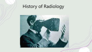
![Wilhelm Conrad Röntgen
German] mechanical engineer and physicist
who, on 8 November 1895, produced and
detected electromagnetic radiation in
a wavelength range known as X-rays or
Röntgen rays.
About six weeks after his discovery, he took
a picture—a radiograph—using X-rays of
his wife Anna Bertha's hand
By 1896 doctors around the world were using x-rays to take pictures of patients](https://image.slidesharecdn.com/cancerimaging-210421223447/85/Cancer-imaging-4-320.jpg)
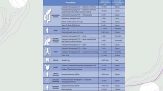
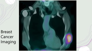
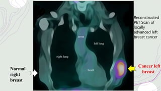
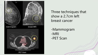
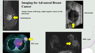
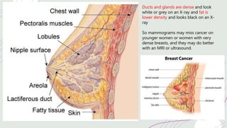
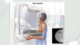
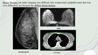
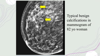
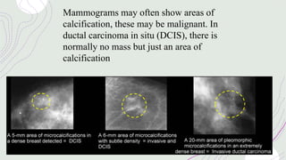
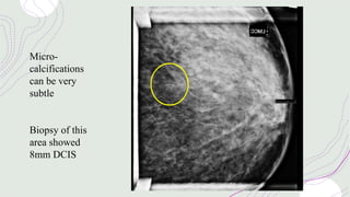
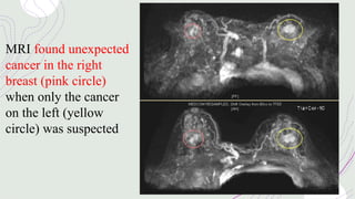
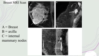
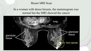
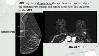
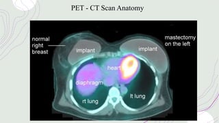
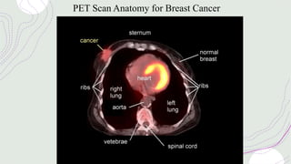
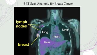
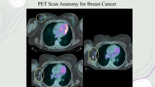
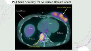
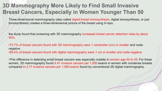
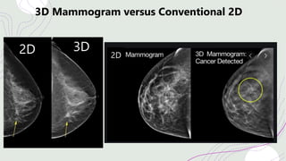
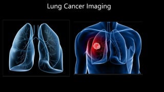
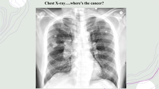

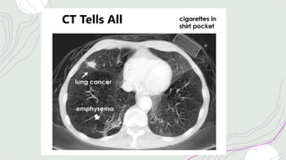
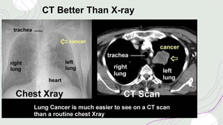
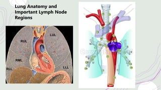
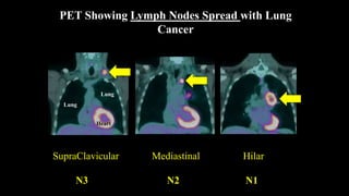
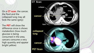
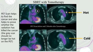
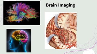
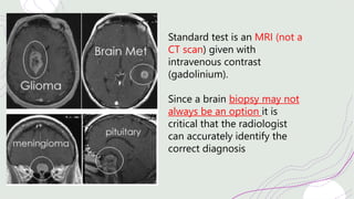
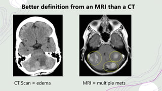
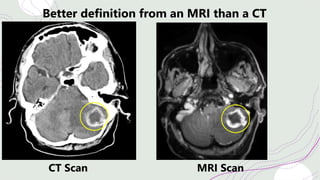
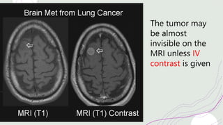
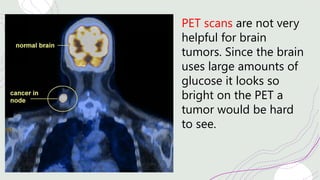
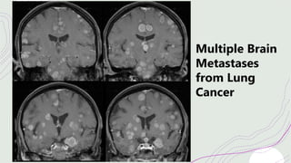
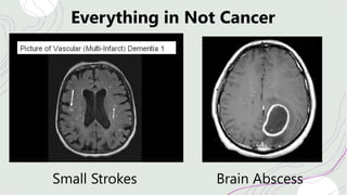
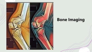
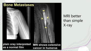
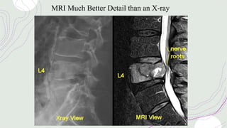
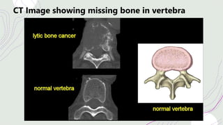
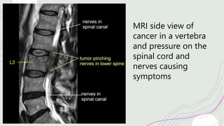
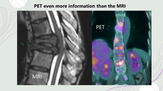
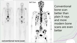

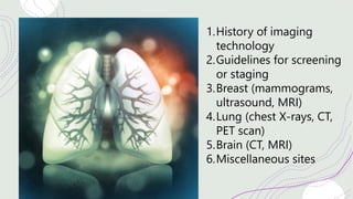
Cancer imaging has advanced significantly with improvements in technologies such as CT, MRI, PET, and ultrasound. Mammography remains the standard breast cancer screening tool, though its accuracy depends on breast density. MRI and ultrasound provide additional information. PET scans help evaluate cancer spread and treatment response. Lung cancer screening involves low-dose CT scans. CT and PET scans precisely evaluate lung tumors and lymph node involvement. Brain tumors are best imaged with MRI which provides detailed soft tissue contrast compared to CT. Bone scans effectively detect bone metastases earlier than plain films. PET scans can further characterize bone lesions found on other imaging.


![ONFH[AVN HIP] -TRIPLE REGIME -A NOVAL SURGICAL CONCEPT .pptx](https://cdn.slidesharecdn.com/ss_thumbnails/onfhavnhip2026koaconcalicutdrgokuldevdrmashraf-260210064517-213ec005-thumbnail.jpg?width=640&height=640&fit=bounds)


