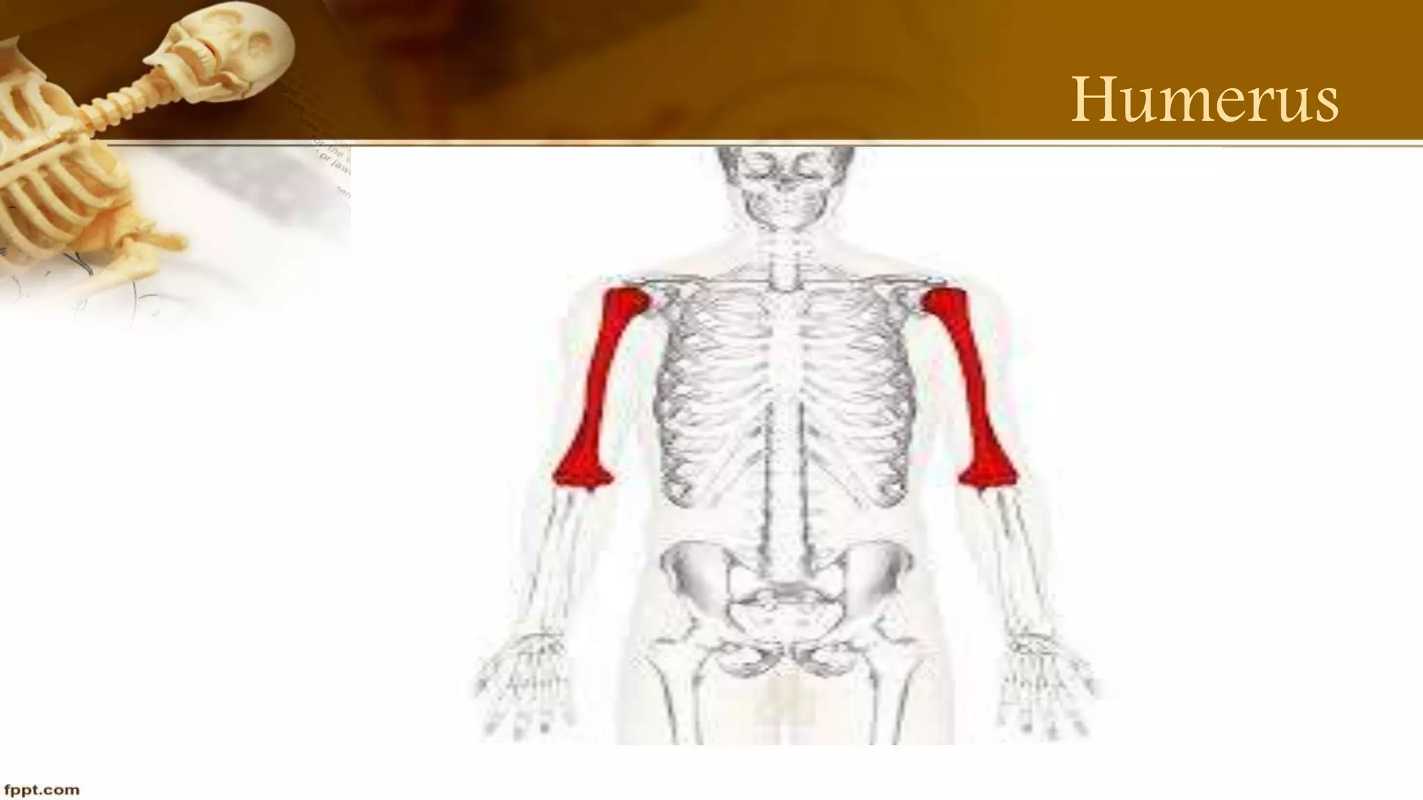
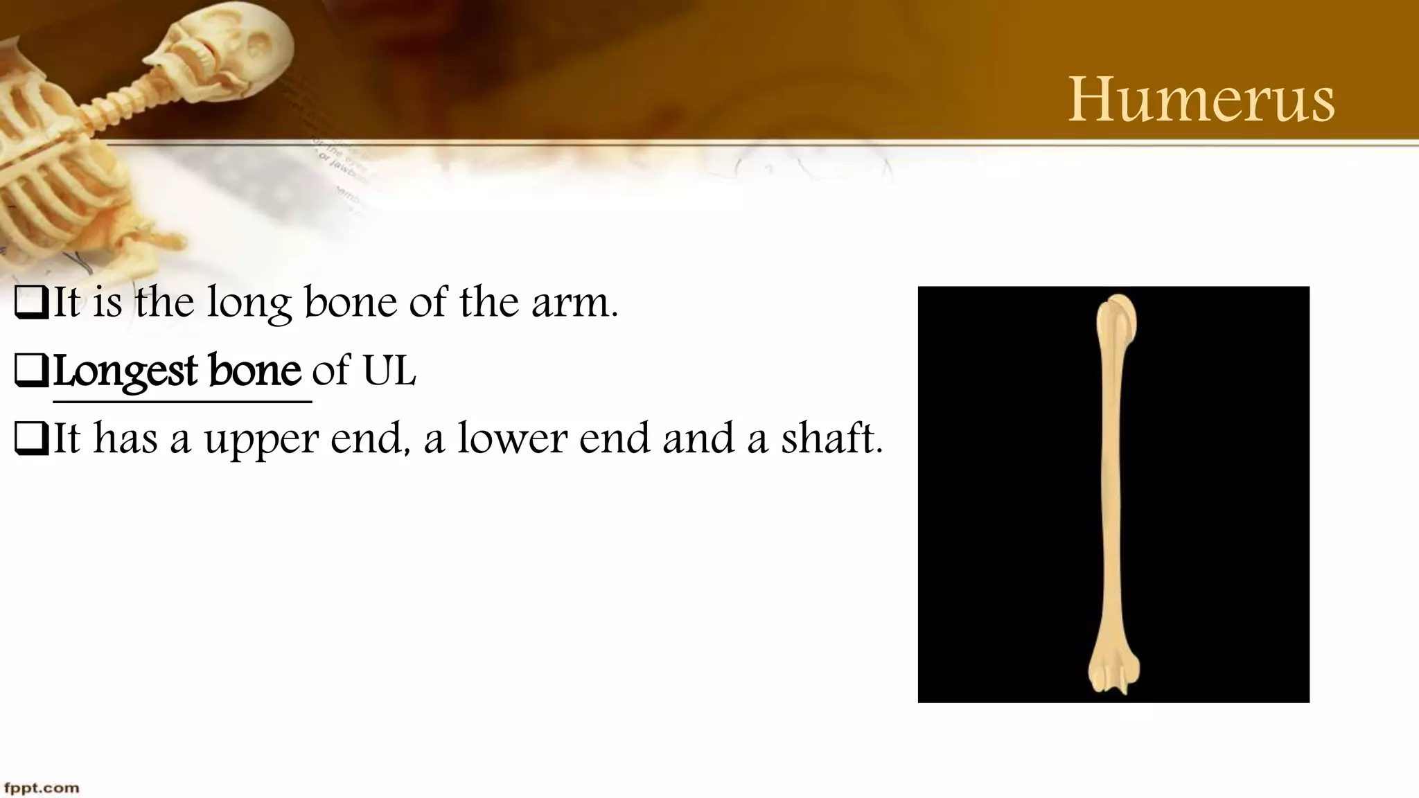
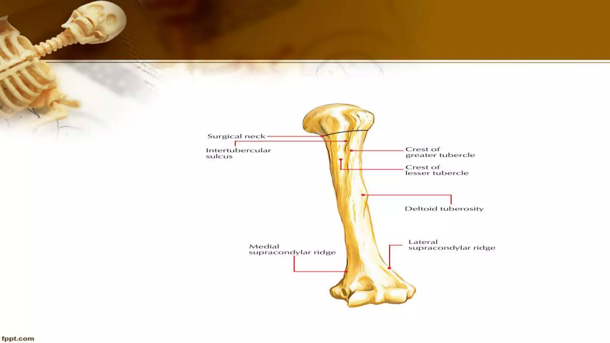
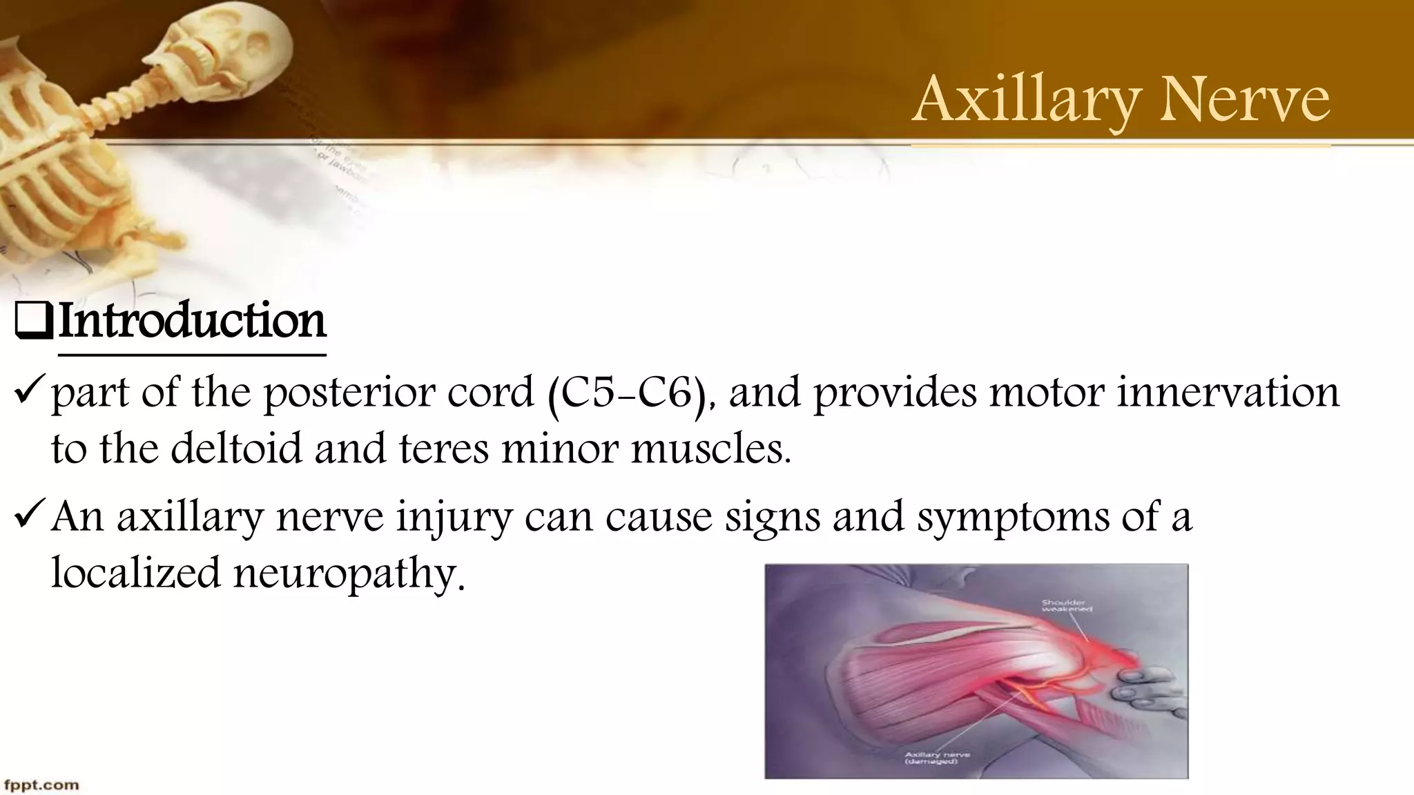
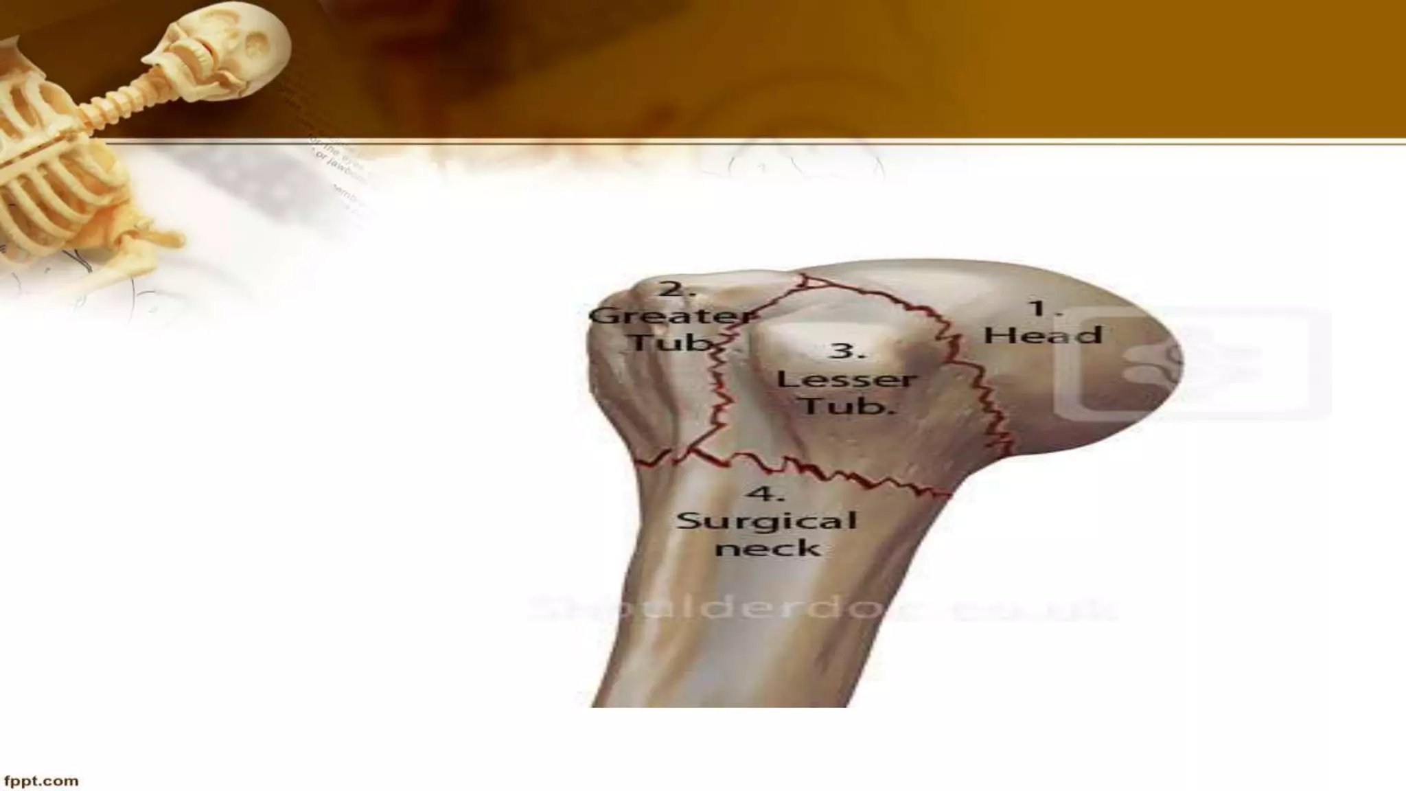
UL Bone
Humerus
Part- 2
BY Purvi Shah
B.P.T
Humerus
Humerus
It is the long bone of the arm.
Longest bone of UL
It has a upper end, a lower end and a shaft.
The Upper End
The head of the humerus is the articular surface of the upper
extremity, which is an irregular hemisphere.
The Anatomical Neck
The Surgical Neck
The Greater Tubercle
The Lesser Tubercle
The intertubercular sulcus (Bicipital groove)
The Shaft
The shaft is rounded in upper half and triangular in the lower half
Borders
1.Anterior border
2.Lateral border
3.Medial border
Surfaces
1.Anteolateral surface2.Anteromedial surface 3.Posterior surface
The Lower End
The lower end of humerus forms the condyle which is expanded
from side to side.
It has articular parts and non articular parts.
Articular Part
Capitulum : Rounded Projection
Trochlea : Pulley shaped
The Lower End
Non articular surface
Medial epicondyle
Lateral epicondyle
Medial supracondylar ridge
Lateral supracondylar ridge
Coronoid fossa
Radial fossa
Olecranon fossa
Functions
The humerus serves as an attachment to 13 muscles which
contribute to the movements of the hand and elbow, and therefore
the function of the upper limb.
Articulations
Glenohumeral joint
Elbow joint
Attachments
Muscles
Subscapularis is inserted into the lesser tubercle
Supraspinatus is inserted into upper impression on GT
Infraspinatus is inserted into middle impression on GT
Teres minor is inserted into lower impression on GT
Pectoralis major is inserted lateral lip of intertubercular sulcus
Attachments
Muscles
LD is inserted into floor of intertubercular sulcus
Teres major is inserted into medial lip of intertubercular sulcus
Deltoid is inserted into deltoid tuberosity
Coracobrachialis is inserted into rough area on the middle of medial
border
Brachialis is inserted arises lower halves of the anteromedial &
anterolateral surfaces of the shaft
Attachments
Muscles
Brachioradialis arises from the upper two third of lateral
supracondylar ridge
ECRL arises from the lower one third of lateral supracondylar ridge
Pronator teres arises from lower one third of medial suprcondylar
ridge
Superficial flexors muscles of forearm arises by a common origin from
the ant aspect of medial epicondyle
Attachments
Muscles
The superficial extensors muscles of forearm arises by a common
origin from the lateral epicondyle
Anconeus arises from the posterior surface of the lateral epicondyle
Lateral head of triceps arises from oblique ridge on upper part of
posterior above radial groove
Attachments
Ligaments
Capsular ligaments of shoulder joint is attached to the anatomical
neck except on medial side of humerus
Capsular ligaments of elbow joint is attached to lower end along a
line that reaches the upper limits of radial and coronoid fossae.
Ossification
Ossifies from the one primary centres and 7 secondary centres
The upper end ossifies from 3 secondary centres
The lower end ossifies from 4 centres which from 2 epiphyses
Clinical Anatomy
Three nerves are directly related to the humerus and therefore
liable to injury
Axillary nerve at surgical neck
Radial nerve at radial groove
Ulnar nerve behind the medial epicondylar
Axillary Nerve
Introduction
part of the posterior cord (C5-C6), and provides motor innervation
to the deltoid and teres minor muscles.
An axillary nerve injury can cause signs and symptoms of a
localized neuropathy.
Axillary nerve
Axillary nerve injury
Signs and symptoms : Pain to the area of the deltoid and anterior
shoulder
 Loss of movement and/or lack of sensation in the shoulder area
 Reported or observed weakness to the deltoid and teres minor
muscles (Abduction and External rotation).
Radial Nerve
Introduction
The radial nerve is one of the terminal branches of the posterior
cord.
Root : C5, 6, 7, 8 & T1
Symptoms : pain
weakness in the wrist, hand, or fingers
loss of function in the wrist, hand, or fingers
Ulnar Nerve
Introduction
The ulnar nerve originates from C8-T1 nerve roots which form the
medial cord of the brachial plexus.
Symptoms : Burning feeling in hand, arm or finger
 Increased arm/finger numbness or tingling while typing or writing.
 “Pins and needles” sensation (prickling) in the hand, arm or fingers.
Weakness (loss of strength) in the hand, arm or fingers.
Nerve Injury
Nerve injury
Investigation : MRI
EMG/NCV
Surgical management : Neurolysis
Neurorrhaphy
Nerve Grafting
Neurotization
Clinical Anatomy
The common sites of fracture are;
Surgical neck
The shaft
The supracondylar region
The humerus has a poor blood supply at the junction of it’s upper
&middle thirds
Fractures at this sites show delayed union
or non union.
Thank You

Purvi shah humerus anatomy ppt

  • 1.
  • 2.
  • 3.
    Humerus It is thelong bone of the arm. Longest bone of UL It has a upper end, a lower end and a shaft.
  • 5.
    The Upper End Thehead of the humerus is the articular surface of the upper extremity, which is an irregular hemisphere. The Anatomical Neck The Surgical Neck The Greater Tubercle The Lesser Tubercle The intertubercular sulcus (Bicipital groove)
  • 6.
    The Shaft The shaftis rounded in upper half and triangular in the lower half Borders 1.Anterior border 2.Lateral border 3.Medial border Surfaces 1.Anteolateral surface2.Anteromedial surface 3.Posterior surface
  • 7.
    The Lower End Thelower end of humerus forms the condyle which is expanded from side to side. It has articular parts and non articular parts. Articular Part Capitulum : Rounded Projection Trochlea : Pulley shaped
  • 8.
    The Lower End Nonarticular surface Medial epicondyle Lateral epicondyle Medial supracondylar ridge Lateral supracondylar ridge Coronoid fossa Radial fossa Olecranon fossa
  • 10.
    Functions The humerus servesas an attachment to 13 muscles which contribute to the movements of the hand and elbow, and therefore the function of the upper limb.
  • 11.
  • 13.
    Attachments Muscles Subscapularis is insertedinto the lesser tubercle Supraspinatus is inserted into upper impression on GT Infraspinatus is inserted into middle impression on GT Teres minor is inserted into lower impression on GT Pectoralis major is inserted lateral lip of intertubercular sulcus
  • 14.
    Attachments Muscles LD is insertedinto floor of intertubercular sulcus Teres major is inserted into medial lip of intertubercular sulcus Deltoid is inserted into deltoid tuberosity Coracobrachialis is inserted into rough area on the middle of medial border Brachialis is inserted arises lower halves of the anteromedial & anterolateral surfaces of the shaft
  • 15.
    Attachments Muscles Brachioradialis arises fromthe upper two third of lateral supracondylar ridge ECRL arises from the lower one third of lateral supracondylar ridge Pronator teres arises from lower one third of medial suprcondylar ridge Superficial flexors muscles of forearm arises by a common origin from the ant aspect of medial epicondyle
  • 16.
    Attachments Muscles The superficial extensorsmuscles of forearm arises by a common origin from the lateral epicondyle Anconeus arises from the posterior surface of the lateral epicondyle Lateral head of triceps arises from oblique ridge on upper part of posterior above radial groove
  • 17.
    Attachments Ligaments Capsular ligaments ofshoulder joint is attached to the anatomical neck except on medial side of humerus Capsular ligaments of elbow joint is attached to lower end along a line that reaches the upper limits of radial and coronoid fossae.
  • 18.
    Ossification Ossifies from theone primary centres and 7 secondary centres The upper end ossifies from 3 secondary centres The lower end ossifies from 4 centres which from 2 epiphyses
  • 19.
    Clinical Anatomy Three nervesare directly related to the humerus and therefore liable to injury Axillary nerve at surgical neck Radial nerve at radial groove Ulnar nerve behind the medial epicondylar
  • 20.
    Axillary Nerve Introduction part ofthe posterior cord (C5-C6), and provides motor innervation to the deltoid and teres minor muscles. An axillary nerve injury can cause signs and symptoms of a localized neuropathy.
  • 21.
    Axillary nerve Axillary nerveinjury Signs and symptoms : Pain to the area of the deltoid and anterior shoulder  Loss of movement and/or lack of sensation in the shoulder area  Reported or observed weakness to the deltoid and teres minor muscles (Abduction and External rotation).
  • 22.
    Radial Nerve Introduction The radialnerve is one of the terminal branches of the posterior cord. Root : C5, 6, 7, 8 & T1 Symptoms : pain weakness in the wrist, hand, or fingers loss of function in the wrist, hand, or fingers
  • 23.
    Ulnar Nerve Introduction The ulnarnerve originates from C8-T1 nerve roots which form the medial cord of the brachial plexus. Symptoms : Burning feeling in hand, arm or finger  Increased arm/finger numbness or tingling while typing or writing.  “Pins and needles” sensation (prickling) in the hand, arm or fingers. Weakness (loss of strength) in the hand, arm or fingers.
  • 24.
    Nerve Injury Nerve injury Investigation: MRI EMG/NCV Surgical management : Neurolysis Neurorrhaphy Nerve Grafting Neurotization
  • 25.
    Clinical Anatomy The commonsites of fracture are; Surgical neck The shaft The supracondylar region The humerus has a poor blood supply at the junction of it’s upper &middle thirds Fractures at this sites show delayed union or non union.
  • 28.