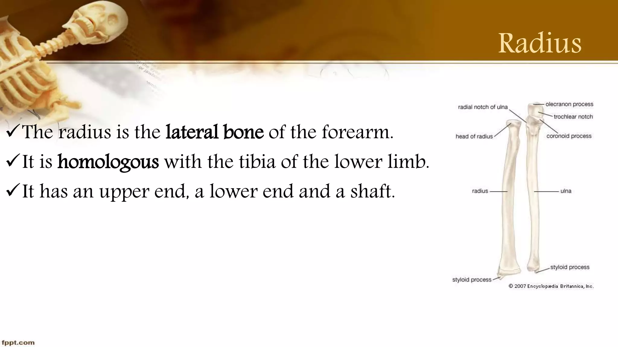
Radius & Ulna Anatomy
Part-3
By Purvi Shah
BPT
Radius
Radius
The radius is the lateral bone of the forearm.
It is homologous with the tibia of the lower limb.
It has an upper end, a lower end and a shaft.
The Upper End
The Head is disc shaped & is covered with hyaline cartilage
Head of radius fits into a socket formed by radial notch of ulna
and annular ligament, thus forming superior radioulnar joint.
The Neck is enclosed by narrow lower margin of annular
ligament.
The Tuberosity lies just below the medial part of the neck.
It has a rough posterior part and a smooth anterior part.
The Upper End
The Shaft
It has three borders and three surfaces.
Borders
The Anterior Border
The Posterior Border(mirror image of the anterior border)
The Medial or Interosseous Border(sharpest border)
The Shaft
Surfaces
The anterior surface lies between anterior and interosseous
borders.
The posterior surface lies between the posterior and interosseous
borders.
The lateral surface lies between the anterior posterior borders.
The Lower End
It is widest part of the bone;it has a five surfaces.
The anterior surface (The radial artery palpated against this
surface)
The posterior surface(Presents 4 grooves for extensors tendons)
The medial surface(occupied by the ulnar notch for the head of
ulna)
The lateral surface (prolonged downwards to from styloid process)
The inferior surface(bears a triangular area for the scaphoid bone;
Muscle Attachments
 Biceps attaches to the radial tuberosity.
 Supinator,flexor pollicis longus and the flexor digitorum
superficialis attach to the upper third part of the shaft of the
radius.
EPB,abductor pollicis longus and pronator teres all attach to the
mid shaft of the radius.
Pronator quadratus muscle attach to the distal quarter of the
radial shaft.
Muscle Attachments
Articulation
Elbow Joint
Wrist Joint
Ossification
The shaft ossifies from a primary centre.
The lower end ossifies from a secondary centre.
The Upper end ossifies from a secondary centre.
Clinical Anatomy
 The Radius commonly gets fractured about 2 cm above its lower end
Colles’s fracture.
Mechanism of injury: Fall on outstretched hand(FOOSH)
Distal fragments is displaced upwards and backwards.
Reverse of the colles’s fracture: Smith fracture
Distal fragments being palmar flexed rather than dorsiflexion.
Distal End Of Radius Fracture
Clinical Anatomy
Congenital absence of the radius is rare anomaly.
Resulting gross radial deviation of the hand and the thumb is often
absent.
Radioulnar synostosis is also rare condition in which radius and
ulna are fused together.
Ulna
Ulna
The ulna is the medial bone of forearm.
It is homologous with fibula of lower limb.
It has a upper end, shaft, lower end.
The ulna is a long bone larger proximally than distally.
Upper End
Olecranon process projects upwards from the shaft.
It has superior, anterior, posterior, medial, lateral surfaces.
Coronoid process projects forwards from the shaft just below the
olecranon process.
It has four surfaces; superior, anterior, posterior, medial, lateral
surfaces.
The trochlear notch forms an articular surface.
The radial notch articulate with the head of radius.
The Shaft
It has a 3 Borders and 3 surfaces.
Borders
Interosseous border/Lateral border(sharpest)
Anterior border(thick and rounded)
Posterior border(subcutaneous)
The Shaft
Surfaces
Anterior surface
Medial surface
Posterior surface
Lower End
Head of the ulna
The lateral, distal end of the ulna is the head of the ulna.
 It articulates with the ulnar notch on the radius and with the
triangular articular disc in the Wrist Joint.
Styloid process
It projects downwards from the posteromedial side of lower end of
the ulna.
Muscle Attachments
Triceps inserts into the posterior of the olecranon process.
Brachialis inserts to anterior, inferior coronoid process.
Supinator arises from the supinator crest
FDS arises from a tubercle at the upper end of coronoid process.
Pronator teres arises medial margin of the coronoid process.
Muscle Attachments
FDP arises from the shaft of ulna.
Pronator quadratus origin from oblique ridge on lower part
anterior surface.
FCU arise from medial side of olecranon process.
ECU arises from posterior border
Anconeus is inserted lateral aspect of olecranon process.
Ligament Attachments
Capsular ligament
Annular ligament
Ulna collateral ligament
Articulation
Elbow joint
Radio-ulna joint
Ossification
The shaft and the most of the upper end ossify from a primary
centre.
The superior part of olecranon ossifies from a secondary centre.
The lower end ossifies from a secondary centre.
Clinical Anatomy
Shaft of ulna may fracture either alone or along with that of the
radius.
Fracture of olecranon is common caused by FOOSH
Dislocation of elbow is produced by FOOSH
Madelung’s deformity is dorsal subluxation of lower end of ulna.
Clinical Anatomy
Thank You

Purvi shah radius & ulna anatomy ppt

  • 1.
    Radius & UlnaAnatomy Part-3 By Purvi Shah BPT
  • 2.
  • 3.
    Radius The radius isthe lateral bone of the forearm. It is homologous with the tibia of the lower limb. It has an upper end, a lower end and a shaft.
  • 4.
    The Upper End TheHead is disc shaped & is covered with hyaline cartilage Head of radius fits into a socket formed by radial notch of ulna and annular ligament, thus forming superior radioulnar joint. The Neck is enclosed by narrow lower margin of annular ligament. The Tuberosity lies just below the medial part of the neck. It has a rough posterior part and a smooth anterior part.
  • 5.
  • 6.
    The Shaft It hasthree borders and three surfaces. Borders The Anterior Border The Posterior Border(mirror image of the anterior border) The Medial or Interosseous Border(sharpest border)
  • 7.
    The Shaft Surfaces The anteriorsurface lies between anterior and interosseous borders. The posterior surface lies between the posterior and interosseous borders. The lateral surface lies between the anterior posterior borders.
  • 8.
    The Lower End Itis widest part of the bone;it has a five surfaces. The anterior surface (The radial artery palpated against this surface) The posterior surface(Presents 4 grooves for extensors tendons) The medial surface(occupied by the ulnar notch for the head of ulna) The lateral surface (prolonged downwards to from styloid process) The inferior surface(bears a triangular area for the scaphoid bone;
  • 9.
    Muscle Attachments  Bicepsattaches to the radial tuberosity.  Supinator,flexor pollicis longus and the flexor digitorum superficialis attach to the upper third part of the shaft of the radius. EPB,abductor pollicis longus and pronator teres all attach to the mid shaft of the radius. Pronator quadratus muscle attach to the distal quarter of the radial shaft.
  • 10.
  • 11.
  • 12.
    Ossification The shaft ossifiesfrom a primary centre. The lower end ossifies from a secondary centre. The Upper end ossifies from a secondary centre.
  • 13.
    Clinical Anatomy  TheRadius commonly gets fractured about 2 cm above its lower end Colles’s fracture. Mechanism of injury: Fall on outstretched hand(FOOSH) Distal fragments is displaced upwards and backwards. Reverse of the colles’s fracture: Smith fracture Distal fragments being palmar flexed rather than dorsiflexion.
  • 14.
    Distal End OfRadius Fracture
  • 16.
    Clinical Anatomy Congenital absenceof the radius is rare anomaly. Resulting gross radial deviation of the hand and the thumb is often absent. Radioulnar synostosis is also rare condition in which radius and ulna are fused together.
  • 17.
  • 18.
    Ulna The ulna isthe medial bone of forearm. It is homologous with fibula of lower limb. It has a upper end, shaft, lower end. The ulna is a long bone larger proximally than distally.
  • 19.
    Upper End Olecranon processprojects upwards from the shaft. It has superior, anterior, posterior, medial, lateral surfaces. Coronoid process projects forwards from the shaft just below the olecranon process. It has four surfaces; superior, anterior, posterior, medial, lateral surfaces. The trochlear notch forms an articular surface. The radial notch articulate with the head of radius.
  • 20.
    The Shaft It hasa 3 Borders and 3 surfaces. Borders Interosseous border/Lateral border(sharpest) Anterior border(thick and rounded) Posterior border(subcutaneous)
  • 21.
  • 22.
    Lower End Head ofthe ulna The lateral, distal end of the ulna is the head of the ulna.  It articulates with the ulnar notch on the radius and with the triangular articular disc in the Wrist Joint. Styloid process It projects downwards from the posteromedial side of lower end of the ulna.
  • 23.
    Muscle Attachments Triceps insertsinto the posterior of the olecranon process. Brachialis inserts to anterior, inferior coronoid process. Supinator arises from the supinator crest FDS arises from a tubercle at the upper end of coronoid process. Pronator teres arises medial margin of the coronoid process.
  • 24.
    Muscle Attachments FDP arisesfrom the shaft of ulna. Pronator quadratus origin from oblique ridge on lower part anterior surface. FCU arise from medial side of olecranon process. ECU arises from posterior border Anconeus is inserted lateral aspect of olecranon process.
  • 26.
    Ligament Attachments Capsular ligament Annularligament Ulna collateral ligament
  • 27.
  • 28.
    Ossification The shaft andthe most of the upper end ossify from a primary centre. The superior part of olecranon ossifies from a secondary centre. The lower end ossifies from a secondary centre.
  • 29.
    Clinical Anatomy Shaft ofulna may fracture either alone or along with that of the radius. Fracture of olecranon is common caused by FOOSH Dislocation of elbow is produced by FOOSH Madelung’s deformity is dorsal subluxation of lower end of ulna.
  • 30.
  • 31.