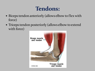
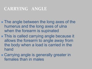
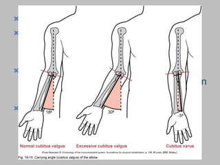
ELBOW JOINT
Dr. Heta Patel(PT)
Assistant Professor
ARIP
CHARUSAT
 Hinge variety of Synovial Joint
 Distal end of humerus and Proximal
end of Radius & Ulna
 Formed by three joints
 Humeroulnar joint (ulna trochlear)
 Humeroradial joint (radio
capitellar)
 Proximal radio ulnar joint
 Most upper extremity movements
involve the elbow & radioulnar joints
 These two joints are usually grouped
together due to close anatomical
relationship
The long mid center of the humerus is the
body (shaft), and the humeral condyle isits
expanded distal end.
Articularpartof the humeral condyle is
made up of trochlear and thecapitulum.
Trochlear is more medial andarticulates
with theulna.
Capitulum is more lateral andwill
articulate with head of radius.
Lateral epicondyle is a small projectionon
lateral aspect of distalhumerus.
Medial epicondyle (larger and more
prominent) is located on medial edgeof
distal humerus.
Anterior depressions:
Coronoid fossa and radial
fossa.
Posteriordepression: is the
olecranon fossa.
Lateral view of elbow shows
proximal radius and ulnawith
radial head and neck and
radial tuberosity.
HUMERO-ULNAR PART
 It is articulation between trochlea of humerus and
trochlear notch of ulna
 The medial edge of trochlea is 6mm beyond the
lateral edge
 Plane of the joint is 2cms distal to intercondylar line
slopes downwards and medially
 Two nonarticular depressions coronoid fossa and
olecrenon fossa are present in relation to this
articulation
 Trochlear notch of ulna is reciprocally saddle shape
and formed by the articular surfaces of olecrenon and
coronoid processes
HUMERO-RADIAL PART
 Structurally it is a ball and
socket type of joint.
 The ball is represented by
capitulum of
humerus and socket is
by articular surface
of disc like head of the radius
 In full flexion head of the
radius lodges radial fossa above
the capitulum.
 the annular ligament binds the head of
the radius to the radial notch of the
ulna forming the proximal radioulnar
joint – this is a pivot joint allowing for
pronation and supination
LIGAMENTS OF ELBOW JOINT
 Capsular ligament
 Ulnar collateral ligament or Medial
ligament
 Radial collateral ligament or Lateral
ligament
FIBROUS CAPSULE
 Fibrous capsule completely envelop the joint.
 It is attached to the lower end of humerus in
a continuous line, which excludes the two
epicondyles but include three fossae.
 Cushions of extra-synovial fat fill up the three
fossae.
ULNARCOLLATERALLIGAMENT
 UCL is triangular in shape and
extends from medial epicondyle
to medial margin of trochlear
notch. The ligament consists of
three bands, Anterior Posterior
andInferior
 UCL is overlapped by Triceps
,FCU , FDS and Ulnar nerve.
ULNARCOLLATERALLIGAMENT
RADIAL COLLATERAL LIGAMENT
 RCL is triangular and extends from lateral
epicondyle to annular ligament. It is related
to supinator and ECRB
RADIAL COLLATERAL LIGAMENT
SYNOVIAL MEMBRANE
RELATIONS
RELATIONS OF ELBOW JOINT
 In front-Brachialis, tendon of biceps, median
nerve and brachial artery.
 Behind- Triceps and anconeus.
 Medially-common origin of superficial flexors,
Flexor carpi ulnaris and ulnar nerve.
 Laterally- common origin of superficial
extensors, supinator, extensor carpi radialis
brevis, radial nerve with its superficial and
deep branches
ANTERIOR RELATIONS
 Flexion
 Movement of forearm to
shoulder by bending theelbow
to decrease itsangle
 Extension
 Movement of forearm awayfrom
shoulder by straightening the
elbow to increase itsangle
 Pronation
 Internal rotary movementof
radiuson ulna that results in
hand moving from palm-up
to palm-down position
 Supination
 External rotary movement
of radius on ulna thatresults
in hand moving from palm-
down to palm-upposition
 Elbow flexors
 Biceps brachii
 Brachialis
 Brachioradialis
 Weak assistance from
Pronator teres
 Elbow extensor
 Triceps brachii
 Anconeus provides
assistance
 Radioulnar pronators
 Pronator teres
 Pronator quadratus
 Brachioradialis
 Radioulnar supinators
 Biceps brachii
 Supinator muscle
 Brachioradialis
Anterior
 Primarily flexion &
pronation
Biceps brachii
Brachialis
Brachioradialis
Pronator teres
Pronator quadratus
 Posterior
 Primarily extension
& supination
 Triceps brachii
 Anconeus
 Supinator
 All elbow joint and muscles areinnervated from
median, musculotaneous, ulnar & radial nerves of
brachial plexus
 Biceps tendon anteriorly (allowselbow to flex with
force)
 Triceps tendon posteriorly (allowselbow toextend
with force)
 The articularsurfacesare connected together bya
capsule
 Anterior part – from radial and coronoid fossa of
humerus tocoronoid processof ulnaand annular
ligament of radius
 Posteriorpart – from capitulum, olecranon fossa, and
lateral epicondyle of humerus to annular ligament of
radius, olecranon of ulna, and posterior to radial
notch.
CARRYING ANGLE
 The angle between the long axes of the
humerus and the long axes of ulna
when the forearm is supinated
 This is called carrying angle because it
allows the forearm to angle away from
the body when a load is carried in the
hand
 Carrying angle is generally greater in
females than in males
n
 Carry Angle of the Elbow
 The angle between the long axes of the
humerus and the ulna when the arm is in
an anatomical position
 This is called this because it allows the
forearm to angle away from the body whe
a load is carried in the hand
 Carrying angle is generally greater in
females than in males
Ossification of Bones.
 Capitelum - (1-2yrs)
 Medial epicondyle - 4yrs
 Trochlea - 8 yrs
 Lateral epicondyle - 10yrs
Fracture Classification.
DISTAL HUMERUS:
FRACTURES:
SUPRACONDYLAR
INTERCONDYLAR
CONDYLAR
EPICONDYLAR
FRACTURES OF
PROXIMAL ULNA:
OLECRANON
FRACTURE
CORONOID PROCESS
FRACTURE
FRACTURES
OF RADIUS:
RADIAL HEAD
RADIAL NECK
 “Tennis elbow" - common problem usually
involving extensor digitorum muscle near
its origin on lateral epicondyle
 known lateral epicondylitis
 associated with gripping & lifting
activities
Tennis Elbow
Somewhat less common
 Also known as golfer's elbow
Associated with medial wrist flexor & pronator group
near their origin on medial epicondyle.
Involves muscles which cross elbow but act
primarily on wrist & hand
Medial Epicondylitis
Elbow joint with applied anatomy

Elbow joint with applied anatomy

  • 1.
    ELBOW JOINT Dr. HetaPatel(PT) Assistant Professor ARIP CHARUSAT
  • 2.
     Hinge varietyof Synovial Joint  Distal end of humerus and Proximal end of Radius & Ulna  Formed by three joints  Humeroulnar joint (ulna trochlear)  Humeroradial joint (radio capitellar)  Proximal radio ulnar joint  Most upper extremity movements involve the elbow & radioulnar joints  These two joints are usually grouped together due to close anatomical relationship
  • 3.
    The long midcenter of the humerus is the body (shaft), and the humeral condyle isits expanded distal end. Articularpartof the humeral condyle is made up of trochlear and thecapitulum. Trochlear is more medial andarticulates with theulna. Capitulum is more lateral andwill articulate with head of radius. Lateral epicondyle is a small projectionon lateral aspect of distalhumerus. Medial epicondyle (larger and more prominent) is located on medial edgeof distal humerus.
  • 4.
    Anterior depressions: Coronoid fossaand radial fossa. Posteriordepression: is the olecranon fossa. Lateral view of elbow shows proximal radius and ulnawith radial head and neck and radial tuberosity.
  • 5.
    HUMERO-ULNAR PART  Itis articulation between trochlea of humerus and trochlear notch of ulna  The medial edge of trochlea is 6mm beyond the lateral edge  Plane of the joint is 2cms distal to intercondylar line slopes downwards and medially  Two nonarticular depressions coronoid fossa and olecrenon fossa are present in relation to this articulation  Trochlear notch of ulna is reciprocally saddle shape and formed by the articular surfaces of olecrenon and coronoid processes
  • 6.
    HUMERO-RADIAL PART  Structurallyit is a ball and socket type of joint.  The ball is represented by capitulum of humerus and socket is by articular surface of disc like head of the radius  In full flexion head of the radius lodges radial fossa above the capitulum.
  • 7.
     the annularligament binds the head of the radius to the radial notch of the ulna forming the proximal radioulnar joint – this is a pivot joint allowing for pronation and supination
  • 8.
    LIGAMENTS OF ELBOWJOINT  Capsular ligament  Ulnar collateral ligament or Medial ligament  Radial collateral ligament or Lateral ligament
  • 9.
    FIBROUS CAPSULE  Fibrouscapsule completely envelop the joint.  It is attached to the lower end of humerus in a continuous line, which excludes the two epicondyles but include three fossae.  Cushions of extra-synovial fat fill up the three fossae.
  • 10.
    ULNARCOLLATERALLIGAMENT  UCL istriangular in shape and extends from medial epicondyle to medial margin of trochlear notch. The ligament consists of three bands, Anterior Posterior andInferior  UCL is overlapped by Triceps ,FCU , FDS and Ulnar nerve.
  • 11.
  • 12.
    RADIAL COLLATERAL LIGAMENT RCL is triangular and extends from lateral epicondyle to annular ligament. It is related to supinator and ECRB
  • 13.
  • 14.
  • 15.
  • 16.
    RELATIONS OF ELBOWJOINT  In front-Brachialis, tendon of biceps, median nerve and brachial artery.  Behind- Triceps and anconeus.  Medially-common origin of superficial flexors, Flexor carpi ulnaris and ulnar nerve.  Laterally- common origin of superficial extensors, supinator, extensor carpi radialis brevis, radial nerve with its superficial and deep branches
  • 17.
  • 18.
     Flexion  Movementof forearm to shoulder by bending theelbow to decrease itsangle  Extension  Movement of forearm awayfrom shoulder by straightening the elbow to increase itsangle
  • 19.
     Pronation  Internalrotary movementof radiuson ulna that results in hand moving from palm-up to palm-down position  Supination  External rotary movement of radius on ulna thatresults in hand moving from palm- down to palm-upposition
  • 20.
     Elbow flexors Biceps brachii  Brachialis  Brachioradialis  Weak assistance from Pronator teres  Elbow extensor  Triceps brachii  Anconeus provides assistance  Radioulnar pronators  Pronator teres  Pronator quadratus  Brachioradialis  Radioulnar supinators  Biceps brachii  Supinator muscle  Brachioradialis
  • 21.
    Anterior  Primarily flexion& pronation Biceps brachii Brachialis Brachioradialis Pronator teres Pronator quadratus
  • 22.
     Posterior  Primarilyextension & supination  Triceps brachii  Anconeus  Supinator
  • 23.
     All elbowjoint and muscles areinnervated from median, musculotaneous, ulnar & radial nerves of brachial plexus
  • 24.
     Biceps tendonanteriorly (allowselbow to flex with force)  Triceps tendon posteriorly (allowselbow toextend with force)
  • 25.
     The articularsurfacesareconnected together bya capsule  Anterior part – from radial and coronoid fossa of humerus tocoronoid processof ulnaand annular ligament of radius  Posteriorpart – from capitulum, olecranon fossa, and lateral epicondyle of humerus to annular ligament of radius, olecranon of ulna, and posterior to radial notch.
  • 27.
    CARRYING ANGLE  Theangle between the long axes of the humerus and the long axes of ulna when the forearm is supinated  This is called carrying angle because it allows the forearm to angle away from the body when a load is carried in the hand  Carrying angle is generally greater in females than in males
  • 28.
    n  Carry Angleof the Elbow  The angle between the long axes of the humerus and the ulna when the arm is in an anatomical position  This is called this because it allows the forearm to angle away from the body whe a load is carried in the hand  Carrying angle is generally greater in females than in males
  • 29.
  • 30.
     Capitelum -(1-2yrs)  Medial epicondyle - 4yrs  Trochlea - 8 yrs  Lateral epicondyle - 10yrs
  • 31.
  • 32.
    DISTAL HUMERUS: FRACTURES: SUPRACONDYLAR INTERCONDYLAR CONDYLAR EPICONDYLAR FRACTURES OF PROXIMALULNA: OLECRANON FRACTURE CORONOID PROCESS FRACTURE FRACTURES OF RADIUS: RADIAL HEAD RADIAL NECK
  • 36.
     “Tennis elbow"- common problem usually involving extensor digitorum muscle near its origin on lateral epicondyle  known lateral epicondylitis  associated with gripping & lifting activities Tennis Elbow
  • 37.
    Somewhat less common Also known as golfer's elbow Associated with medial wrist flexor & pronator group near their origin on medial epicondyle. Involves muscles which cross elbow but act primarily on wrist & hand Medial Epicondylitis