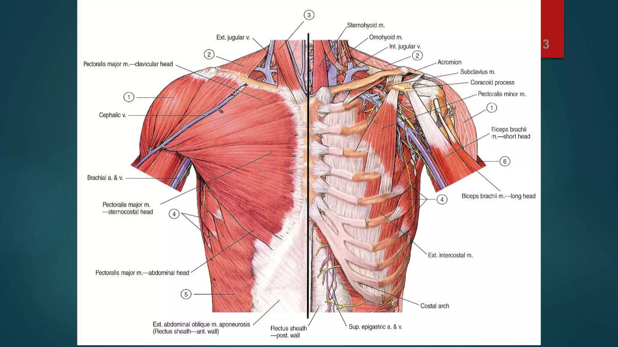
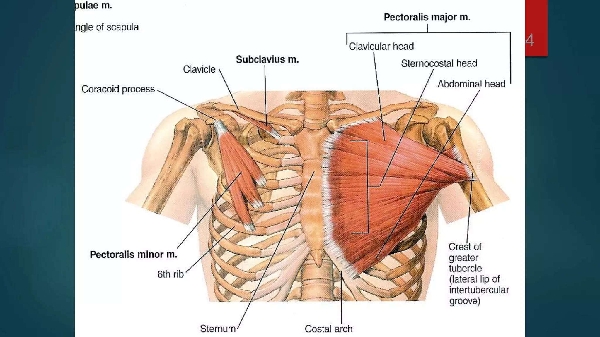
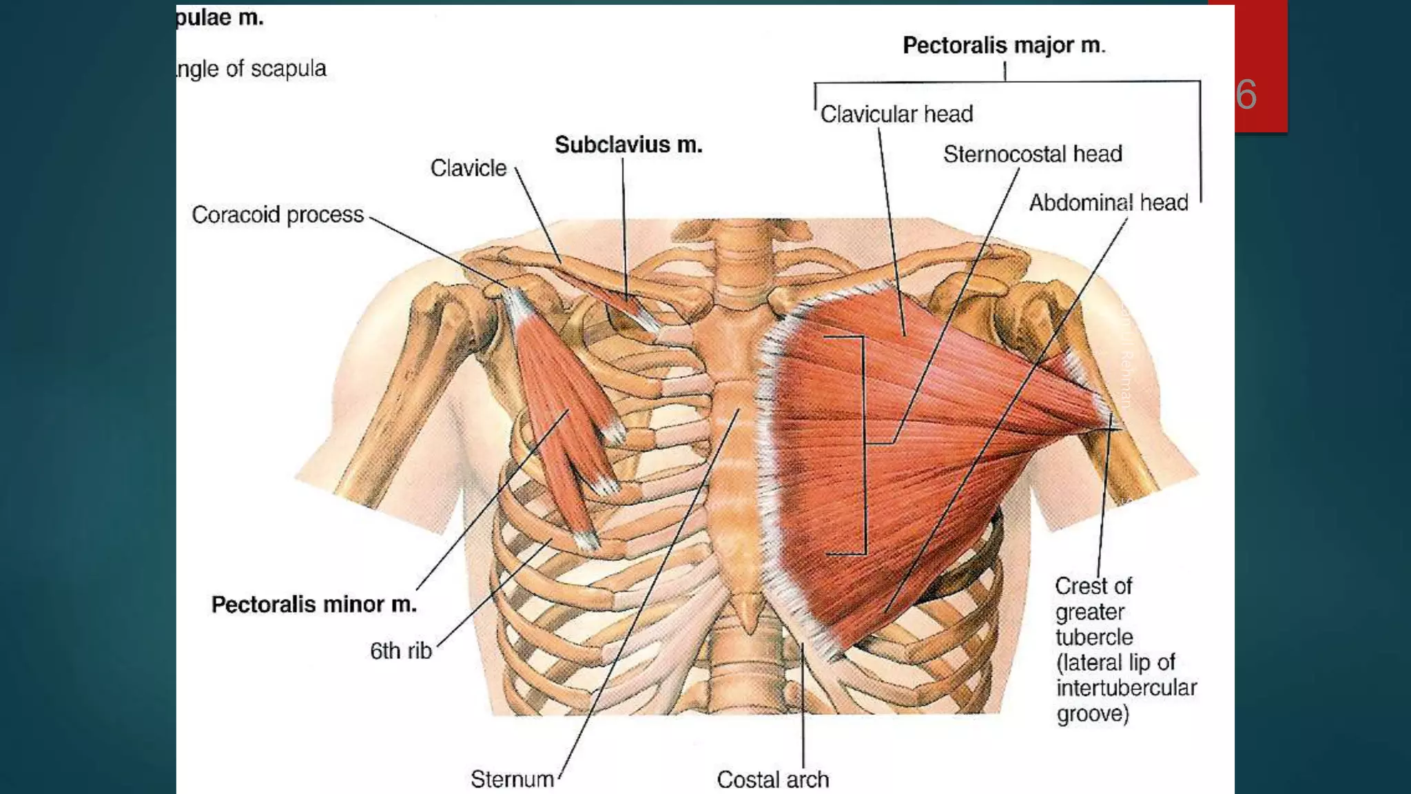
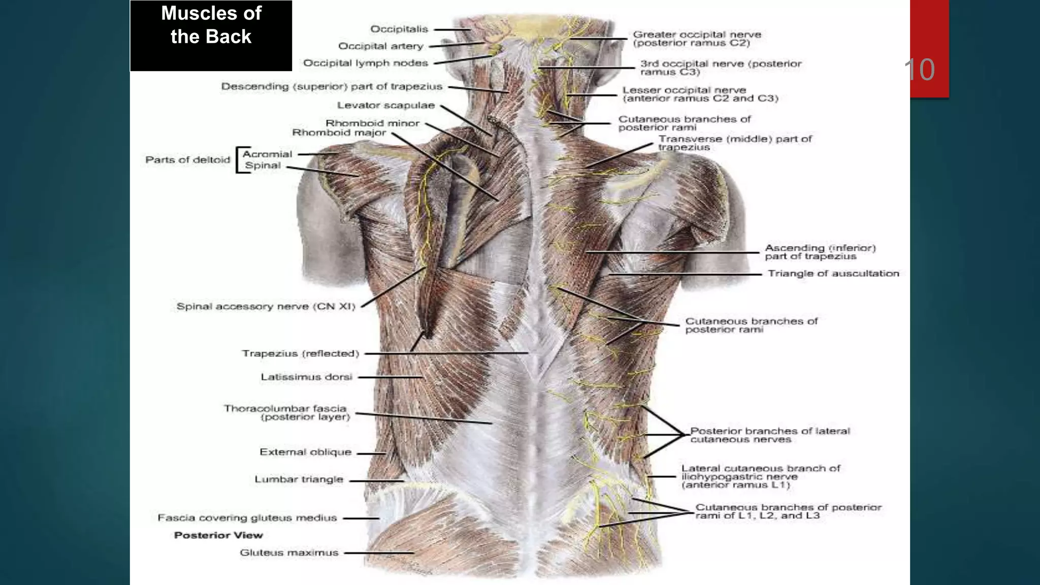
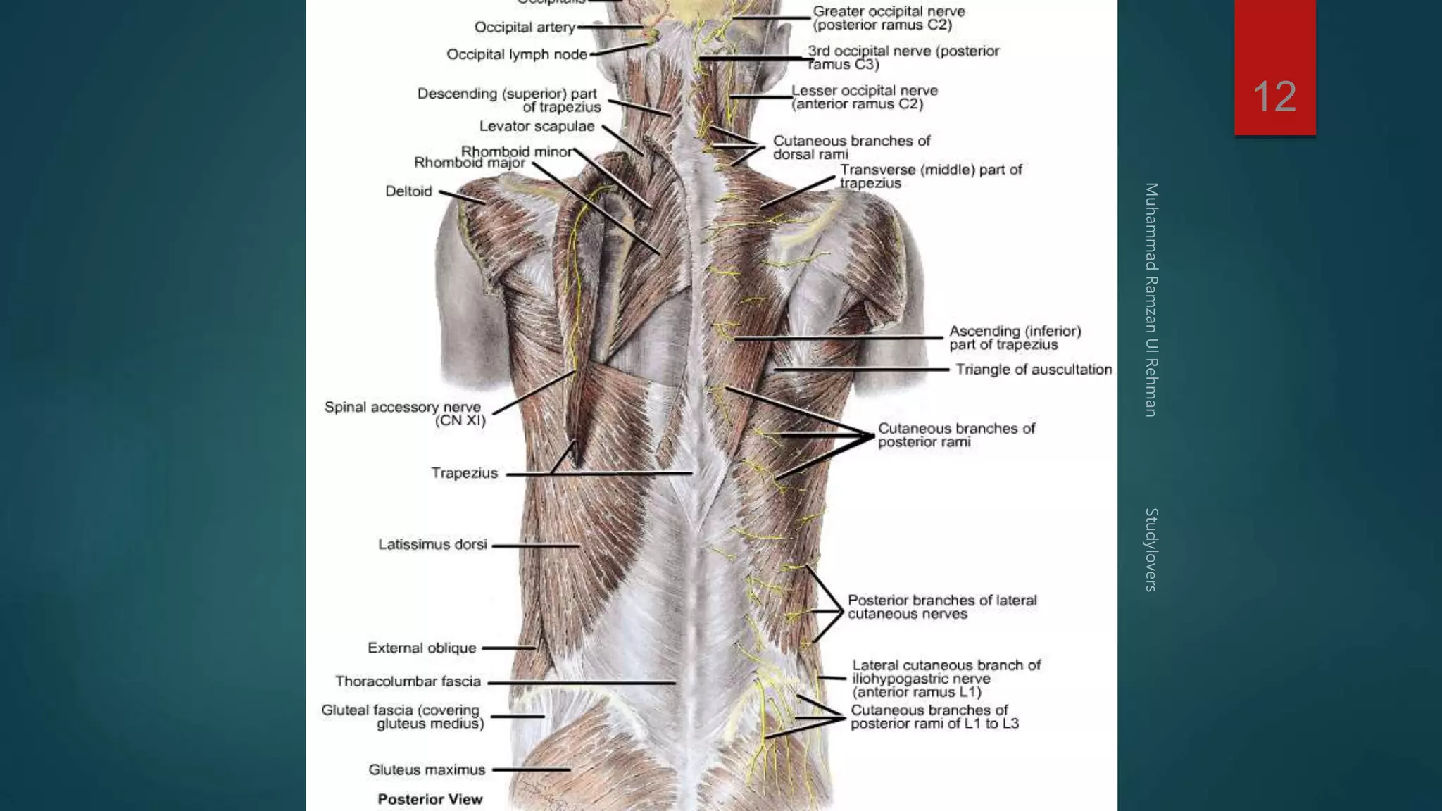
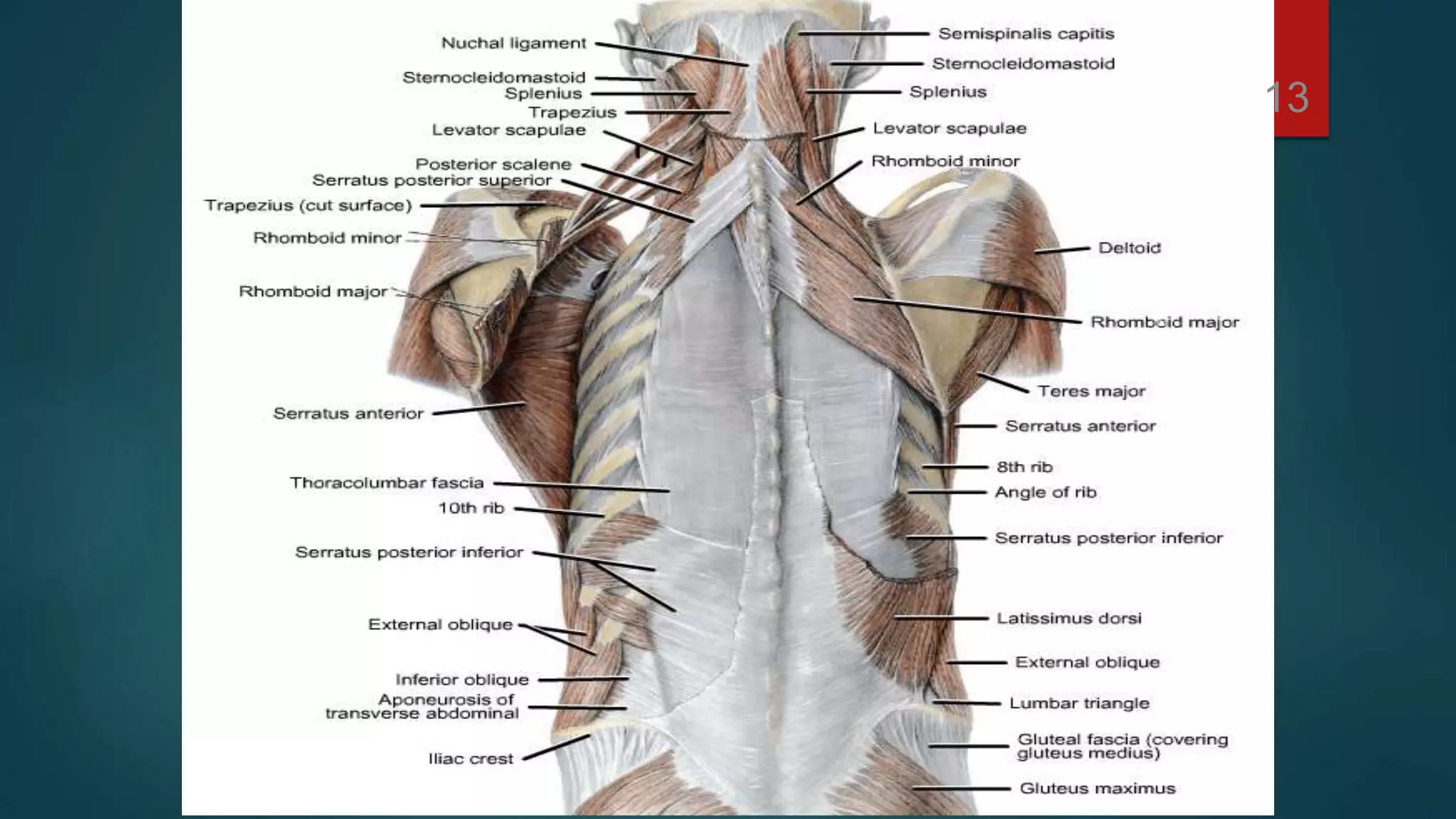
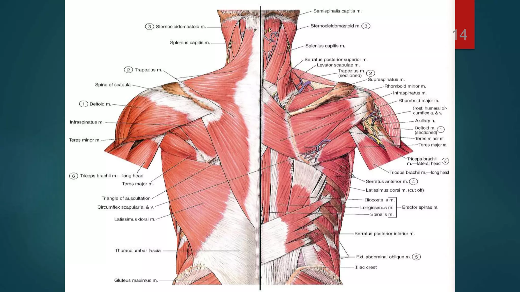
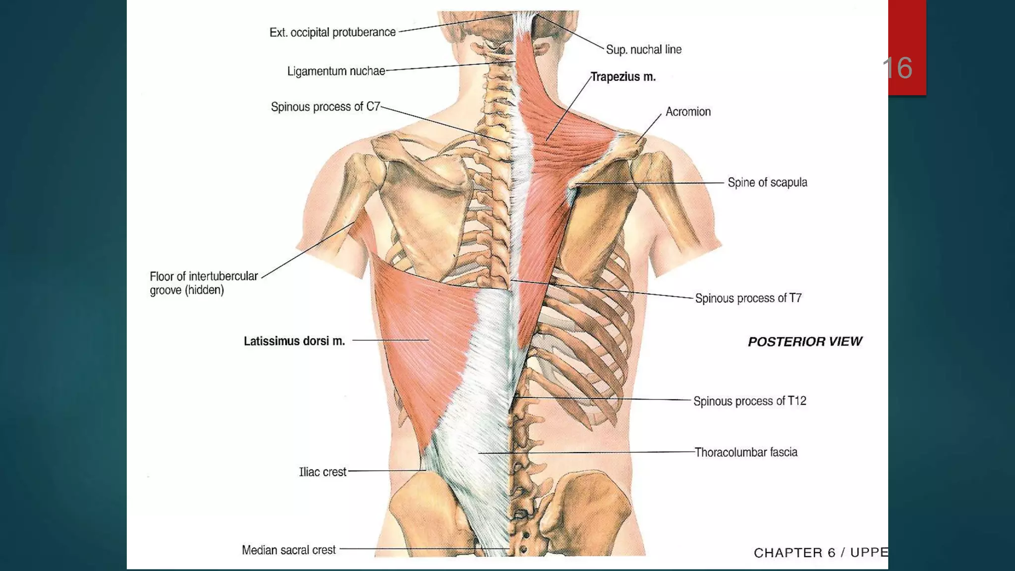
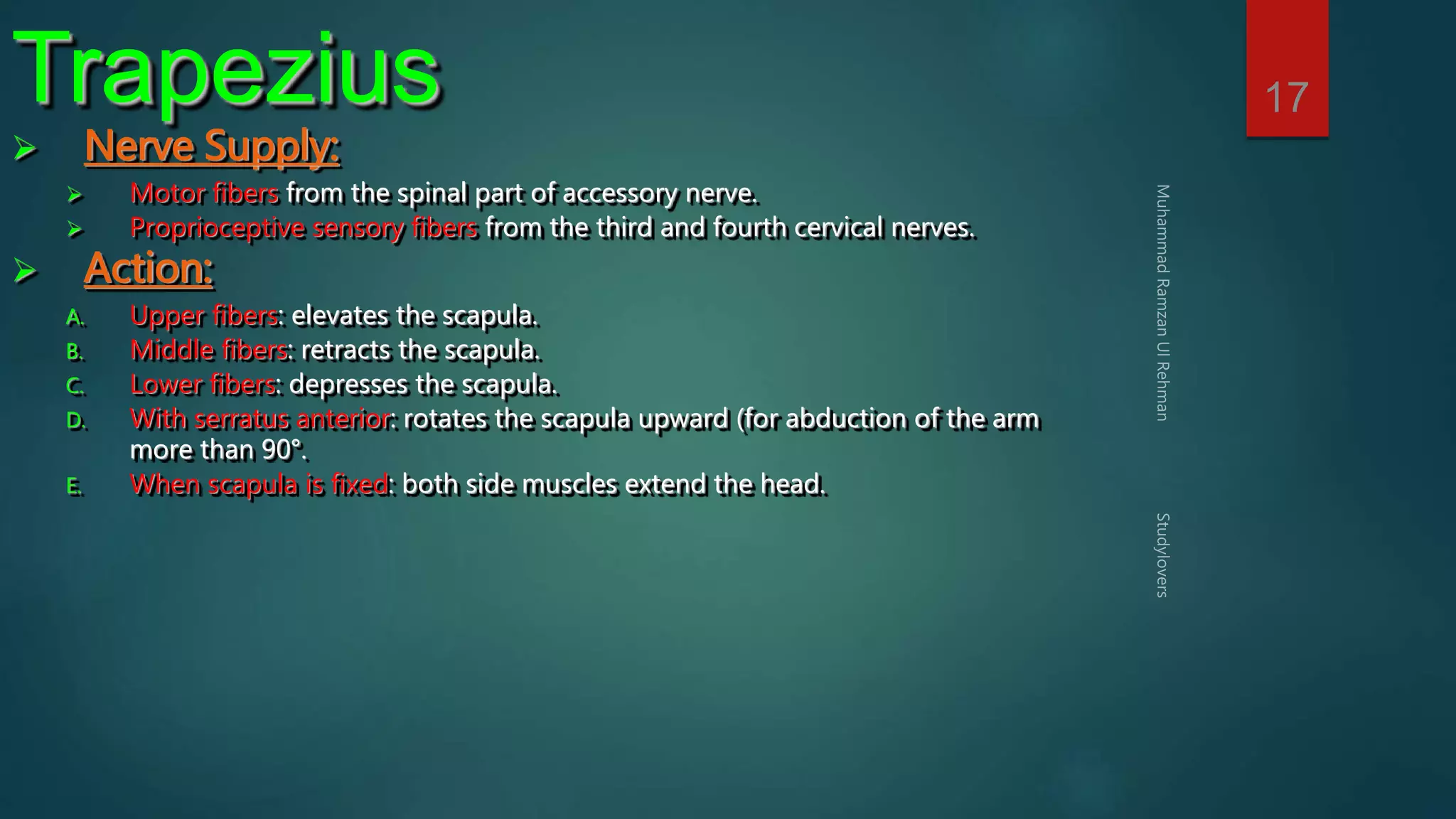
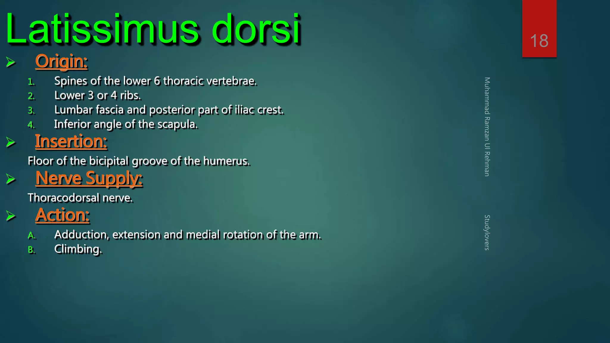
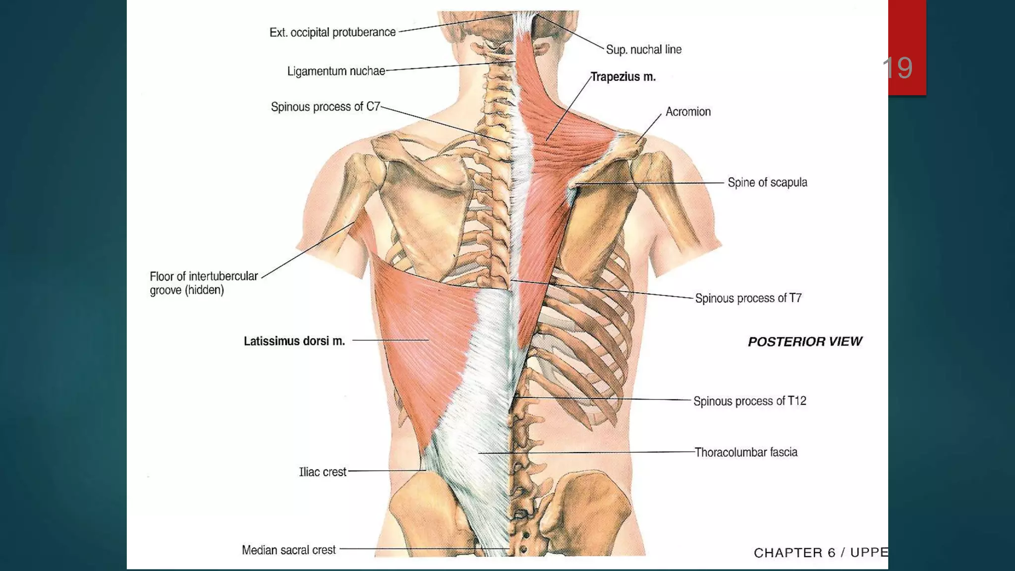
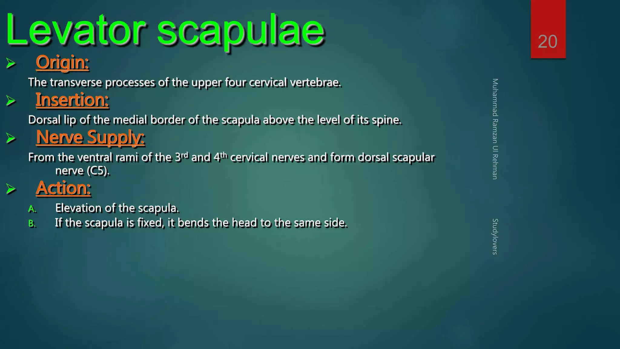
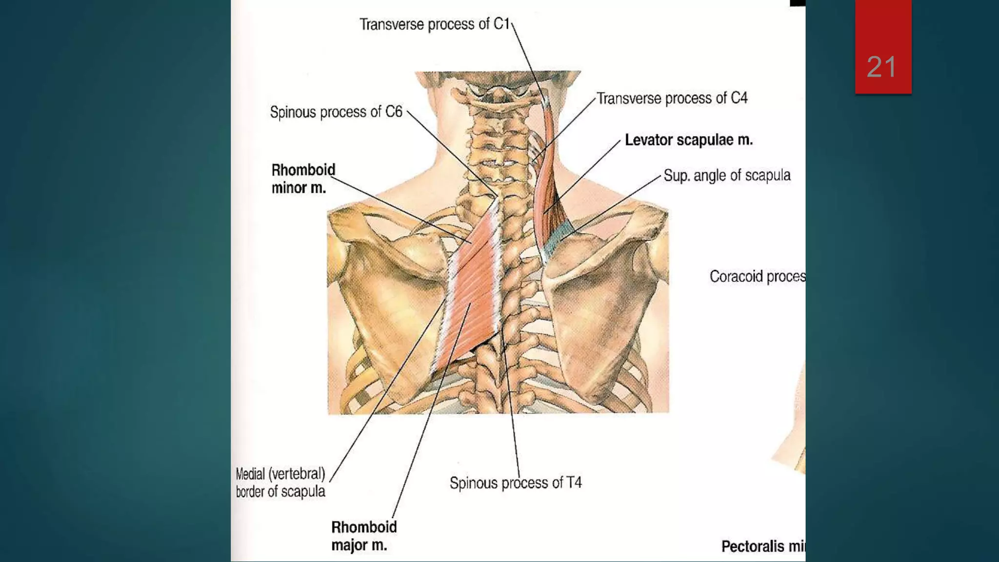
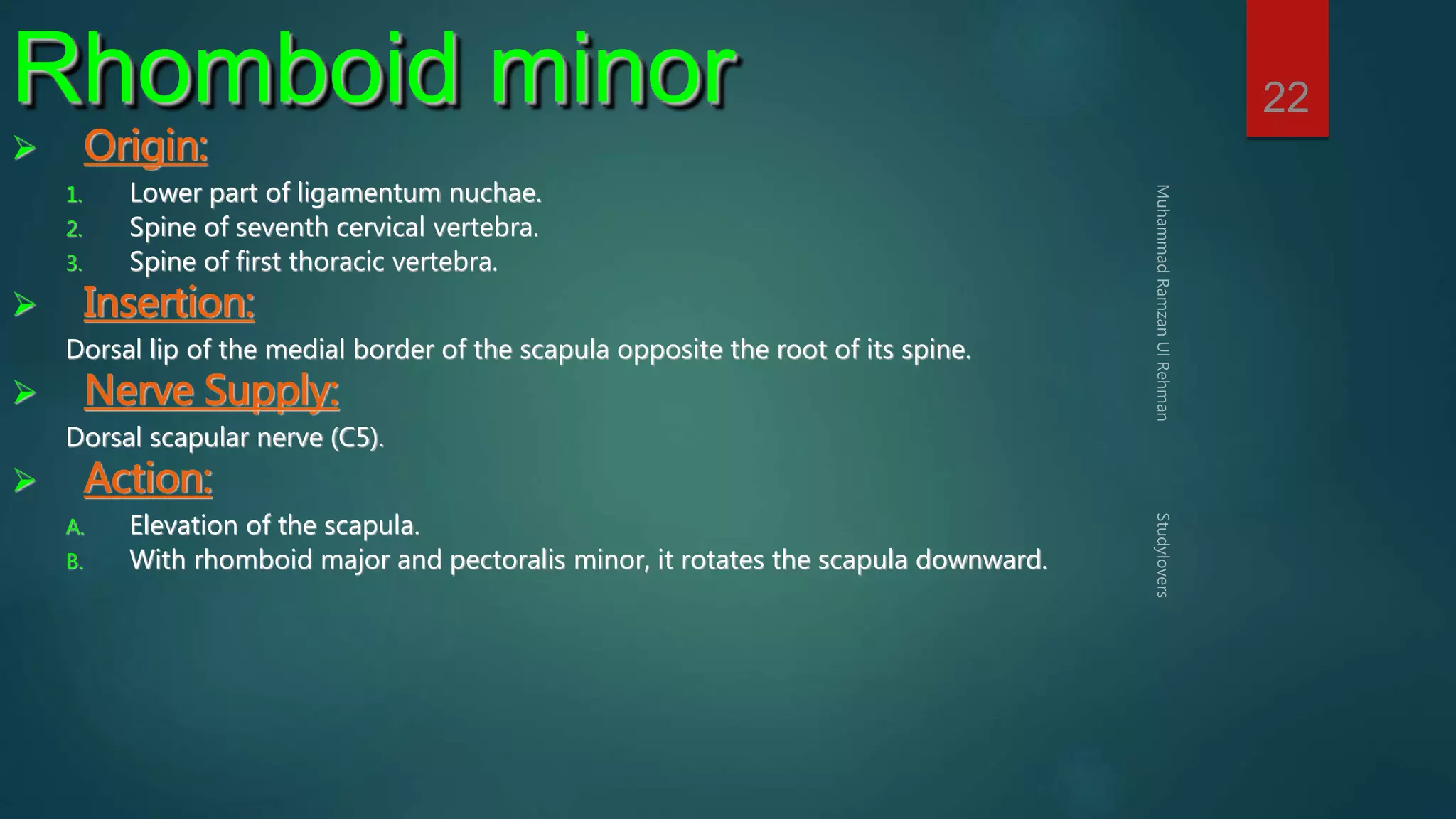
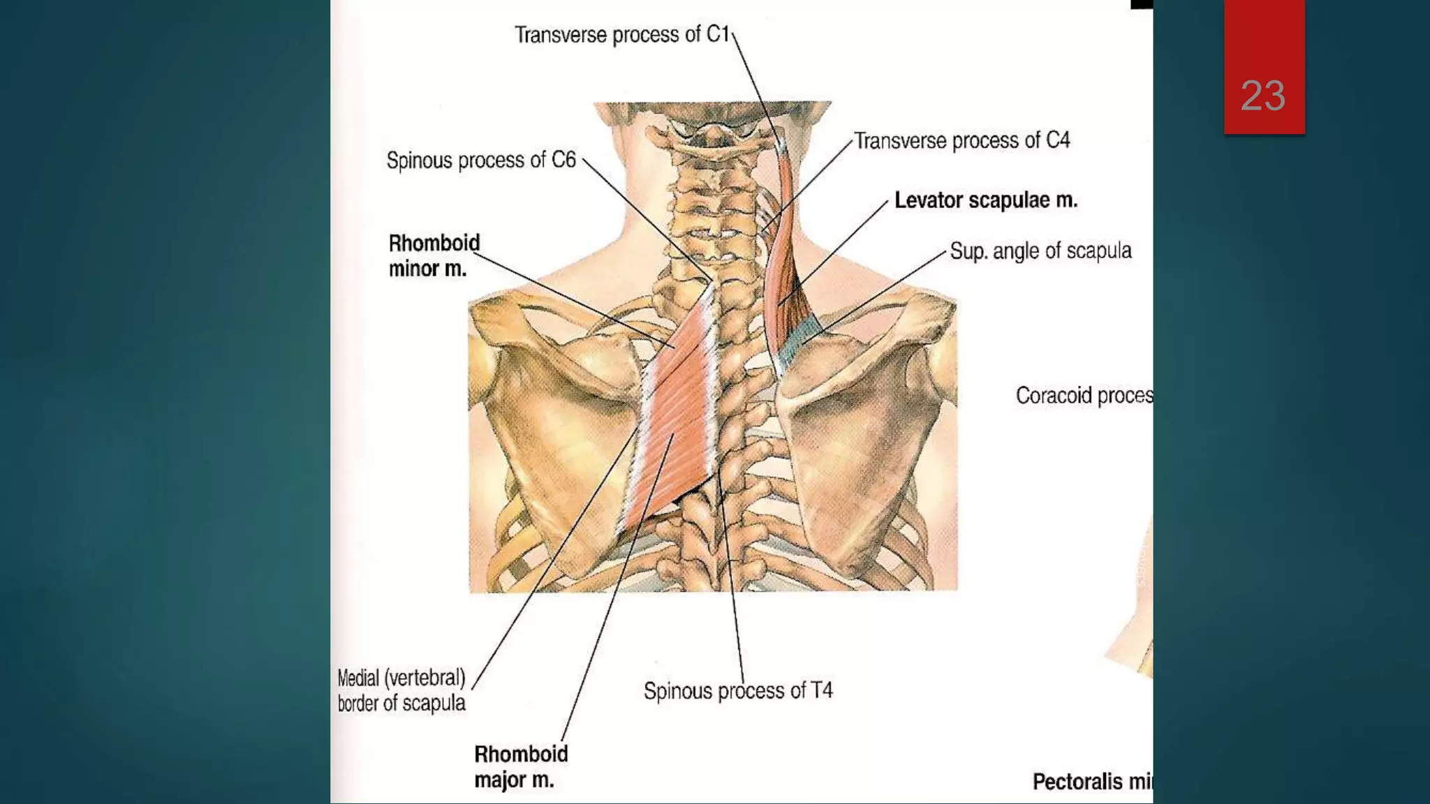
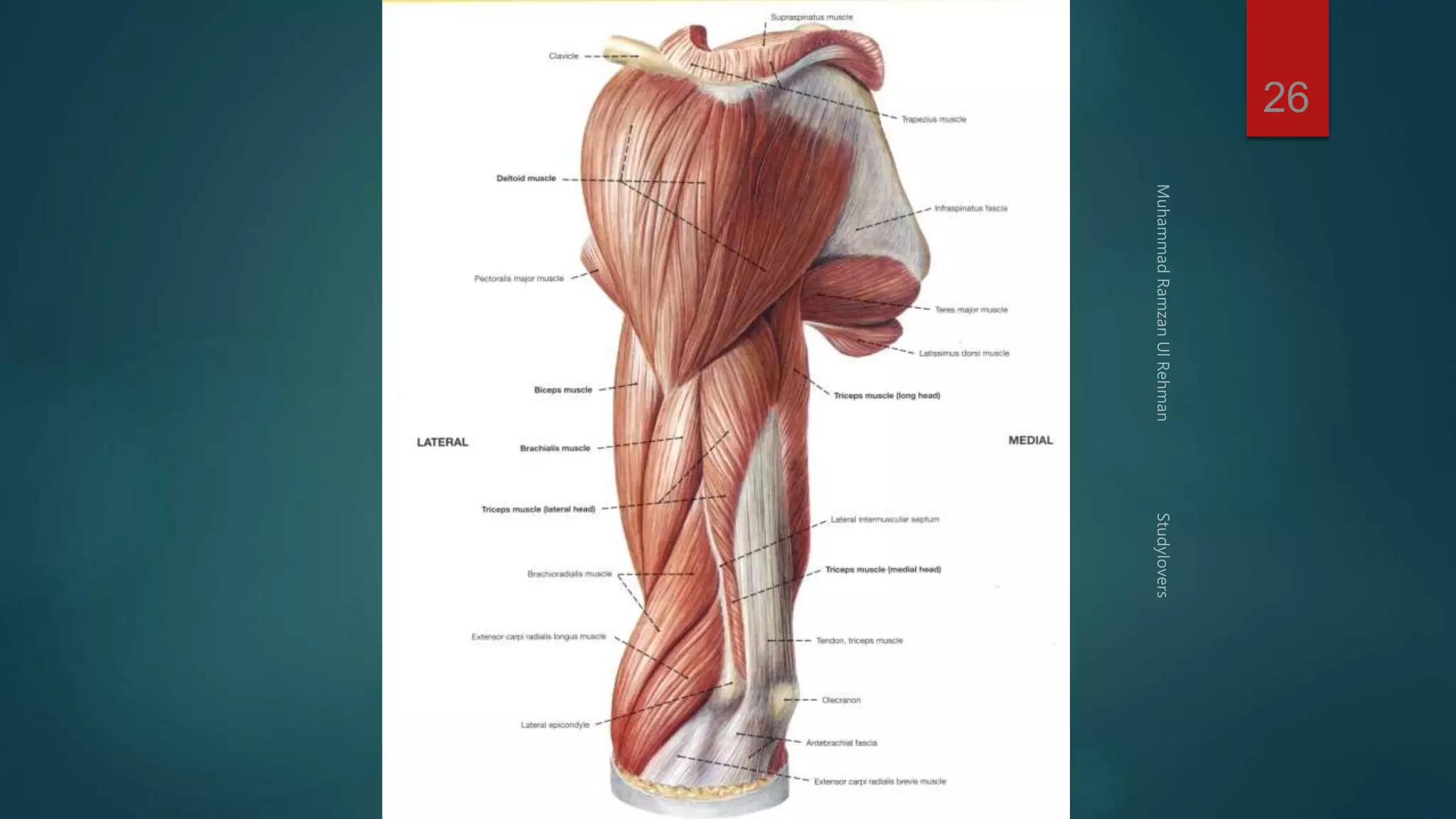
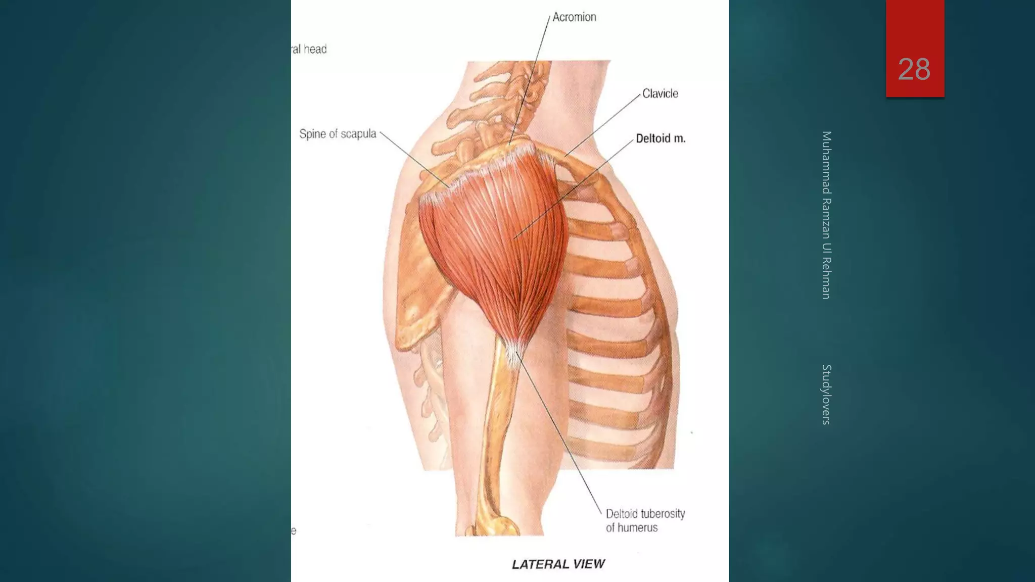
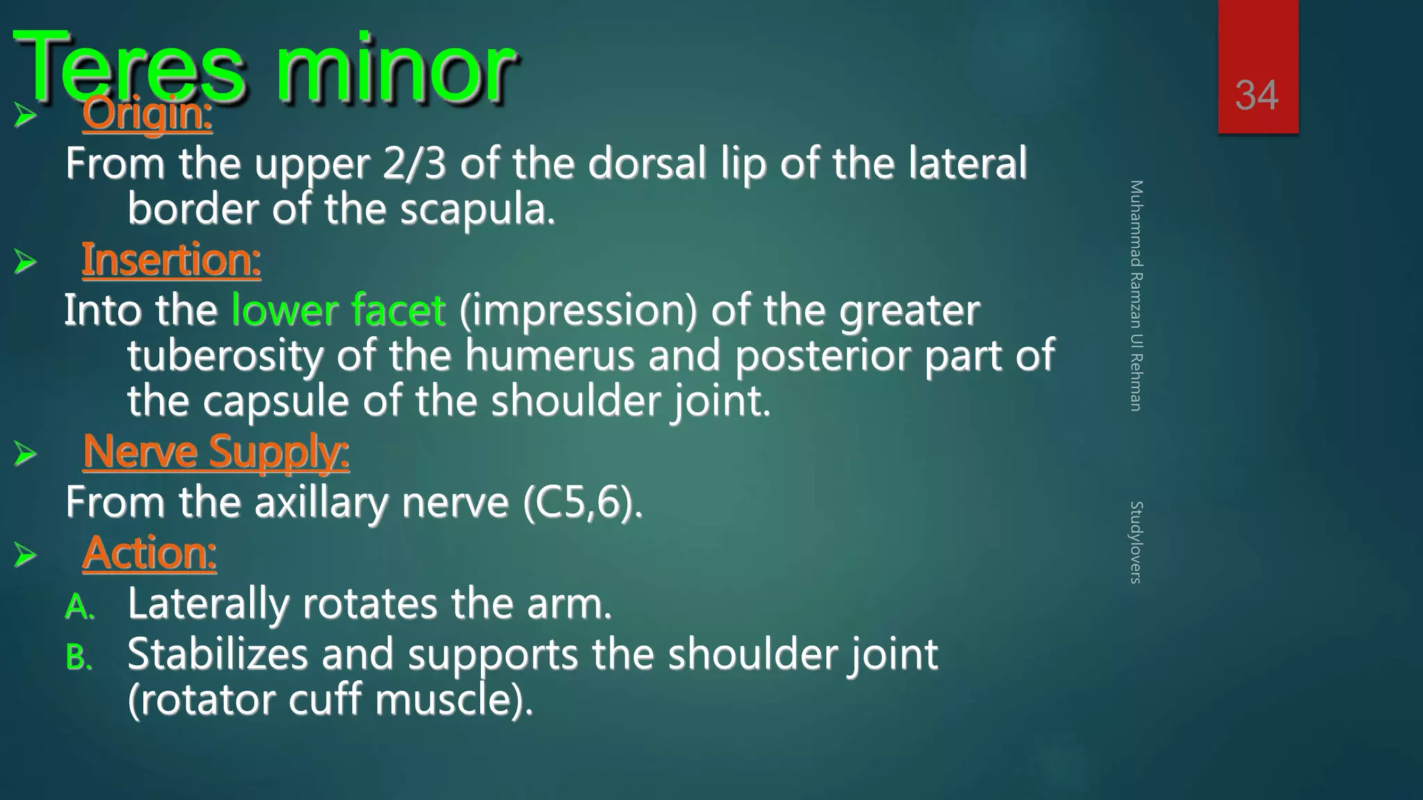
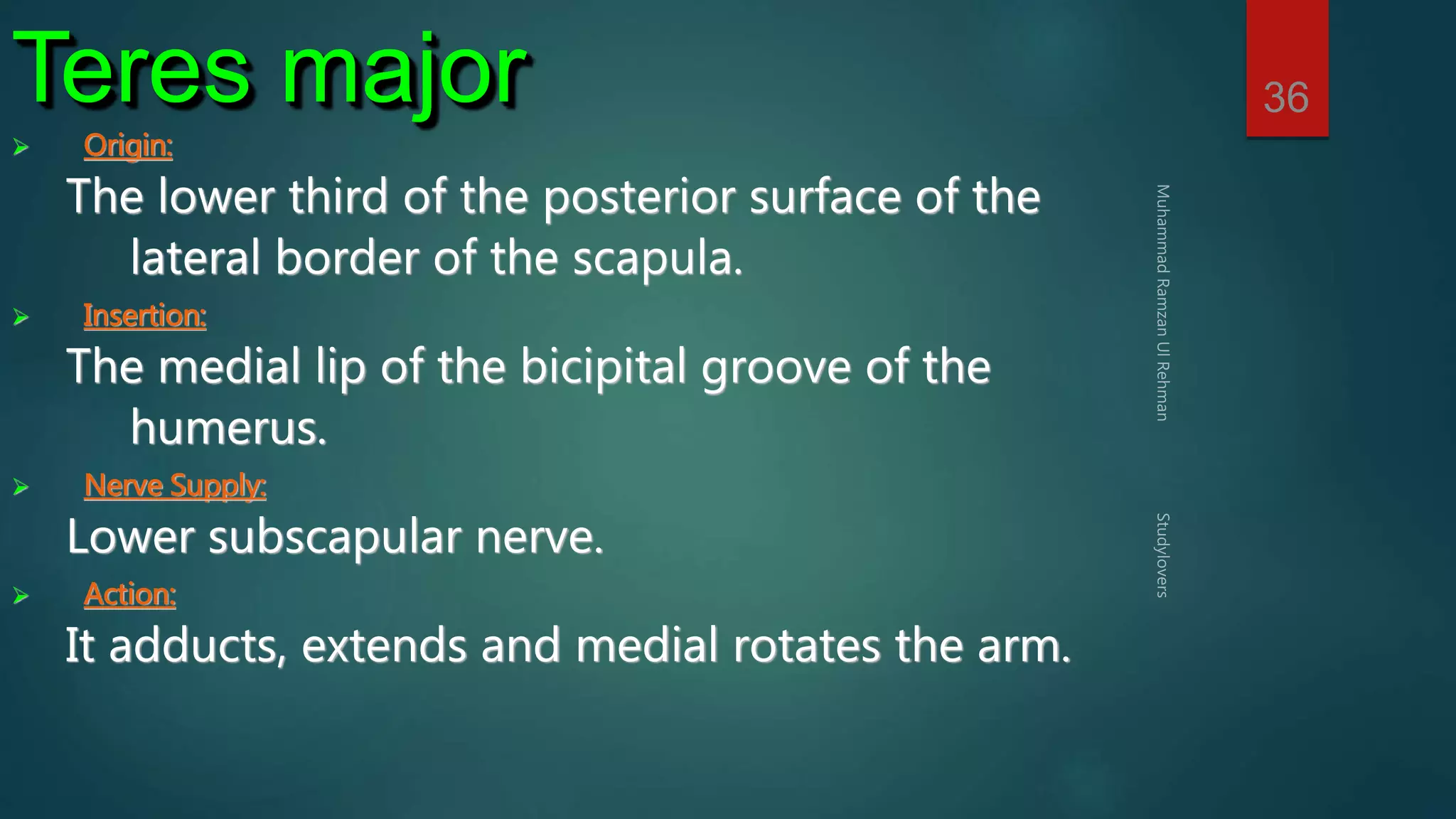
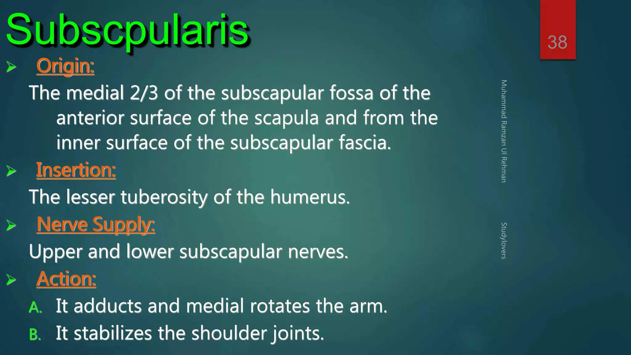
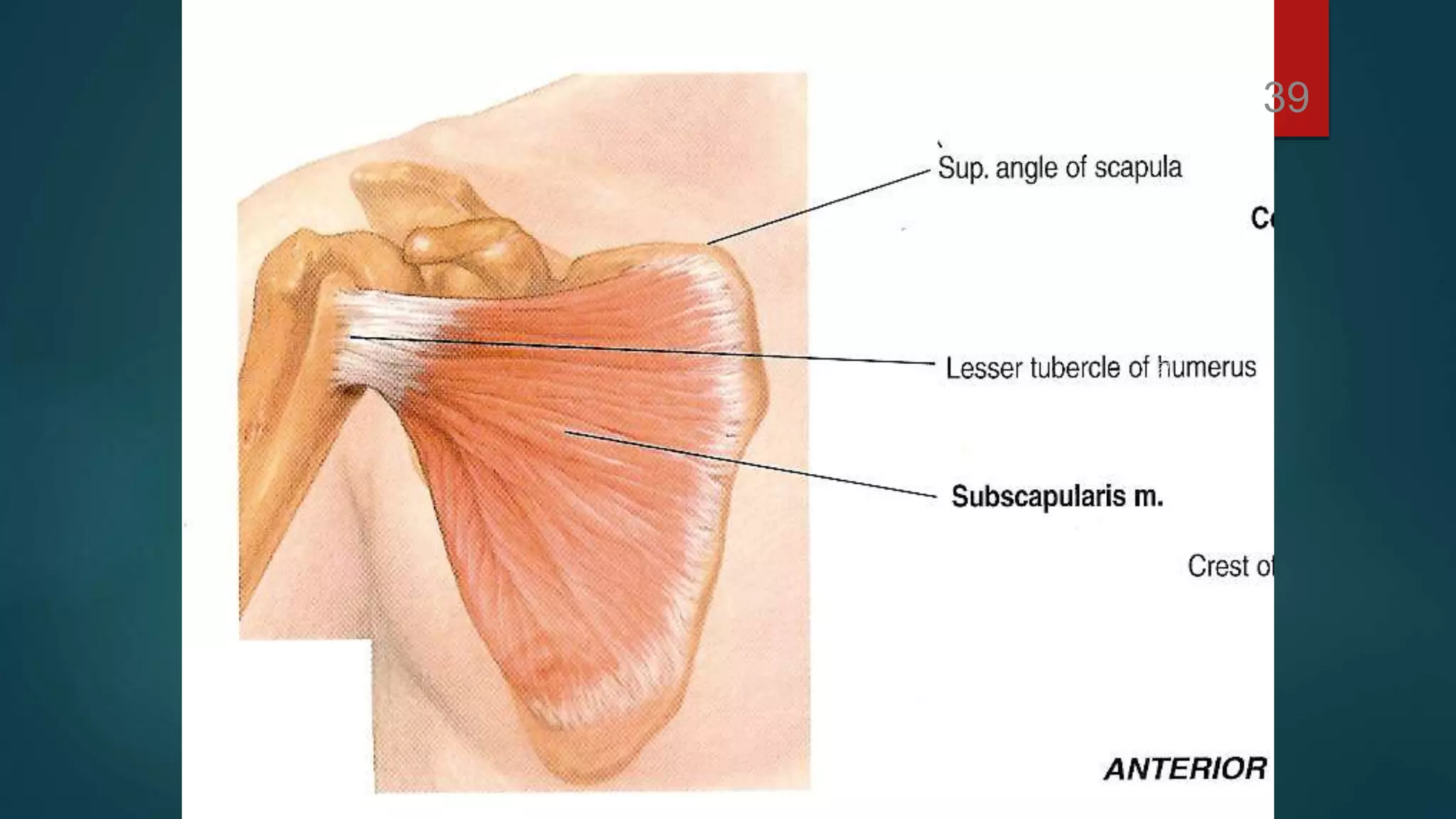
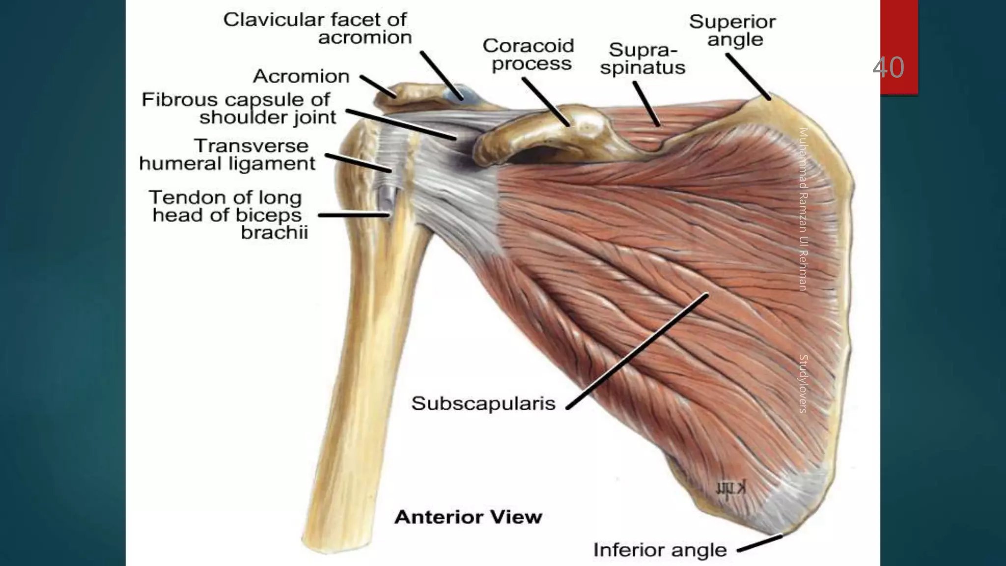
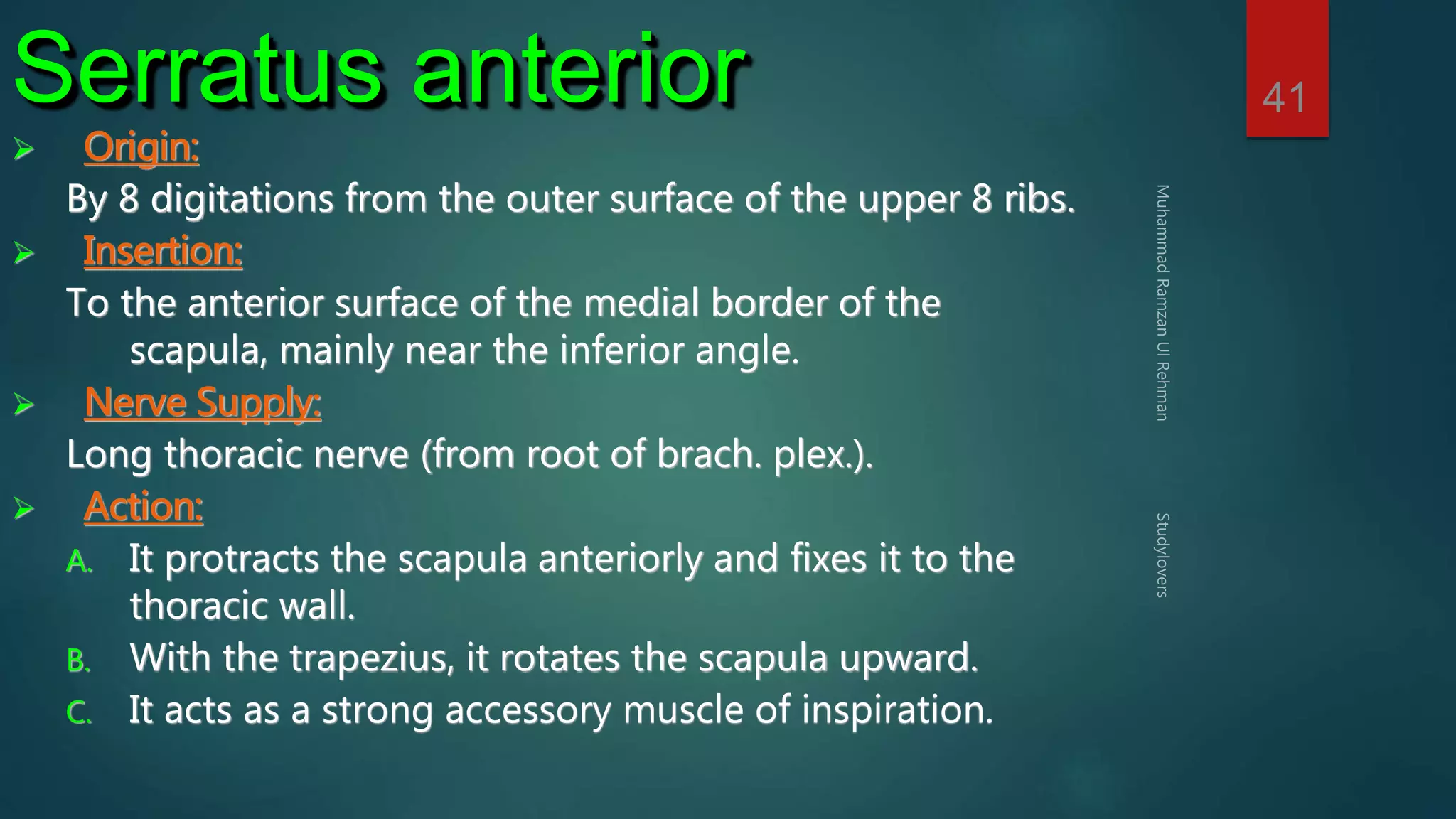
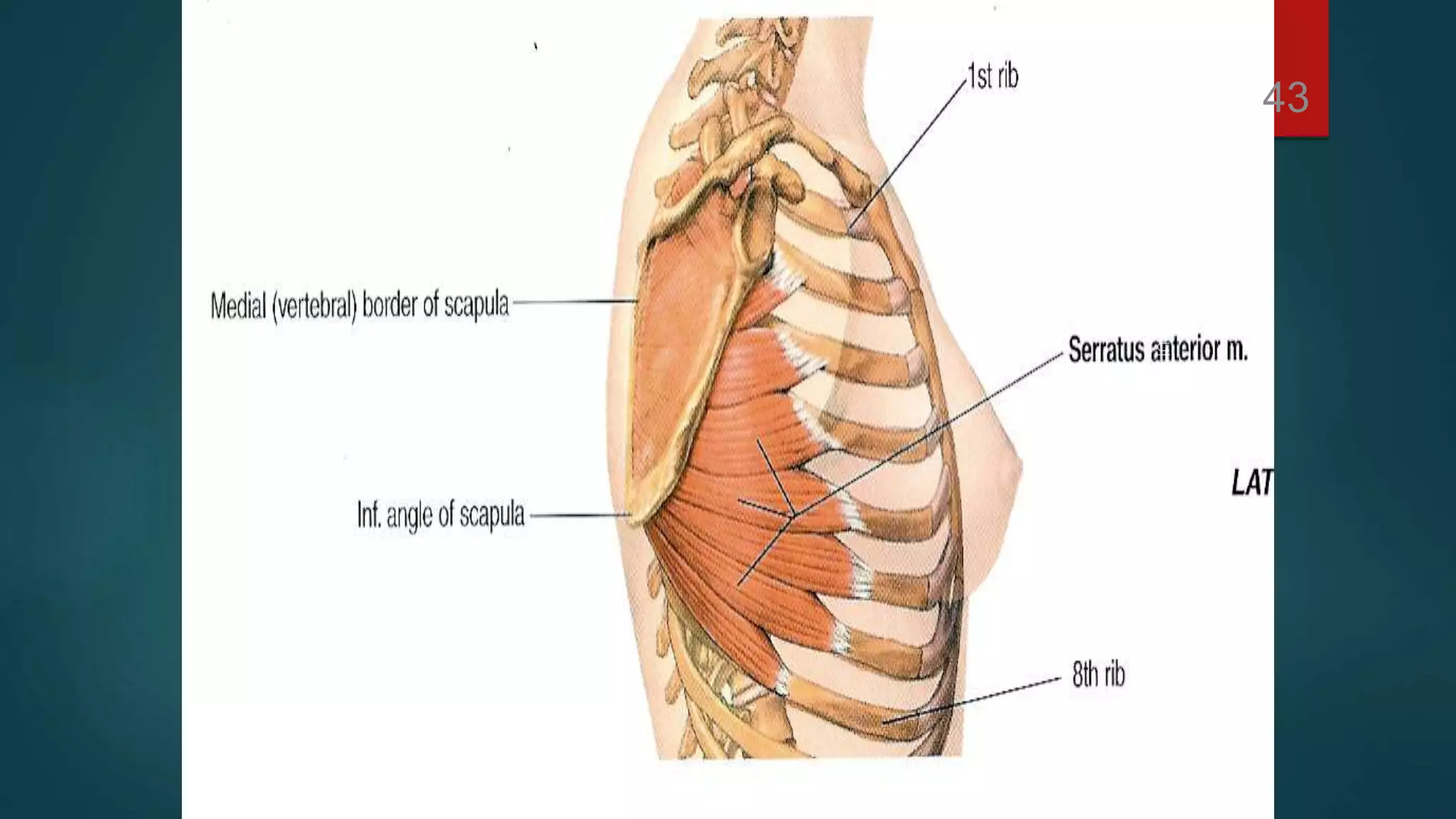
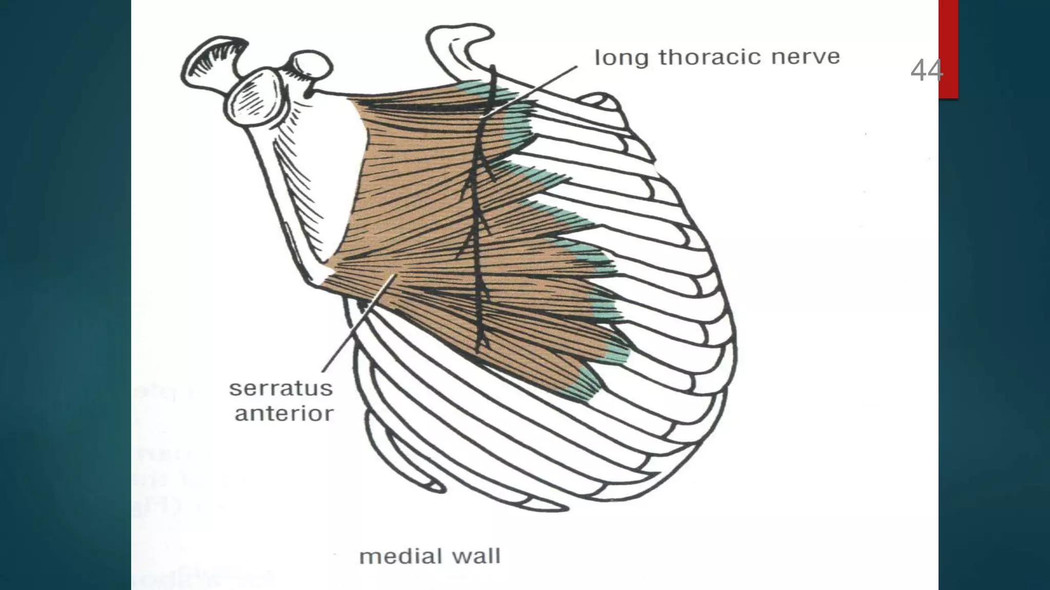
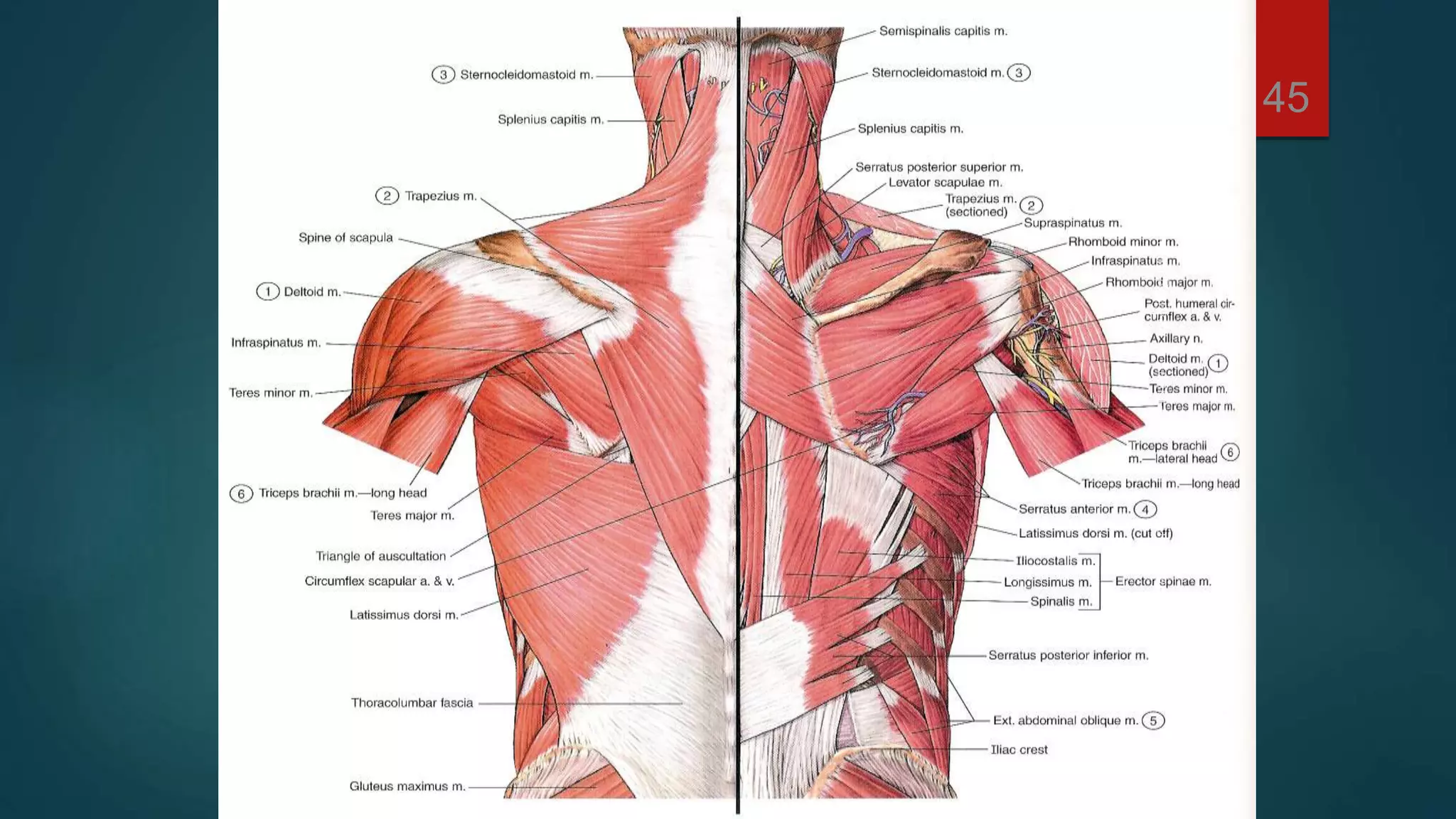
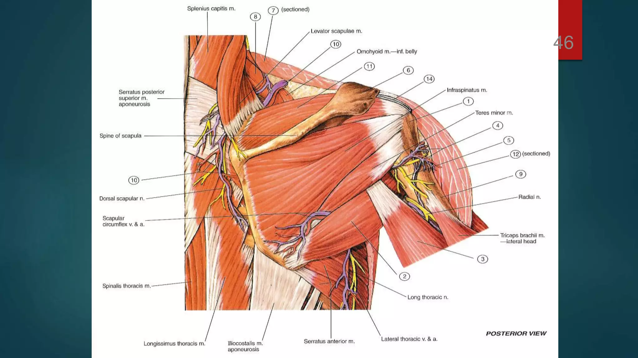
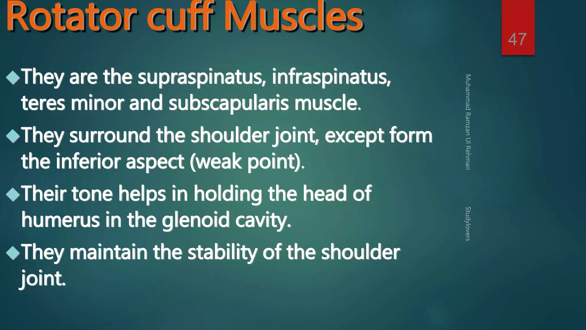
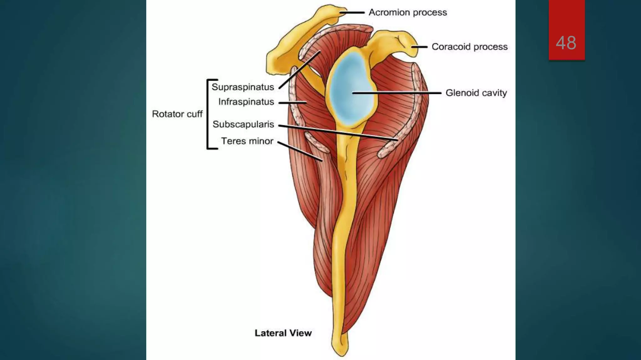
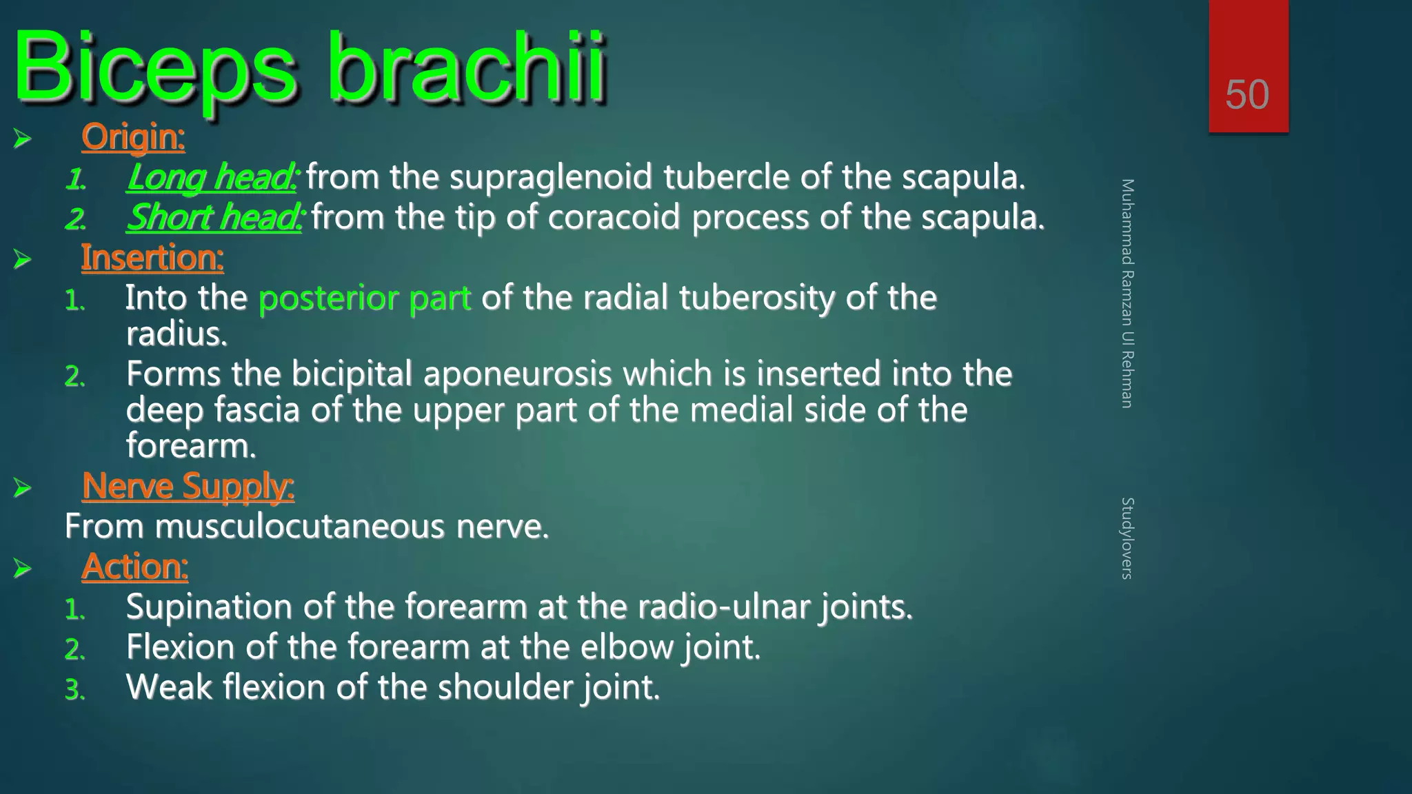
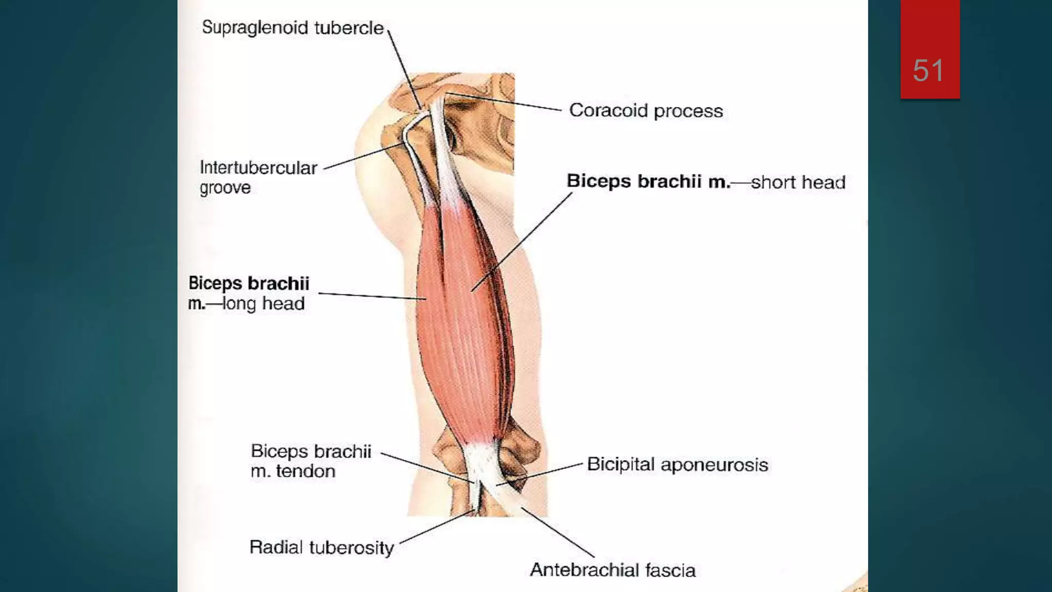
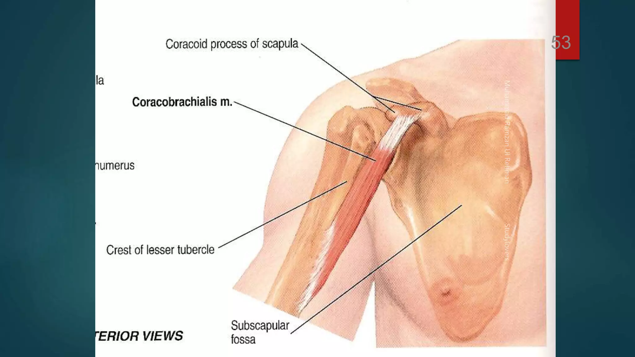
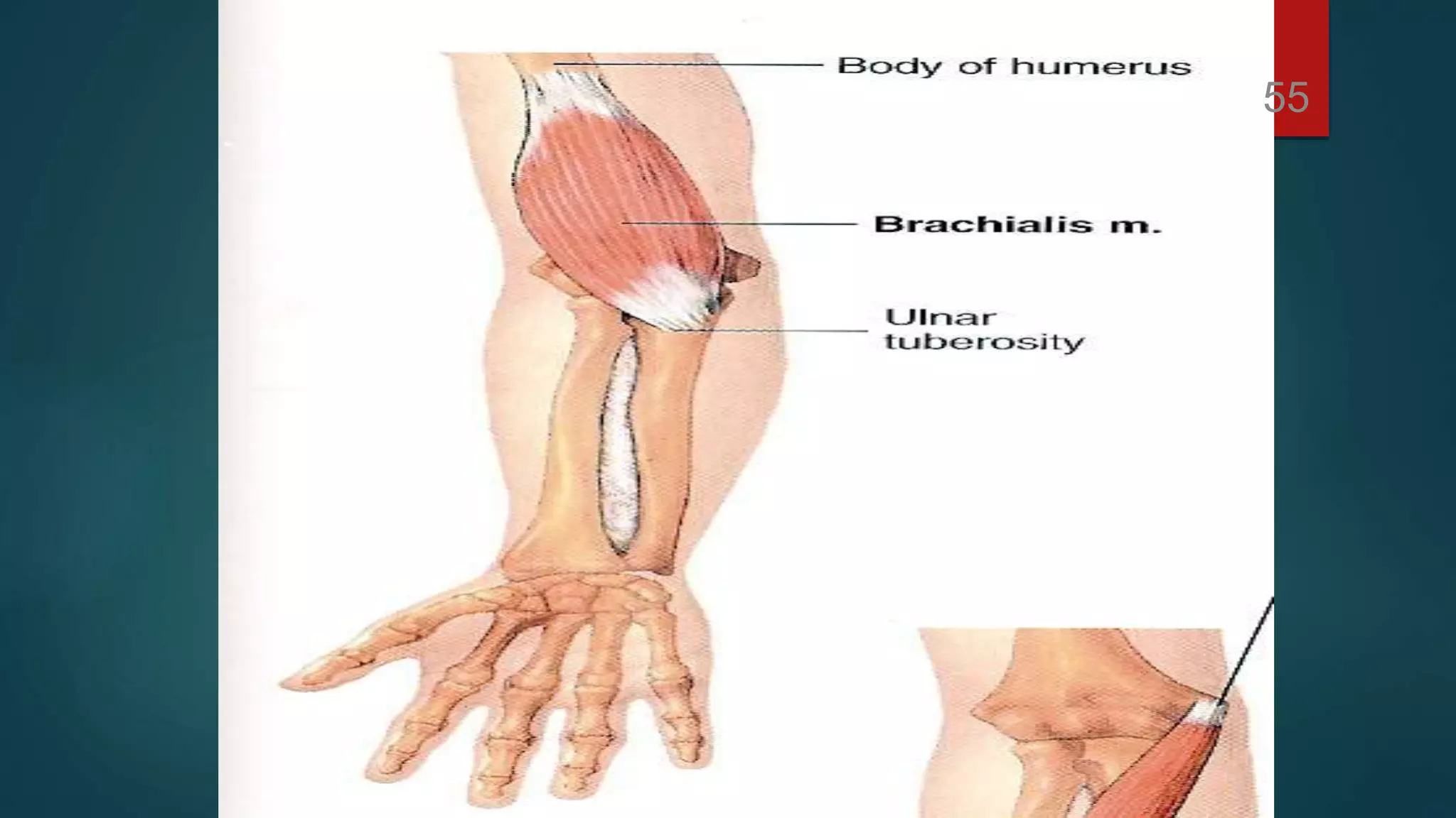
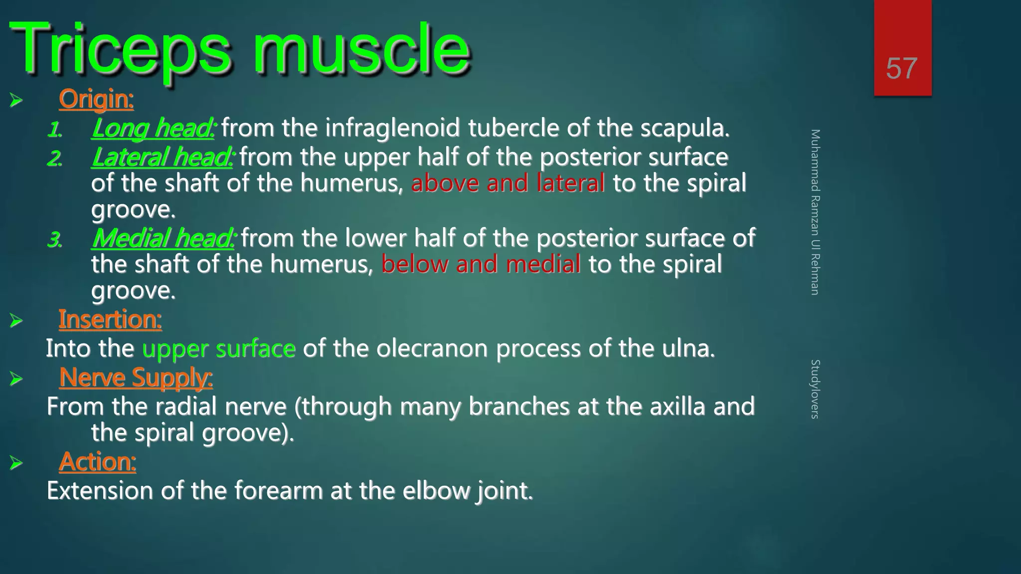
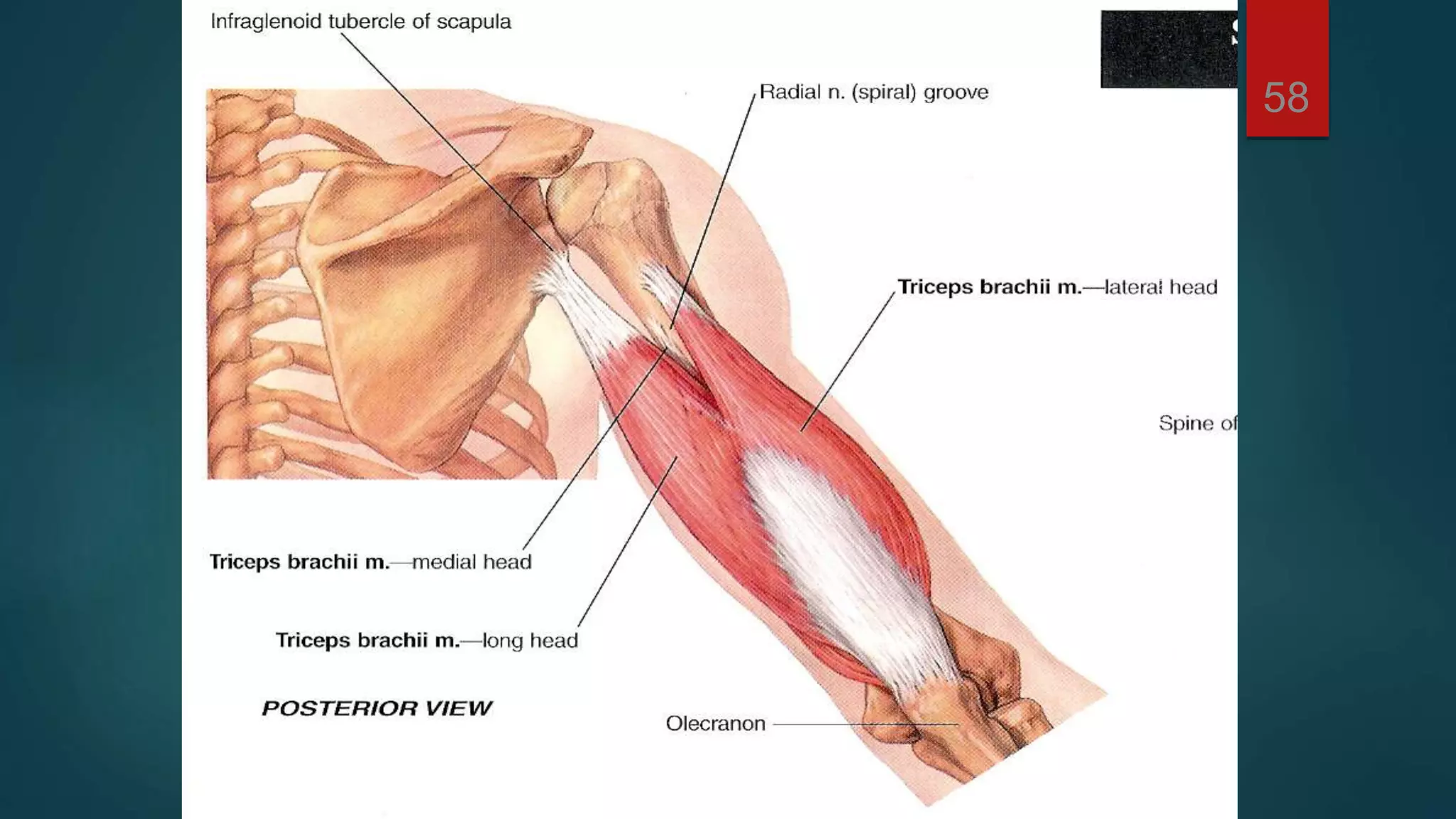
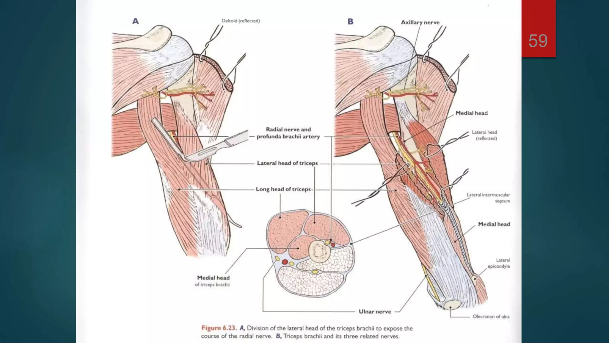
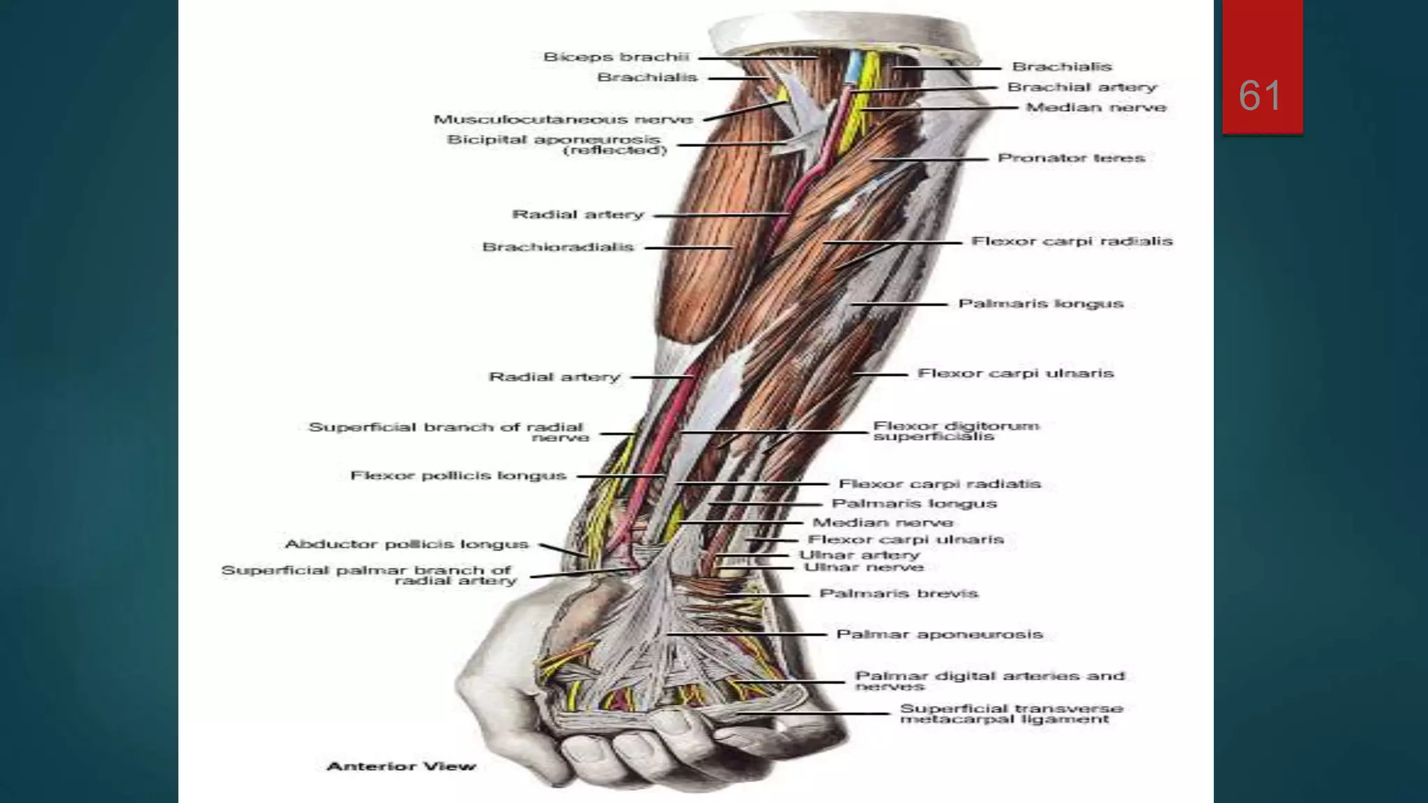
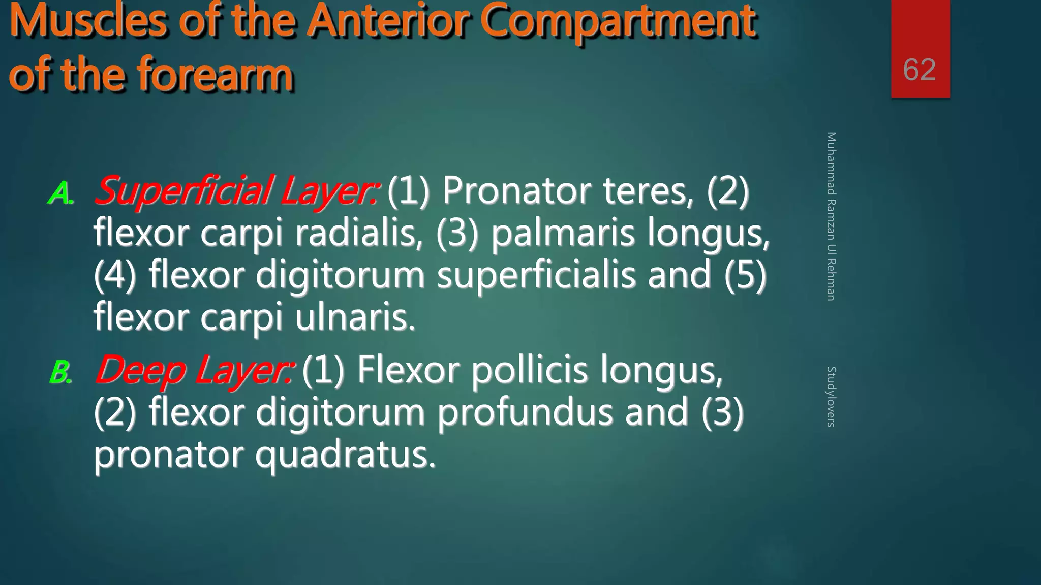
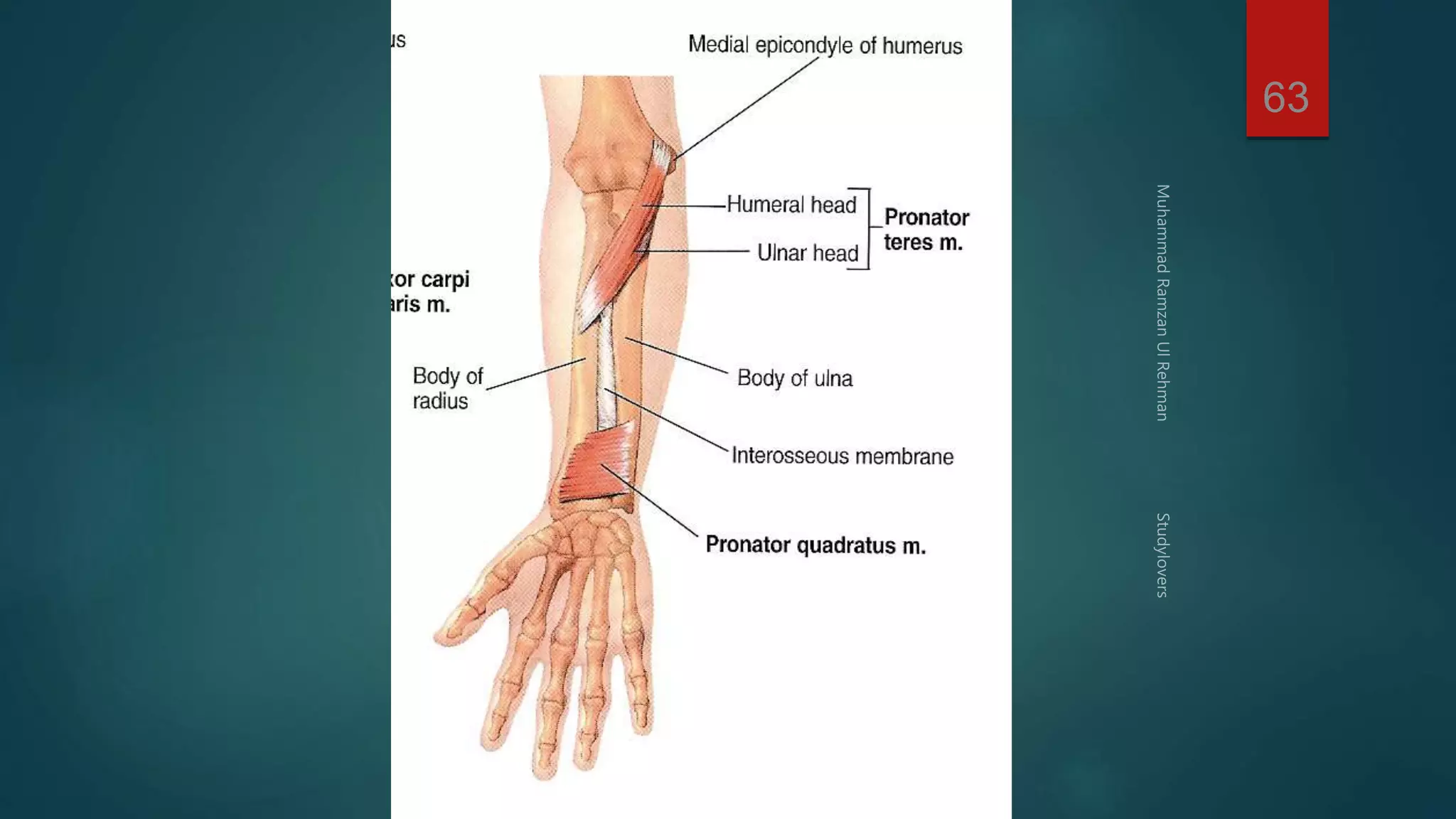
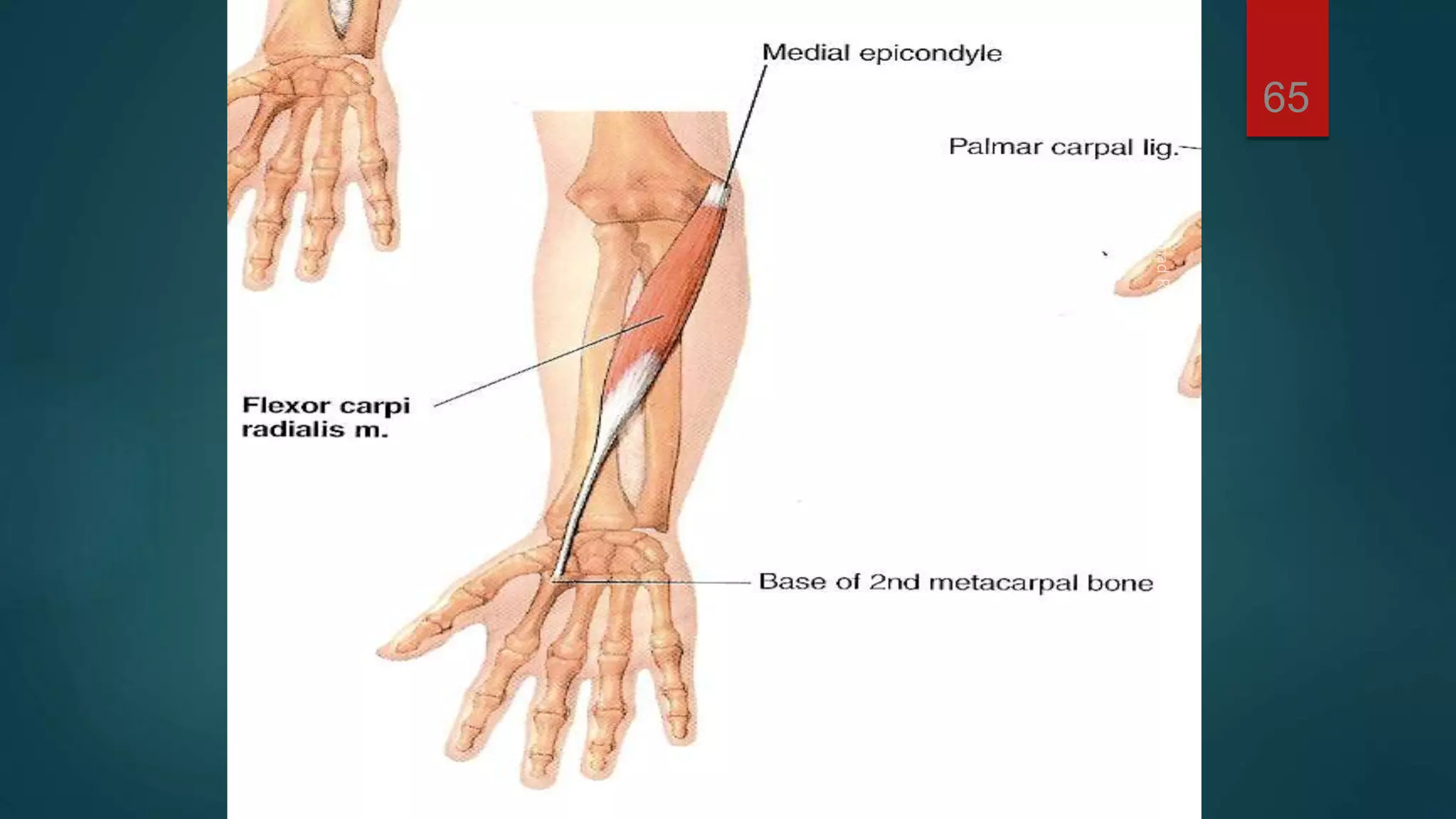
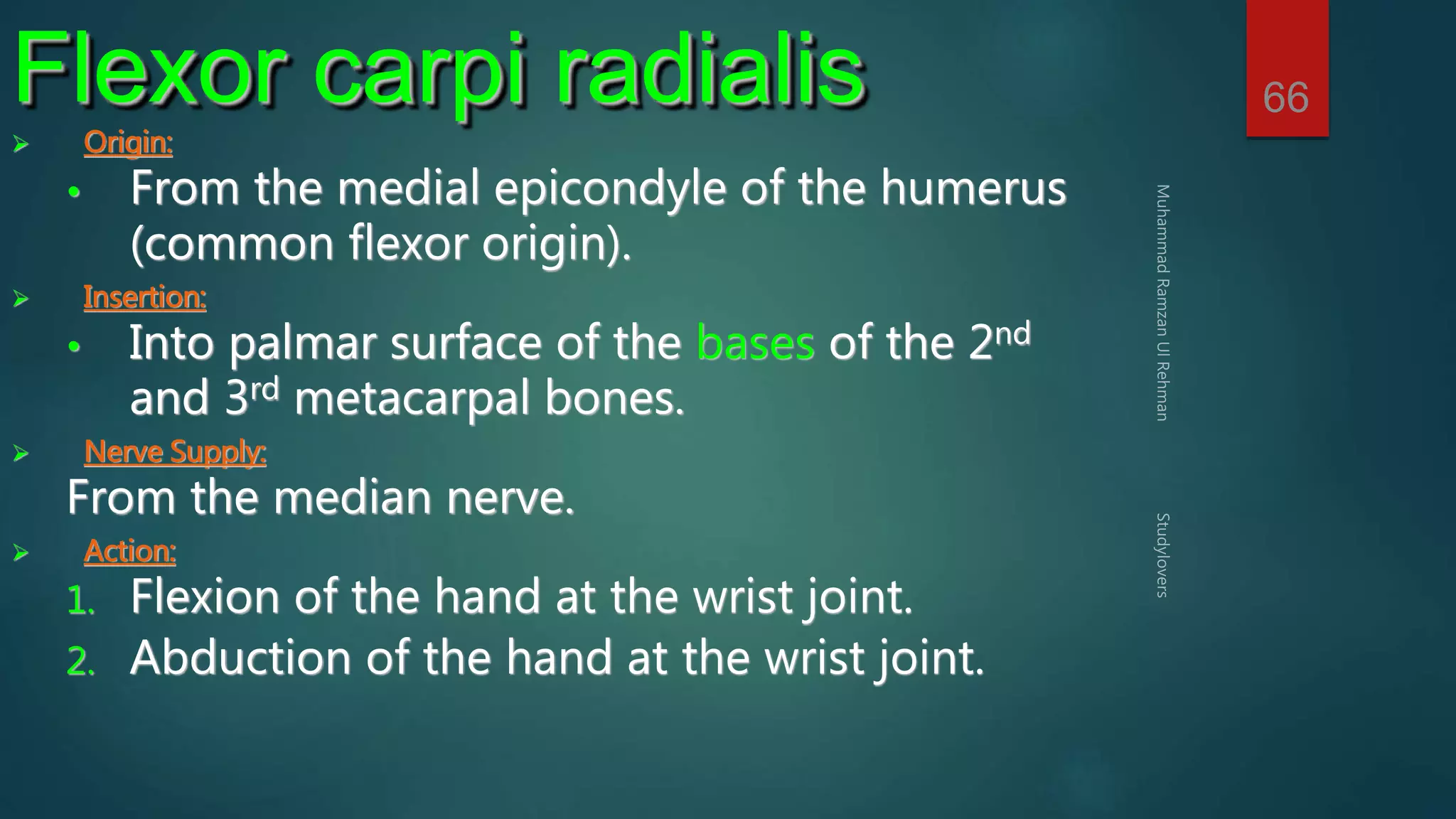
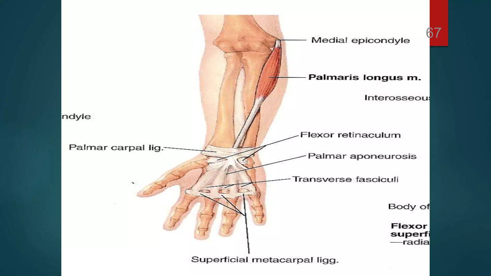
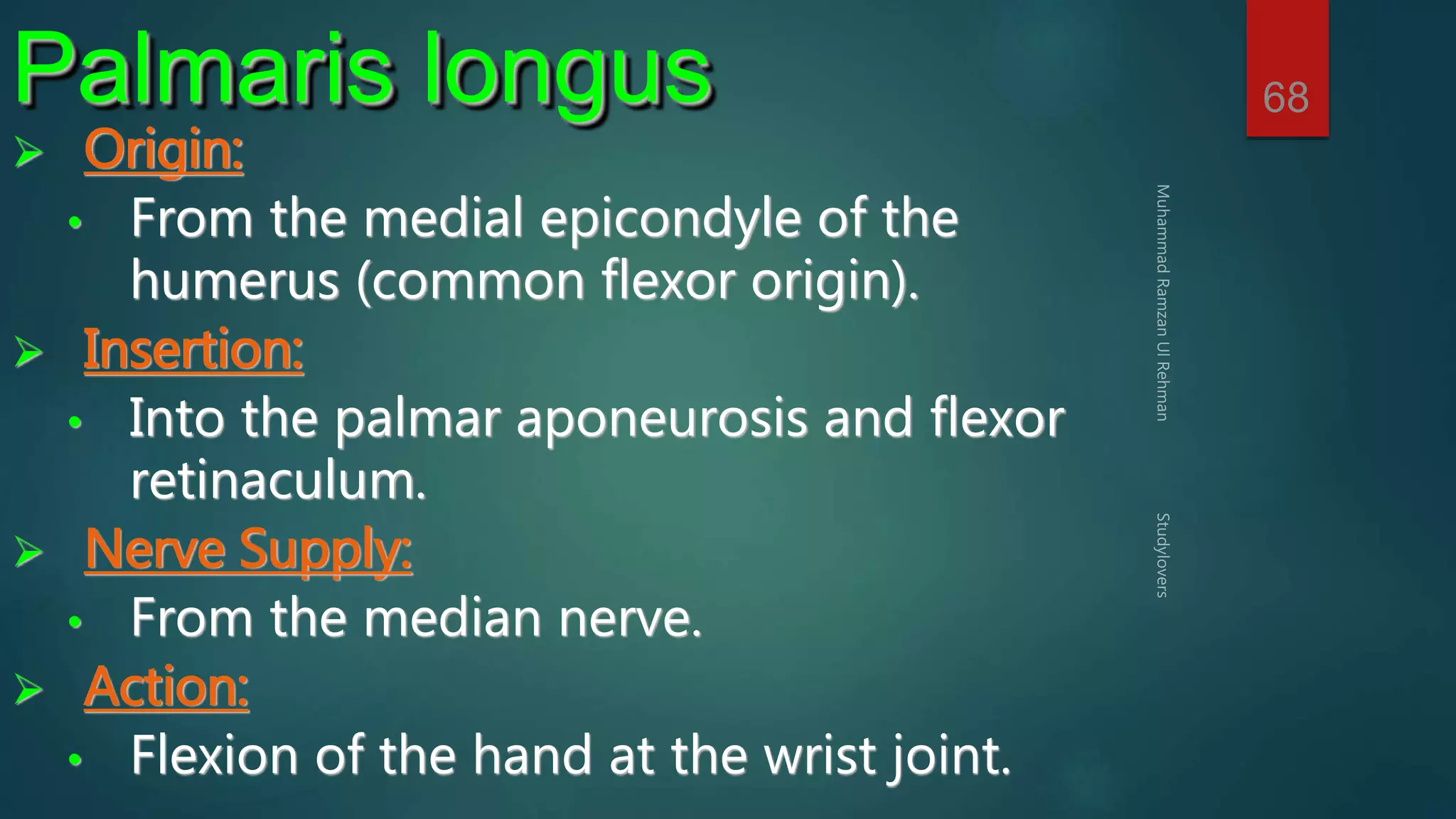
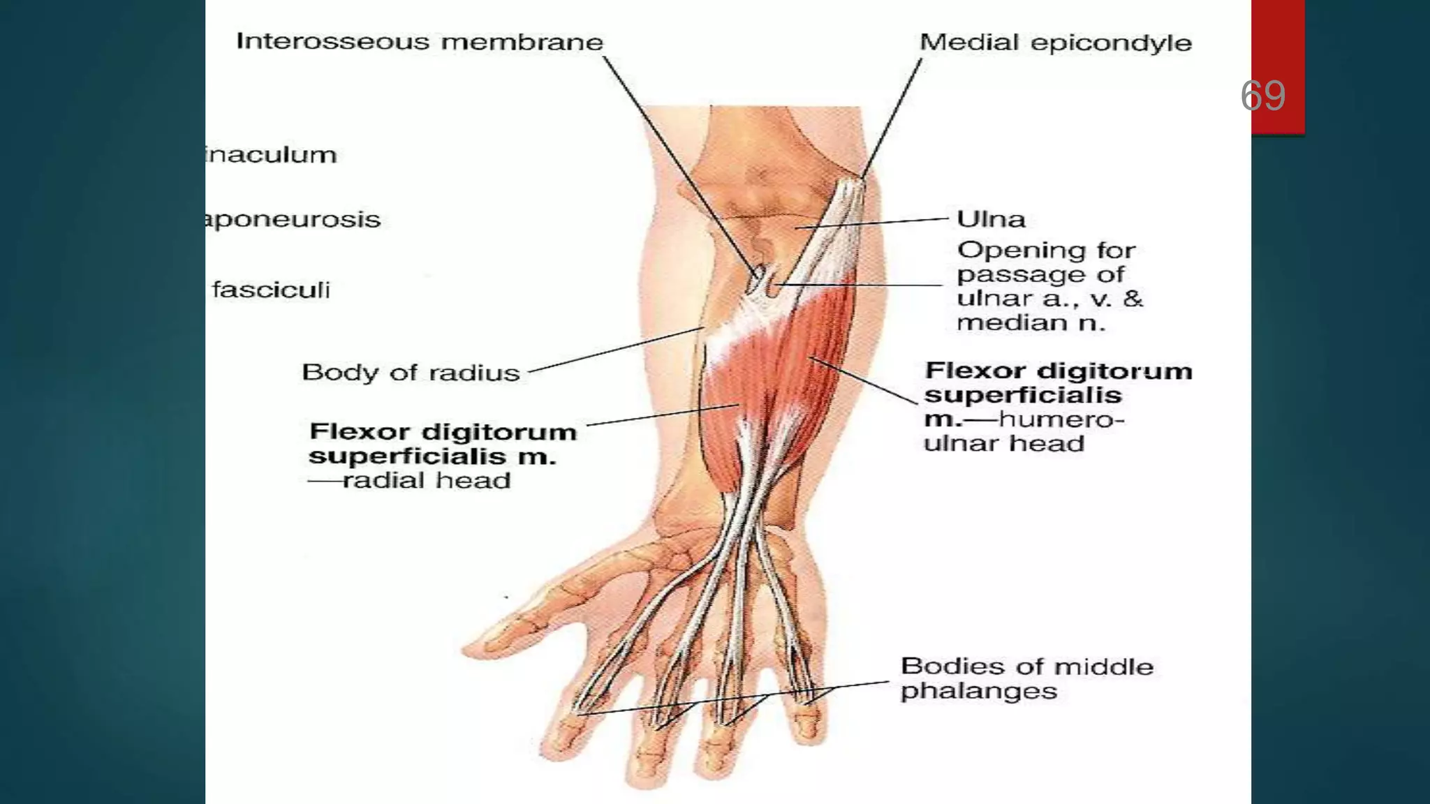
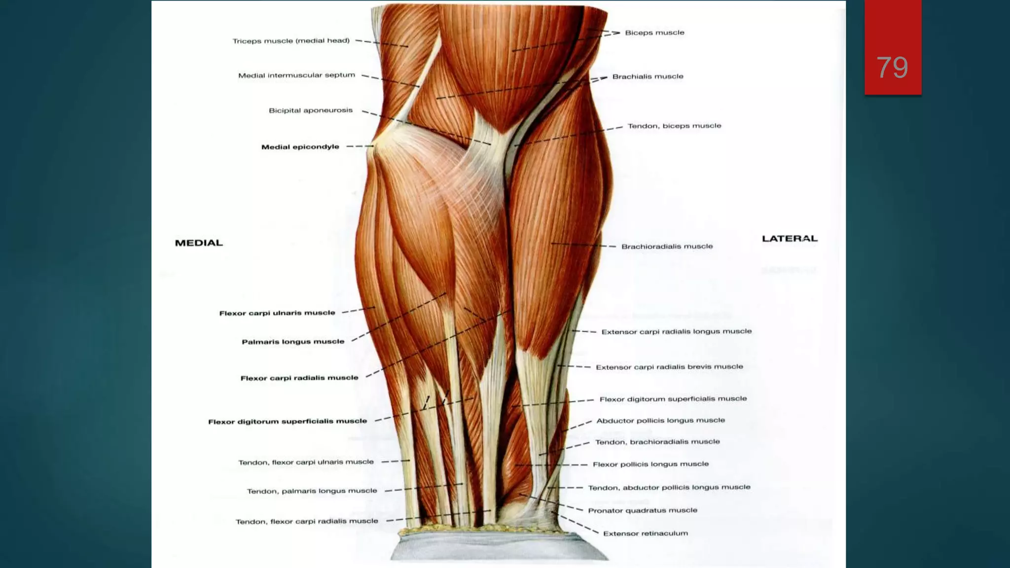
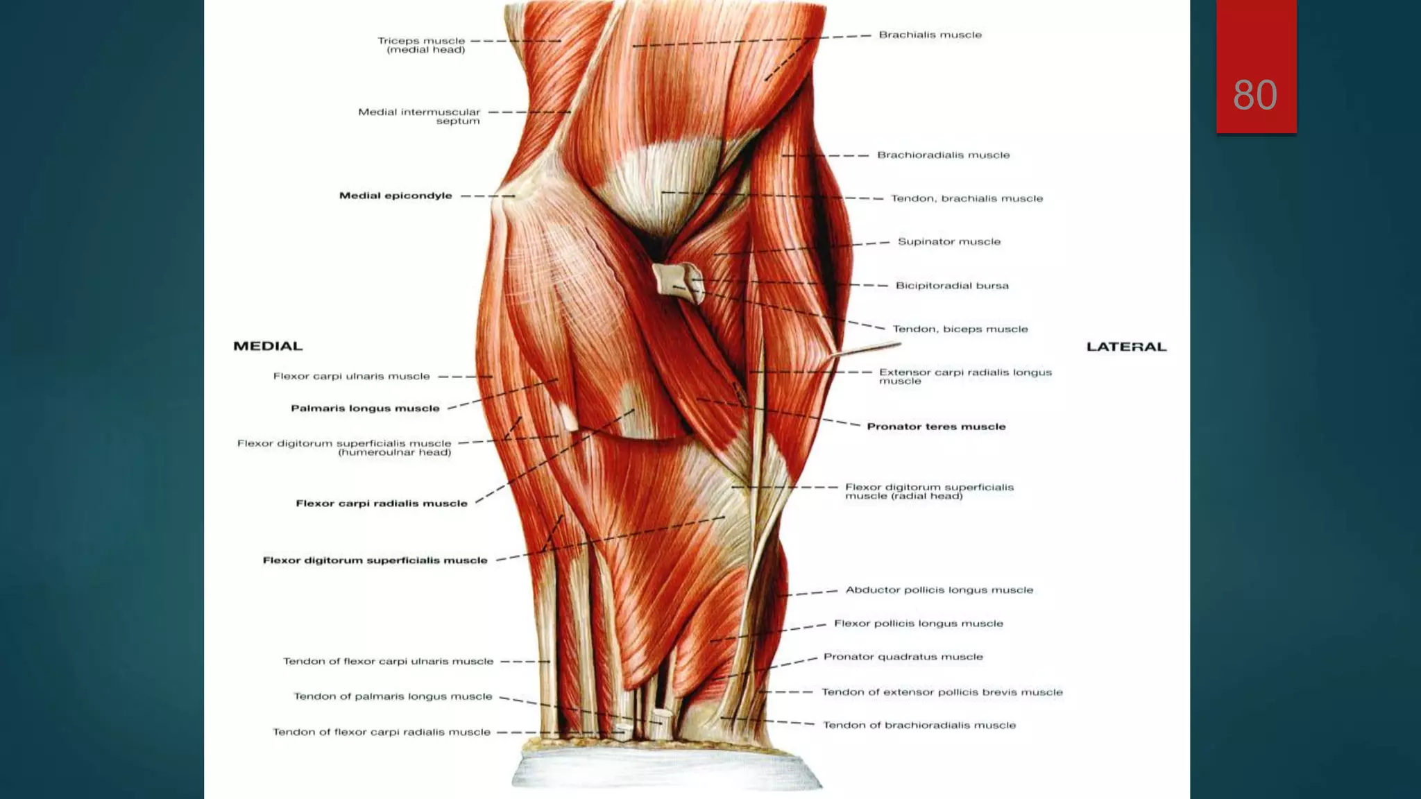
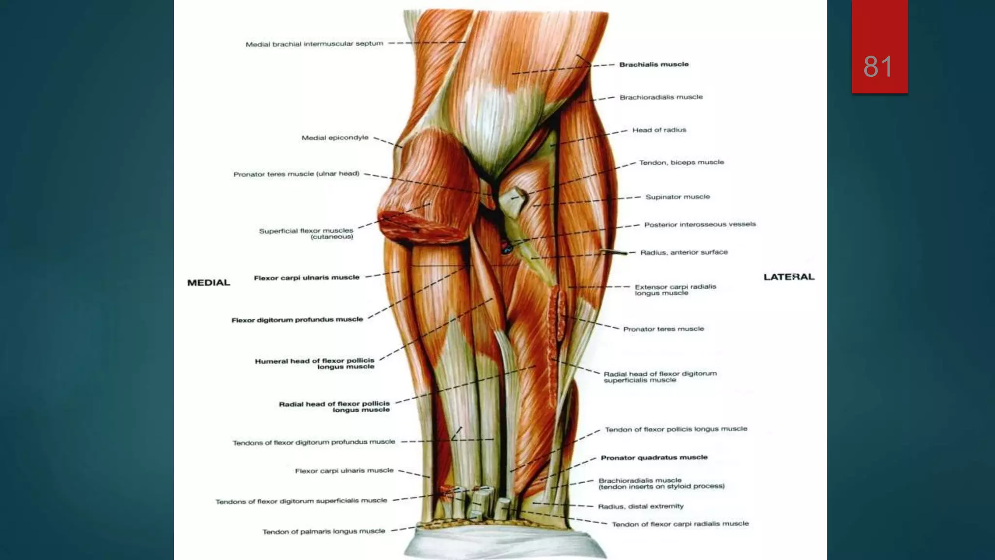
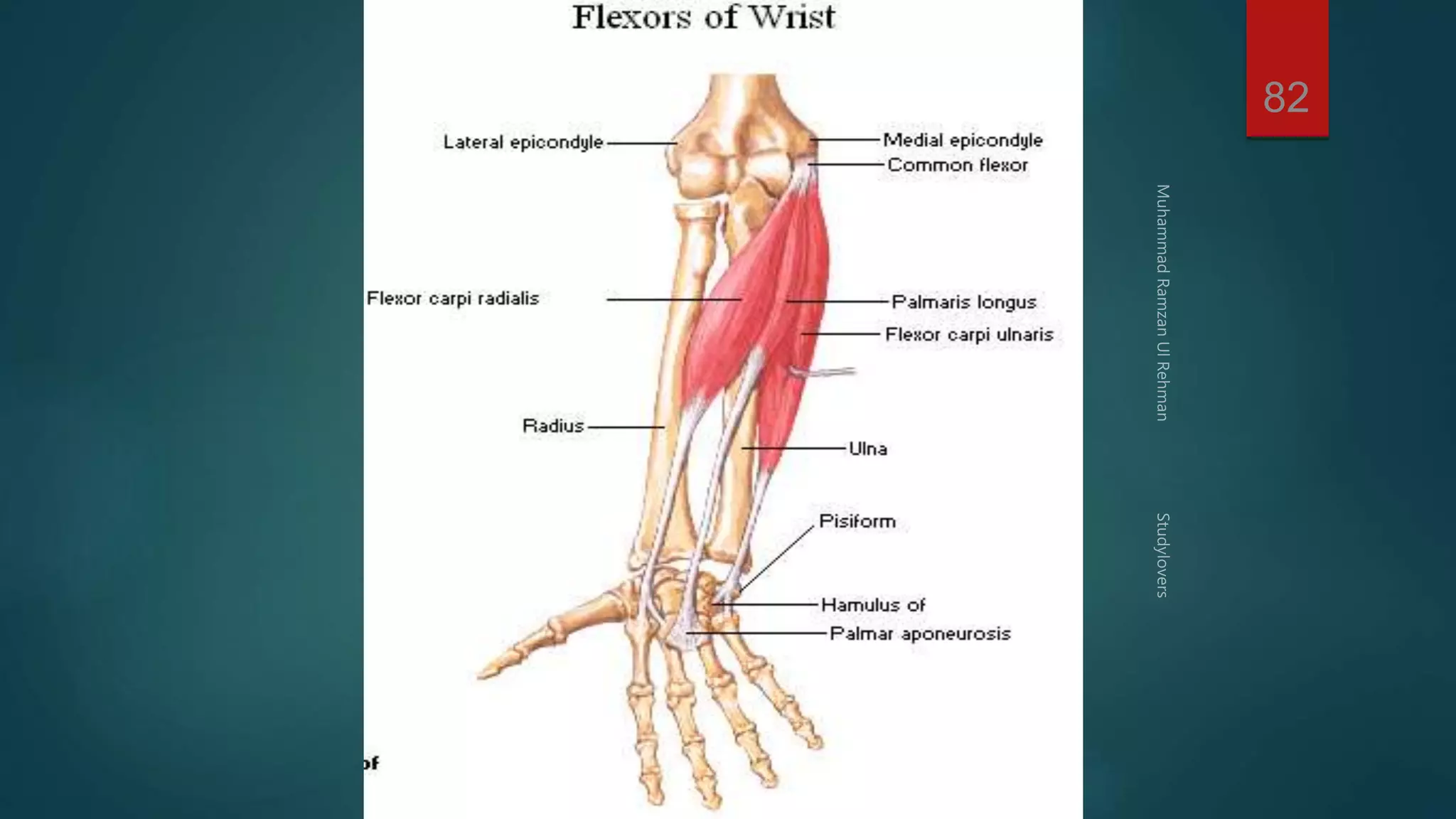
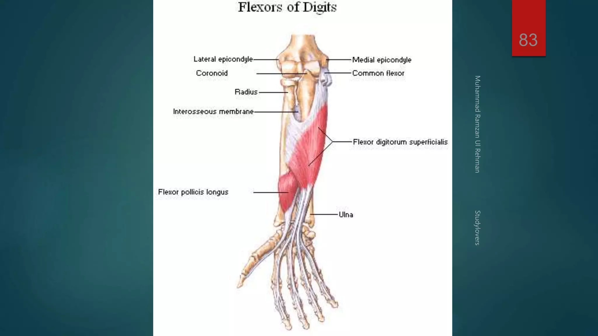
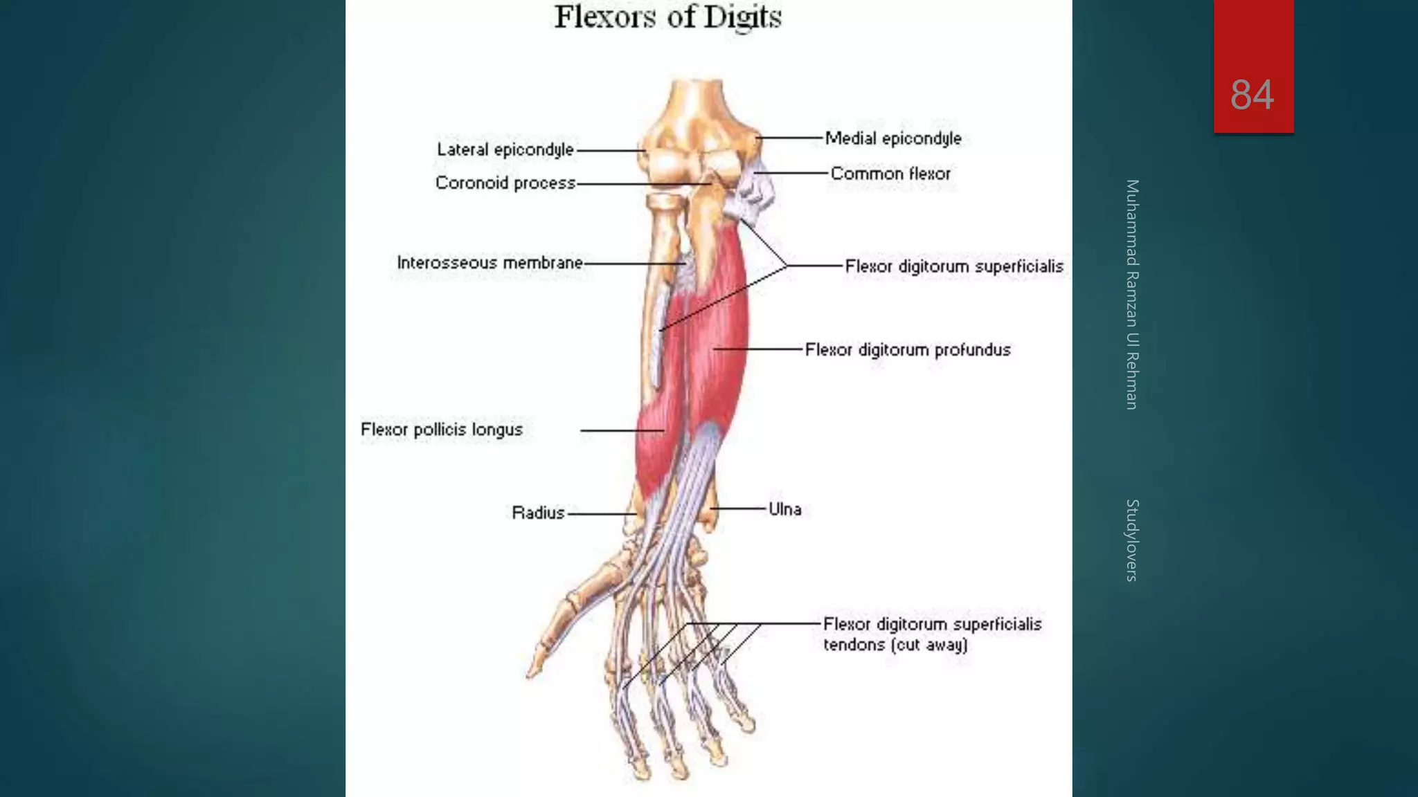
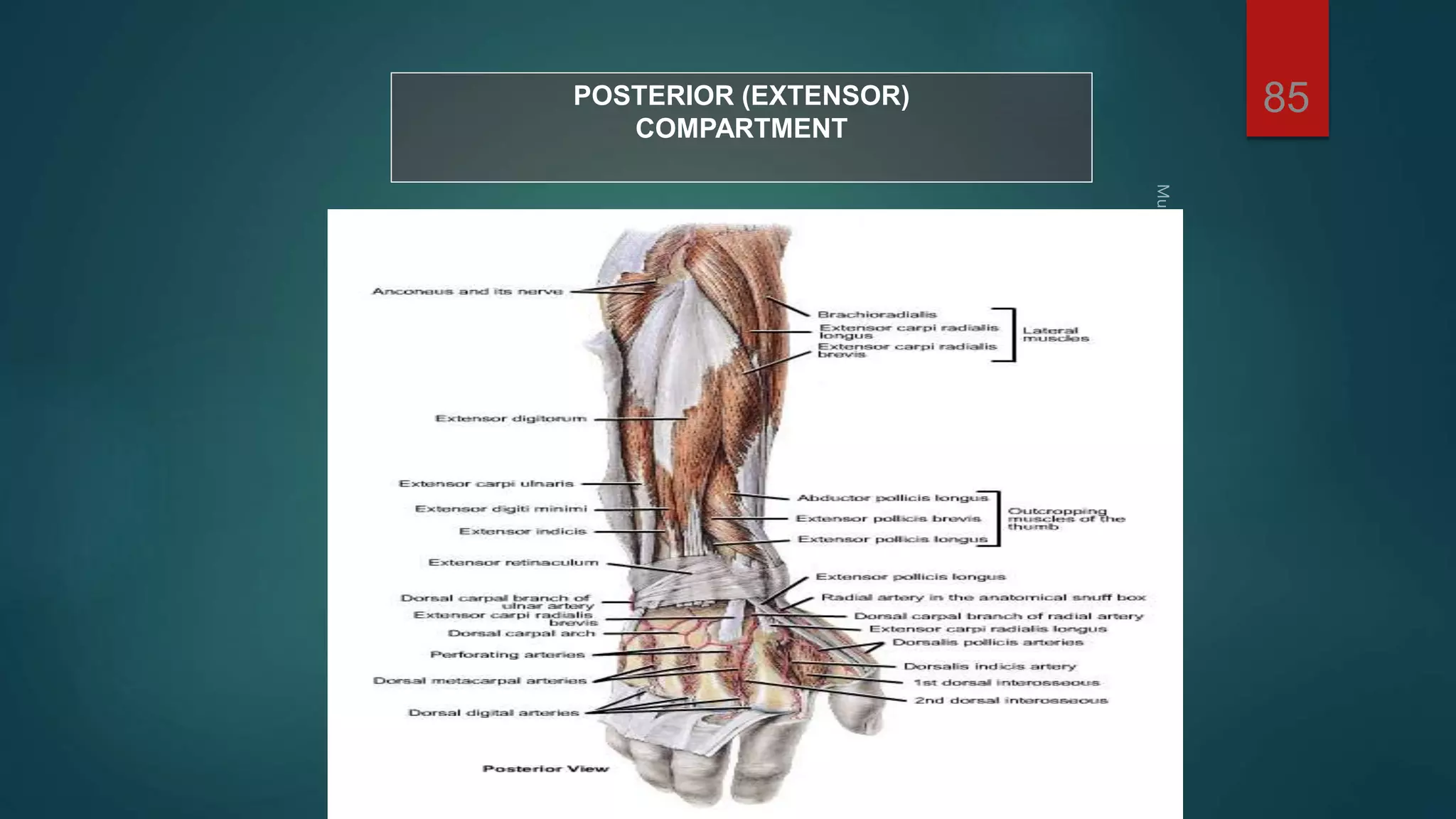
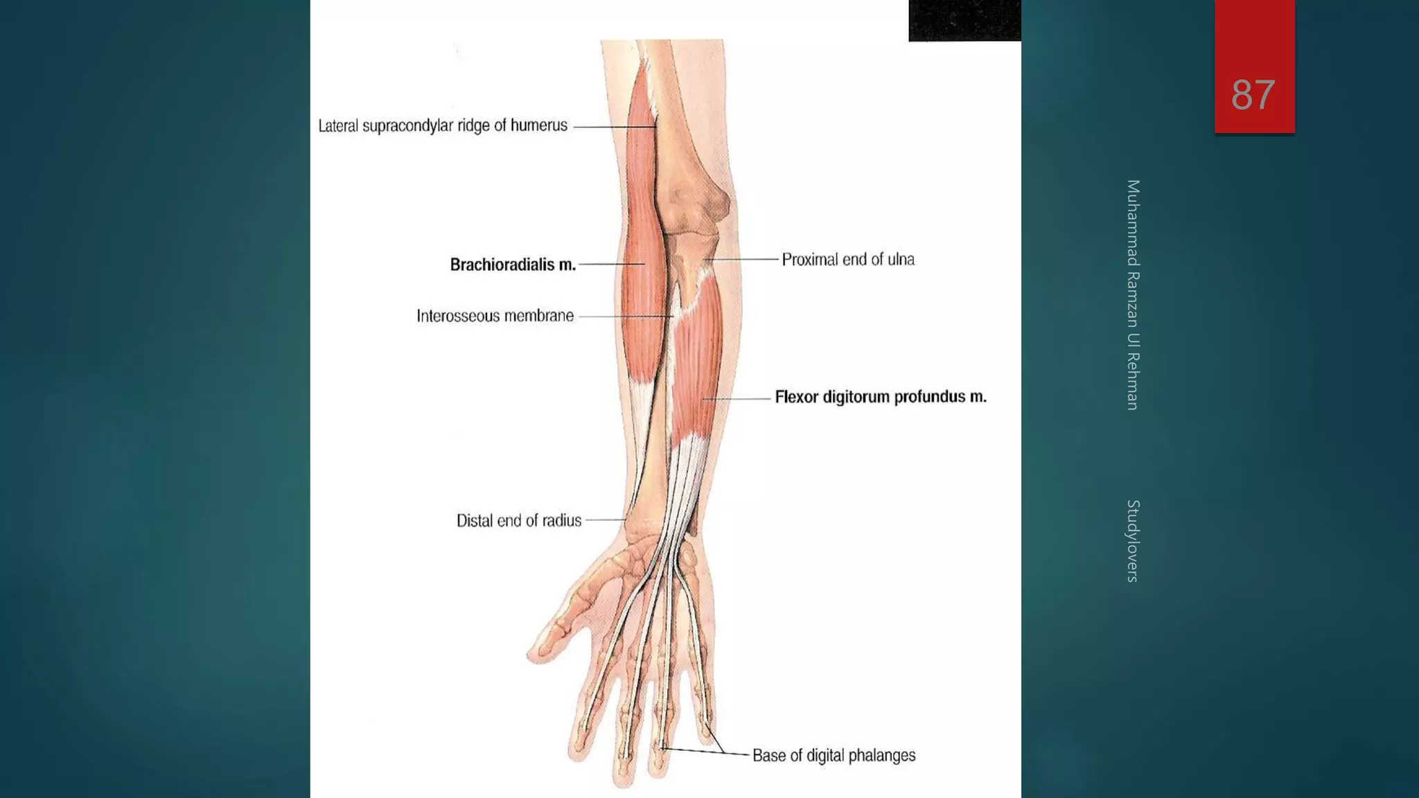
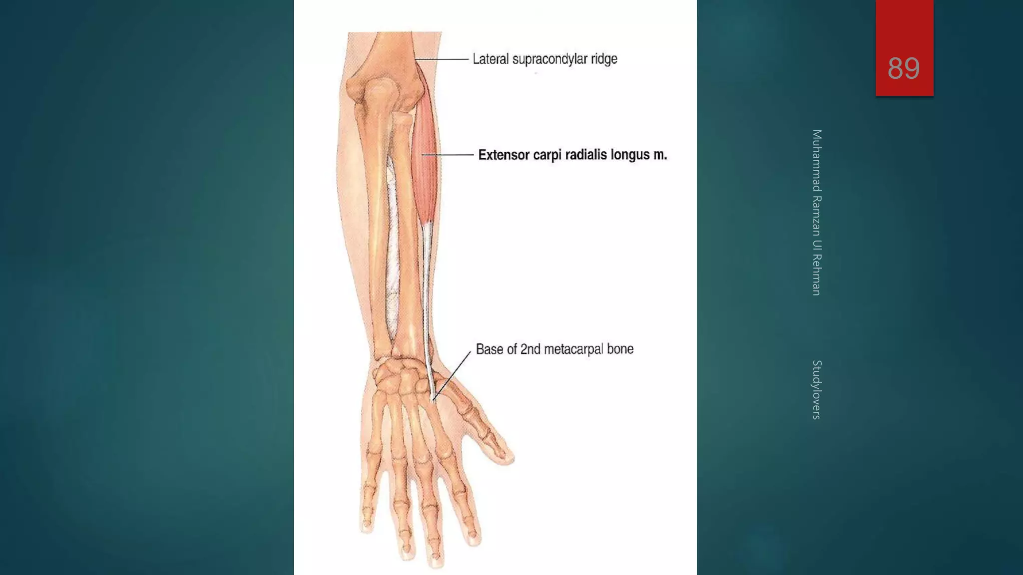
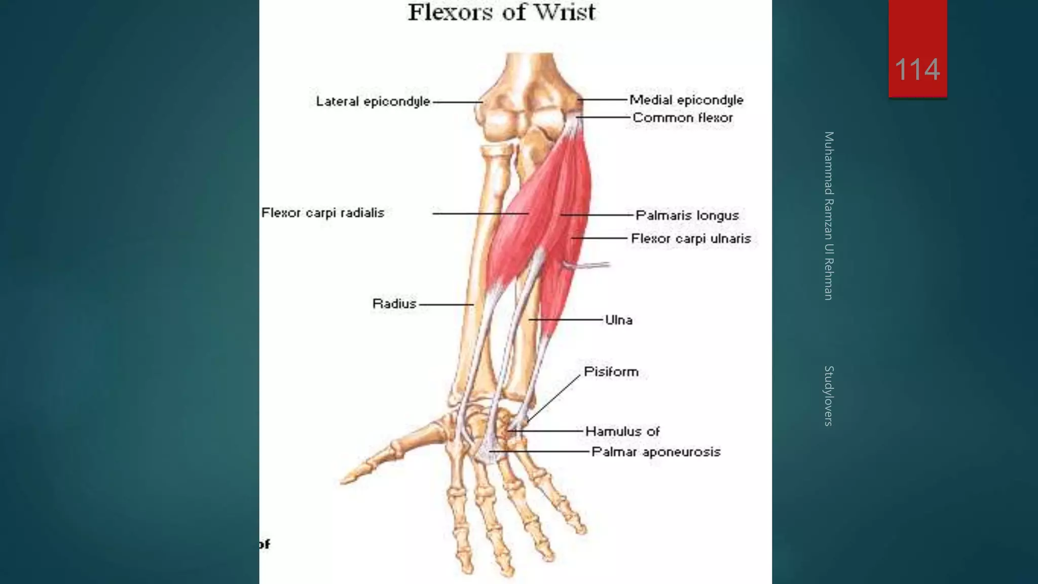
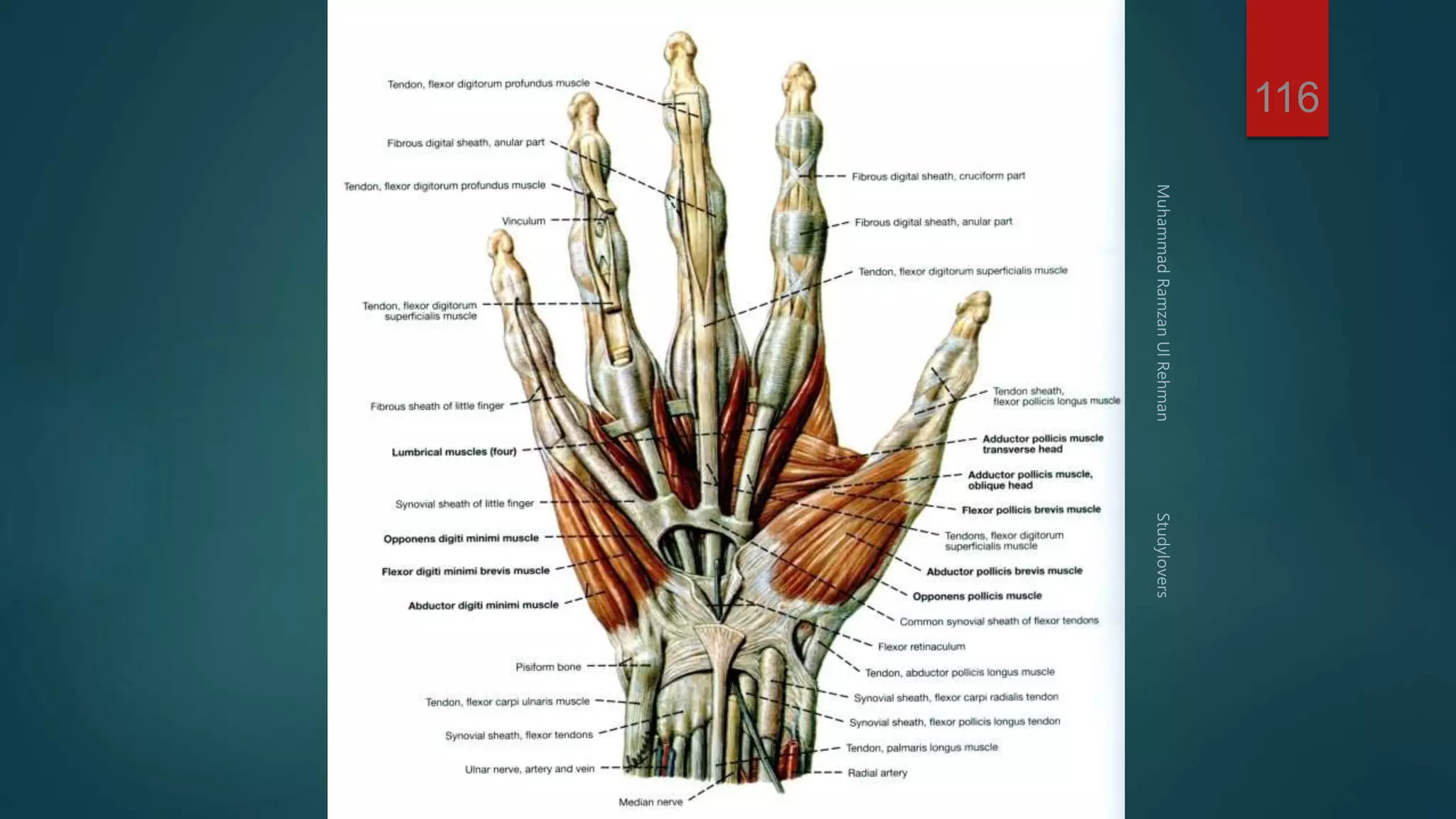
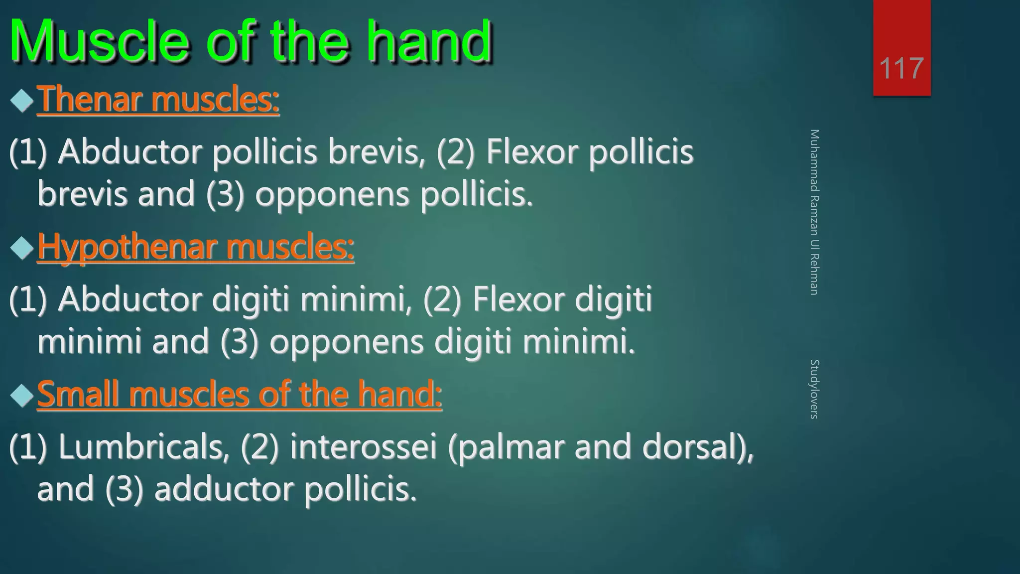
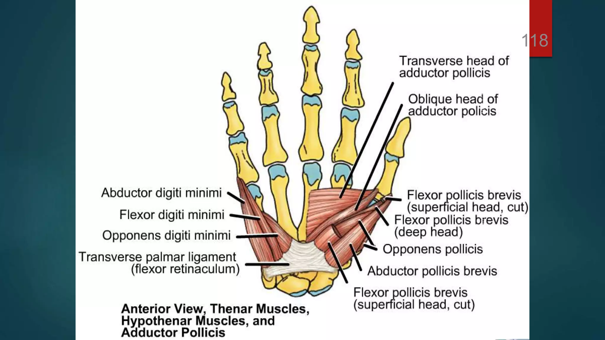
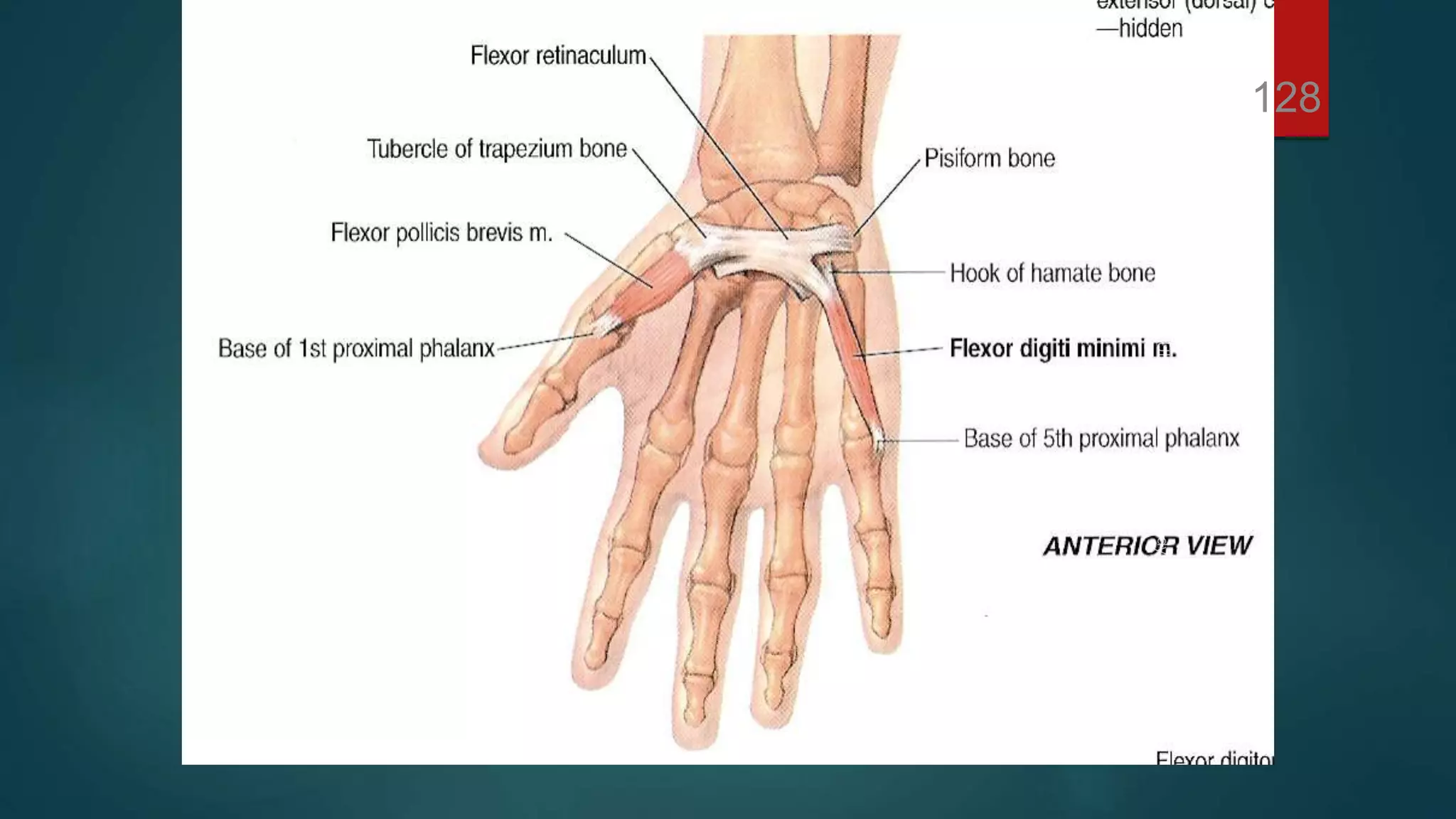
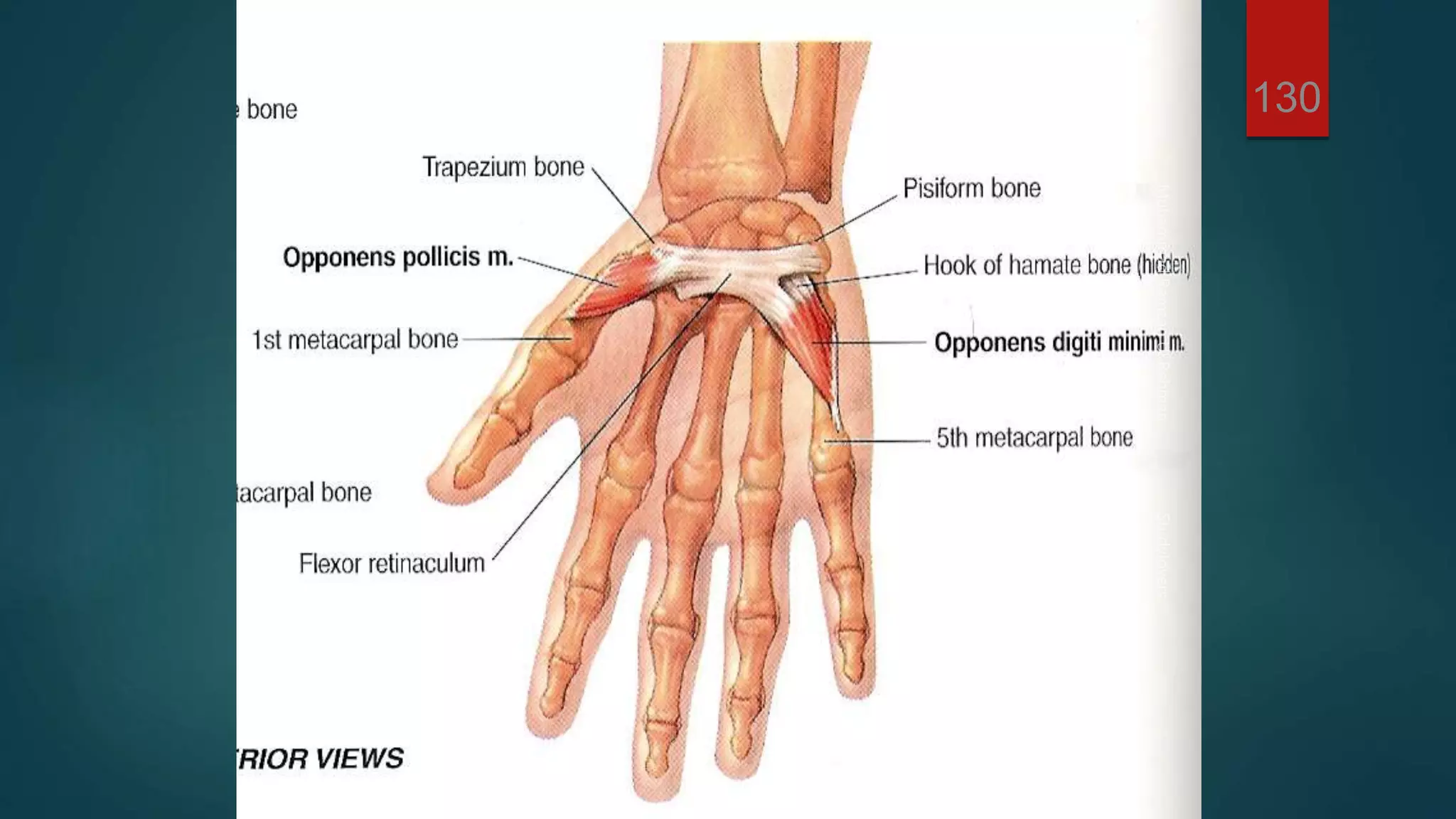
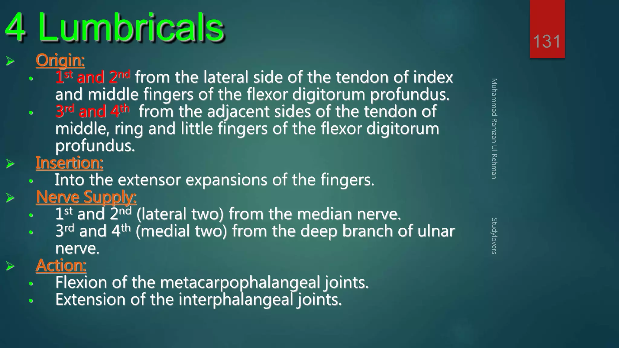
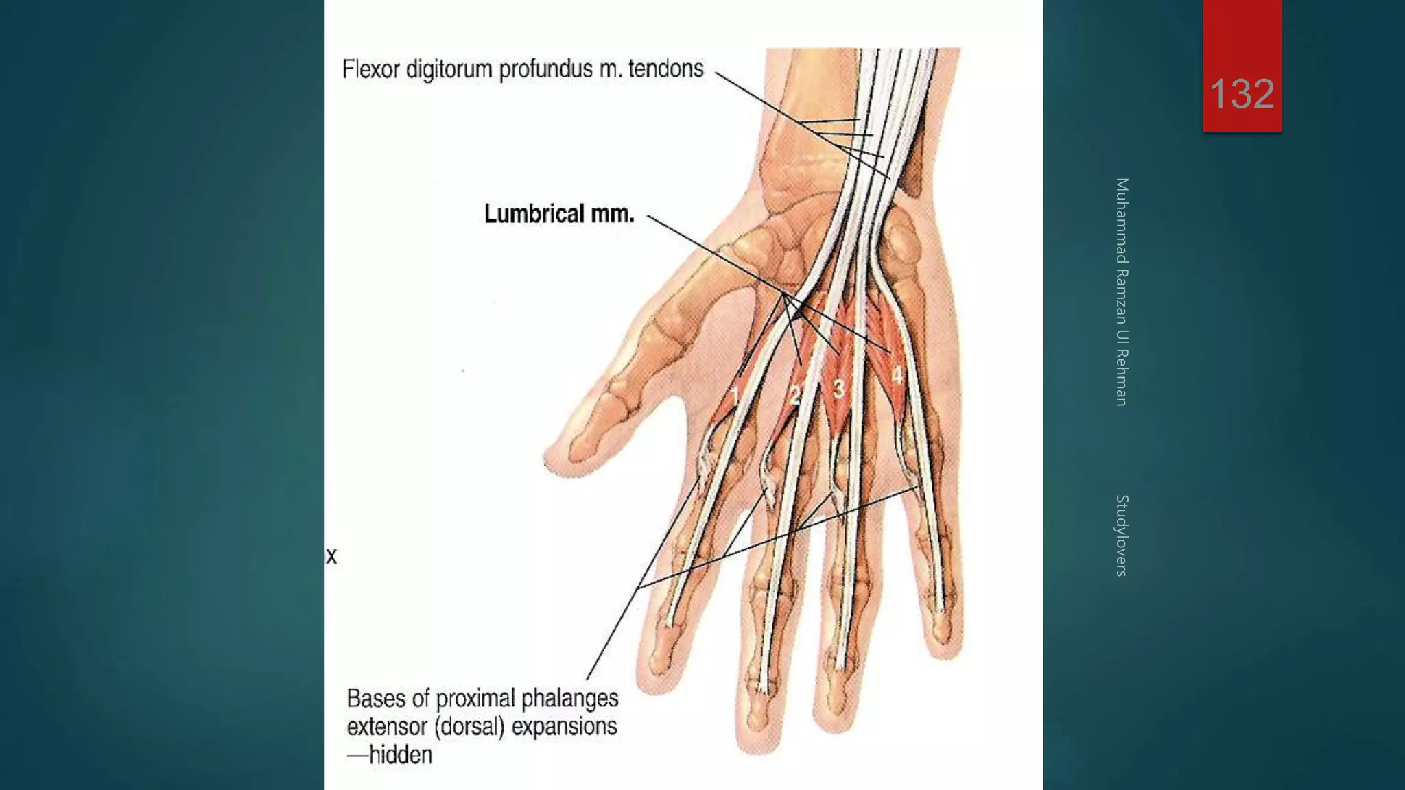
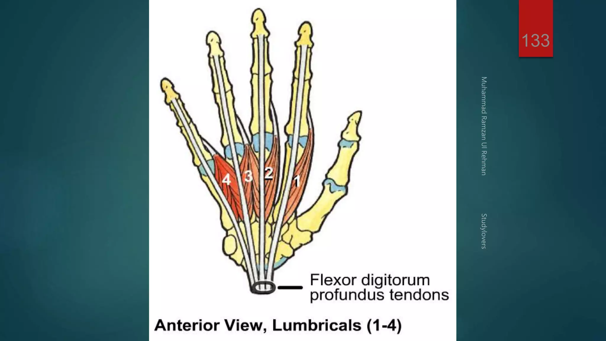
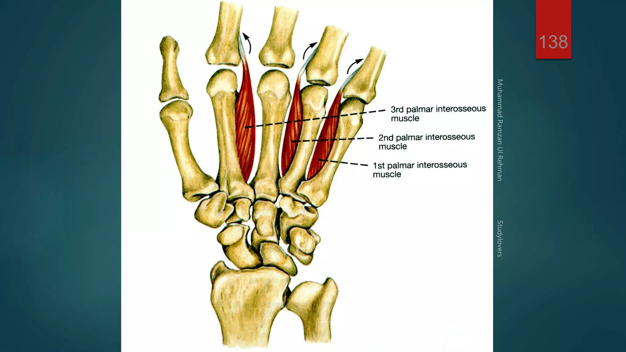
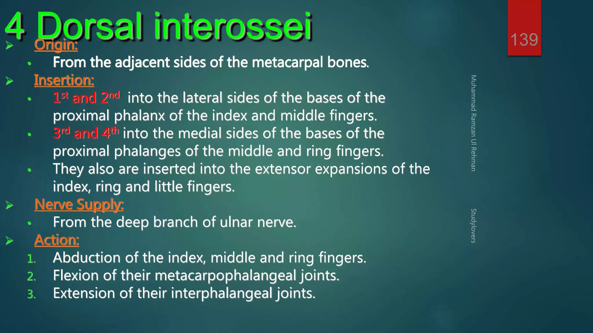
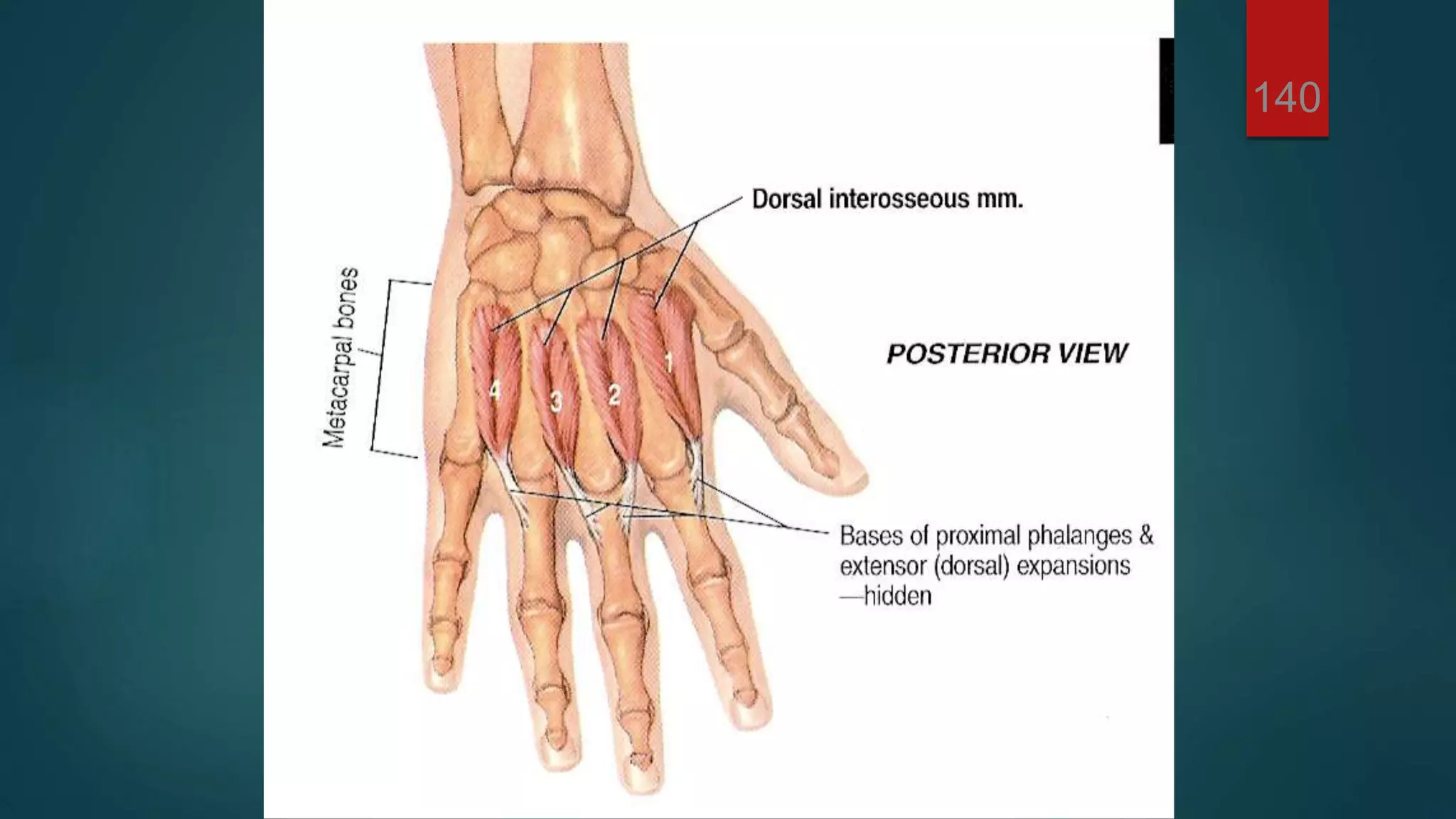
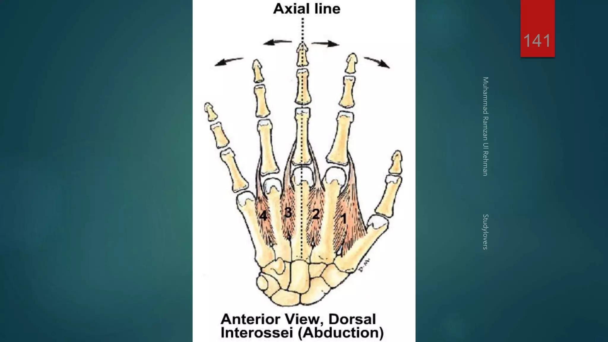
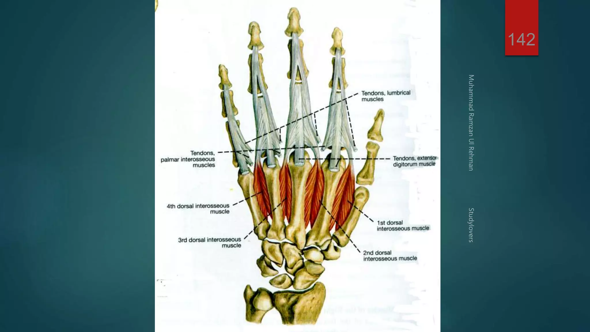
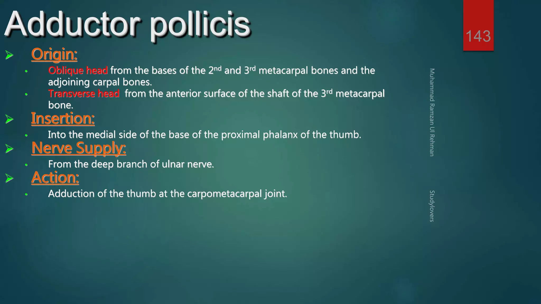
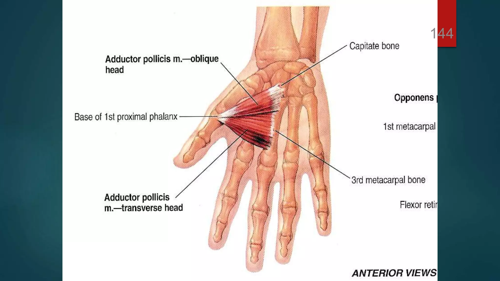
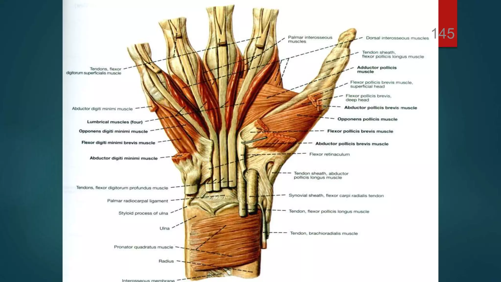
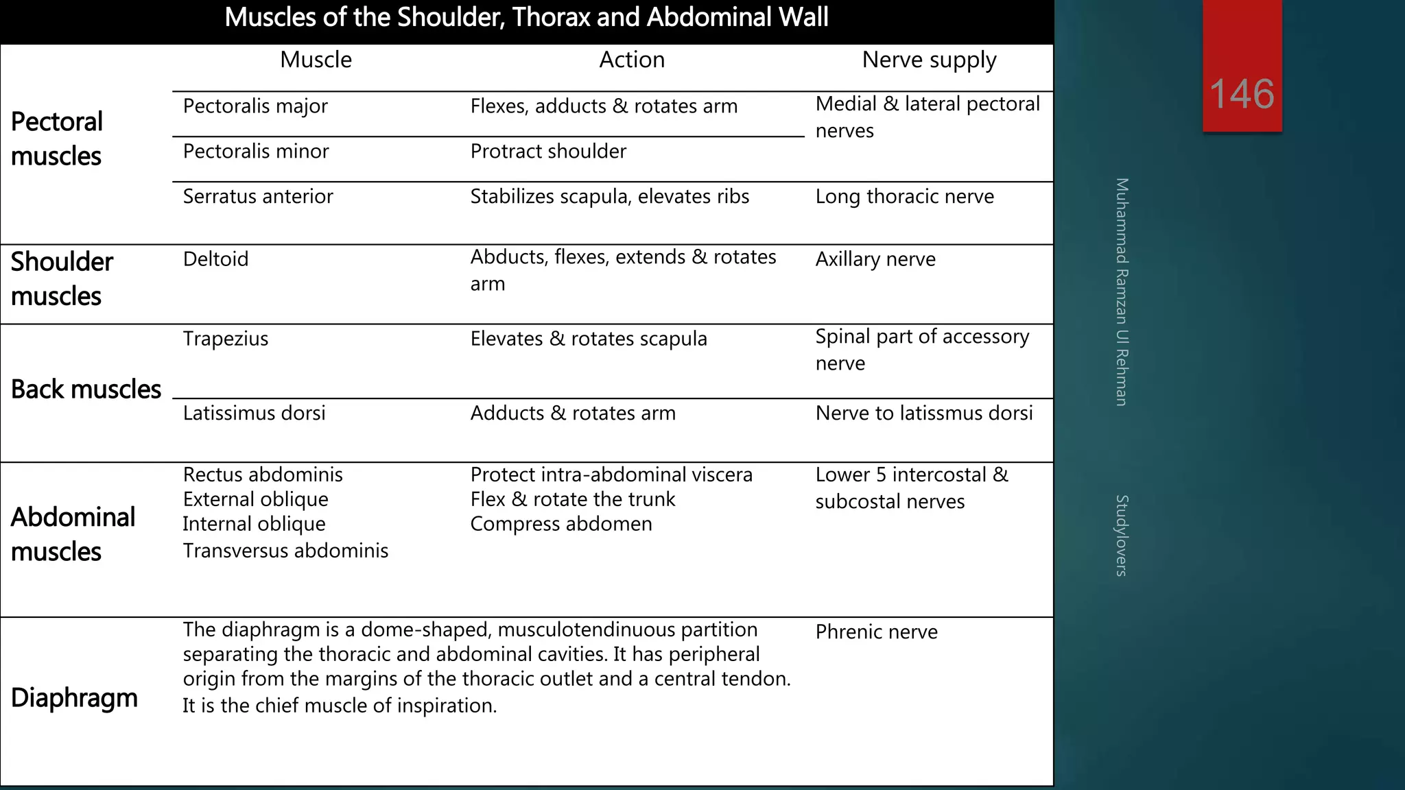
SlideShare a Scribd company logo
1
1 
Muhammad Ramzan-Ul- 
Rehman
3
4
5 Pectoralis major 
 Origin: 
1. Medial half of the clavicle 
2. The sternum. 
3. Upper six costal cartilage. 
 Insertion: 
Lateral lip of bicipital groove of the humerus. 
 Nerve Supply: 
Medial and lateral pectoral nerves. 
 Action: 
A. Adduction, flexion and medial rotation of the arm. 
B. Climbing. 
C. It is an accessory muscle of inspiration.
6
7 Pectoralis minor 
 Origin: 
3rd, 4th and 5th ribs near their cartilage. 
 Insertion: 
Anterior border of the coracoid process of the scapula. 
 Nerve Supply: 
Medial pectoral nerves. 
 Action: 
A. Depression and protraction of the scapula. 
B. If the insertion is fixed, it elevates the 3rd, 4th and 5th ribs. 
C. It rotates the scapula downward (with rhomboids).
8
9 Subclavius 
 Origin: 
The upper surface of the first costal cartilage. 
 Insertion: 
Middle third of the inferior surface of the clavicle. 
 Nerve Supply: 
Nerve to subclavius (from upper trunk of brachial 
plexus). 
 Action: 
A. It depresses the clavicle. 
B. It steadies the clavicle during movement of the 
shoulder girdle.
Muscles of 
the Back 
10
11 Layers of Back Muscles 
First Layer: 
Trapezius and latissimus dorsi. 
Second Layer: 
Levator scapulae, rhomboid minor and 
rhomboid major. 
Third Layer: 
Serratus posterior superior and serratus 
posterior inferior.
12
13
14
15 Trapezius 
 Origin: 
1. Medial third of superior nuchal line of occipital b. 
2. External occipital protuberance. 
3. Ligamentum nuchae. 
4. Spine of the seventh cervical vertebra. 
5. Spines and supraspinous ligaments of all thoracic 
vertebrae 
 Insertion: 
1. Upper fibers into the posterior aspect of the lateral 
third of the clavicle. 
2. Middle fibers into the medial border of the acromion 
process of the scapula. 
3. Lower fibers into the spine of the scapula.
16
17 Trapezius 
 Nerve Supply: 
 Motor fibers from the spinal part of accessory nerve. 
 Proprioceptive sensory fibers from the third and fourth cervical nerves. 
 Action: 
A. Upper fibers: elevates the scapula. 
B. Middle fibers: retracts the scapula. 
C. Lower fibers: depresses the scapula. 
D. With serratus anterior: rotates the scapula upward (for abduction of the arm 
more than 90°. 
E. When scapula is fixed: both side muscles extend the head.
18 Latissimus dorsi 
 Origin: 
1. Spines of the lower 6 thoracic vertebrae. 
2. Lower 3 or 4 ribs. 
3. Lumbar fascia and posterior part of iliac crest. 
4. Inferior angle of the scapula. 
 Insertion: 
Floor of the bicipital groove of the humerus. 
 Nerve Supply: 
Thoracodorsal nerve. 
 Action: 
A. Adduction, extension and medial rotation of the arm. 
B. Climbing.
19
20 Levator scapulae 
 Origin: 
The transverse processes of the upper four cervical vertebrae. 
 Insertion: 
Dorsal lip of the medial border of the scapula above the level of its spine. 
 Nerve Supply: 
From the ventral rami of the 3rd and 4th cervical nerves and form dorsal scapular 
nerve (C5). 
 Action: 
A. Elevation of the scapula. 
B. If the scapula is fixed, it bends the head to the same side.
21
22 Rhomboid minor 
 Origin: 
1. Lower part of ligamentum nuchae. 
2. Spine of seventh cervical vertebra. 
3. Spine of first thoracic vertebra. 
 Insertion: 
Dorsal lip of the medial border of the scapula opposite the root of its spine. 
 Nerve Supply: 
Dorsal scapular nerve (C5). 
 Action: 
A. Elevation of the scapula. 
B. With rhomboid major and pectoralis minor, it rotates the scapula downward.
23
24 Rhomboid major 
 Origin: 
1. From 2nd to 5th spines of the thoracic vertebrae. 
2. And their supraspinous ligaments. 
 Insertion: 
Dorsal lip of the medial border of the scapula 
below the level of its spine. 
 Nerve Supply: 
Dorsal scapular nerve (C5). 
 Action: 
A. Elevation of the scapula. 
B. With rhomboid major and pectoralis minor, it 
rotates the scapula downward.
25 
Muscles:. 
Deltoid. 
Supraspinatus. 
Infraspinatus 
Teres Minor. 
Teres Major. 
Subscapularis. 
Serratus Anterior. 
Muscles of 
the scapular 
region
26
27 Deltoid 
 Origin: 
1. Anterior fibers from the lateral third of the 
anterior border of the clavicle. 
2. Middle fibers from the lateral border of the 
acromion process of the scapula. 
3. Posterior fibers from the lower border of the 
spine of the scapula. 
 Insertion: 
Into the deltoid tuberosity, on the middle of 
the lateral surface of the shaft of the 
humerus.
28
29 Deltoid 
 Nerve Supply: 
From the axillary nerve (C5,6). 
 Action: 
A. Anterior fibers: flexes and medially 
rotates the arm. 
B. Posterior fibers: extends and laterally 
rotates the arm. 
C. Middle fibers: abducts the arm till the 
level of the head (from 15° to 90°).
30 Supraspinatus 
 Origin: 
From the supraspinous fossa of the scapula. 
 Insertion: 
Into the upper facet (impression) of the greater 
tuberosity of the humerus and upper part of the 
capsule of the shoulder joint. 
 Nerve Supply: 
From the suprascapular nerve (C5,6). 
 Action: 
A. Starts abduction of the arm (from 0° to 15°). 
B. Stabilizes and supports the shoulder joint (rotator 
cuff muscle).
31
32 Infraspinatus 
 Origin: 
From the infraspinous fossa of the scapula. 
 Insertion: 
Into the middle facet (impression) of the greater 
tuberosity of the humerus and posterior part of 
the capsule of the shoulder joint. 
 Nerve Supply: 
From the suprascapular nerve (C5,6). 
 Action: 
A. Laterally rotates the arm. 
B. Stabilizes and supports the shoulder joint 
(rotator cuff muscle).
33
34 Teres minor  Origin: 
From the upper 2/3 of the dorsal lip of the lateral 
border of the scapula. 
 Insertion: 
Into the lower facet (impression) of the greater 
tuberosity of the humerus and posterior part of 
the capsule of the shoulder joint. 
 Nerve Supply: 
From the axillary nerve (C5,6). 
 Action: 
A. Laterally rotates the arm. 
B. Stabilizes and supports the shoulder joint 
(rotator cuff muscle).
35
36 Teres major 
 Origin: 
The lower third of the posterior surface of the 
lateral border of the scapula. 
 Insertion: 
The medial lip of the bicipital groove of the 
humerus. 
 Nerve Supply: 
Lower subscapular nerve. 
 Action: 
It adducts, extends and medial rotates the arm.
37
38 Subscpularis 
 Origin: 
The medial 2/3 of the subscapular fossa of the 
anterior surface of the scapula and from the 
inner surface of the subscapular fascia. 
 Insertion: 
The lesser tuberosity of the humerus. 
 Nerve Supply: 
Upper and lower subscapular nerves. 
 Action: 
A. It adducts and medial rotates the arm. 
B. It stabilizes the shoulder joints.
39
40
41 Serratus anterior 
 Origin: 
By 8 digitations from the outer surface of the upper 8 ribs. 
 Insertion: 
To the anterior surface of the medial border of the 
scapula, mainly near the inferior angle. 
 Nerve Supply: 
Long thoracic nerve (from root of brach. plex.). 
 Action: 
A. It protracts the scapula anteriorly and fixes it to the 
thoracic wall. 
B. With the trapezius, it rotates the scapula upward. 
C. It acts as a strong accessory muscle of inspiration.
42
43
44
45
46
47 
Rotator cuff Muscles 
They are the supraspinatus, infraspinatus, 
teres minor and subscapularis muscle. 
They surround the shoulder joint, except form 
the inferior aspect (weak point). 
Their tone helps in holding the head of 
humerus in the glenoid cavity. 
They maintain the stability of the shoulder 
joint.
48
49
50 Biceps brachii 
 Origin: 
1. Long head: from the supraglenoid tubercle of the scapula. 
2. Short head: from the tip of coracoid process of the scapula. 
 Insertion: 
1. Into the posterior part of the radial tuberosity of the 
radius. 
2. Forms the bicipital aponeurosis which is inserted into the 
deep fascia of the upper part of the medial side of the 
forearm. 
 Nerve Supply: 
From musculocutaneous nerve. 
 Action: 
1. Supination of the forearm at the radio-ulnar joints. 
2. Flexion of the forearm at the elbow joint. 
3. Weak flexion of the shoulder joint.
51
52 Coracobrachialis 
 Origin: 
From the tip of coracoid process of the scapula (with short head of biceps). 
 Insertion: 
Into the middle third of the medial side of the shaft of the humerus. 
 Nerve Supply: 
From musculocutaneous nerve. 
 Action: 
1. Flexion of the shoulder joint. 
2. Weak adduction of the shoulder joint.
53
54 Brachialis 
 Origin: 
From the lower half of the anterior surface of the shaft of the humerus and the 
adjoining parts of the medial and lateral intermuscular septa. 
 Insertion: 
Into the anterior surface of the coronoid process of the ulna. 
 Nerve Supply: 
1. Majority of the muscle from musculocutaneous nerve. 
2. Small lateral part by the radial nerve. 
 Action: 
Strong flexion of the elbow joint.
55
56
57 Triceps muscle 
 Origin: 
1. Long head: from the infraglenoid tubercle of the scapula. 
2. Lateral head: from the upper half of the posterior surface 
of the shaft of the humerus, above and lateral to the spiral 
groove. 
3. Medial head: from the lower half of the posterior surface of 
the shaft of the humerus, below and medial to the spiral 
groove. 
 Insertion: 
Into the upper surface of the olecranon process of the ulna. 
 Nerve Supply: 
From the radial nerve (through many branches at the axilla and 
the spiral groove). 
 Action: 
Extension of the forearm at the elbow joint.
58
59
FOREARM 
60
61
62 
Muscles of the Anterior Compartment 
of the forearm 
A. Superficial Layer: (1) Pronator teres, (2) 
flexor carpi radialis, (3) palmaris longus, 
(4) flexor digitorum superficialis and (5) 
flexor carpi ulnaris. 
B. Deep Layer: (1) Flexor pollicis longus, 
(2) flexor digitorum profundus and (3) 
pronator quadratus.
63
64 Pronator teres 
 Origin: 
1. Humeral head: from the medial epicondyle of the humerus (common flexor 
origin). 
2. Ulnar head: from the medial border of the coronoid process of the ulna. 
 Insertion: 
o Into the pronator tuberosity on the lateral surface of the middle part of the 
shaft of the radius. 
 Nerve Supply: 
From the median nerve. 
 Action: 
1. Pronation of the forearm at the radio-ulnar joints. 
2. Flexion of the forearm at the elbow joint.
65
66 Flexor carpi radialis 
 Origin: 
• From the medial epicondyle of the humerus 
(common flexor origin). 
 Insertion: 
• Into palmar surface of the bases of the 2nd 
and 3rd metacarpal bones. 
 Nerve Supply: 
From the median nerve. 
 Action: 
1. Flexion of the hand at the wrist joint. 
2. Abduction of the hand at the wrist joint.
67
68 Palmaris longus 
 Origin: 
• From the medial epicondyle of the 
humerus (common flexor origin). 
 Insertion: 
• Into the palmar aponeurosis and flexor 
retinaculum. 
 Nerve Supply: 
• From the median nerve. 
 Action: 
• Flexion of the hand at the wrist joint.
69
70 
Flexor digitorum superficialis 
 Origin: 
1. Humero-ulnar head: from the medial epicondyle of the 
humerus (common flexor origin) and from the medial 
border of the coronoid process of the ulna. 
2. Radial head: from the oblique line on the anterior surface 
of the shaft of the radius. 
 Insertion: 
• Its tendon divides into 4 tendons which are inserted into 
the sides of the middle phalanges of the medial 4 fingers. 
 Nerve Supply: 
• From the median nerve. 
 Action: 
1. Flexion of the proximal interphalageal joints and 
metacarpophalangeal joints of the medial 4 fingers. 
2. Helps of flexion of the hand at the wrist joint.
71
72 Flexor carpi ulnaris 
 Origin: 
1. Humeral head: from the medial epicondyle of the 
humerus (common flexor origin). 
2. Ulnar head: from the medial aspect of the olecranon 
process and form the posterior border of the shaft of 
the ulna. 
 Insertion: 
• Into the pisiform, hook of hamate and base of the 5th 
metacarpal bone. 
 Nerve Supply: 
• From the ULNAR nerve. 
 Action: 
1. Flexion of the hand at the wrist joint. 
2. Adduction of the hand at the wrist joint.
73
74 Flexor pollicis longus 
 Origin: 
• From the anterior surface of the middle of 
the shaft of the radius and adjoining part of 
the interosseus membrane. 
 Insertion: 
• Into the palmar surface of the base of the 
distal phalanx of the thumb. 
 Nerve Supply: 
• From the anterior interosseus nerve (branch 
of the median nerve). 
 Action: 
• Flexion of the thumb.
75
76 
Flexor digitorum profundus 
 Origin: 
• From the upper ¾ of the anterior surface of the shaft of the ulna 
and adjoining part of the interosseus membrane. 
 Insertion: 
• The muscle divides into 4 tendons which pierce the tendons of 
the flexor digitorum superficialis and are inserted into the bases 
of the distal phalanges of the medial 4 fingers. 
 Nerve Supply: 
• Its lateral half: from the anterior interosseus nerve (branch of the 
median nerve). 
• It medial half: from the ulnar nerve. 
 Action: 
1. Flexion of the distal interphalangeal joints of the medial 4 
fingers. 
2. Helps in flexion of the proximal interphalangeal joints and 
metacarpophalangeal joints of the medial 4 fingers. 
3. Helps of flexion of the hand at the wrist joint.
77
78 Pronator quadratus 
 Origin: 
• From the lower ¼ of the anterior surface of 
the shaft of the ulna. 
 Insertion: 
• Into the lower ¼ of the anterior surface of 
the shaft of the radius. 
 Nerve Supply: 
• From the anterior interosseus nerve (branch 
of the median nerve). 
 Action: 
• Pronation of the forearm at the radio-ulnar 
joints.
79
80
81
82
83
84
POSTERIOR (EXTENSOR) 
COMPARTMENT 
85
86 
Muscles of the posterior 
compartment of the forearm 
A. Superficial Layer: (1) Brachioradialis, (2) 
extensor carpi radialis longus, (3) extensor 
carpi radialis brevis, (4) extensor digitorum, 
(5) extensor digiti minimi, (6) extensor carpi 
ulnaris and (7) anconeus. 
B. Deep Layer: (1) Supinator, (2) abductor 
pollicis longus, (3) extensor pollicis brevis, 
(4) extensor pollicis longus and (5) extensor 
indicis.
87
88 Brachioradialis 
 Origin: 
• from the upper 2/3 of the lateral supracondylar 
ridge of the humerus. 
 Insertion: 
• Into the base of the styloid process of the 
radius. 
 Nerve Supply: 
• From the radial nerve. 
 Action: 
1. Flexion of the forearm (specially in midprone 
position). 
2. Restoration of the forearm into the midprone 
position.
89
90 
Extensor carpi radialis longus 
 Origin: 
• from the lower 1/3 of the lateral 
supracondylar ridge of the humerus. 
 Insertion: 
• Into the posterior surface of the base of 
the 2nd metacarpal bone. 
 Nerve Supply: 
• From the radial nerve. 
 Action: 
1. Extension of the hand at wrist joint. 
2. Abduction of the hand at wrist joint.
91
92 
Extensor carpi radialis brevis 
 Origin: 
• from the lateral epicondyle of the 
humerus (common extensor origin). 
 Insertion: 
• Into the posterior surface of the base of 
the 3rd metacarpal bone. 
 Nerve Supply: 
• From the deep branch of radial nerve. 
 Action: 
1. Extension of the hand at wrist joint. 
2. Abduction of the hand at wrist joint.
93
94 Extensor digitorum 
 Origin: 
• from the lateral epicondyle of the humerus 
(common extensor origin). 
 Insertion: 
• Into the extensor expansion of the medial 4 
fingers. 
 Nerve Supply: 
• From the deep branch of radial nerve. 
 Action: 
1. Extension of metacarpophalangeal and 
interphalangeal joints of the medial 4 fingers. 
2. Extension of the hand at wrist joint.
95
96 Extensor digiti minimi 
 Origin: 
• from the lateral epicondyle of the humerus 
(common extensor origin). 
 Insertion: 
• Into the extensor expansion of the little finger. 
 Nerve Supply: 
• From the deep branch of radial nerve. 
 Action: 
1. Extension of metacarpophalangeal and 
interphalangeal joints of the little finger. 
2. Helps in extension of the hand at wrist joint.
97
98 Extensor carpi ulnaris 
 Origin: 
• from the lateral epicondyle of the humerus 
(common extensor origin). 
 Insertion: 
• Into the posterior surface of the base of the 5th 
metacarpal bone. 
 Nerve Supply: 
• From the deep branch of radial nerve. 
 Action: 
1. Extension of the hand at wrist joint. 
2. Adduction of the hand at wrist joint.
99
100 Anconeus 
 Origin: 
• from the posterior aspect of the lateral 
epicondyle of the humerus. 
 Insertion: 
• Into the lateral surface of the olecranon 
process of the ulna. 
 Nerve Supply: 
• From the radial nerve. 
 Action: 
• It helps the triceps in extension of the elbow 
joint.
101
102 Supinator  Origin: 
• from (1) the lateral epicondyle of the humerus, (2) lateral collateral ligament of 
the elbow joint, (3) annular ligament of the superior radio-ulnar joint, and (4) 
supinator fossa and crest of the ulna. 
 Insertion: 
• Into the upper 1/3 of the lateral surface of the shaft of the radius. 
 Nerve Supply: 
• From the deep branch of the radial nerve. 
 Action: 
• It helps the biceps in supination of the forearm at the radio-ulnar joints.
103
104 Abductor pollicis longus 
 Origin: 
• from the middle of the posterior surface of 
radius and ulna and intervening interosseus 
membrane. 
 Insertion: 
• Into the posterior surface of the base of the 1st 
metacarpal bone. 
 Nerve Supply: 
• From the deep branch of radial (posterior 
interosseus) nerve. 
 Action: 
• Abduction and extension of the thumb at the 
carpo-metacarpal joint.
105
106 Extensor pollicis brevis 
 Origin: 
• from the posterior surface of radius (below 
abductor pollicis longus) and adjoining 
interosseus membrane. 
 Insertion: 
• Into the posterior surface of the base of the 
proximal phalanx of the thumb. 
 Nerve Supply: 
• From the deep branch of radial (posterior 
interosseus) nerve. 
 Action: 
• Extension of the metacarpophalangeal joint of 
the thumb.
107
108 Extensor pollicis longus 
 Origin: 
• from the posterior surface of ulna (below 
abductor pollicis longus) and adjoining 
interosseus membrane. 
 Insertion: 
• Into the posterior surface of the base of the 
distal phalanx of the thumb. 
 Nerve Supply: 
• From the deep branch of radial (posterior 
interosseus) nerve. 
 Action: 
• Extension of the interphalangeal joint of the 
thumb.
109
110 Extensor indicis 
 Origin: 
• from the posterior surface of ulna (below 
extensor pollicis longus) and adjoining 
interosseus membrane. 
 Insertion: 
• Into the extensor expansion of the index 
finger. 
 Nerve Supply: 
• From the deep branch of radial (posterior 
interosseus) nerve. 
 Action: 
• Extension of all joints of the index finger. 
• It helps in extension of the wrist joint.
111
112
113
114
THE HAND 
115
116
117 Muscle of the hand 
Thenar muscles: 
(1) Abductor pollicis brevis, (2) Flexor pollicis 
brevis and (3) opponens pollicis. 
Hypothenar muscles: 
(1) Abductor digiti minimi, (2) Flexor digiti 
minimi and (3) opponens digiti minimi. 
Small muscles of the hand: 
(1) Lumbricals, (2) interossei (palmar and dorsal), 
and (3) adductor pollicis.
118
119 Abductor pollicis brevis 
 Origin: 
• from the scaphoid, trapezium and flexor 
retinaculum. 
 Insertion: 
• Into the lateral aspect of the base of the 
proximal phalanx of the thumb. 
 Nerve Supply: 
• From the median nerve. 
 Action: 
• Abduction of the thumb at the carpo-metacarpal 
joint.
120
121 Flexor pollicis brevis 
 Origin: 
• from the flexor retinaculum. 
 Insertion: 
• Into the lateral aspect of the base of 
the proximal phalanx of the thumb. 
 Nerve Supply: 
• From the median nerve. 
 Action: 
• Flexion of the thumb at the 
metacarpophalangeal joint.
122
123 Opponens pollicis 
 Origin: 
• from the flexor retinaculum. 
 Insertion: 
• Into the lateral border of the shaft of 
the 1st metacarpal bone. 
 Nerve Supply: 
• From the median nerve. 
 Action: 
• Opposition of the thumb against other 
fingers.
124
125 Abductor digiti minimi 
 Origin: 
• from the pisiform and flexor retinaculum. 
 Insertion: 
• Into the medial aspect of the base of the 
proximal phalanx of the little finger. 
 Nerve Supply: 
• From the deep branch of ulnar nerve. 
 Action: 
• Abduction of the little finger at the 
metacarpophalangeal joint.
126
127 Flexor digiti minimi 
 Origin: 
• from the flexor retinaculum. 
 Insertion: 
• Into the medial aspect of the base of the 
proximal phalanx of the little finger. 
 Nerve Supply: 
• From the deep branch of ulnar nerve. 
 Action: 
• Flexion of the little finger at the 
metacarpophalangeal joint.
128
129 Opponens digiti minimi 
 Origin: 
• from the flexor retinaculum. 
 Insertion: 
• Into the medial border of the shaft of 
the 5th metacarpal bone. 
 Nerve Supply: 
• From the deep branch of ulnar nerve. 
 Action: 
• Opposition of the little finger against 
the thumb.
130
131 4 Lumbricals 
 Origin: 
• 1st and 2nd from the lateral side of the tendon of index 
and middle fingers of the flexor digitorum profundus. 
• 3rd and 4th from the adjacent sides of the tendon of 
middle, ring and little fingers of the flexor digitorum 
profundus. 
 Insertion: 
• Into the extensor expansions of the fingers. 
 Nerve Supply: 
• 1st and 2nd (lateral two) from the median nerve. 
• 3rd and 4th (medial two) from the deep branch of ulnar 
nerve. 
 Action: 
• Flexion of the metacarpophalangeal joints. 
• Extension of the interphalangeal joints.
132
133
134
135 3 Palmar interossei  Origin: 
• 1st from the medial side of the metacarpal bone of index 
finger. 
• 2nd and 3rd from the lateral side of the metacarpal bones of 
the ring and little fingers. 
 Insertion: 
• 1st into the medial side of the base of the proximal phalanx 
of the index finger. 
• 2nd and 3rd into the lateral side of the base of the proximal 
phalanges of the ring and little fingers. 
• They also are inserted into the extensor expansions of the 
index, ring and little fingers. 
 Nerve Supply: 
• From the deep branch of ulnar nerve. 
 Action: 
1. Adduction of the index, ring and little fingers. 
2. Flexion of their metacarpophalangeal joints. 
3. Extension of their interphalangeal joints.
136
137
138
139 4 Dorsal interossei  Origin: 
• From the adjacent sides of the metacarpal bones. 
 Insertion: 
• 1st and 2nd into the lateral sides of the bases of the 
proximal phalanx of the index and middle fingers. 
• 3rd and 4th into the medial sides of the bases of the 
proximal phalanges of the middle and ring fingers. 
• They also are inserted into the extensor expansions of the 
index, ring and little fingers. 
 Nerve Supply: 
• From the deep branch of ulnar nerve. 
 Action: 
1. Abduction of the index, middle and ring fingers. 
2. Flexion of their metacarpophalangeal joints. 
3. Extension of their interphalangeal joints.
140
141
142
143 Adductor pollicis 
 Origin: 
• Oblique head from the bases of the 2nd and 3rd metacarpal bones and the 
adjoining carpal bones. 
• Transverse head from the anterior surface of the shaft of the 3rd metacarpal 
bone. 
 Insertion: 
• Into the medial side of the base of the proximal phalanx of the thumb. 
 Nerve Supply: 
• From the deep branch of ulnar nerve. 
 Action: 
• Adduction of the thumb at the carpometacarpal joint.
144
145
146 
Muscles of the Shoulder, Thorax and Abdominal Wall 
Pectoral 
muscles 
Muscle Action Nerve supply 
Pectoralis major Flexes, adducts & rotates arm Medial & lateral pectoral 
nerves 
Pectoralis minor Protract shoulder 
Serratus anterior Stabilizes scapula, elevates ribs Long thoracic nerve 
Shoulder 
muscles 
Deltoid Abducts, flexes, extends & rotates 
arm 
Axillary nerve 
Back muscles 
Trapezius Elevates & rotates scapula Spinal part of accessory 
nerve 
Latissimus dorsi Adducts & rotates arm Nerve to latissmus dorsi 
Abdominal 
muscles 
Rectus abdominis 
External oblique 
Internal oblique 
Transversus abdominis 
Protect intra-abdominal viscera 
Flex & rotate the trunk 
Compress abdomen 
Lower 5 intercostal & 
subcostal nerves 
Diaphragm 
The diaphragm is a dome-shaped, musculotendinuous partition 
separating the thoracic and abdominal cavities. It has peripheral 
origin from the margins of the thoracic outlet and a central tendon. 
It is the chief muscle of inspiration. 
Phrenic nerve
147 
Muscle Action Nerve supply 
Musculo-cutaneous 
nerve 
Front of arm Coracobrachialis flexes & adducts the arm 
Biceps brachii supinates & flexes the forearm 
Brachialis flexes the forearm 
Back of arm Ttriceps brachii extends forearm Radial nerve 
Median & ulnar 
nerves 
Pronator teres pronate forearm 
Pronator quadratus 
Front of forearm 
flexes & abducts wrist 
flexes & adducts wrist 
Flexor carpi radialis Flexor carpi ulnaris 
Flexor digitorum superficialis & profundus flex wrist & fingers 
Radial nerve 
Back of forearm Supinator supinates forearm 
extends & abducts wrist 
extends & adducts wrist 
Extensor carpi radialis 
Extensor carpi ulnaris 
Ext digitorum extends wrist & fingers 
Extensor poilicis brevis extend thumb 
Extensor poilicis longus 
Abductor poilicis longus abducts thumb
Wish you Good Luck 
. Thank you . 
148
1 
Muhammad Ramzan-Ul- 
Rehman

More Related Content

What's hot

Hip joint anatomy
Hip joint anatomyHip joint anatomy
Hip joint anatomy
adityachakri
 
2. front of the thigh ii
2. front of the thigh ii2. front of the thigh ii
2. front of the thigh ii
Dr. Mohammad Mahmoud
 
8. hip joint
8. hip joint8. hip joint
8. hip joint
Dr. Mohammad Mahmoud
 
Muscles of the forearm
Muscles of the forearmMuscles of the forearm
Muscles of the forearmMohaned Lehya
 
Upper limb muscles
Upper limb musclesUpper limb muscles
Upper limb muscles
Jay Patel
 
Knee joint
Knee jointKnee joint
Knee joint
Idris Siddiqui
 
Sciatic nerve
Sciatic nerveSciatic nerve
Sciatic nerve
Prabhat Kumar Shantanu
 
Cubital fossa
Cubital fossaCubital fossa
Cubital fossa
Idris Siddiqui
 
2. shoulder joint & its applied anatomy 07[1]
2. shoulder joint & its applied anatomy   07[1]2. shoulder joint & its applied anatomy   07[1]
2. shoulder joint & its applied anatomy 07[1]MBBS IMS MSU
 
Brachial Plexus Anatomy
Brachial Plexus AnatomyBrachial Plexus Anatomy
Brachial Plexus Anatomy
Abdulaziz Alanzi
 
Anatomy of elbow joint
Anatomy of elbow jointAnatomy of elbow joint
Anatomy of elbow joint
BipulBorthakur
 
Radioulnar joints
Radioulnar jointsRadioulnar joints
Radioulnar joints
Idris Siddiqui
 
Slideshow: Humerus
Slideshow: HumerusSlideshow: Humerus
Slideshow: Humerus
The Funky Professor
 
Slideshow: Scapula
Slideshow: ScapulaSlideshow: Scapula
Slideshow: Scapula
The Funky Professor
 
Slideshow: Clavicle
Slideshow: ClavicleSlideshow: Clavicle
Slideshow: Clavicle
The Funky Professor
 
5.posterior compartment of the thigh
5.posterior compartment of the thigh5.posterior compartment of the thigh
5.posterior compartment of the thigh
Dr. Mohammad Mahmoud
 
Slideshow: Rotator Cuff
Slideshow: Rotator CuffSlideshow: Rotator Cuff
Slideshow: Rotator Cuff
The Funky Professor
 
Femoral triangle
Femoral triangleFemoral triangle
Femoral triangle
Idris Siddiqui
 

What's hot (20)

Hip joint anatomy
Hip joint anatomyHip joint anatomy
Hip joint anatomy
 
Muscles of the back
Muscles of the back Muscles of the back
Muscles of the back
 
2. front of the thigh ii
2. front of the thigh ii2. front of the thigh ii
2. front of the thigh ii
 
8. hip joint
8. hip joint8. hip joint
8. hip joint
 
Muscles of the forearm
Muscles of the forearmMuscles of the forearm
Muscles of the forearm
 
Upper limb muscles
Upper limb musclesUpper limb muscles
Upper limb muscles
 
Knee joint
Knee jointKnee joint
Knee joint
 
Lumbar plexus
Lumbar plexusLumbar plexus
Lumbar plexus
 
Sciatic nerve
Sciatic nerveSciatic nerve
Sciatic nerve
 
Cubital fossa
Cubital fossaCubital fossa
Cubital fossa
 
2. shoulder joint & its applied anatomy 07[1]
2. shoulder joint & its applied anatomy   07[1]2. shoulder joint & its applied anatomy   07[1]
2. shoulder joint & its applied anatomy 07[1]
 
Brachial Plexus Anatomy
Brachial Plexus AnatomyBrachial Plexus Anatomy
Brachial Plexus Anatomy
 
Anatomy of elbow joint
Anatomy of elbow jointAnatomy of elbow joint
Anatomy of elbow joint
 
Radioulnar joints
Radioulnar jointsRadioulnar joints
Radioulnar joints
 
Slideshow: Humerus
Slideshow: HumerusSlideshow: Humerus
Slideshow: Humerus
 
Slideshow: Scapula
Slideshow: ScapulaSlideshow: Scapula
Slideshow: Scapula
 
Slideshow: Clavicle
Slideshow: ClavicleSlideshow: Clavicle
Slideshow: Clavicle
 
5.posterior compartment of the thigh
5.posterior compartment of the thigh5.posterior compartment of the thigh
5.posterior compartment of the thigh
 
Slideshow: Rotator Cuff
Slideshow: Rotator CuffSlideshow: Rotator Cuff
Slideshow: Rotator Cuff
 
Femoral triangle
Femoral triangleFemoral triangle
Femoral triangle
 

Viewers also liked

Clinical anatomy of the upper limb
Clinical anatomy of the upper limb Clinical anatomy of the upper limb
Clinical anatomy of the upper limb
Muhammad Ramzan Ul Rehman
 
Long cases in medicine notes
Long cases in medicine notesLong cases in medicine notes
Long cases in medicine notes
Muhammad Ramzan Ul Rehman
 
Mediastinum 2 (Anatomy of the Thorax)
Mediastinum 2 (Anatomy of the Thorax)Mediastinum 2 (Anatomy of the Thorax)
Mediastinum 2 (Anatomy of the Thorax)
Dr. Sherif Fahmy
 
Clinical anatomy of the lower limb
Clinical anatomy of the lower limbClinical anatomy of the lower limb
Clinical anatomy of the lower limb
Muhammad Ramzan Ul Rehman
 
Folding of the Embryo
Folding of the EmbryoFolding of the Embryo
Folding of the Embryo
Muhammad Ramzan Ul Rehman
 
The Lungs (Anatomy of the Thorax)
The Lungs (Anatomy of the Thorax)The Lungs (Anatomy of the Thorax)
The Lungs (Anatomy of the Thorax)
Dr. Sherif Fahmy
 
Blood supply of abdomen
Blood supply of abdomenBlood supply of abdomen
Blood supply of abdomen
Muhammad Ramzan Ul Rehman
 
Placenta embryology
Placenta embryologyPlacenta embryology
Placenta embryology
Muhammad Ramzan Ul Rehman
 
Upper limb nerve injuries
Upper limb nerve injuriesUpper limb nerve injuries
Upper limb nerve injuries
Muhammad Ramzan Ul Rehman
 
THORAX OS powerpoint
THORAX OS powerpointTHORAX OS powerpoint
THORAX OS powerpointMarime S
 
Thorax and Abdomen Information
Thorax and Abdomen InformationThorax and Abdomen Information
Thorax and Abdomen Information
chadweaver
 
Git blood supply
Git blood supply Git blood supply
Git blood supply
Muhammad Ramzan Ul Rehman
 
Anatomy of thorax (2)
Anatomy of thorax (2)Anatomy of thorax (2)
Anatomy of thorax (2)
Dr Rajinder Dhaliwal
 
Anatomy of the thorax
Anatomy of the thoraxAnatomy of the thorax
Anatomy of the thoraxWill Wilson
 
Anatomy of thorax
Anatomy of thoraxAnatomy of thorax
Anatomy of thorax
Zeeshan Awan
 
Regional anatomy of the human thorax rs-2011
Regional anatomy of the human thorax rs-2011Regional anatomy of the human thorax rs-2011
Regional anatomy of the human thorax rs-2011AHS_anatomy2
 
Inguinal cannal
Inguinal cannal Inguinal cannal
Inguinal cannal
Muhammad Ramzan Ul Rehman
 
Thorax anatomy presentation
Thorax anatomy presentationThorax anatomy presentation
Thorax anatomy presentation
Muhammad Ramzan Ul Rehman
 
Anatomy of thorax /certified fixed orthodontic courses by Indian dental academy
Anatomy of thorax /certified fixed orthodontic courses by Indian dental academy Anatomy of thorax /certified fixed orthodontic courses by Indian dental academy
Anatomy of thorax /certified fixed orthodontic courses by Indian dental academy
Indian dental academy
 

Viewers also liked (20)

Clinical anatomy of the upper limb
Clinical anatomy of the upper limb Clinical anatomy of the upper limb
Clinical anatomy of the upper limb
 
Long cases in medicine notes
Long cases in medicine notesLong cases in medicine notes
Long cases in medicine notes
 
Peritoneum
PeritoneumPeritoneum
Peritoneum
 
Mediastinum 2 (Anatomy of the Thorax)
Mediastinum 2 (Anatomy of the Thorax)Mediastinum 2 (Anatomy of the Thorax)
Mediastinum 2 (Anatomy of the Thorax)
 
Clinical anatomy of the lower limb
Clinical anatomy of the lower limbClinical anatomy of the lower limb
Clinical anatomy of the lower limb
 
Folding of the Embryo
Folding of the EmbryoFolding of the Embryo
Folding of the Embryo
 
The Lungs (Anatomy of the Thorax)
The Lungs (Anatomy of the Thorax)The Lungs (Anatomy of the Thorax)
The Lungs (Anatomy of the Thorax)
 
Blood supply of abdomen
Blood supply of abdomenBlood supply of abdomen
Blood supply of abdomen
 
Placenta embryology
Placenta embryologyPlacenta embryology
Placenta embryology
 
Upper limb nerve injuries
Upper limb nerve injuriesUpper limb nerve injuries
Upper limb nerve injuries
 
THORAX OS powerpoint
THORAX OS powerpointTHORAX OS powerpoint
THORAX OS powerpoint
 
Thorax and Abdomen Information
Thorax and Abdomen InformationThorax and Abdomen Information
Thorax and Abdomen Information
 
Git blood supply
Git blood supply Git blood supply
Git blood supply
 
Anatomy of thorax (2)
Anatomy of thorax (2)Anatomy of thorax (2)
Anatomy of thorax (2)
 
Anatomy of the thorax
Anatomy of the thoraxAnatomy of the thorax
Anatomy of the thorax
 
Anatomy of thorax
Anatomy of thoraxAnatomy of thorax
Anatomy of thorax
 
Regional anatomy of the human thorax rs-2011
Regional anatomy of the human thorax rs-2011Regional anatomy of the human thorax rs-2011
Regional anatomy of the human thorax rs-2011
 
Inguinal cannal
Inguinal cannal Inguinal cannal
Inguinal cannal
 
Thorax anatomy presentation
Thorax anatomy presentationThorax anatomy presentation
Thorax anatomy presentation
 
Anatomy of thorax /certified fixed orthodontic courses by Indian dental academy
Anatomy of thorax /certified fixed orthodontic courses by Indian dental academy Anatomy of thorax /certified fixed orthodontic courses by Indian dental academy
Anatomy of thorax /certified fixed orthodontic courses by Indian dental academy
 

Similar to Upper limb muscles

1. Muscles of upper limb .pdf
1. Muscles of upper limb .pdf1. Muscles of upper limb .pdf
1. Muscles of upper limb .pdf
ssuserbf4af22
 
Muscles of upper
Muscles of upperMuscles of upper
Muscles of upper
Pave Medicine
 
muscels of the upper Limb edited(1).pptx
muscels of the upper Limb edited(1).pptxmuscels of the upper Limb edited(1).pptx
muscels of the upper Limb edited(1).pptx
PhebeLois1
 
Muscles of back, Suboccipital triangle
Muscles of back, Suboccipital triangle Muscles of back, Suboccipital triangle
Muscles of back, Suboccipital triangle
Prabhakar Yadav
 
Muscles of human body
Muscles of human bodyMuscles of human body
Muscles of human body
Nikhil Vaishnav
 
Muscles of the scapular region
Muscles of the scapular regionMuscles of the scapular region
Muscles of the scapular region
DestinyGOSWAMI
 
.UPPER LIMB MUSCLES-I.pptx
.UPPER LIMB MUSCLES-I.pptx.UPPER LIMB MUSCLES-I.pptx
.UPPER LIMB MUSCLES-I.pptx
NimraFaraz
 
Muscles Of Fore Limb Of Cattle
Muscles Of Fore Limb Of CattleMuscles Of Fore Limb Of Cattle
Muscles Of Fore Limb Of Cattle
VivekKumar2638
 
Suboccipital triangle
Suboccipital triangleSuboccipital triangle
Suboccipital triangle
Chitransha03
 
Spine Anatomy.
Spine Anatomy. Spine Anatomy.
Spine Anatomy.
Dr.Md.Monsur Rahman
 
Muscles of neck. final presentation
Muscles of neck. final presentationMuscles of neck. final presentation
Muscles of neck. final presentation
Zubair Khalid
 
Superficial muscles of back
Superficial muscles of backSuperficial muscles of back
Superficial muscles of back
Shaifaly madan rustagi
 
Muscles of neck
Muscles of neckMuscles of neck
Muscles of neck
Dr. Raj Maheshwari
 
عملي تشريح سنة اولي ترم اول
عملي تشريح سنة اولي ترم اولعملي تشريح سنة اولي ترم اول
عملي تشريح سنة اولي ترم اول
ebrahim mahmoud
 
تشريح اولي عملي
تشريح اولي عمليتشريح اولي عملي
تشريح اولي عملي
ebrahim mahmoud
 
Muscular system of horse, Equine muscular system
Muscular system of horse, Equine muscular systemMuscular system of horse, Equine muscular system
Muscular system of horse, Equine muscular system
Vet J
 
6-Pectoral Region and Axilla pdf.pptx hj
6-Pectoral Region and Axilla pdf.pptx hj6-Pectoral Region and Axilla pdf.pptx hj
6-Pectoral Region and Axilla pdf.pptx hj
rk7424934
 
Muscles-of-back of human being with supplypdf
Muscles-of-back of human being with supplypdfMuscles-of-back of human being with supplypdf
Muscles-of-back of human being with supplypdf
mukeshgupta500425
 
Muscles Of The Pectoral Region
Muscles Of The Pectoral RegionMuscles Of The Pectoral Region
Muscles Of The Pectoral RegionMBBS IMS MSU
 

Similar to Upper limb muscles (20)

1. Muscles of upper limb .pdf
1. Muscles of upper limb .pdf1. Muscles of upper limb .pdf
1. Muscles of upper limb .pdf
 
Muscles of upper
Muscles of upperMuscles of upper
Muscles of upper
 
muscels of the upper Limb edited(1).pptx
muscels of the upper Limb edited(1).pptxmuscels of the upper Limb edited(1).pptx
muscels of the upper Limb edited(1).pptx
 
Muscles of back, Suboccipital triangle
Muscles of back, Suboccipital triangle Muscles of back, Suboccipital triangle
Muscles of back, Suboccipital triangle
 
Muscles of human body
Muscles of human bodyMuscles of human body
Muscles of human body
 
Muscles of the scapular region
Muscles of the scapular regionMuscles of the scapular region
Muscles of the scapular region
 
.UPPER LIMB MUSCLES-I.pptx
.UPPER LIMB MUSCLES-I.pptx.UPPER LIMB MUSCLES-I.pptx
.UPPER LIMB MUSCLES-I.pptx
 
Muscles Of Fore Limb Of Cattle
Muscles Of Fore Limb Of CattleMuscles Of Fore Limb Of Cattle
Muscles Of Fore Limb Of Cattle
 
Suboccipital triangle
Suboccipital triangleSuboccipital triangle
Suboccipital triangle
 
Spine Anatomy.
Spine Anatomy. Spine Anatomy.
Spine Anatomy.
 
Shoulder girdle1
Shoulder girdle1Shoulder girdle1
Shoulder girdle1
 
Muscles of neck. final presentation
Muscles of neck. final presentationMuscles of neck. final presentation
Muscles of neck. final presentation
 
Superficial muscles of back
Superficial muscles of backSuperficial muscles of back
Superficial muscles of back
 
Muscles of neck
Muscles of neckMuscles of neck
Muscles of neck
 
عملي تشريح سنة اولي ترم اول
عملي تشريح سنة اولي ترم اولعملي تشريح سنة اولي ترم اول
عملي تشريح سنة اولي ترم اول
 
تشريح اولي عملي
تشريح اولي عمليتشريح اولي عملي
تشريح اولي عملي
 
Muscular system of horse, Equine muscular system
Muscular system of horse, Equine muscular systemMuscular system of horse, Equine muscular system
Muscular system of horse, Equine muscular system
 
6-Pectoral Region and Axilla pdf.pptx hj
6-Pectoral Region and Axilla pdf.pptx hj6-Pectoral Region and Axilla pdf.pptx hj
6-Pectoral Region and Axilla pdf.pptx hj
 
Muscles-of-back of human being with supplypdf
Muscles-of-back of human being with supplypdfMuscles-of-back of human being with supplypdf
Muscles-of-back of human being with supplypdf
 
Muscles Of The Pectoral Region
Muscles Of The Pectoral RegionMuscles Of The Pectoral Region
Muscles Of The Pectoral Region
 

More from Muhammad Ramzan Ul Rehman

Pharmacomics unit 1
Pharmacomics unit 1Pharmacomics unit 1
Pharmacomics unit 1
Muhammad Ramzan Ul Rehman
 
female breast anatomy and physiology
female breast anatomy and physiology female breast anatomy and physiology
female breast anatomy and physiology
Muhammad Ramzan Ul Rehman
 
Mbbs part 2 histology slides
Mbbs part 2 histology slidesMbbs part 2 histology slides
Mbbs part 2 histology slides
Muhammad Ramzan Ul Rehman
 
Glycolysis and Gluconeogensis
Glycolysis and GluconeogensisGlycolysis and Gluconeogensis
Glycolysis and Gluconeogensis
Muhammad Ramzan Ul Rehman
 
Diabetic ketoacidosis
Diabetic ketoacidosisDiabetic ketoacidosis
Diabetic ketoacidosis
Muhammad Ramzan Ul Rehman
 
Genetics Biochemistry
Genetics Biochemistry Genetics Biochemistry
Genetics Biochemistry
Muhammad Ramzan Ul Rehman
 
Physiology ospe 2nd year mbbs
Physiology ospe 2nd year mbbsPhysiology ospe 2nd year mbbs
Physiology ospe 2nd year mbbs
Muhammad Ramzan Ul Rehman
 
Eye physiology from guyton and halls physiology Part 4
Eye physiology from guyton and halls physiology Part 4Eye physiology from guyton and halls physiology Part 4
Eye physiology from guyton and halls physiology Part 4
Muhammad Ramzan Ul Rehman
 
Eye physiology from guyton and halls physiology Part 3
Eye physiology from guyton and halls physiology Part 3Eye physiology from guyton and halls physiology Part 3
Eye physiology from guyton and halls physiology Part 3
Muhammad Ramzan Ul Rehman
 
Eye physiology from guyton and halls physiology Part 2
Eye physiology from guyton and halls physiology Part 2Eye physiology from guyton and halls physiology Part 2
Eye physiology from guyton and halls physiology Part 2
Muhammad Ramzan Ul Rehman
 
Eye physiology from guyton and halls physiology Part 1
Eye physiology from guyton and halls physiology Part 1Eye physiology from guyton and halls physiology Part 1
Eye physiology from guyton and halls physiology Part 1
Muhammad Ramzan Ul Rehman
 
Eye physiology from guyton and halls physiology Part 5
Eye physiology from guyton and halls physiology Part 5Eye physiology from guyton and halls physiology Part 5
Eye physiology from guyton and halls physiology Part 5
Muhammad Ramzan Ul Rehman
 
Histo slides mbbs part 2 for uhs
Histo slides mbbs part 2 for uhsHisto slides mbbs part 2 for uhs
Histo slides mbbs part 2 for uhs
Muhammad Ramzan Ul Rehman
 
viva questions of Pelvis and Perinium anatomy
viva questions of Pelvis and Perinium anatomyviva questions of Pelvis and Perinium anatomy
viva questions of Pelvis and Perinium anatomy
Muhammad Ramzan Ul Rehman
 
Pelvis blood and nerve supply
Pelvis blood and nerve supplyPelvis blood and nerve supply
Pelvis blood and nerve supply
Muhammad Ramzan Ul Rehman
 
Anatomy abdomen Mcqs
Anatomy abdomen McqsAnatomy abdomen Mcqs
Anatomy abdomen Mcqs
Muhammad Ramzan Ul Rehman
 
Pharyngeal arches pouches and clefts
Pharyngeal arches pouches and cleftsPharyngeal arches pouches and clefts
Pharyngeal arches pouches and clefts
Muhammad Ramzan Ul Rehman
 
Histology slides for MBBS part 1 uhs
Histology slides for MBBS part 1 uhsHistology slides for MBBS part 1 uhs
Histology slides for MBBS part 1 uhs
Muhammad Ramzan Ul Rehman
 
Identification points of general histology slides
Identification points of general histology slidesIdentification points of general histology slides
Identification points of general histology slides
Muhammad Ramzan Ul Rehman
 

More from Muhammad Ramzan Ul Rehman (20)

Pharmacomics unit 1
Pharmacomics unit 1Pharmacomics unit 1
Pharmacomics unit 1
 
female breast anatomy and physiology
female breast anatomy and physiology female breast anatomy and physiology
female breast anatomy and physiology
 
Mbbs part 2 histology slides
Mbbs part 2 histology slidesMbbs part 2 histology slides
Mbbs part 2 histology slides
 
Glycolysis and Gluconeogensis
Glycolysis and GluconeogensisGlycolysis and Gluconeogensis
Glycolysis and Gluconeogensis
 
Diabetic ketoacidosis
Diabetic ketoacidosisDiabetic ketoacidosis
Diabetic ketoacidosis
 
Genetics Biochemistry
Genetics Biochemistry Genetics Biochemistry
Genetics Biochemistry
 
Physiology ospe 2nd year mbbs
Physiology ospe 2nd year mbbsPhysiology ospe 2nd year mbbs
Physiology ospe 2nd year mbbs
 
Eye physiology from guyton and halls physiology Part 4
Eye physiology from guyton and halls physiology Part 4Eye physiology from guyton and halls physiology Part 4
Eye physiology from guyton and halls physiology Part 4
 
Eye physiology from guyton and halls physiology Part 3
Eye physiology from guyton and halls physiology Part 3Eye physiology from guyton and halls physiology Part 3
Eye physiology from guyton and halls physiology Part 3
 
Eye physiology from guyton and halls physiology Part 2
Eye physiology from guyton and halls physiology Part 2Eye physiology from guyton and halls physiology Part 2
Eye physiology from guyton and halls physiology Part 2
 
Eye physiology from guyton and halls physiology Part 1
Eye physiology from guyton and halls physiology Part 1Eye physiology from guyton and halls physiology Part 1
Eye physiology from guyton and halls physiology Part 1
 
Eye physiology from guyton and halls physiology Part 5
Eye physiology from guyton and halls physiology Part 5Eye physiology from guyton and halls physiology Part 5
Eye physiology from guyton and halls physiology Part 5
 
Histo slides mbbs part 2 for uhs
Histo slides mbbs part 2 for uhsHisto slides mbbs part 2 for uhs
Histo slides mbbs part 2 for uhs
 
viva questions of Pelvis and Perinium anatomy
viva questions of Pelvis and Perinium anatomyviva questions of Pelvis and Perinium anatomy
viva questions of Pelvis and Perinium anatomy
 
Pelvis blood and nerve supply
Pelvis blood and nerve supplyPelvis blood and nerve supply
Pelvis blood and nerve supply
 
Anatomy abdomen Mcqs
Anatomy abdomen McqsAnatomy abdomen Mcqs
Anatomy abdomen Mcqs
 
Pharyngeal arches pouches and clefts
Pharyngeal arches pouches and cleftsPharyngeal arches pouches and clefts
Pharyngeal arches pouches and clefts
 
Glycolysis
GlycolysisGlycolysis
Glycolysis
 
Histology slides for MBBS part 1 uhs
Histology slides for MBBS part 1 uhsHistology slides for MBBS part 1 uhs
Histology slides for MBBS part 1 uhs
 
Identification points of general histology slides
Identification points of general histology slidesIdentification points of general histology slides
Identification points of general histology slides
 

Recently uploaded

1.4 modern child centered education - mahatma gandhi-2.pptx
1.4 modern child centered education - mahatma gandhi-2.pptx1.4 modern child centered education - mahatma gandhi-2.pptx
1.4 modern child centered education - mahatma gandhi-2.pptx
JosvitaDsouza2
 
Home assignment II on Spectroscopy 2024 Answers.pdf
Home assignment II on Spectroscopy 2024 Answers.pdfHome assignment II on Spectroscopy 2024 Answers.pdf
Home assignment II on Spectroscopy 2024 Answers.pdf
Tamralipta Mahavidyalaya
 
Operation Blue Star - Saka Neela Tara
Operation Blue Star   -  Saka Neela TaraOperation Blue Star   -  Saka Neela Tara
Operation Blue Star - Saka Neela Tara
Balvir Singh
 
Instructions for Submissions thorugh G- Classroom.pptx
Instructions for Submissions thorugh G- Classroom.pptxInstructions for Submissions thorugh G- Classroom.pptx
Instructions for Submissions thorugh G- Classroom.pptx
Jheel Barad
 
Language Across the Curriculm LAC B.Ed.
Language Across the  Curriculm LAC B.Ed.Language Across the  Curriculm LAC B.Ed.
Language Across the Curriculm LAC B.Ed.
Atul Kumar Singh
 
CLASS 11 CBSE B.St Project AIDS TO TRADE - INSURANCE
CLASS 11 CBSE B.St Project AIDS TO TRADE - INSURANCECLASS 11 CBSE B.St Project AIDS TO TRADE - INSURANCE
CLASS 11 CBSE B.St Project AIDS TO TRADE - INSURANCE
BhavyaRajput3
 
The geography of Taylor Swift - some ideas
The geography of Taylor Swift - some ideasThe geography of Taylor Swift - some ideas
The geography of Taylor Swift - some ideas
GeoBlogs
 
Ethnobotany and Ethnopharmacology ......
Ethnobotany and Ethnopharmacology ......Ethnobotany and Ethnopharmacology ......
Ethnobotany and Ethnopharmacology ......
Ashokrao Mane college of Pharmacy Peth-Vadgaon
 
Mule 4.6 & Java 17 Upgrade | MuleSoft Mysore Meetup #46
Mule 4.6 & Java 17 Upgrade | MuleSoft Mysore Meetup #46Mule 4.6 & Java 17 Upgrade | MuleSoft Mysore Meetup #46
Mule 4.6 & Java 17 Upgrade | MuleSoft Mysore Meetup #46
MysoreMuleSoftMeetup
 
Synthetic Fiber Construction in lab .pptx
Synthetic Fiber Construction in lab .pptxSynthetic Fiber Construction in lab .pptx
Synthetic Fiber Construction in lab .pptx
Pavel ( NSTU)
 
Unit 8 - Information and Communication Technology (Paper I).pdf
Unit 8 - Information and Communication Technology (Paper I).pdfUnit 8 - Information and Communication Technology (Paper I).pdf
Unit 8 - Information and Communication Technology (Paper I).pdf
Thiyagu K
 
Students, digital devices and success - Andreas Schleicher - 27 May 2024..pptx
Students, digital devices and success - Andreas Schleicher - 27 May 2024..pptxStudents, digital devices and success - Andreas Schleicher - 27 May 2024..pptx
Students, digital devices and success - Andreas Schleicher - 27 May 2024..pptx
EduSkills OECD
 
Additional Benefits for Employee Website.pdf
Additional Benefits for Employee Website.pdfAdditional Benefits for Employee Website.pdf
Additional Benefits for Employee Website.pdf
joachimlavalley1
 
GIÁO ÁN DẠY THÊM (KẾ HOẠCH BÀI BUỔI 2) - TIẾNG ANH 8 GLOBAL SUCCESS (2 CỘT) N...
GIÁO ÁN DẠY THÊM (KẾ HOẠCH BÀI BUỔI 2) - TIẾNG ANH 8 GLOBAL SUCCESS (2 CỘT) N...GIÁO ÁN DẠY THÊM (KẾ HOẠCH BÀI BUỔI 2) - TIẾNG ANH 8 GLOBAL SUCCESS (2 CỘT) N...
GIÁO ÁN DẠY THÊM (KẾ HOẠCH BÀI BUỔI 2) - TIẾNG ANH 8 GLOBAL SUCCESS (2 CỘT) N...
Nguyen Thanh Tu Collection
 
MARUTI SUZUKI- A Successful Joint Venture in India.pptx
MARUTI SUZUKI- A Successful Joint Venture in India.pptxMARUTI SUZUKI- A Successful Joint Venture in India.pptx
MARUTI SUZUKI- A Successful Joint Venture in India.pptx
bennyroshan06
 
How to Break the cycle of negative Thoughts
How to Break the cycle of negative ThoughtsHow to Break the cycle of negative Thoughts
How to Break the cycle of negative Thoughts
Col Mukteshwar Prasad
 
Digital Tools and AI for Teaching Learning and Research
Digital Tools and AI for Teaching Learning and ResearchDigital Tools and AI for Teaching Learning and Research
Digital Tools and AI for Teaching Learning and Research
Vikramjit Singh
 
2024.06.01 Introducing a competency framework for languag learning materials ...
2024.06.01 Introducing a competency framework for languag learning materials ...2024.06.01 Introducing a competency framework for languag learning materials ...
2024.06.01 Introducing a competency framework for languag learning materials ...
Sandy Millin
 
How to Split Bills in the Odoo 17 POS Module
How to Split Bills in the Odoo 17 POS ModuleHow to Split Bills in the Odoo 17 POS Module
How to Split Bills in the Odoo 17 POS Module
Celine George
 
Basic phrases for greeting and assisting costumers
Basic phrases for greeting and assisting costumersBasic phrases for greeting and assisting costumers
Basic phrases for greeting and assisting costumers
PedroFerreira53928
 

Recently uploaded (20)

1.4 modern child centered education - mahatma gandhi-2.pptx
1.4 modern child centered education - mahatma gandhi-2.pptx1.4 modern child centered education - mahatma gandhi-2.pptx
1.4 modern child centered education - mahatma gandhi-2.pptx
 
Home assignment II on Spectroscopy 2024 Answers.pdf
Home assignment II on Spectroscopy 2024 Answers.pdfHome assignment II on Spectroscopy 2024 Answers.pdf
Home assignment II on Spectroscopy 2024 Answers.pdf
 
Operation Blue Star - Saka Neela Tara
Operation Blue Star   -  Saka Neela TaraOperation Blue Star   -  Saka Neela Tara
Operation Blue Star - Saka Neela Tara
 
Instructions for Submissions thorugh G- Classroom.pptx
Instructions for Submissions thorugh G- Classroom.pptxInstructions for Submissions thorugh G- Classroom.pptx
Instructions for Submissions thorugh G- Classroom.pptx
 
Language Across the Curriculm LAC B.Ed.
Language Across the  Curriculm LAC B.Ed.Language Across the  Curriculm LAC B.Ed.
Language Across the Curriculm LAC B.Ed.
 
CLASS 11 CBSE B.St Project AIDS TO TRADE - INSURANCE
CLASS 11 CBSE B.St Project AIDS TO TRADE - INSURANCECLASS 11 CBSE B.St Project AIDS TO TRADE - INSURANCE
CLASS 11 CBSE B.St Project AIDS TO TRADE - INSURANCE
 
The geography of Taylor Swift - some ideas
The geography of Taylor Swift - some ideasThe geography of Taylor Swift - some ideas
The geography of Taylor Swift - some ideas
 
Ethnobotany and Ethnopharmacology ......
Ethnobotany and Ethnopharmacology ......Ethnobotany and Ethnopharmacology ......
Ethnobotany and Ethnopharmacology ......
 
Mule 4.6 & Java 17 Upgrade | MuleSoft Mysore Meetup #46
Mule 4.6 & Java 17 Upgrade | MuleSoft Mysore Meetup #46Mule 4.6 & Java 17 Upgrade | MuleSoft Mysore Meetup #46
Mule 4.6 & Java 17 Upgrade | MuleSoft Mysore Meetup #46
 
Synthetic Fiber Construction in lab .pptx
Synthetic Fiber Construction in lab .pptxSynthetic Fiber Construction in lab .pptx
Synthetic Fiber Construction in lab .pptx
 
Unit 8 - Information and Communication Technology (Paper I).pdf
Unit 8 - Information and Communication Technology (Paper I).pdfUnit 8 - Information and Communication Technology (Paper I).pdf
Unit 8 - Information and Communication Technology (Paper I).pdf
 
Students, digital devices and success - Andreas Schleicher - 27 May 2024..pptx
Students, digital devices and success - Andreas Schleicher - 27 May 2024..pptxStudents, digital devices and success - Andreas Schleicher - 27 May 2024..pptx
Students, digital devices and success - Andreas Schleicher - 27 May 2024..pptx
 
Additional Benefits for Employee Website.pdf
Additional Benefits for Employee Website.pdfAdditional Benefits for Employee Website.pdf
Additional Benefits for Employee Website.pdf
 
GIÁO ÁN DẠY THÊM (KẾ HOẠCH BÀI BUỔI 2) - TIẾNG ANH 8 GLOBAL SUCCESS (2 CỘT) N...
GIÁO ÁN DẠY THÊM (KẾ HOẠCH BÀI BUỔI 2) - TIẾNG ANH 8 GLOBAL SUCCESS (2 CỘT) N...GIÁO ÁN DẠY THÊM (KẾ HOẠCH BÀI BUỔI 2) - TIẾNG ANH 8 GLOBAL SUCCESS (2 CỘT) N...
GIÁO ÁN DẠY THÊM (KẾ HOẠCH BÀI BUỔI 2) - TIẾNG ANH 8 GLOBAL SUCCESS (2 CỘT) N...
 
MARUTI SUZUKI- A Successful Joint Venture in India.pptx
MARUTI SUZUKI- A Successful Joint Venture in India.pptxMARUTI SUZUKI- A Successful Joint Venture in India.pptx
MARUTI SUZUKI- A Successful Joint Venture in India.pptx
 
How to Break the cycle of negative Thoughts
How to Break the cycle of negative ThoughtsHow to Break the cycle of negative Thoughts
How to Break the cycle of negative Thoughts
 
Digital Tools and AI for Teaching Learning and Research
Digital Tools and AI for Teaching Learning and ResearchDigital Tools and AI for Teaching Learning and Research
Digital Tools and AI for Teaching Learning and Research
 
2024.06.01 Introducing a competency framework for languag learning materials ...
2024.06.01 Introducing a competency framework for languag learning materials ...2024.06.01 Introducing a competency framework for languag learning materials ...
2024.06.01 Introducing a competency framework for languag learning materials ...
 
How to Split Bills in the Odoo 17 POS Module
How to Split Bills in the Odoo 17 POS ModuleHow to Split Bills in the Odoo 17 POS Module
How to Split Bills in the Odoo 17 POS Module
 
Basic phrases for greeting and assisting costumers
Basic phrases for greeting and assisting costumersBasic phrases for greeting and assisting costumers
Basic phrases for greeting and assisting costumers
 

Upper limb muscles

  • 1. 1
  • 3. 3
  • 4. 4
  • 5. 5 Pectoralis major  Origin: 1. Medial half of the clavicle 2. The sternum. 3. Upper six costal cartilage.  Insertion: Lateral lip of bicipital groove of the humerus.  Nerve Supply: Medial and lateral pectoral nerves.  Action: A. Adduction, flexion and medial rotation of the arm. B. Climbing. C. It is an accessory muscle of inspiration.
  • 6. 6
  • 7. 7 Pectoralis minor  Origin: 3rd, 4th and 5th ribs near their cartilage.  Insertion: Anterior border of the coracoid process of the scapula.  Nerve Supply: Medial pectoral nerves.  Action: A. Depression and protraction of the scapula. B. If the insertion is fixed, it elevates the 3rd, 4th and 5th ribs. C. It rotates the scapula downward (with rhomboids).
  • 8. 8
  • 9. 9 Subclavius  Origin: The upper surface of the first costal cartilage.  Insertion: Middle third of the inferior surface of the clavicle.  Nerve Supply: Nerve to subclavius (from upper trunk of brachial plexus).  Action: A. It depresses the clavicle. B. It steadies the clavicle during movement of the shoulder girdle.
  • 10. Muscles of the Back 10
  • 11. 11 Layers of Back Muscles First Layer: Trapezius and latissimus dorsi. Second Layer: Levator scapulae, rhomboid minor and rhomboid major. Third Layer: Serratus posterior superior and serratus posterior inferior.
  • 12. 12
  • 13. 13
  • 14. 14
  • 15. 15 Trapezius  Origin: 1. Medial third of superior nuchal line of occipital b. 2. External occipital protuberance. 3. Ligamentum nuchae. 4. Spine of the seventh cervical vertebra. 5. Spines and supraspinous ligaments of all thoracic vertebrae  Insertion: 1. Upper fibers into the posterior aspect of the lateral third of the clavicle. 2. Middle fibers into the medial border of the acromion process of the scapula. 3. Lower fibers into the spine of the scapula.
  • 16. 16
  • 17. 17 Trapezius  Nerve Supply:  Motor fibers from the spinal part of accessory nerve.  Proprioceptive sensory fibers from the third and fourth cervical nerves.  Action: A. Upper fibers: elevates the scapula. B. Middle fibers: retracts the scapula. C. Lower fibers: depresses the scapula. D. With serratus anterior: rotates the scapula upward (for abduction of the arm more than 90°. E. When scapula is fixed: both side muscles extend the head.
  • 18. 18 Latissimus dorsi  Origin: 1. Spines of the lower 6 thoracic vertebrae. 2. Lower 3 or 4 ribs. 3. Lumbar fascia and posterior part of iliac crest. 4. Inferior angle of the scapula.  Insertion: Floor of the bicipital groove of the humerus.  Nerve Supply: Thoracodorsal nerve.  Action: A. Adduction, extension and medial rotation of the arm. B. Climbing.
  • 19. 19
  • 20. 20 Levator scapulae  Origin: The transverse processes of the upper four cervical vertebrae.  Insertion: Dorsal lip of the medial border of the scapula above the level of its spine.  Nerve Supply: From the ventral rami of the 3rd and 4th cervical nerves and form dorsal scapular nerve (C5).  Action: A. Elevation of the scapula. B. If the scapula is fixed, it bends the head to the same side.
  • 21. 21
  • 22. 22 Rhomboid minor  Origin: 1. Lower part of ligamentum nuchae. 2. Spine of seventh cervical vertebra. 3. Spine of first thoracic vertebra.  Insertion: Dorsal lip of the medial border of the scapula opposite the root of its spine.  Nerve Supply: Dorsal scapular nerve (C5).  Action: A. Elevation of the scapula. B. With rhomboid major and pectoralis minor, it rotates the scapula downward.
  • 23. 23
  • 24. 24 Rhomboid major  Origin: 1. From 2nd to 5th spines of the thoracic vertebrae. 2. And their supraspinous ligaments.  Insertion: Dorsal lip of the medial border of the scapula below the level of its spine.  Nerve Supply: Dorsal scapular nerve (C5).  Action: A. Elevation of the scapula. B. With rhomboid major and pectoralis minor, it rotates the scapula downward.
  • 25. 25 Muscles:. Deltoid. Supraspinatus. Infraspinatus Teres Minor. Teres Major. Subscapularis. Serratus Anterior. Muscles of the scapular region
  • 26. 26
  • 27. 27 Deltoid  Origin: 1. Anterior fibers from the lateral third of the anterior border of the clavicle. 2. Middle fibers from the lateral border of the acromion process of the scapula. 3. Posterior fibers from the lower border of the spine of the scapula.  Insertion: Into the deltoid tuberosity, on the middle of the lateral surface of the shaft of the humerus.
  • 28. 28
  • 29. 29 Deltoid  Nerve Supply: From the axillary nerve (C5,6).  Action: A. Anterior fibers: flexes and medially rotates the arm. B. Posterior fibers: extends and laterally rotates the arm. C. Middle fibers: abducts the arm till the level of the head (from 15° to 90°).
  • 30. 30 Supraspinatus  Origin: From the supraspinous fossa of the scapula.  Insertion: Into the upper facet (impression) of the greater tuberosity of the humerus and upper part of the capsule of the shoulder joint.  Nerve Supply: From the suprascapular nerve (C5,6).  Action: A. Starts abduction of the arm (from 0° to 15°). B. Stabilizes and supports the shoulder joint (rotator cuff muscle).
  • 31. 31
  • 32. 32 Infraspinatus  Origin: From the infraspinous fossa of the scapula.  Insertion: Into the middle facet (impression) of the greater tuberosity of the humerus and posterior part of the capsule of the shoulder joint.  Nerve Supply: From the suprascapular nerve (C5,6).  Action: A. Laterally rotates the arm. B. Stabilizes and supports the shoulder joint (rotator cuff muscle).
  • 33. 33
  • 34. 34 Teres minor  Origin: From the upper 2/3 of the dorsal lip of the lateral border of the scapula.  Insertion: Into the lower facet (impression) of the greater tuberosity of the humerus and posterior part of the capsule of the shoulder joint.  Nerve Supply: From the axillary nerve (C5,6).  Action: A. Laterally rotates the arm. B. Stabilizes and supports the shoulder joint (rotator cuff muscle).
  • 35. 35
  • 36. 36 Teres major  Origin: The lower third of the posterior surface of the lateral border of the scapula.  Insertion: The medial lip of the bicipital groove of the humerus.  Nerve Supply: Lower subscapular nerve.  Action: It adducts, extends and medial rotates the arm.
  • 37. 37
  • 38. 38 Subscpularis  Origin: The medial 2/3 of the subscapular fossa of the anterior surface of the scapula and from the inner surface of the subscapular fascia.  Insertion: The lesser tuberosity of the humerus.  Nerve Supply: Upper and lower subscapular nerves.  Action: A. It adducts and medial rotates the arm. B. It stabilizes the shoulder joints.
  • 39. 39
  • 40. 40
  • 41. 41 Serratus anterior  Origin: By 8 digitations from the outer surface of the upper 8 ribs.  Insertion: To the anterior surface of the medial border of the scapula, mainly near the inferior angle.  Nerve Supply: Long thoracic nerve (from root of brach. plex.).  Action: A. It protracts the scapula anteriorly and fixes it to the thoracic wall. B. With the trapezius, it rotates the scapula upward. C. It acts as a strong accessory muscle of inspiration.
  • 42. 42
  • 43. 43
  • 44. 44
  • 45. 45
  • 46. 46
  • 47. 47 Rotator cuff Muscles They are the supraspinatus, infraspinatus, teres minor and subscapularis muscle. They surround the shoulder joint, except form the inferior aspect (weak point). Their tone helps in holding the head of humerus in the glenoid cavity. They maintain the stability of the shoulder joint.
  • 48. 48
  • 49. 49
  • 50. 50 Biceps brachii  Origin: 1. Long head: from the supraglenoid tubercle of the scapula. 2. Short head: from the tip of coracoid process of the scapula.  Insertion: 1. Into the posterior part of the radial tuberosity of the radius. 2. Forms the bicipital aponeurosis which is inserted into the deep fascia of the upper part of the medial side of the forearm.  Nerve Supply: From musculocutaneous nerve.  Action: 1. Supination of the forearm at the radio-ulnar joints. 2. Flexion of the forearm at the elbow joint. 3. Weak flexion of the shoulder joint.
  • 51. 51
  • 52. 52 Coracobrachialis  Origin: From the tip of coracoid process of the scapula (with short head of biceps).  Insertion: Into the middle third of the medial side of the shaft of the humerus.  Nerve Supply: From musculocutaneous nerve.  Action: 1. Flexion of the shoulder joint. 2. Weak adduction of the shoulder joint.
  • 53. 53
  • 54. 54 Brachialis  Origin: From the lower half of the anterior surface of the shaft of the humerus and the adjoining parts of the medial and lateral intermuscular septa.  Insertion: Into the anterior surface of the coronoid process of the ulna.  Nerve Supply: 1. Majority of the muscle from musculocutaneous nerve. 2. Small lateral part by the radial nerve.  Action: Strong flexion of the elbow joint.
  • 55. 55
  • 56. 56
  • 57. 57 Triceps muscle  Origin: 1. Long head: from the infraglenoid tubercle of the scapula. 2. Lateral head: from the upper half of the posterior surface of the shaft of the humerus, above and lateral to the spiral groove. 3. Medial head: from the lower half of the posterior surface of the shaft of the humerus, below and medial to the spiral groove.  Insertion: Into the upper surface of the olecranon process of the ulna.  Nerve Supply: From the radial nerve (through many branches at the axilla and the spiral groove).  Action: Extension of the forearm at the elbow joint.
  • 58. 58
  • 59. 59
  • 61. 61
  • 62. 62 Muscles of the Anterior Compartment of the forearm A. Superficial Layer: (1) Pronator teres, (2) flexor carpi radialis, (3) palmaris longus, (4) flexor digitorum superficialis and (5) flexor carpi ulnaris. B. Deep Layer: (1) Flexor pollicis longus, (2) flexor digitorum profundus and (3) pronator quadratus.
  • 63. 63
  • 64. 64 Pronator teres  Origin: 1. Humeral head: from the medial epicondyle of the humerus (common flexor origin). 2. Ulnar head: from the medial border of the coronoid process of the ulna.  Insertion: o Into the pronator tuberosity on the lateral surface of the middle part of the shaft of the radius.  Nerve Supply: From the median nerve.  Action: 1. Pronation of the forearm at the radio-ulnar joints. 2. Flexion of the forearm at the elbow joint.
  • 65. 65
  • 66. 66 Flexor carpi radialis  Origin: • From the medial epicondyle of the humerus (common flexor origin).  Insertion: • Into palmar surface of the bases of the 2nd and 3rd metacarpal bones.  Nerve Supply: From the median nerve.  Action: 1. Flexion of the hand at the wrist joint. 2. Abduction of the hand at the wrist joint.
  • 67. 67
  • 68. 68 Palmaris longus  Origin: • From the medial epicondyle of the humerus (common flexor origin).  Insertion: • Into the palmar aponeurosis and flexor retinaculum.  Nerve Supply: • From the median nerve.  Action: • Flexion of the hand at the wrist joint.
  • 69. 69
  • 70. 70 Flexor digitorum superficialis  Origin: 1. Humero-ulnar head: from the medial epicondyle of the humerus (common flexor origin) and from the medial border of the coronoid process of the ulna. 2. Radial head: from the oblique line on the anterior surface of the shaft of the radius.  Insertion: • Its tendon divides into 4 tendons which are inserted into the sides of the middle phalanges of the medial 4 fingers.  Nerve Supply: • From the median nerve.  Action: 1. Flexion of the proximal interphalageal joints and metacarpophalangeal joints of the medial 4 fingers. 2. Helps of flexion of the hand at the wrist joint.
  • 71. 71
  • 72. 72 Flexor carpi ulnaris  Origin: 1. Humeral head: from the medial epicondyle of the humerus (common flexor origin). 2. Ulnar head: from the medial aspect of the olecranon process and form the posterior border of the shaft of the ulna.  Insertion: • Into the pisiform, hook of hamate and base of the 5th metacarpal bone.  Nerve Supply: • From the ULNAR nerve.  Action: 1. Flexion of the hand at the wrist joint. 2. Adduction of the hand at the wrist joint.
  • 73. 73
  • 74. 74 Flexor pollicis longus  Origin: • From the anterior surface of the middle of the shaft of the radius and adjoining part of the interosseus membrane.  Insertion: • Into the palmar surface of the base of the distal phalanx of the thumb.  Nerve Supply: • From the anterior interosseus nerve (branch of the median nerve).  Action: • Flexion of the thumb.
  • 75. 75
  • 76. 76 Flexor digitorum profundus  Origin: • From the upper ¾ of the anterior surface of the shaft of the ulna and adjoining part of the interosseus membrane.  Insertion: • The muscle divides into 4 tendons which pierce the tendons of the flexor digitorum superficialis and are inserted into the bases of the distal phalanges of the medial 4 fingers.  Nerve Supply: • Its lateral half: from the anterior interosseus nerve (branch of the median nerve). • It medial half: from the ulnar nerve.  Action: 1. Flexion of the distal interphalangeal joints of the medial 4 fingers. 2. Helps in flexion of the proximal interphalangeal joints and metacarpophalangeal joints of the medial 4 fingers. 3. Helps of flexion of the hand at the wrist joint.
  • 77. 77
  • 78. 78 Pronator quadratus  Origin: • From the lower ¼ of the anterior surface of the shaft of the ulna.  Insertion: • Into the lower ¼ of the anterior surface of the shaft of the radius.  Nerve Supply: • From the anterior interosseus nerve (branch of the median nerve).  Action: • Pronation of the forearm at the radio-ulnar joints.
  • 79. 79
  • 80. 80
  • 81. 81
  • 82. 82
  • 83. 83
  • 84. 84
  • 86. 86 Muscles of the posterior compartment of the forearm A. Superficial Layer: (1) Brachioradialis, (2) extensor carpi radialis longus, (3) extensor carpi radialis brevis, (4) extensor digitorum, (5) extensor digiti minimi, (6) extensor carpi ulnaris and (7) anconeus. B. Deep Layer: (1) Supinator, (2) abductor pollicis longus, (3) extensor pollicis brevis, (4) extensor pollicis longus and (5) extensor indicis.
  • 87. 87
  • 88. 88 Brachioradialis  Origin: • from the upper 2/3 of the lateral supracondylar ridge of the humerus.  Insertion: • Into the base of the styloid process of the radius.  Nerve Supply: • From the radial nerve.  Action: 1. Flexion of the forearm (specially in midprone position). 2. Restoration of the forearm into the midprone position.
  • 89. 89
  • 90. 90 Extensor carpi radialis longus  Origin: • from the lower 1/3 of the lateral supracondylar ridge of the humerus.  Insertion: • Into the posterior surface of the base of the 2nd metacarpal bone.  Nerve Supply: • From the radial nerve.  Action: 1. Extension of the hand at wrist joint. 2. Abduction of the hand at wrist joint.
  • 91. 91
  • 92. 92 Extensor carpi radialis brevis  Origin: • from the lateral epicondyle of the humerus (common extensor origin).  Insertion: • Into the posterior surface of the base of the 3rd metacarpal bone.  Nerve Supply: • From the deep branch of radial nerve.  Action: 1. Extension of the hand at wrist joint. 2. Abduction of the hand at wrist joint.
  • 93. 93
  • 94. 94 Extensor digitorum  Origin: • from the lateral epicondyle of the humerus (common extensor origin).  Insertion: • Into the extensor expansion of the medial 4 fingers.  Nerve Supply: • From the deep branch of radial nerve.  Action: 1. Extension of metacarpophalangeal and interphalangeal joints of the medial 4 fingers. 2. Extension of the hand at wrist joint.
  • 95. 95
  • 96. 96 Extensor digiti minimi  Origin: • from the lateral epicondyle of the humerus (common extensor origin).  Insertion: • Into the extensor expansion of the little finger.  Nerve Supply: • From the deep branch of radial nerve.  Action: 1. Extension of metacarpophalangeal and interphalangeal joints of the little finger. 2. Helps in extension of the hand at wrist joint.
  • 97. 97
  • 98. 98 Extensor carpi ulnaris  Origin: • from the lateral epicondyle of the humerus (common extensor origin).  Insertion: • Into the posterior surface of the base of the 5th metacarpal bone.  Nerve Supply: • From the deep branch of radial nerve.  Action: 1. Extension of the hand at wrist joint. 2. Adduction of the hand at wrist joint.
  • 99. 99
  • 100. 100 Anconeus  Origin: • from the posterior aspect of the lateral epicondyle of the humerus.  Insertion: • Into the lateral surface of the olecranon process of the ulna.  Nerve Supply: • From the radial nerve.  Action: • It helps the triceps in extension of the elbow joint.
  • 101. 101
  • 102. 102 Supinator  Origin: • from (1) the lateral epicondyle of the humerus, (2) lateral collateral ligament of the elbow joint, (3) annular ligament of the superior radio-ulnar joint, and (4) supinator fossa and crest of the ulna.  Insertion: • Into the upper 1/3 of the lateral surface of the shaft of the radius.  Nerve Supply: • From the deep branch of the radial nerve.  Action: • It helps the biceps in supination of the forearm at the radio-ulnar joints.
  • 103. 103
  • 104. 104 Abductor pollicis longus  Origin: • from the middle of the posterior surface of radius and ulna and intervening interosseus membrane.  Insertion: • Into the posterior surface of the base of the 1st metacarpal bone.  Nerve Supply: • From the deep branch of radial (posterior interosseus) nerve.  Action: • Abduction and extension of the thumb at the carpo-metacarpal joint.
  • 105. 105
  • 106. 106 Extensor pollicis brevis  Origin: • from the posterior surface of radius (below abductor pollicis longus) and adjoining interosseus membrane.  Insertion: • Into the posterior surface of the base of the proximal phalanx of the thumb.  Nerve Supply: • From the deep branch of radial (posterior interosseus) nerve.  Action: • Extension of the metacarpophalangeal joint of the thumb.
  • 107. 107
  • 108. 108 Extensor pollicis longus  Origin: • from the posterior surface of ulna (below abductor pollicis longus) and adjoining interosseus membrane.  Insertion: • Into the posterior surface of the base of the distal phalanx of the thumb.  Nerve Supply: • From the deep branch of radial (posterior interosseus) nerve.  Action: • Extension of the interphalangeal joint of the thumb.
  • 109. 109
  • 110. 110 Extensor indicis  Origin: • from the posterior surface of ulna (below extensor pollicis longus) and adjoining interosseus membrane.  Insertion: • Into the extensor expansion of the index finger.  Nerve Supply: • From the deep branch of radial (posterior interosseus) nerve.  Action: • Extension of all joints of the index finger. • It helps in extension of the wrist joint.
  • 111. 111
  • 112. 112
  • 113. 113
  • 114. 114
  • 116. 116
  • 117. 117 Muscle of the hand Thenar muscles: (1) Abductor pollicis brevis, (2) Flexor pollicis brevis and (3) opponens pollicis. Hypothenar muscles: (1) Abductor digiti minimi, (2) Flexor digiti minimi and (3) opponens digiti minimi. Small muscles of the hand: (1) Lumbricals, (2) interossei (palmar and dorsal), and (3) adductor pollicis.
  • 118. 118
  • 119. 119 Abductor pollicis brevis  Origin: • from the scaphoid, trapezium and flexor retinaculum.  Insertion: • Into the lateral aspect of the base of the proximal phalanx of the thumb.  Nerve Supply: • From the median nerve.  Action: • Abduction of the thumb at the carpo-metacarpal joint.
  • 120. 120
  • 121. 121 Flexor pollicis brevis  Origin: • from the flexor retinaculum.  Insertion: • Into the lateral aspect of the base of the proximal phalanx of the thumb.  Nerve Supply: • From the median nerve.  Action: • Flexion of the thumb at the metacarpophalangeal joint.
  • 122. 122
  • 123. 123 Opponens pollicis  Origin: • from the flexor retinaculum.  Insertion: • Into the lateral border of the shaft of the 1st metacarpal bone.  Nerve Supply: • From the median nerve.  Action: • Opposition of the thumb against other fingers.
  • 124. 124
  • 125. 125 Abductor digiti minimi  Origin: • from the pisiform and flexor retinaculum.  Insertion: • Into the medial aspect of the base of the proximal phalanx of the little finger.  Nerve Supply: • From the deep branch of ulnar nerve.  Action: • Abduction of the little finger at the metacarpophalangeal joint.
  • 126. 126
  • 127. 127 Flexor digiti minimi  Origin: • from the flexor retinaculum.  Insertion: • Into the medial aspect of the base of the proximal phalanx of the little finger.  Nerve Supply: • From the deep branch of ulnar nerve.  Action: • Flexion of the little finger at the metacarpophalangeal joint.
  • 128. 128
  • 129. 129 Opponens digiti minimi  Origin: • from the flexor retinaculum.  Insertion: • Into the medial border of the shaft of the 5th metacarpal bone.  Nerve Supply: • From the deep branch of ulnar nerve.  Action: • Opposition of the little finger against the thumb.
  • 130. 130
  • 131. 131 4 Lumbricals  Origin: • 1st and 2nd from the lateral side of the tendon of index and middle fingers of the flexor digitorum profundus. • 3rd and 4th from the adjacent sides of the tendon of middle, ring and little fingers of the flexor digitorum profundus.  Insertion: • Into the extensor expansions of the fingers.  Nerve Supply: • 1st and 2nd (lateral two) from the median nerve. • 3rd and 4th (medial two) from the deep branch of ulnar nerve.  Action: • Flexion of the metacarpophalangeal joints. • Extension of the interphalangeal joints.
  • 132. 132
  • 133. 133
  • 134. 134
  • 135. 135 3 Palmar interossei  Origin: • 1st from the medial side of the metacarpal bone of index finger. • 2nd and 3rd from the lateral side of the metacarpal bones of the ring and little fingers.  Insertion: • 1st into the medial side of the base of the proximal phalanx of the index finger. • 2nd and 3rd into the lateral side of the base of the proximal phalanges of the ring and little fingers. • They also are inserted into the extensor expansions of the index, ring and little fingers.  Nerve Supply: • From the deep branch of ulnar nerve.  Action: 1. Adduction of the index, ring and little fingers. 2. Flexion of their metacarpophalangeal joints. 3. Extension of their interphalangeal joints.
  • 136. 136
  • 137. 137
  • 138. 138
  • 139. 139 4 Dorsal interossei  Origin: • From the adjacent sides of the metacarpal bones.  Insertion: • 1st and 2nd into the lateral sides of the bases of the proximal phalanx of the index and middle fingers. • 3rd and 4th into the medial sides of the bases of the proximal phalanges of the middle and ring fingers. • They also are inserted into the extensor expansions of the index, ring and little fingers.  Nerve Supply: • From the deep branch of ulnar nerve.  Action: 1. Abduction of the index, middle and ring fingers. 2. Flexion of their metacarpophalangeal joints. 3. Extension of their interphalangeal joints.
  • 140. 140
  • 141. 141
  • 142. 142
  • 143. 143 Adductor pollicis  Origin: • Oblique head from the bases of the 2nd and 3rd metacarpal bones and the adjoining carpal bones. • Transverse head from the anterior surface of the shaft of the 3rd metacarpal bone.  Insertion: • Into the medial side of the base of the proximal phalanx of the thumb.  Nerve Supply: • From the deep branch of ulnar nerve.  Action: • Adduction of the thumb at the carpometacarpal joint.
  • 144. 144
  • 145. 145
  • 146. 146 Muscles of the Shoulder, Thorax and Abdominal Wall Pectoral muscles Muscle Action Nerve supply Pectoralis major Flexes, adducts & rotates arm Medial & lateral pectoral nerves Pectoralis minor Protract shoulder Serratus anterior Stabilizes scapula, elevates ribs Long thoracic nerve Shoulder muscles Deltoid Abducts, flexes, extends & rotates arm Axillary nerve Back muscles Trapezius Elevates & rotates scapula Spinal part of accessory nerve Latissimus dorsi Adducts & rotates arm Nerve to latissmus dorsi Abdominal muscles Rectus abdominis External oblique Internal oblique Transversus abdominis Protect intra-abdominal viscera Flex & rotate the trunk Compress abdomen Lower 5 intercostal & subcostal nerves Diaphragm The diaphragm is a dome-shaped, musculotendinuous partition separating the thoracic and abdominal cavities. It has peripheral origin from the margins of the thoracic outlet and a central tendon. It is the chief muscle of inspiration. Phrenic nerve
  • 147. 147 Muscle Action Nerve supply Musculo-cutaneous nerve Front of arm Coracobrachialis flexes & adducts the arm Biceps brachii supinates & flexes the forearm Brachialis flexes the forearm Back of arm Ttriceps brachii extends forearm Radial nerve Median & ulnar nerves Pronator teres pronate forearm Pronator quadratus Front of forearm flexes & abducts wrist flexes & adducts wrist Flexor carpi radialis Flexor carpi ulnaris Flexor digitorum superficialis & profundus flex wrist & fingers Radial nerve Back of forearm Supinator supinates forearm extends & abducts wrist extends & adducts wrist Extensor carpi radialis Extensor carpi ulnaris Ext digitorum extends wrist & fingers Extensor poilicis brevis extend thumb Extensor poilicis longus Abductor poilicis longus abducts thumb
  • 148. Wish you Good Luck . Thank you . 148