Femur
•Download as PPTX, PDF•
15 likes•2,077 views

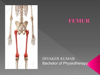
The femur or thigh bone, is the proximal bone of the hindlimb in tetrapod vertebrates. The head of the femur articulates with the acetabulum in the pelvic bone forming the hip joint, while the distal part of the femur articulates with the tibia and kneecap forming the knee joint. By most measures the femur is the strongest bone in the body. The femur is also the longest bone in the human body.

More Related Content
What's hot
What's hot (20)
Similar to Femur
Similar to Femur (20)
Recently uploaded
Recently uploaded (20)
Femur
- 1. DIVAKER KUMAR Bachelor of Physiotherapy
- 2. Thigh bone Longest and the strongest bone in the body It has a Shaft Upper end Lower end
- 3. Includes • Head • Neck • Greater trochanter(=knob like) • Lesser trochanter • Intertrochanteric line • Intertrochanteric crest
- 4. • Shaft is more or less cylindrical • More expanded inferiorly than superiorly • Convex forwards • Directed obliquely downwards and medially
- 5. • Includes : 2 large Condyles – Medial & Lateral Posteriorly separated by a deep gap- Intercondyla r fossa or notch
- 6. • Head is directed medially upwards and slightly forwards • Shaft is directed obliquely downwards and medially so that the lower surfaces of the 2 condyles of the femur lie in the same horizontal plane
- 7. • Upper end bears a rounded head whereas the lower end is expanded to form 2 large condyles • Head is directed medially • Cylindrical shaft is convex forwards
- 8. Head of the Femur: • Forms more than half of a sphere • Directed medially, upwards and slightly forwards • Pit/ Fovea – at the centre of the head – Round ligament of the head( Ligamentum teres femoris) • Blood supply: Small, Medial part – by Medial epiphyseal arteries( Posterior division of the obturator artery & ascending branch of medial circumflex femoral artery) Larger lateral part – Lateral epiphyseal arteries( retinacular branches of medial circumflex femoral artery)- Necrosis due to fracture of the neck
- 9. Neck of the Femur: • Connects head with the shaft, about 3.7 cms in length • Neck shaft angle - 125⁰ in adults : facilitates the movement of the hip joint, strengthened by Calcar femorale • 2 borders , 2 surfaces • Borders: Upper border: concave, horizontal, meets the shaft at the greater trochanter Lower border: straight and oblique, at the lesser trochanter • Surfaces : Anterior: meets shaft at the intertrochanteric line, entirely intracapsular; oblique bony ridges for attachment of retinacular fibers of fibrous capsule Posterior : only a little more than it’s medial half is intracapsular, groove for tendon of obturator externus
- 10. • Angle of femoral torsion/ Anteversion: between the transverse diameters of upper end and lower end of femur • Blood supply: Intracapsular part- retinacular arteries( chiefly from trochanteric anastamosis) – produce longitudinal grooves and foramina directed towards the head, mainly on the anterior & postero-superior surfaces Extracapsular part- ascending branch of medial circumflex femoral artery
- 11. Greater Trochanter: • Large quadrangular prominence located at the upper part of junction of neck with the shaft Upper border with an apex 3 surfaces: Anterior, medial & Lateral • Apex- inturned posterior part of posterior border • Anterior surface- rough in it’s lateral part • Medial surface- rough impression above, deep trochanteric fossa below • Lateral surface- crossed by a ridge directed downwards and forwards
- 12. Attachments : • Piriformis – apex • Gluteus minimus – rough lateral part of the anterior surface • Obturator internus, 2 gemelli- upper rough impression on the medial surface • Obturator externus- trochanteric fossa • Gluteus medius- ridge on the lateral surface
- 13. Lesser Trochanter: • Conical eminence • Directed medially, backwards from the junction of postero- inferior part of neck with the shaft • Attachments: Psoas major- apex and medial part of rough anterior surface Iliacus – anterior surface of the base and on the area below it Smooth posterior surface is covered by a bursa that lies deep to the upper horizontal fibers of Adductor magnus
- 14. Intertrochanteric line: • Marks the junction of the anterior surface of the neck with the shaft • Prominent roughened ridge • begins above at the antero- superior angle of the greater trochanter as a tubercle and continuous below with the spiral line in front of the lesser trochanter • Attachments: Origin to highest fibers of vastus lateralis from the upper end From the lower end, vastus medialis
- 15. Intertrochanteric crest : • Marks the junction between posterior surface of the neck with the shaft of the femur • Smooth rounded ridge, begins above at the postero-superior angle of greater trochanter and ends at the lesser trochanter • Quadrate tubercle – rounded elevation , a little above it’s middle • Attachments: Quadrate tubercle- Insertion to quadratus femoris
- 16. • In the middle one third, 3 borders : Medial, lateral and posterior 3 surfaces : Anterior, medial, Lateral • Borders: Medial, Lateral borders – Ill- defined Posterior border- broad roughened ridge: Linea aspera • Surfaces: Medial & Lateral surfaces- more backwards than the sides
- 17. Linea Aspera (Rough line) • 2 distinct lips- medial, lateral • In the upper one-third, Lips diverge to enclose an additional posterior surface Thus 4 borders: Lateral- Spiral Line, Medial- Gluteal Tuberosity & 4 surfaces • Lower one- third, 2 lips diverge as supracondylar lines: enclose an additional popliteal surface
- 18. Attachments to Linea Aspera & shaft: • Medial , popliteal surfaces- bare, except that the popliteal surface is encroached by the medial head of gastrocnemius • Gluteus maximus- Gluteal tuberosity • Plantaris – lower part of lateral supracondylar line, upper part – lateral head of gastrocnemius • Popliteal surface- covered with fat and forms the floor of popliteal fossa
- 19. • Condyles- posteriorly separated by a deep gap, intercondylar fossa/ notch • Posteriorly, project backwards much beyond the plane of the popliteal surface
- 20. Articular surface: • 2 condyles are partially covered by a large articular surface • It is divisible into patellar and tibial parts • Patellar surface: covers anterior surfaces and extend more on the lateral than the medial condyle, vertical groove between the condyles • Tibial surface: occupy the inferior & posterior surfaces, part on the medial condyle is longer and convex medially • Both surfaces are separated by 2 faint grooves
- 21. Lateral condyle: • Flat laterally, more in line with the shaft • Less prominent but stouter and stronger • Greater part in weight transmission to the tibia • Most prominent point- Lateral epicondyle • Just below the epicondyle- Popliteal groove Medial condyle: • Convex medially • More prominent • Prominence- Lateral epicondyle • Posterosuperior to the epicondyle- Adductor tubercle( landmark- epiphyseal line for lower end of femur passes through it)
- 22. Attachments: • Popliteus- origin from the popliteal groove • Adductor tubercle- insertion of Hamstring part of or ischial head of Adductor magnus M L
- 23. Intercondylar fossa: • Separates lower & posterior parts of the condyles • Extent: Anteriorly- Patellar articular surface Posteriorly- intercondylar line, separates notch from the popliteal surface
- 24. Nutrient Artery to femur: • Derived from 2nd perforating artery • If absent, derived from 1st & 3rd perforating arteries • Nutrient foramen- medial to linea aspera, directed upwards
- 25. FOR MORE INFORMATION DIVAKER_SHAH <INSTAGRAM>