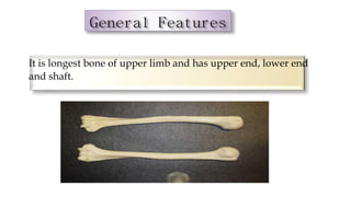
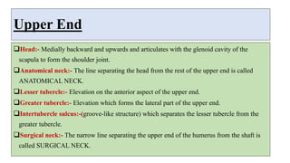
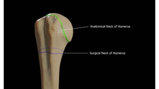
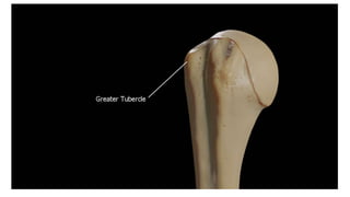
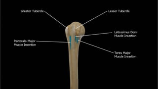
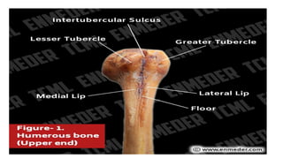
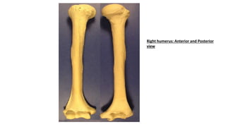
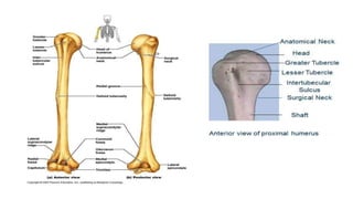
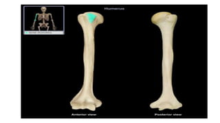
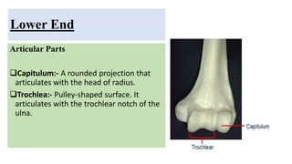
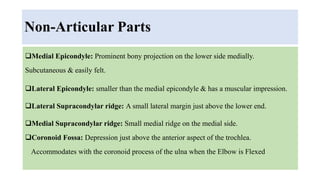
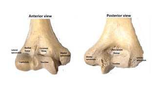
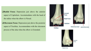
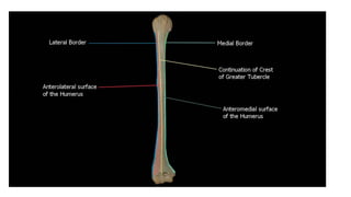
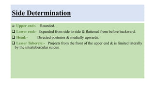
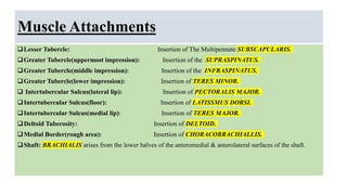
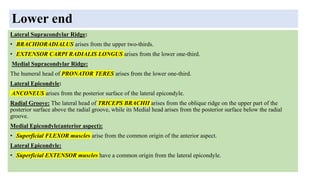
2. It is longest bone of upper limb and has upper end, lower end
and shaft.
Introduction
3. Upper End
Head:- Medially backward and upwards and articulates with the glenoid cavity of the
scapula to form the shoulder joint.
Anatomical neck:- The line separating the head from the rest of the upper end is called
ANATOMICAL NECK.
Lesser tubercle:- Elevation on the anterior aspect of the upper end.
Greater tubercle:- Elevation which forms the lateral part of the upper end.
Intertubercle sulcus:-(groove-like structure) which separates the lesser tubercle from the
greater tubercle.
Surgical neck:- The narrow line separating the upper end of the humerus from the shaft is
called SURGICAL NECK.
11. Lower End
Articular Parts
Capitulum:- A rounded projection that
articulates with the head of radius.
Trochlea:- Pulley-shaped surface. It
articulates with the trochlear notch of the
ulna.
12. Non-Articular Parts
Medial Epicondyle: Prominent bony projection on the lower side medially.
Subcutaneous & easily felt.
Lateral Epicondyle: smaller than the medial epicondyle & has a muscular impression.
Lateral Supracondylar ridge: A small lateral margin just above the lower end.
Medial Supracondylar ridge: Small medial ridge on the medial side.
Coronoid Fossa: Depression just above the anterior aspect of the trochlea.
Accommodates with the coronoid process of the ulna when the Elbow is Flexed
13.
14. Radial Fossa: Depression just above the anterior
aspect of Capitulum. Accommodates with the head of
the radius when the elbow is Flexed.
Olecranon Fossa: Depression just above the posterior
aspect of Trochlea. Accommodates with the olecranon
process of the ulna when the elbow is Extended.
15. Shaft
• Rounded in the upper half & triangular in the lower half.
• Contains three borders & three surfaces.
Borders:
Anterior Border: The upper one-third forms the Lateral lip of the intertubercular sulcus. The middle part forms
the anterior region of Deltoid tuberosity. The lower half is smooth & rounded.
Lateral Border: Prominent only in the lower region where it forms a lateral supracondylar ridge. In the middle,
it is interrupted by a Radial groove.
Medial Border: Forms the medial lip of the intertubercular sulcus. Rough through its middle & continuous
below with the medial supracondylar ridge.
16. Three Surfaces
The anterolateral surface lies between the anterior and lateral borders. The upper half
of this surface is covered by the deltoid. A little above the middle, it is marked by a V-
shaped deltoid (Greek triangular-shaped)tuberosity. Behind the deltoid tuberosity, the
radial groove runs downwards and forward across the surface.
The anteromedial surface lies between the anterior and medial borders. Its upper one-
third is narrow and forms the floor of the intertubercular sulcus. A nutrient foramen is
seen near the medial border below its middle part.
The posterior surface lies between the medial and lateral borders. Its upper part is
marked by an oblique ridge. The middle one-third is crossed by the radial groove.
17.
18. Side Determination
Upper end:- Rounded.
Lower end:- Expanded from side to side & flattened from before backward.
Head:- Directed posterior & medially upwards.
Lesser Tubercle:- Projects from the front of the upper end & is limited laterally
by the intertubercular sulcus.
19. Muscle Attachments
Lesser Tubercle: Insertion of The Multipennate SUBSCAPULARIS.
Greater Tubercle(uppermost impression): Insertion of the SUPRASPINATUS.
Greater Tubercle(middle impression): Insertion of the INFRASPINATUS.
Greater Tubercle(lower impression): Insertion of TERES MINOR.
Intertubercular Sulcus(lateral lip): Insertion of PECTORALIS MAJOR.
Intertubercular Sulcus(floor): Insertion of LATISSMUS DORSI.
Intertubercular Sulcus(medial lip): Insertion of TERES MAJOR.
Deltoid Tuberosity: Insertion of DELTOID.
Medial Border(rough area): Insertion of CHORACOBRACHIALLIS.
Shaft: BRACHIALIS arises from the lower halves of the anteromedial & anterolateral surfaces of the shaft.
20. Lower end
Lateral Supracondylar Ridge:
• BRACHIORADIALUS arises from the upper two-thirds.
• EXTENSOR CARPI RADIALIS LONGUS arises from the lower one-third.
Medial Supracondylar Ridge:
The humeral head of PRONATOR TERES arises from the lower one-third.
Lateral Epicondyle:
ANCONEUS arises from the posterior surface of the lateral epicondyle.
Radial Groove: The lateral head of TRICEPS BRACHII arises from the oblique ridge on the upper part of the
posterior surface above the radial groove, while its Medial head arises from the posterior surface below the radial
groove.
Medial Epicondyle(anterior aspect):
• Superficial FLEXOR muscles arise from the common origin of the anterior aspect.
Lateral Epicondyle:
• Superficial EXTENSOR muscles have a common origin from the lateral epicondyle.
21. Clinical Anatomy
• The common sites of fracture of humerus are the surgical neck, shaft, and
supracondylar region.
Supracondylar fracture is common at young age. It is produced by a fall on the
outstretched hand.
• The lower fragment is mostly displaced backward, so that the elbow is unduly
prominent, as in dislocation of the elbow joint. This fracture may cause injury to the
median nerve. It may also lead to Volkmann 's ischaemic contracture caused by
occlusion of the brachial Artery.
• • The three bony points of the normal elbow form the equilateral triangle in a flexed
elbow and are in one line in an extended elbow.
• • The humerus has a poor blood supply at the junction of its upper and middle thirds.
Fractures
• at this site show delayed muon or nonunion.
• The head of the humerus commonly dislocates anteroinferiorly.