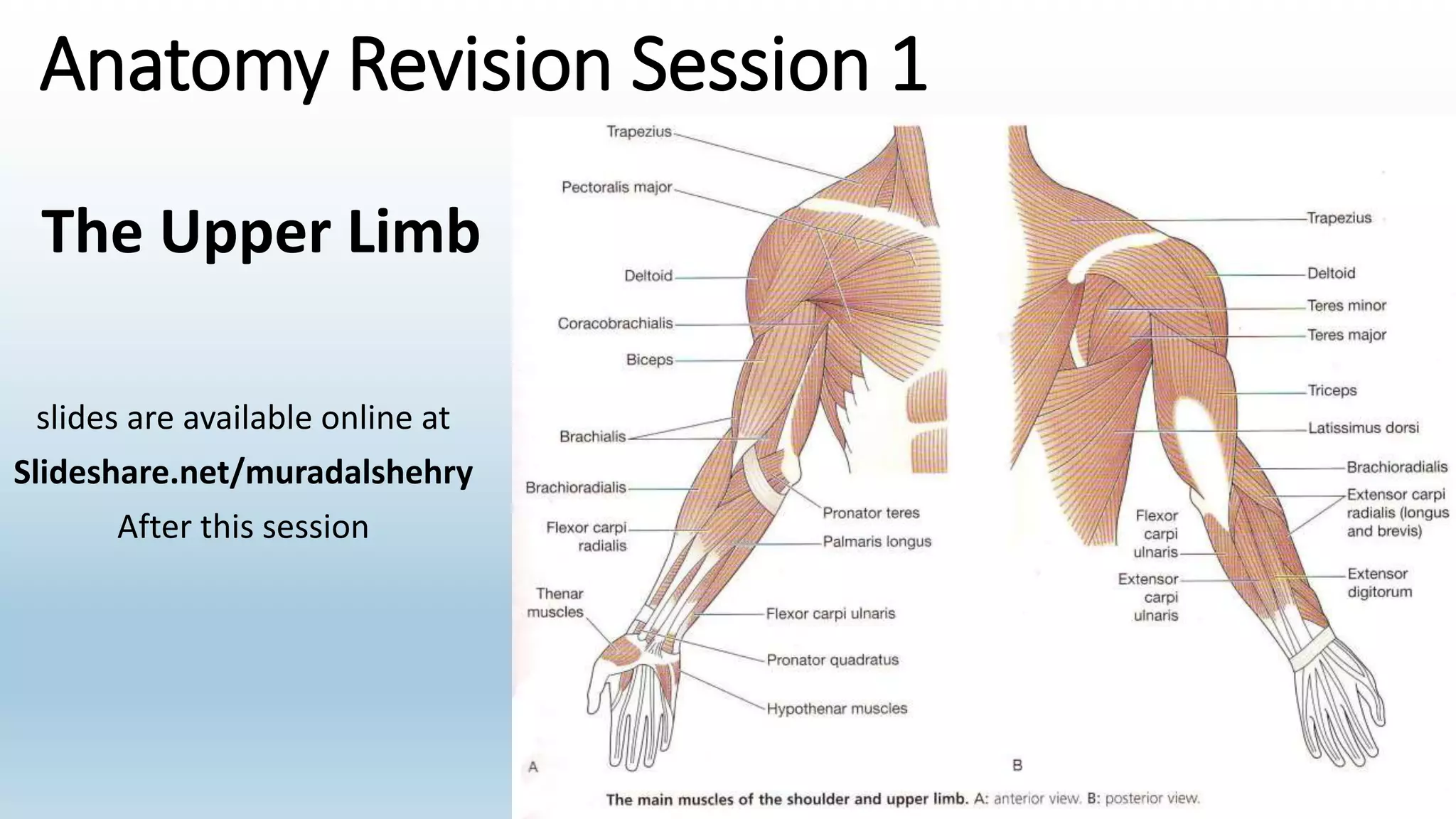
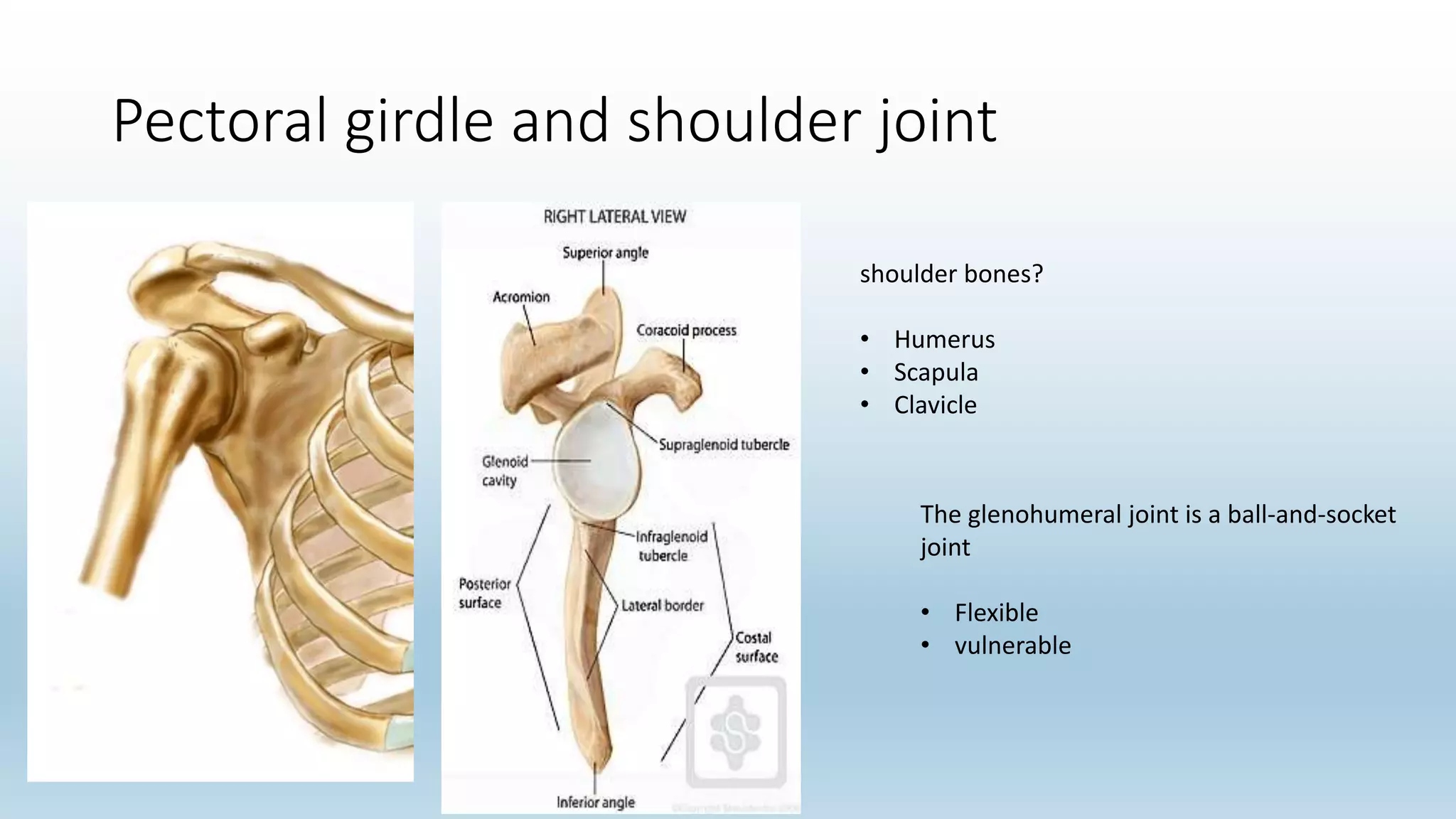
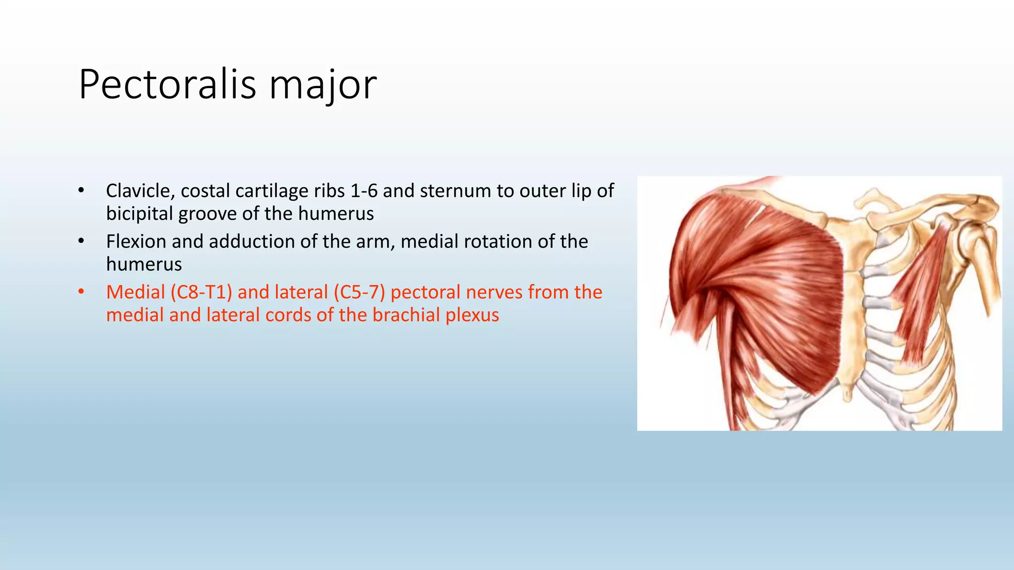
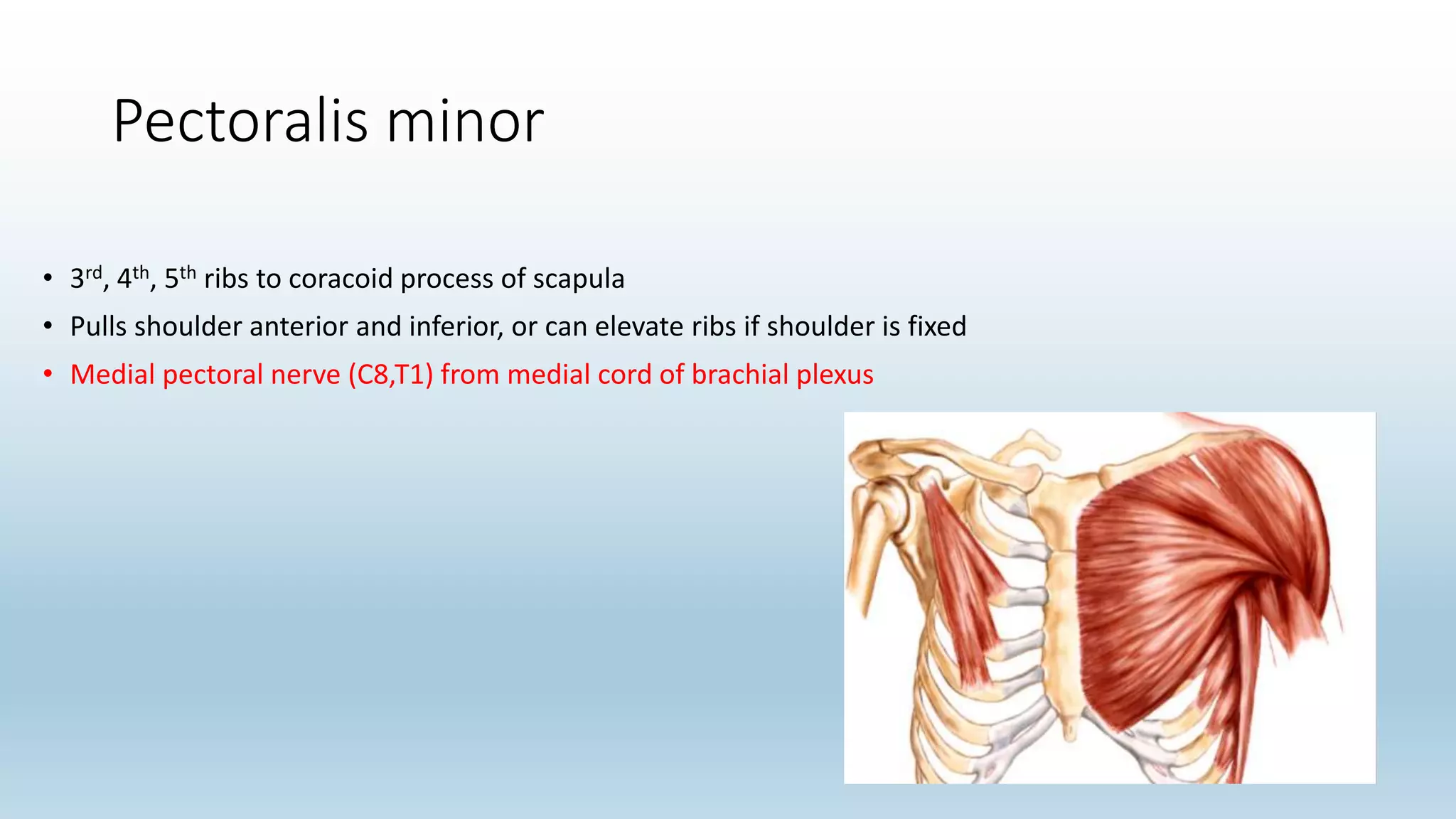
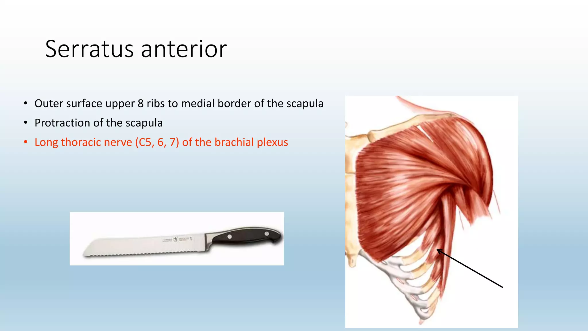
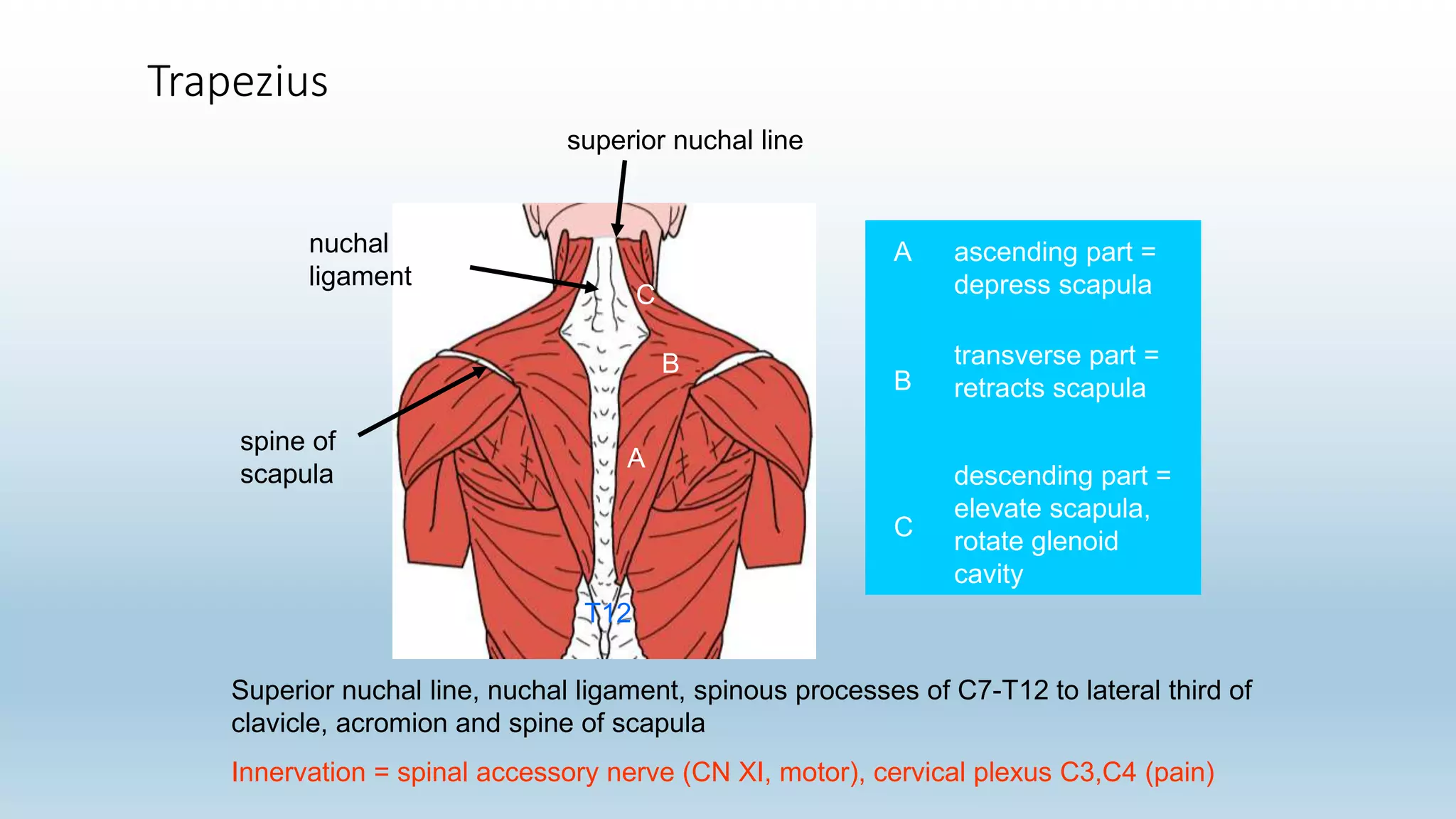
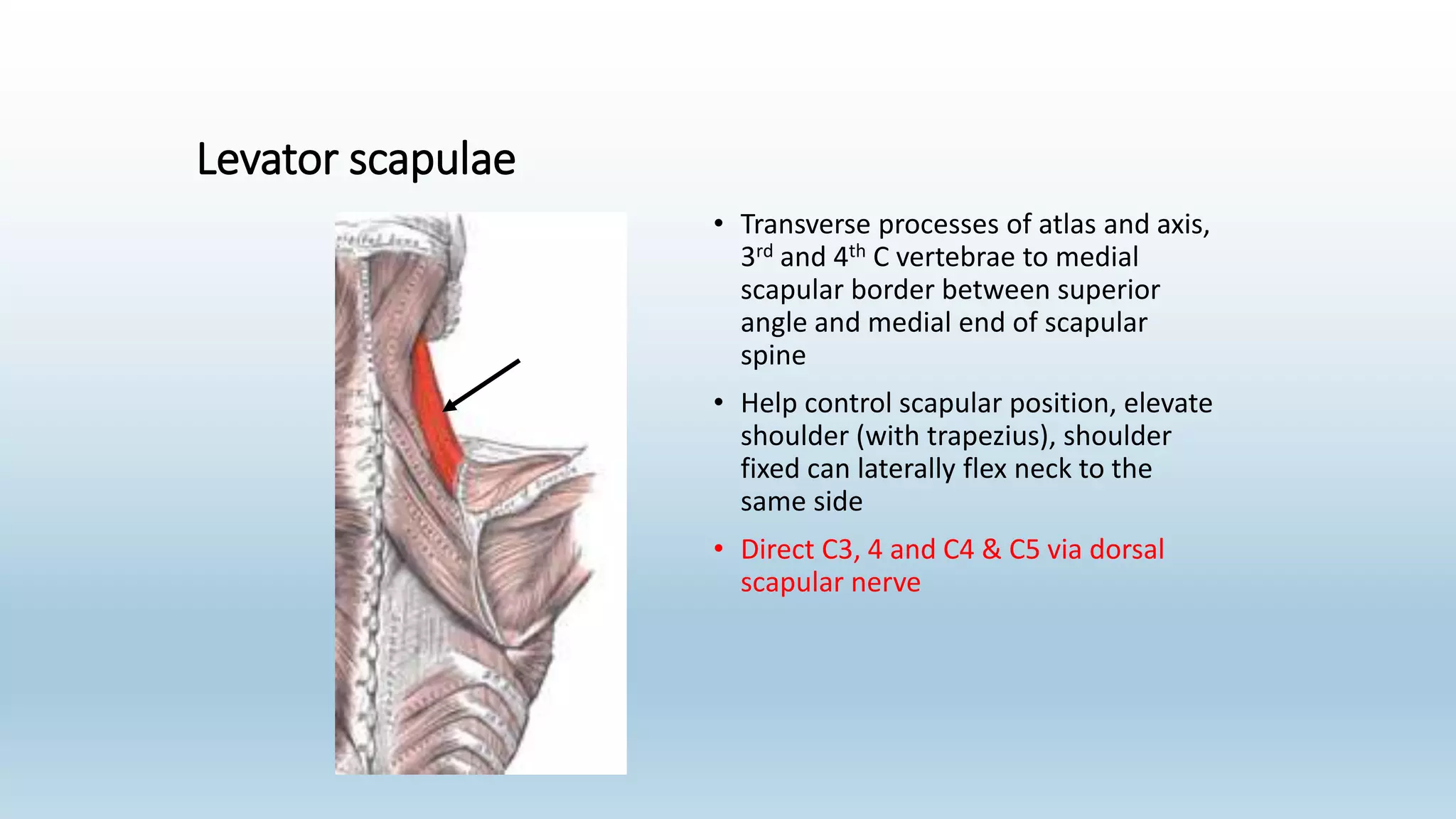
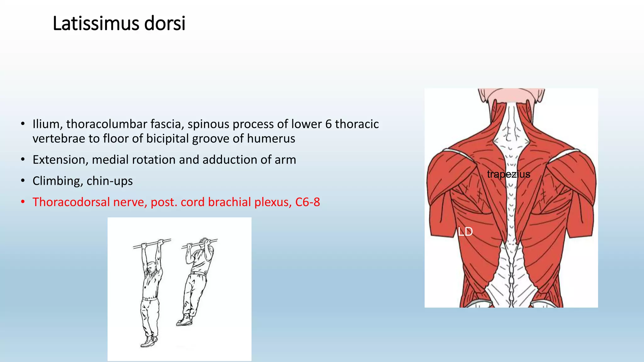
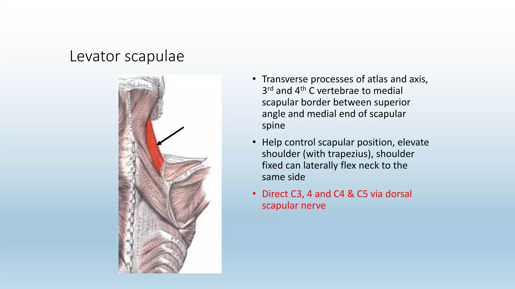
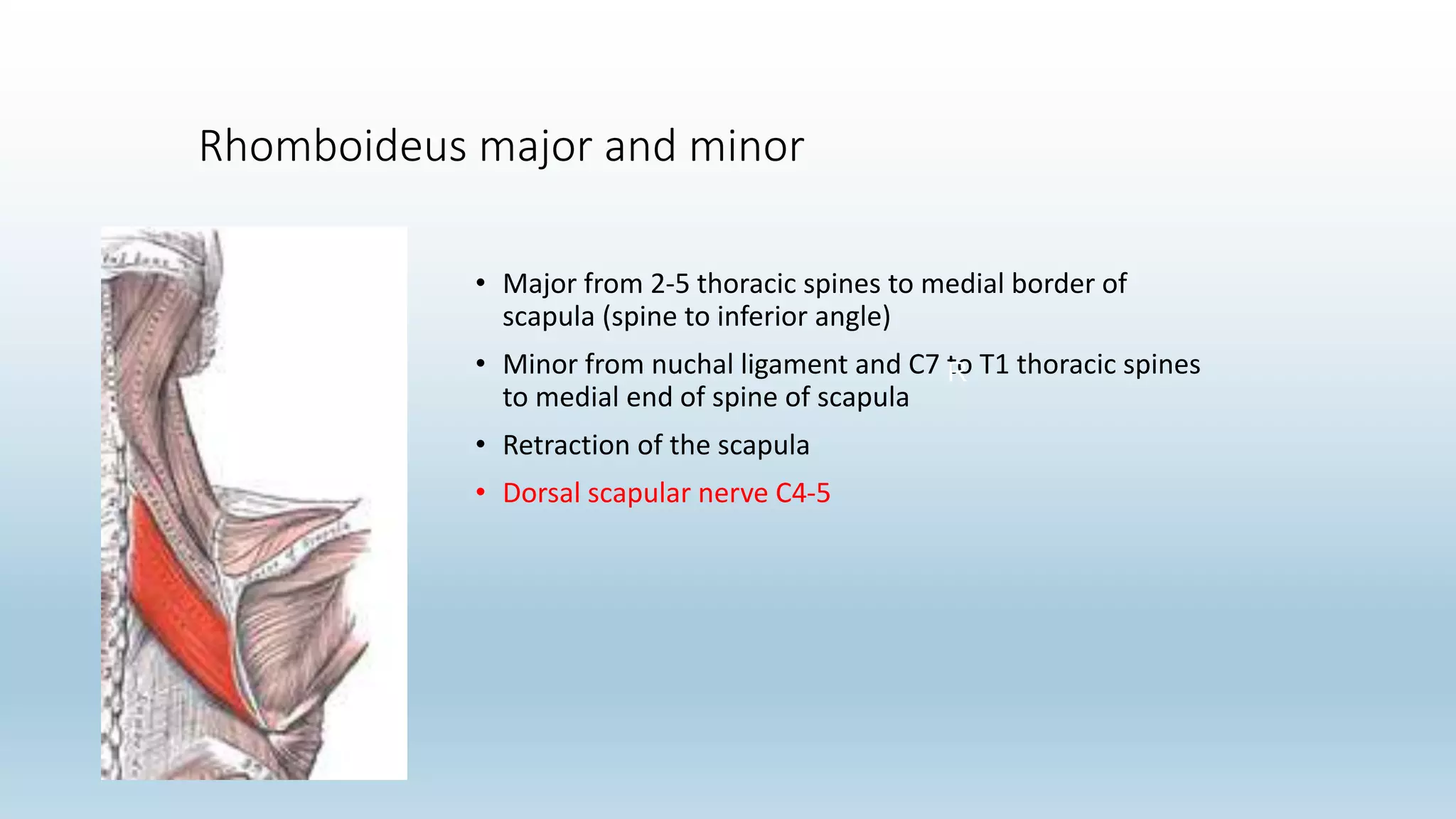
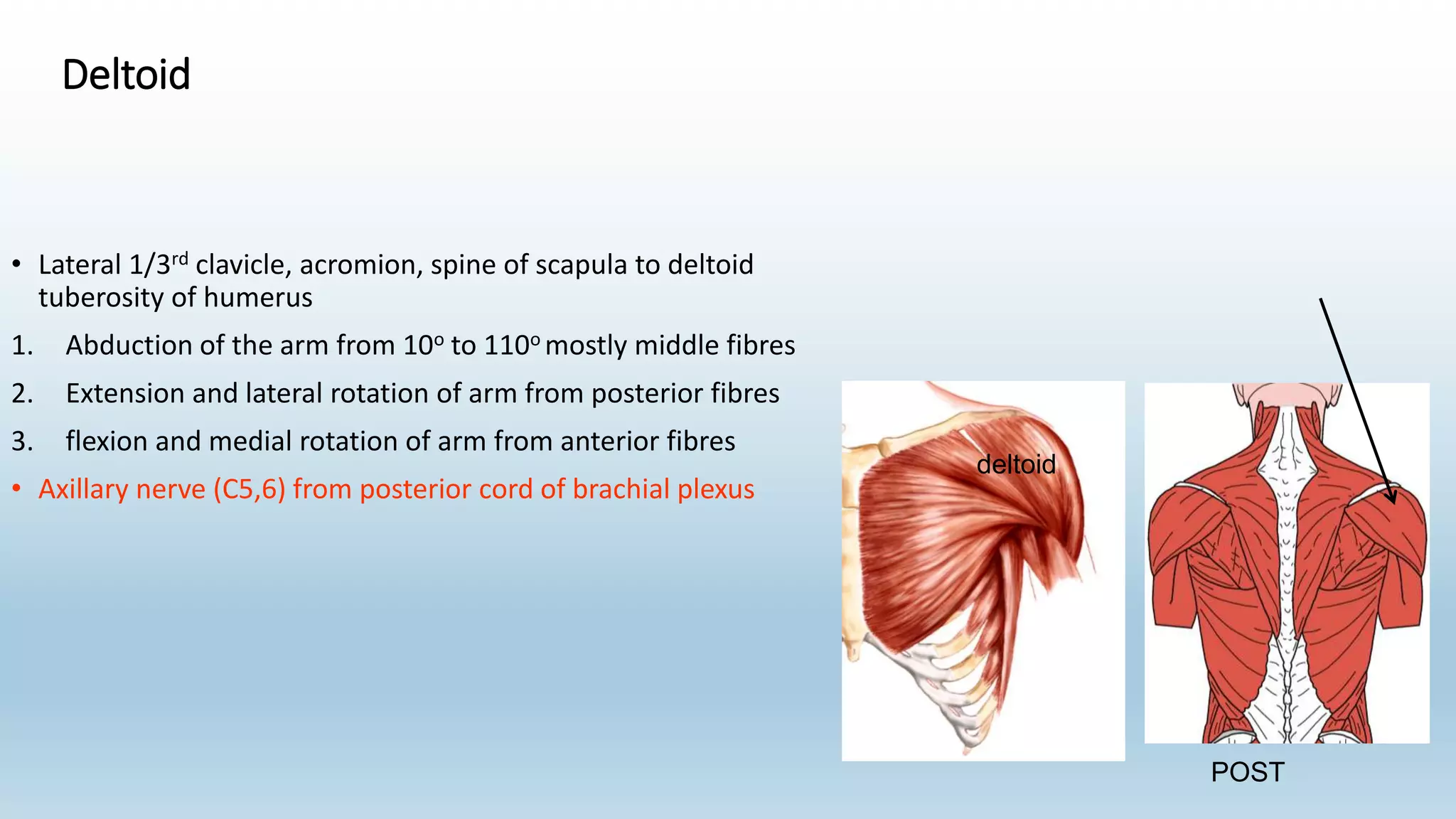
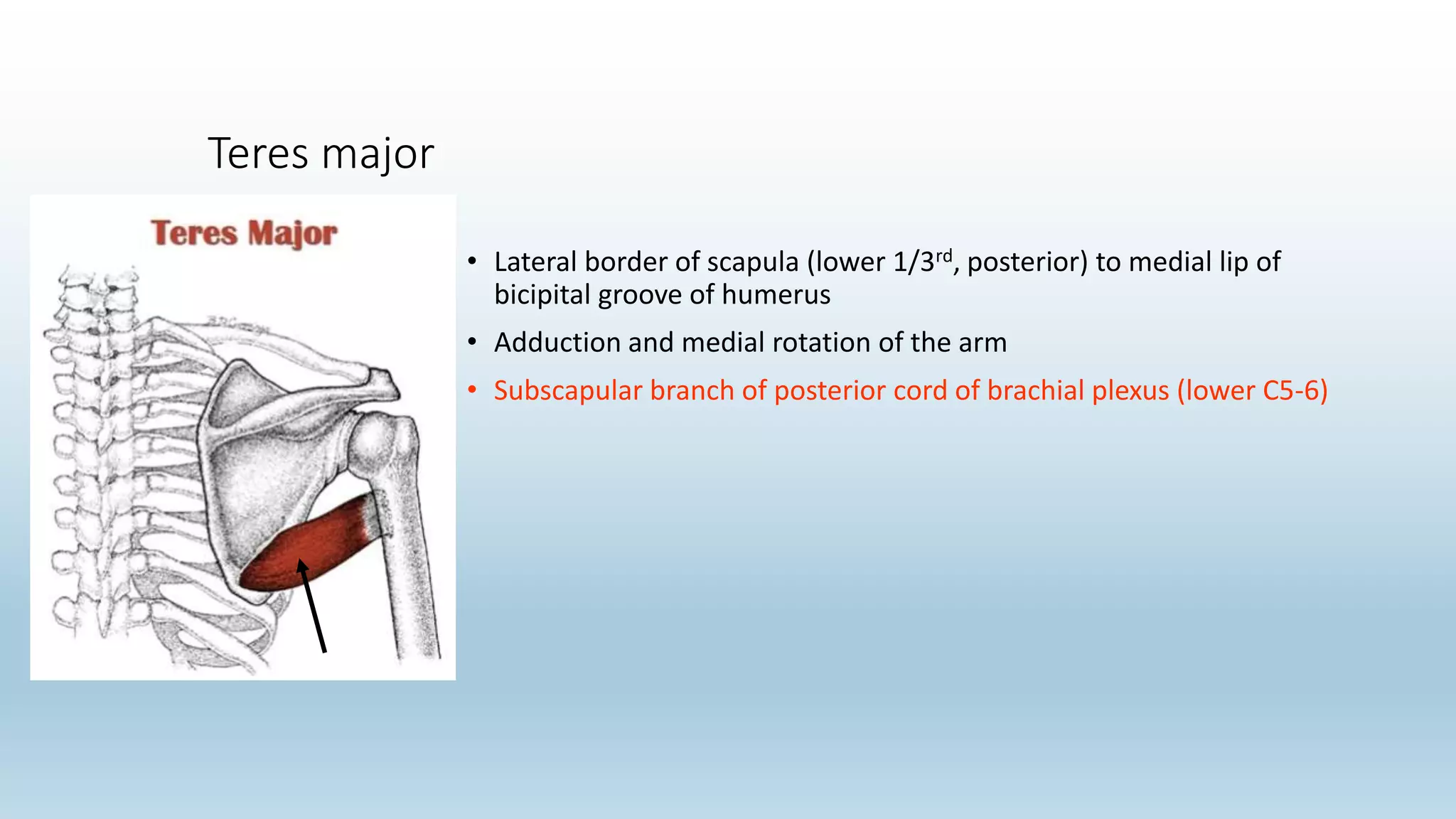
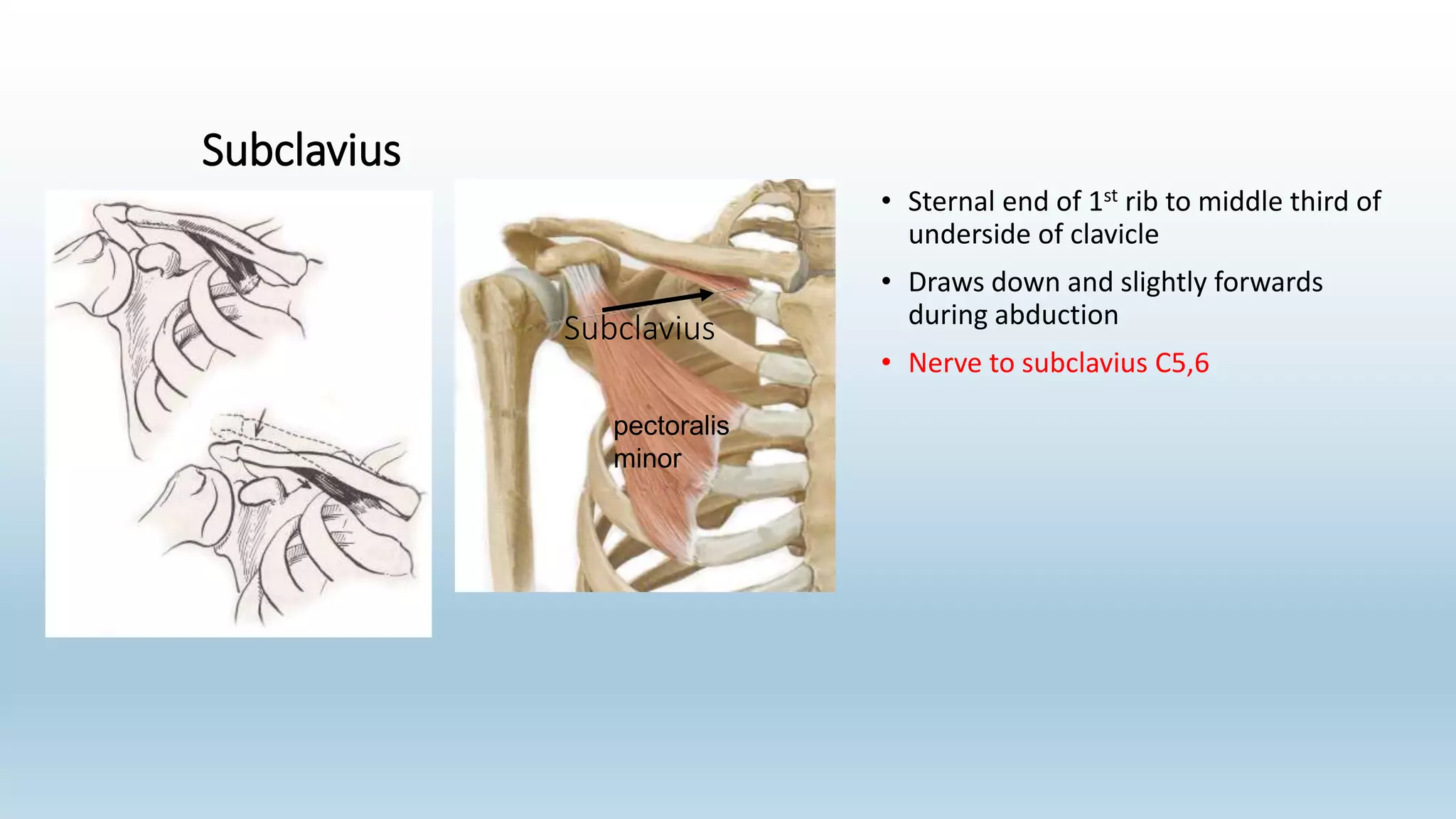
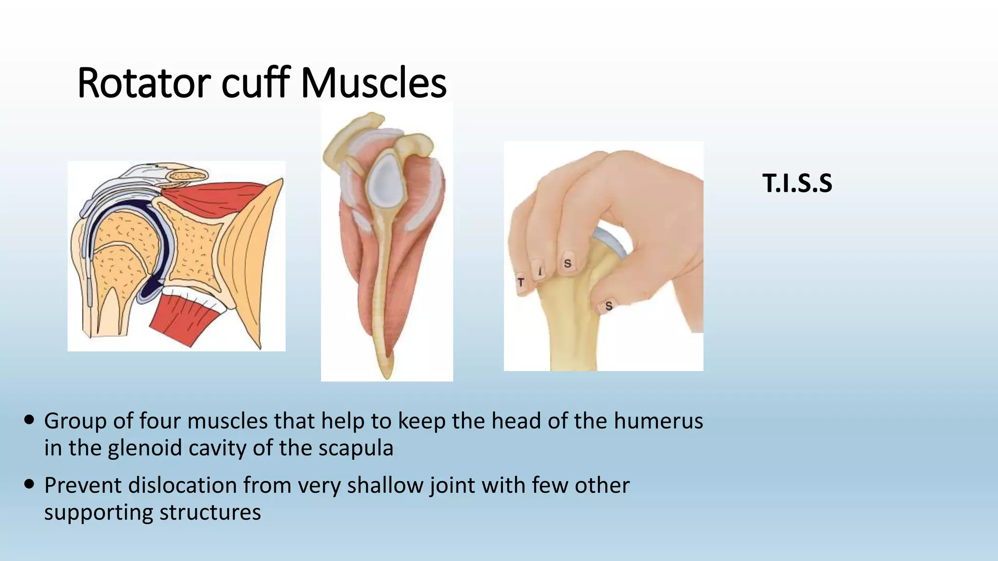
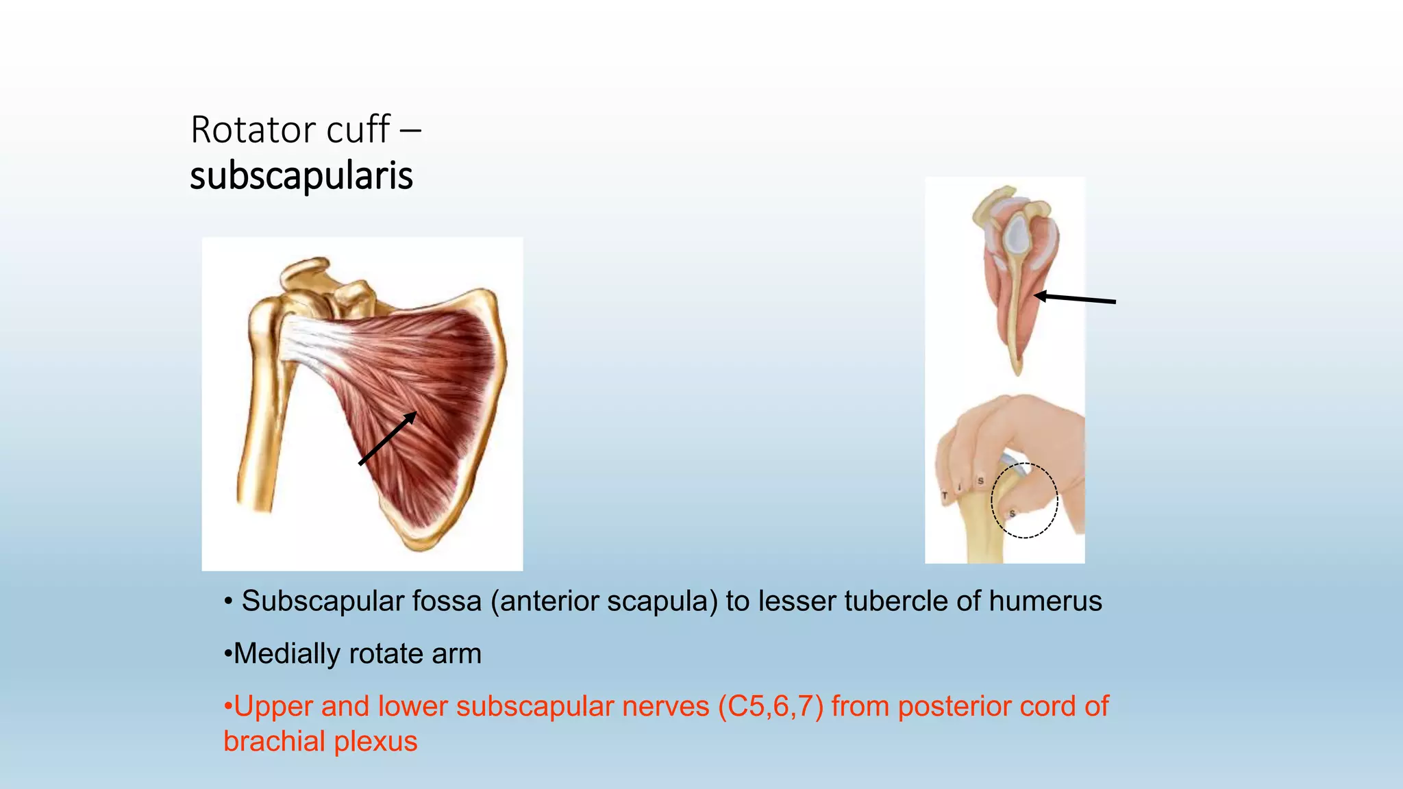
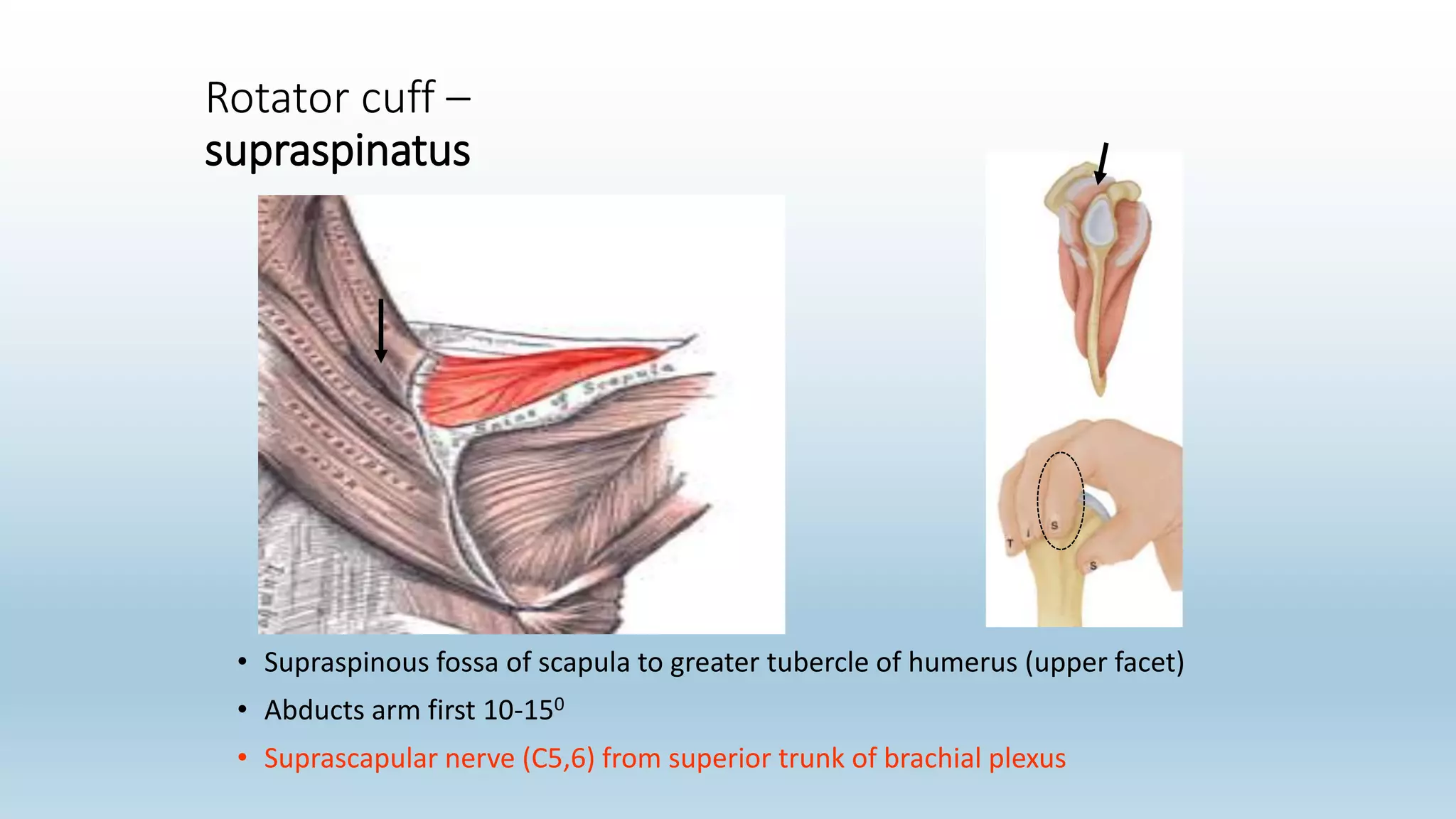
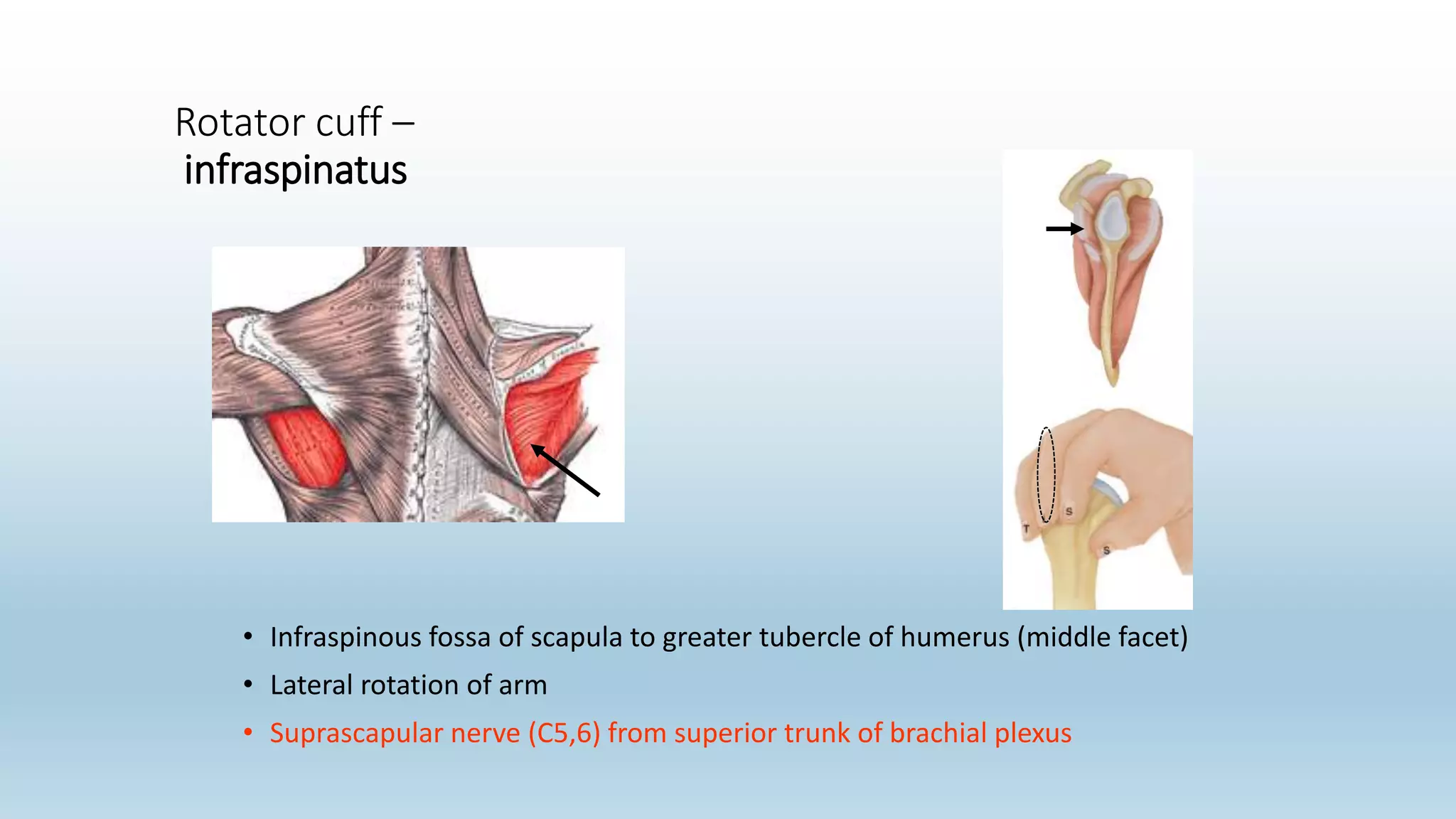
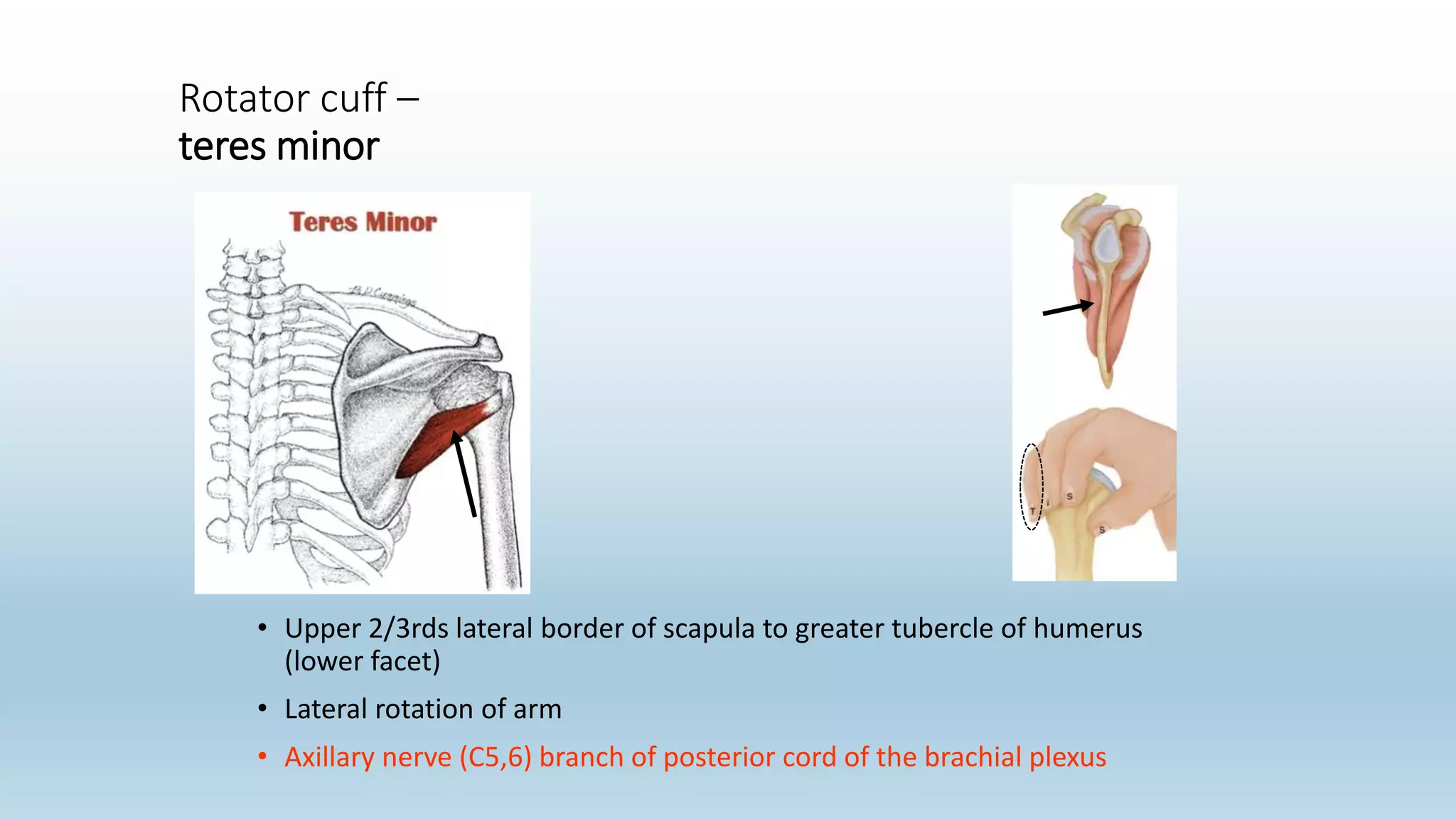
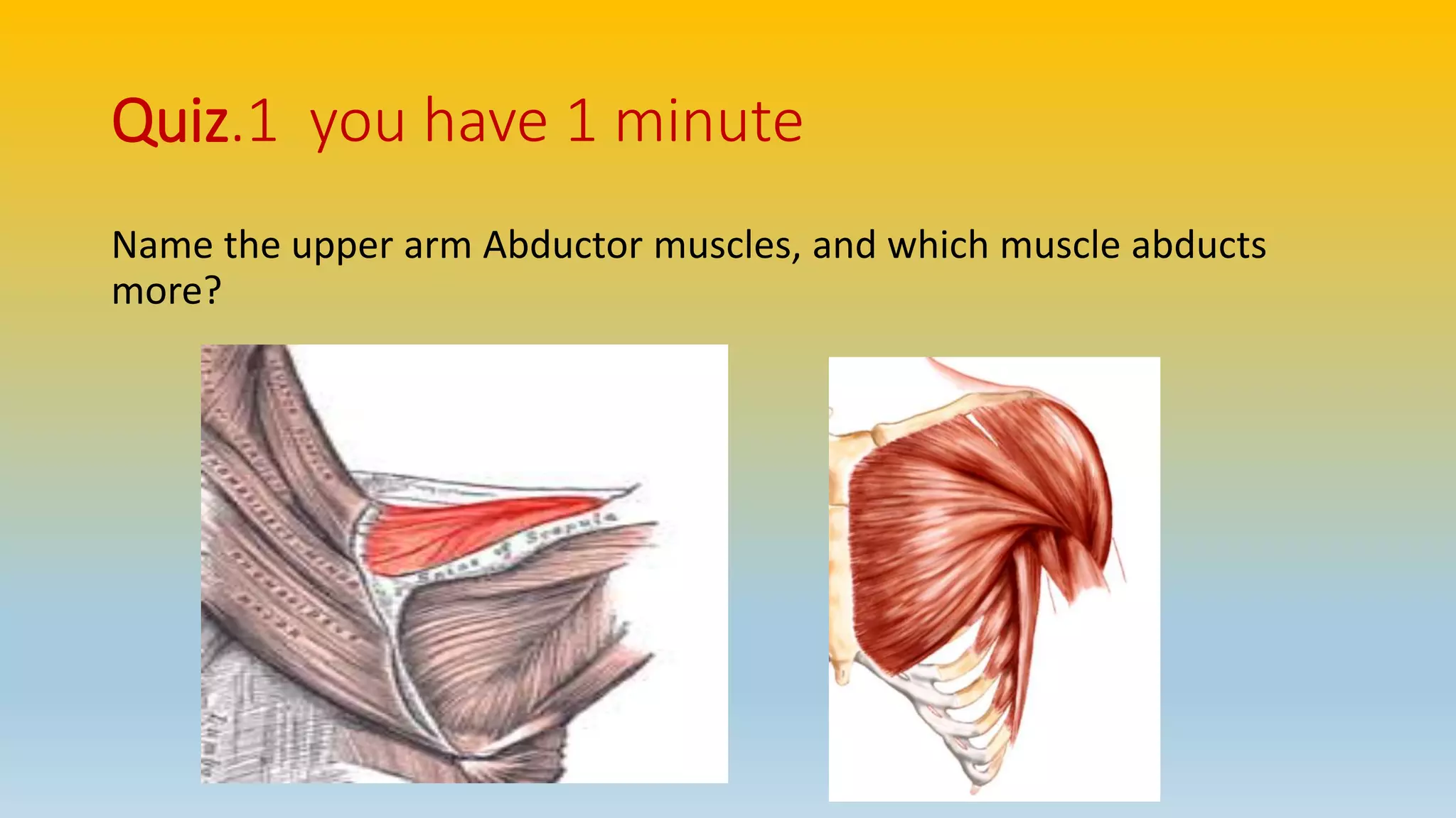
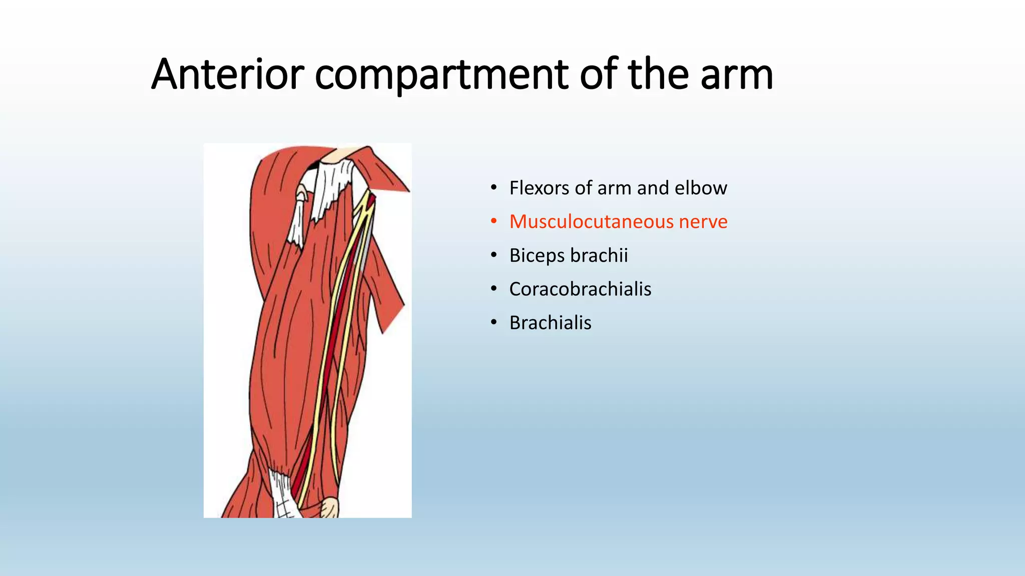
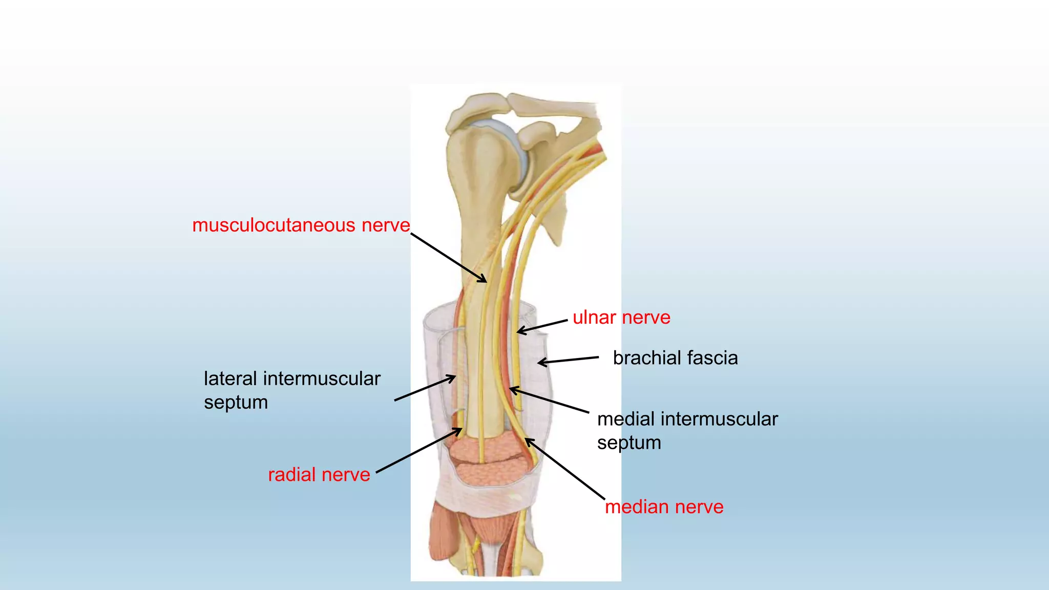
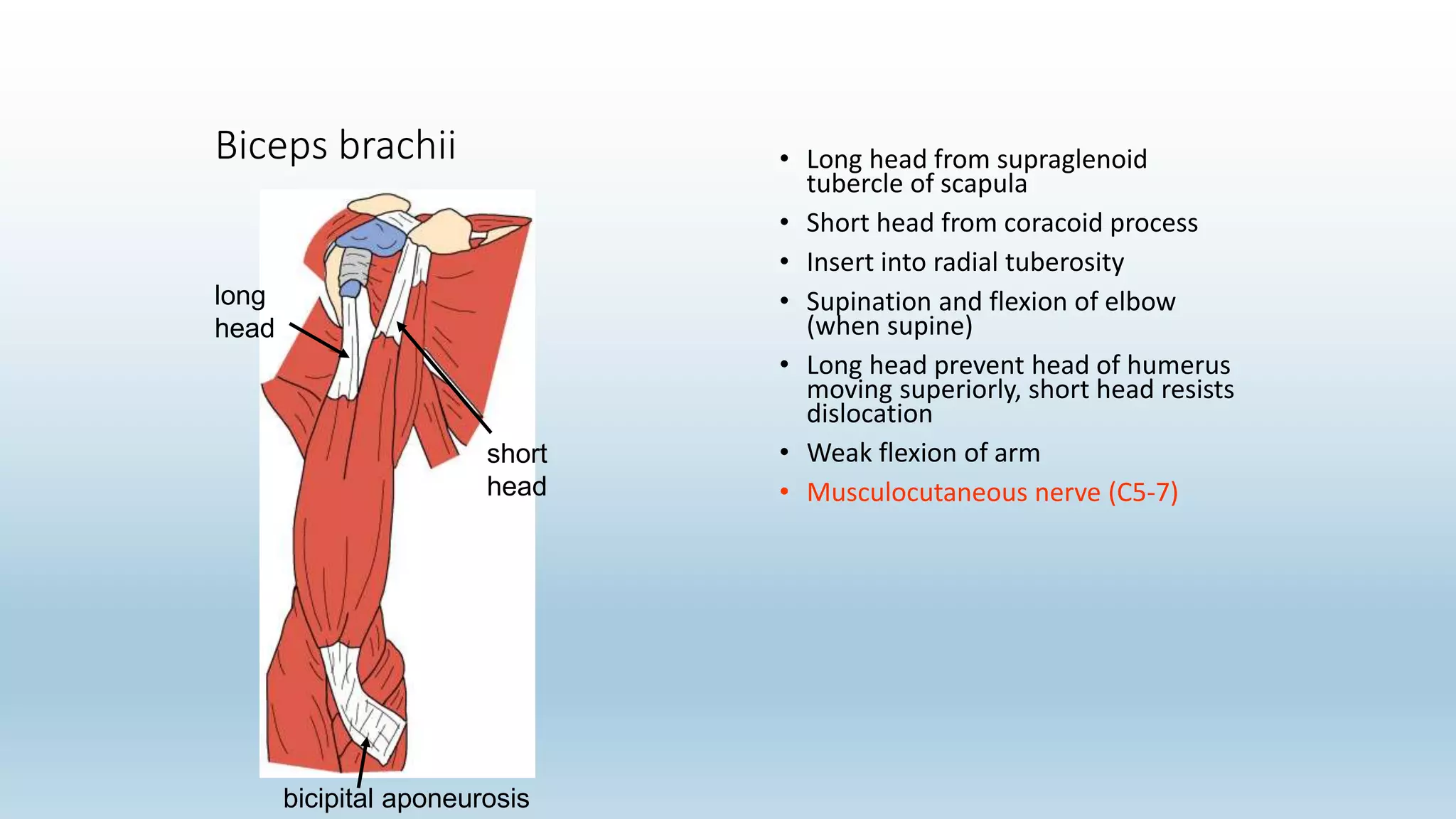
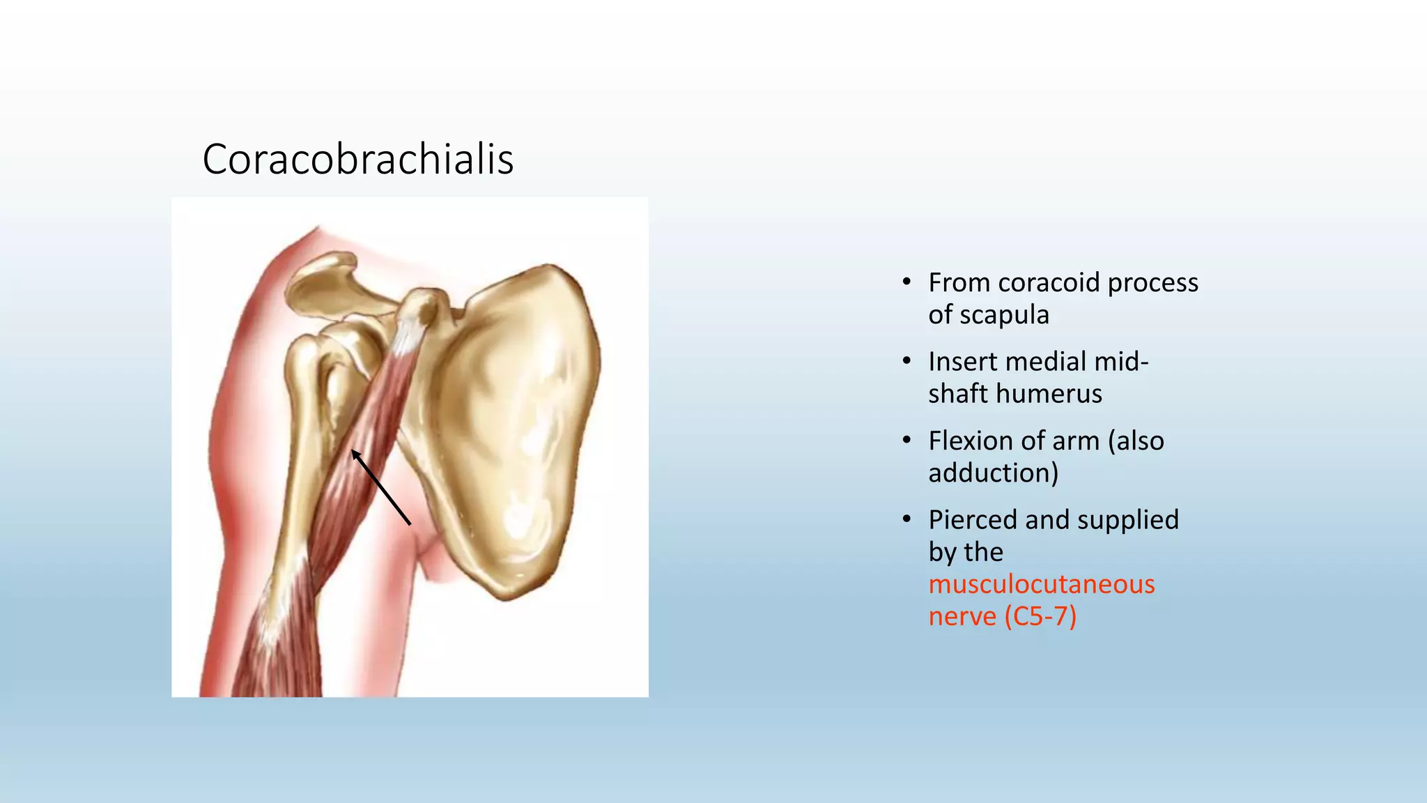
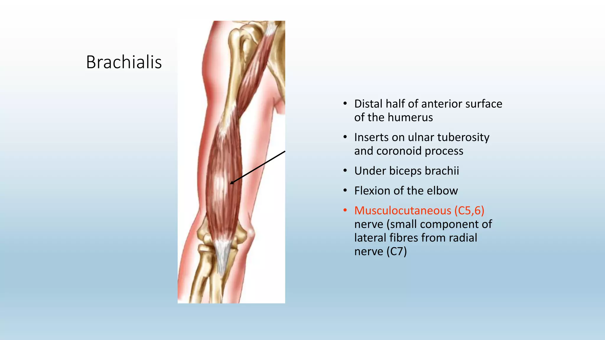
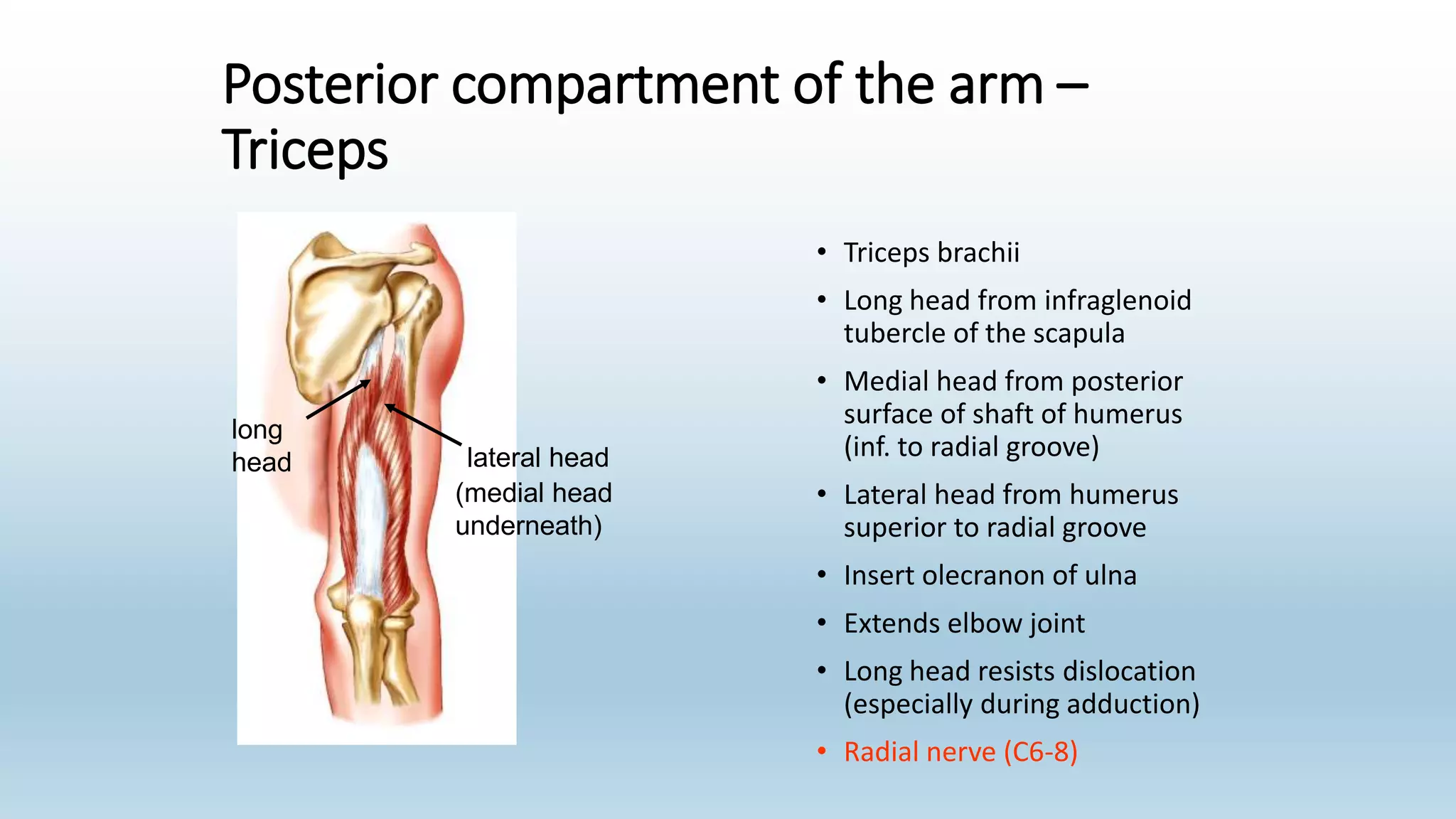
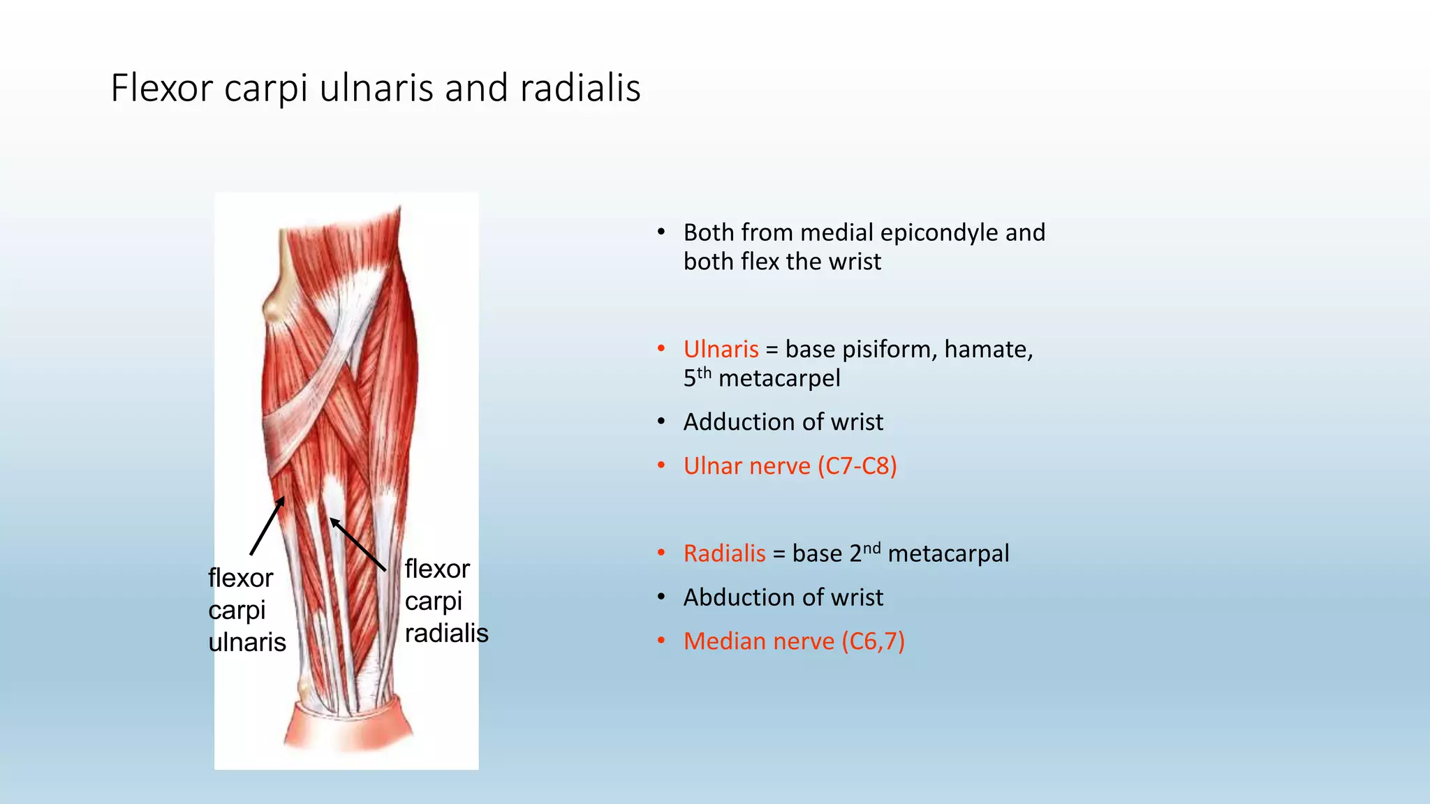
The document summarizes an anatomy revision session on the upper limb. It discusses various muscles of the upper limb including their origins, insertions, innervations and functions. Key muscles covered include the pectoralis major and minor, serratus anterior, deltoid, biceps brachii, brachialis, coracobrachialis, and triceps. It also discusses the rotator cuff muscles and muscles of the forearm including flexor carpi ulnaris and radialis. The session aims to help students identify upper limb muscles and understand their relations to nerves.
this is a presentation on atlanto-axial and atlanto-occipital joints. after reading this, most of you will know about atlas and axis, joint type, anatomy of joint, movements allowed by joint and its clinical considerations.
to download this presentation from this link.
https://mohmmed-ink.blogspot.com/2020/12/joints-of-upper-limb.html
anatomy of the upper limb joints. shoulder, elbow, wrist hand
The Popliteal Fossa is a diamond-shaped space behind the knee joint. It is formed between the muscles in the posterior compartments of the thigh and leg. This anatomical landmark is the major route by which structures pass between the thigh and leg.
List of Images
01. SKULL, ANTERIOR VIEW 15. SKELETON OF UPPER LIMB, ANTERIOR VIEW
02. SKELETON OF HEAD AND NECK, LATERAL VIEW 16. ARTERIES OF UPPER LIMB, ANTERIOR VIEW
03. MUSCULATURE OF HEAD AND NECK, LATERAL VIEW 17. NERVES OF UPPER LIMB, ANTERIOR VIEW
04. ARTERIES OF HEAD AND NECK, LATERAL VIEW 18. MUSCULATURE OF THORAX AND UPPER LIMB, LATERAL VIEW
05. SUPERFICIAL ANATOMY OF HEAD AND NECK, LATERAL VIEW 19. SKELETON OF LOWER LIMB, ANTERIOR VIEW
06. ANATOMY OF HEAD AND NECK, MIDSAGITTAL VIEW 20. ARTERIES OF LOWER LIMB, ANTERIOR AND POSTERIOR VIEWS
07. BRAIN, LATERAL VIEW 21. NERVES OF LOWER LIMB, ANTERIOR AND POSTERIOR VIEWS
08. SKELETON, LATERAL VIEW 22. MUSCULATURE OF LOWER LIMB, LATERAL AND MEDIAL VIEWS
09. SKELETON, ANTERIOR VIEW 23. RESPIRATORY SYSTEM, ANTERIOR VIEW
10. ARTERIES OF THORAX AND ABDOMEN, ANTERIOR VIEW 24. DIGESTIVE SYSTEM, ANTERIOR VIEW
11. AUSCULTATION POINTS 25. LYMPHATICS SYSTEM, ANTERIOR VIEW
12. MUSCULATURE OF THORAX AND ABDOMEN, ANTERIOR VIEW 26. MALE AND FEMALE UROGENITAL SYSTEMS, MIDSAGITTAL VIEW
13. SKELETON, POSTERIOR VIEW 27. ANTERIOR VIEW OF HEART WITH OPENED RIGHT VENTRICLE
14. MUSCULATURE OF BACK, POSTERIOR VIEW
1.INTRODUCTION
Shoulder joint is formed by scapula and clavicle (which is also called as shoulder girdle)and proximal humerus.
2.BONES OF SHOULDER JOINT
3.Joints of the Shoulder Complex
Glenohumeral
Acromioclavicular
Sternoclavicular
Scapulothoracic
4.Muscles of the Shoulder
5.Gateways to the Posterior Scapular Region
6. Movements
Shoulder girdle and brachial plexus anatomyJunaid Ahmad
Anatomy and Functions of muscles and joints
Dr Junaid Ahmad (MBBS FCPS) is the best plastic surgeon in Lahore. He is a well known, trained and expert in his field. He is MBBS and FCPS in Plastic and Recosntructive Surgery. He is a post graduate of the College of Physicians and Surgeons Pakistan which is oldest and best institute for post graduation in this area of the world. He is doing his practice in Lahore, Pakistan. He is always kind to the patients and listens them carefully as it is part of modern clinical skill and training. He is expert in both cosmetic as well as reconstructive surgery. He is also skin cancer and burn expert. A few of Dr Junaid Ahmad expertise are listed here..... call 03104037071
Vertibrae By M Thiru murugan MSc Nursingthiru murugan
Vertebral Column
By,M. Thiru murugan
Vertebral column:
The vertebral column encloses the spinal cord and the fluid surrounding the spinal cord. Also called backbone, spinal column, and spine.
Each vertebra is separated by a disc called intervertebral disc
The vertebrae surround and protect the spinal cord. The spinal cord is divided into segments, each containing a pair of spinal nerves that send messages between the brain and the rest of the body.
Many spinal nerves extend beyond the conus medullaris (the end of the spinal cord) to form a bundle of nerves called the cauda equina.
The vertebral column is made up 26
Cervical vertebrae: These 7 bones are found in the head and neck.
Thoracic vertebrae: These 12 bones are found in the upper back.
Lumbar vertebrae: These 5 bones are found in the lower back.
The sacrum (5) and coccyx (4) are both made up of several fused vertebrae. They help support the weight of the body while sitting.
Parts of the vertebrae:
The vertebrae of the cervical, thoracic, and lumbar spines are independent bones and generally quite similar.
The vertebrae of the sacrum & coccyx are usually fused and unable to move independently.
2 special vertebrae are the atlas (cervical 1) and axis (cervical 2), on which the head rests.
A typical vertebra consists of 2 parts: the vertebral body and the vertebral arch.
Vertebral body: Vertebral body is the thick oval segment of bone forming the front of the vertebra also called the centrum. The cavity of the vertebral body consists of cancellous bone tissue and is encircled by a protective layer of compact bone.
The vertebral arch is posterior, meaning it faces the back of a person.
Together, these enclose the vertebral foramen, which contains the spinal cord.
Because the spinal cord ends in the lumbar spine, and the sacrum and coccyx are fused, they do not contain a central foramen.
The vertebral arch is formed by a pair of pedicles & a pair of laminae, and supports 7 processes (4 articular, 2 transverse, and 1 spinous)
4 articular process: 2 articular process for above vertebrae & 2 articular process for ribs.
2 transverse processes and 1spinous process are posterior to (behind) the vertebral body.
The spinous process comes out the back, The spinous processes of the cervical and lumbar regions can be felt through the skin.
1 transverse process comes out the left, and 1 on the right.
Above & below each vertebra are joints called facet joints. These restrict the range of movement possible
In between each pair of vertebrae are 2 small holes called intervertebral foramina. The spinal nerves leave the spinal cord through these holes.
Cervical spine:
The cervical spine located in the neck area, consists of seven bones (C1 to C7)
The first two cervical spine are unique in shape and function.
first vertebra (C1), also called the atlas, The atlas holds head upright.
The second vertebra (C2), also called the axis, allows the atlas to rotation of head.
Functions:
Protecting spin
The Roman Empire A Historical Colossus.pdfkaushalkr1407
The Roman Empire, a vast and enduring power, stands as one of history's most remarkable civilizations, leaving an indelible imprint on the world. It emerged from the Roman Republic, transitioning into an imperial powerhouse under the leadership of Augustus Caesar in 27 BCE. This transformation marked the beginning of an era defined by unprecedented territorial expansion, architectural marvels, and profound cultural influence.
The empire's roots lie in the city of Rome, founded, according to legend, by Romulus in 753 BCE. Over centuries, Rome evolved from a small settlement to a formidable republic, characterized by a complex political system with elected officials and checks on power. However, internal strife, class conflicts, and military ambitions paved the way for the end of the Republic. Julius Caesar’s dictatorship and subsequent assassination in 44 BCE created a power vacuum, leading to a civil war. Octavian, later Augustus, emerged victorious, heralding the Roman Empire’s birth.
Under Augustus, the empire experienced the Pax Romana, a 200-year period of relative peace and stability. Augustus reformed the military, established efficient administrative systems, and initiated grand construction projects. The empire's borders expanded, encompassing territories from Britain to Egypt and from Spain to the Euphrates. Roman legions, renowned for their discipline and engineering prowess, secured and maintained these vast territories, building roads, fortifications, and cities that facilitated control and integration.
The Roman Empire’s society was hierarchical, with a rigid class system. At the top were the patricians, wealthy elites who held significant political power. Below them were the plebeians, free citizens with limited political influence, and the vast numbers of slaves who formed the backbone of the economy. The family unit was central, governed by the paterfamilias, the male head who held absolute authority.
Culturally, the Romans were eclectic, absorbing and adapting elements from the civilizations they encountered, particularly the Greeks. Roman art, literature, and philosophy reflected this synthesis, creating a rich cultural tapestry. Latin, the Roman language, became the lingua franca of the Western world, influencing numerous modern languages.
Roman architecture and engineering achievements were monumental. They perfected the arch, vault, and dome, constructing enduring structures like the Colosseum, Pantheon, and aqueducts. These engineering marvels not only showcased Roman ingenuity but also served practical purposes, from public entertainment to water supply.
How to Make a Field invisible in Odoo 17Celine George
It is possible to hide or invisible some fields in odoo. Commonly using “invisible” attribute in the field definition to invisible the fields. This slide will show how to make a field invisible in odoo 17.
Introduction to AI for Nonprofits with Tapp NetworkTechSoup
Dive into the world of AI! Experts Jon Hill and Tareq Monaur will guide you through AI's role in enhancing nonprofit websites and basic marketing strategies, making it easy to understand and apply.
Acetabularia Information For Class 9 .docxvaibhavrinwa19
Acetabularia acetabulum is a single-celled green alga that in its vegetative state is morphologically differentiated into a basal rhizoid and an axially elongated stalk, which bears whorls of branching hairs. The single diploid nucleus resides in the rhizoid.
A Strategic Approach: GenAI in EducationPeter Windle
Artificial Intelligence (AI) technologies such as Generative AI, Image Generators and Large Language Models have had a dramatic impact on teaching, learning and assessment over the past 18 months. The most immediate threat AI posed was to Academic Integrity with Higher Education Institutes (HEIs) focusing their efforts on combating the use of GenAI in assessment. Guidelines were developed for staff and students, policies put in place too. Innovative educators have forged paths in the use of Generative AI for teaching, learning and assessments leading to pockets of transformation springing up across HEIs, often with little or no top-down guidance, support or direction.
This Gasta posits a strategic approach to integrating AI into HEIs to prepare staff, students and the curriculum for an evolving world and workplace. We will highlight the advantages of working with these technologies beyond the realm of teaching, learning and assessment by considering prompt engineering skills, industry impact, curriculum changes, and the need for staff upskilling. In contrast, not engaging strategically with Generative AI poses risks, including falling behind peers, missed opportunities and failing to ensure our graduates remain employable. The rapid evolution of AI technologies necessitates a proactive and strategic approach if we are to remain relevant.
June 3, 2024 Anti-Semitism Letter Sent to MIT President Kornbluth and MIT Cor...Levi Shapiro
Letter from the Congress of the United States regarding Anti-Semitism sent June 3rd to MIT President Sally Kornbluth, MIT Corp Chair, Mark Gorenberg
Dear Dr. Kornbluth and Mr. Gorenberg,
The US House of Representatives is deeply concerned by ongoing and pervasive acts of antisemitic
harassment and intimidation at the Massachusetts Institute of Technology (MIT). Failing to act decisively to ensure a safe learning environment for all students would be a grave dereliction of your responsibilities as President of MIT and Chair of the MIT Corporation.
This Congress will not stand idly by and allow an environment hostile to Jewish students to persist. The House believes that your institution is in violation of Title VI of the Civil Rights Act, and the inability or
unwillingness to rectify this violation through action requires accountability.
Postsecondary education is a unique opportunity for students to learn and have their ideas and beliefs challenged. However, universities receiving hundreds of millions of federal funds annually have denied
students that opportunity and have been hijacked to become venues for the promotion of terrorism, antisemitic harassment and intimidation, unlawful encampments, and in some cases, assaults and riots.
The House of Representatives will not countenance the use of federal funds to indoctrinate students into hateful, antisemitic, anti-American supporters of terrorism. Investigations into campus antisemitism by the Committee on Education and the Workforce and the Committee on Ways and Means have been expanded into a Congress-wide probe across all relevant jurisdictions to address this national crisis. The undersigned Committees will conduct oversight into the use of federal funds at MIT and its learning environment under authorities granted to each Committee.
• The Committee on Education and the Workforce has been investigating your institution since December 7, 2023. The Committee has broad jurisdiction over postsecondary education, including its compliance with Title VI of the Civil Rights Act, campus safety concerns over disruptions to the learning environment, and the awarding of federal student aid under the Higher Education Act.
• The Committee on Oversight and Accountability is investigating the sources of funding and other support flowing to groups espousing pro-Hamas propaganda and engaged in antisemitic harassment and intimidation of students. The Committee on Oversight and Accountability is the principal oversight committee of the US House of Representatives and has broad authority to investigate “any matter” at “any time” under House Rule X.
• The Committee on Ways and Means has been investigating several universities since November 15, 2023, when the Committee held a hearing entitled From Ivory Towers to Dark Corners: Investigating the Nexus Between Antisemitism, Tax-Exempt Universities, and Terror Financing. The Committee followed the hearing with letters to those institutions on January 10, 202
Model Attribute Check Company Auto PropertyCeline George
In Odoo, the multi-company feature allows you to manage multiple companies within a single Odoo database instance. Each company can have its own configurations while still sharing common resources such as products, customers, and suppliers.
1. Anatomy Revision Session 1
slides are available online at
Slideshare.net/muradalshehry
After this session
The Upper Limb
2. Anatomy Revision Session 1
Objectives of this session
• To be able to Identify the muscles of the upper limb.
• Know relations of muscles to important nerves.
• There will be a few pop Quiz…. Stay focused ☺
5. Pectoral girdle and shoulder joint
shoulder bones?
• Humerus
• Scapula
• Clavicle
The glenohumeral joint is a ball-and-socket
joint
• Flexible
• vulnerable
6. Pectoralis major
• Clavicle, costal cartilage ribs 1-6 and sternum to outer lip of
bicipital groove of the humerus
• Flexion and adduction of the arm, medial rotation of the
humerus
• Medial (C8-T1) and lateral (C5-7) pectoral nerves from the
medial and lateral cords of the brachial plexus
7. Pectoralis minor
• 3rd, 4th, 5th ribs to coracoid process of scapula
• Pulls shoulder anterior and inferior, or can elevate ribs if shoulder is fixed
• Medial pectoral nerve (C8,T1) from medial cord of brachial plexus
8. Serratus anterior
• Outer surface upper 8 ribs to medial border of the scapula
• Protraction of the scapula
• Long thoracic nerve (C5, 6, 7) of the brachial plexus
9. spine of
scapula
nuchal
ligament
A
B
C
Superior nuchal line, nuchal ligament, spinous processes of C7-T12 to lateral third of
clavicle, acromion and spine of scapula
Innervation = spinal accessory nerve (CN XI, motor), cervical plexus C3,C4 (pain)
A ascending part =
depress scapula
B
transverse part =
retracts scapula
C
descending part =
elevate scapula,
rotate glenoid
cavity
superior nuchal line
T12
Trapezius
10. Levator scapulae
• Transverse processes of atlas and axis,
3rd and 4th C vertebrae to medial
scapular border between superior
angle and medial end of scapular
spine
• Help control scapular position, elevate
shoulder (with trapezius), shoulder
fixed can laterally flex neck to the
same side
• Direct C3, 4 and C4 & C5 via dorsal
scapular nerve
11. Latissimus dorsi
• Ilium, thoracolumbar fascia, spinous process of lower 6 thoracic
vertebrae to floor of bicipital groove of humerus
• Extension, medial rotation and adduction of arm
• Climbing, chin-ups
• Thoracodorsal nerve, post. cord brachial plexus, C6-8
trapezius
LD
12. Levator scapulae
• Transverse processes of atlas and axis,
3rd and 4th C vertebrae to medial
scapular border between superior
angle and medial end of scapular
spine
• Help control scapular position, elevate
shoulder (with trapezius), shoulder
fixed can laterally flex neck to the
same side
• Direct C3, 4 and C4 & C5 via dorsal
scapular nerve
13. Rhomboideus major and minor
• Major from 2-5 thoracic spines to medial border of
scapula (spine to inferior angle)
• Minor from nuchal ligament and C7 to T1 thoracic spines
to medial end of spine of scapula
• Retraction of the scapula
• Dorsal scapular nerve C4-5
R
14. Deltoid
• Lateral 1/3rd clavicle, acromion, spine of scapula to deltoid
tuberosity of humerus
1. Abduction of the arm from 10o to 110o mostly middle fibres
2. Extension and lateral rotation of arm from posterior fibres
3. flexion and medial rotation of arm from anterior fibres
• Axillary nerve (C5,6) from posterior cord of brachial plexus
deltoid
POST
15. Teres major
• Lateral border of scapula (lower 1/3rd, posterior) to medial lip of
bicipital groove of humerus
• Adduction and medial rotation of the arm
• Subscapular branch of posterior cord of brachial plexus (lower C5-6)
16. Subclavius
• Sternal end of 1st rib to middle third of
underside of clavicle
• Draws down and slightly forwards
during abduction
• Nerve to subclavius C5,6
pectoralis
minor
Subclavius
17. Rotator cuff Muscles
Group of four muscles that help to keep the head of the humerus
in the glenoid cavity of the scapula
Prevent dislocation from very shallow joint with few other
supporting structures
T.I.S.S
18. Rotator cuff –
subscapularis
• Subscapular fossa (anterior scapula) to lesser tubercle of humerus
•Medially rotate arm
•Upper and lower subscapular nerves (C5,6,7) from posterior cord of
brachial plexus
19. Rotator cuff –
supraspinatus
• Supraspinous fossa of scapula to greater tubercle of humerus (upper facet)
• Abducts arm first 10-150
• Suprascapular nerve (C5,6) from superior trunk of brachial plexus
20. Rotator cuff –
infraspinatus
• Infraspinous fossa of scapula to greater tubercle of humerus (middle facet)
• Lateral rotation of arm
• Suprascapular nerve (C5,6) from superior trunk of brachial plexus
21. Rotator cuff –
teres minor
• Upper 2/3rds lateral border of scapula to greater tubercle of humerus
(lower facet)
• Lateral rotation of arm
• Axillary nerve (C5,6) branch of posterior cord of the brachial plexus
22. Quiz.1 you have 1 minute
Name the upper arm Abductor muscles, and which muscle abducts
more?
23.
24. Anterior compartment of the arm
• Flexors of arm and elbow
• Musculocutaneous nerve
• Biceps brachii
• Coracobrachialis
• Brachialis
26. Biceps brachii • Long head from supraglenoid
tubercle of scapula
• Short head from coracoid process
• Insert into radial tuberosity
• Supination and flexion of elbow
(when supine)
• Long head prevent head of humerus
moving superiorly, short head resists
dislocation
• Weak flexion of arm
• Musculocutaneous nerve (C5-7)
long
head
short
head
bicipital aponeurosis
27. Coracobrachialis
• From coracoid process
of scapula
• Insert medial mid-
shaft humerus
• Flexion of arm (also
adduction)
• Pierced and supplied
by the
musculocutaneous
nerve (C5-7)
28. Brachialis
• Distal half of anterior surface
of the humerus
• Inserts on ulnar tuberosity
and coronoid process
• Under biceps brachii
• Flexion of the elbow
• Musculocutaneous (C5,6)
nerve (small component of
lateral fibres from radial
nerve (C7)
29. Posterior compartment of the arm –
Triceps
• Triceps brachii
• Long head from infraglenoid
tubercle of the scapula
• Medial head from posterior
surface of shaft of humerus
(inf. to radial groove)
• Lateral head from humerus
superior to radial groove
• Insert olecranon of ulna
• Extends elbow joint
• Long head resists dislocation
(especially during adduction)
• Radial nerve (C6-8)
long
head lateral head
(medial head
underneath)
31. Anterior compartment of the forearm
• Flexion of the wrist, digits
• Pronation
• Common flexor origin = medial epicondyle
of the humerus
• Median nerve dominated except flexor
carpi ulnaris and the ring/little finger side
of flexor digitorum profundus (ulnar nerve)
medial
epicondyle
32. Palmaris longus
• Most superficial
• From medial epicondyle
to palmar fascia
(aponeurosis)
• Tighten aponeurosis and
aids with wrist flexion
• Not present in everyone
• Median nerve (C7,8)
33. Pronator teres
• Medial epicondyle of humerus and
coronoid process of the ulna to
lateral surface of radius
• Pronation (assists elbow flexion)
• Median nerve (C6,7)
biceps
humeral
head
ulnar
head
median nerve
34. Flexor carpi ulnaris and radialis
• Both from medial epicondyle and
both flex the wrist
• Ulnaris = base pisiform, hamate,
5th metacarpel
• Adduction of wrist
• Ulnar nerve (C7-C8)
• Radialis = base 2nd metacarpal
• Abduction of wrist
• Median nerve (C6,7)
flexor
carpi
ulnaris
flexor
carpi
radialis