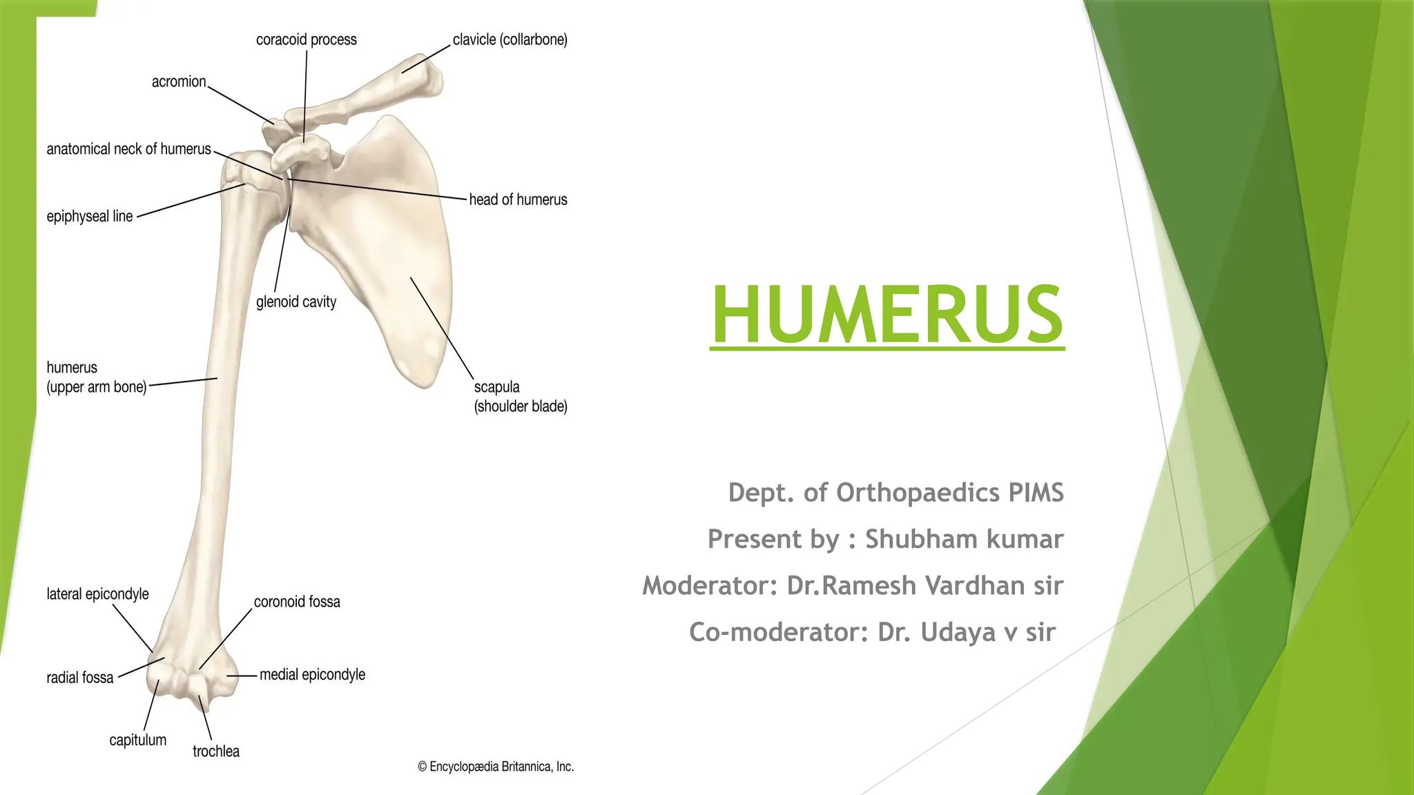
HUMERUS
Dept. of OrthopaedicsPIMS
Present by : Shubham kumar
Moderator: Dr.Ramesh Vardhan sir
Co-moderator: Dr. Udaya v sir
2.
The humerus isa long bone of the
upper arm. It is one of the longest
bones in the body, which makes it
more prone to fractures upon impact.
The word “humerus” comes from the
Latin word for upper arm.
3.
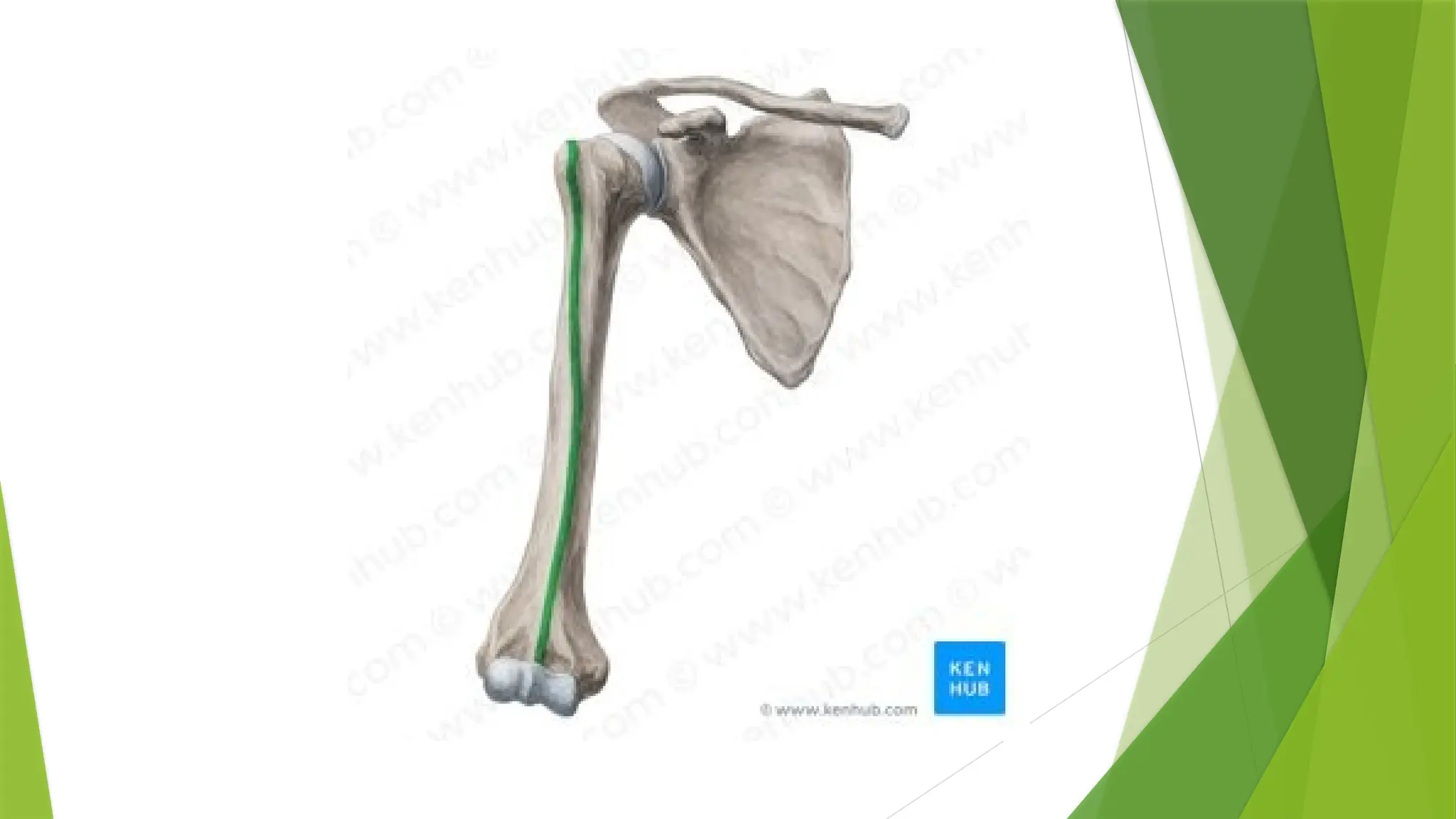
Where is thehumerus located?
The humerus bone is located in the upper arm, between
the shoulder joint and the elbow joint. The shoulder joint,
also known as the glenohumeral joint, is a ball and socket
joint. The ball is the humeral head, and the socket is the
glenoid fossa of the scapula. The joint is supported by
ligaments, and surrounded by the four rotator cuff
muscles and their tendons: the supraspinatus,
infraspinatus, teres minor, and subscapularis. These
muscles originate on the scapula and insert on the
humeral head.
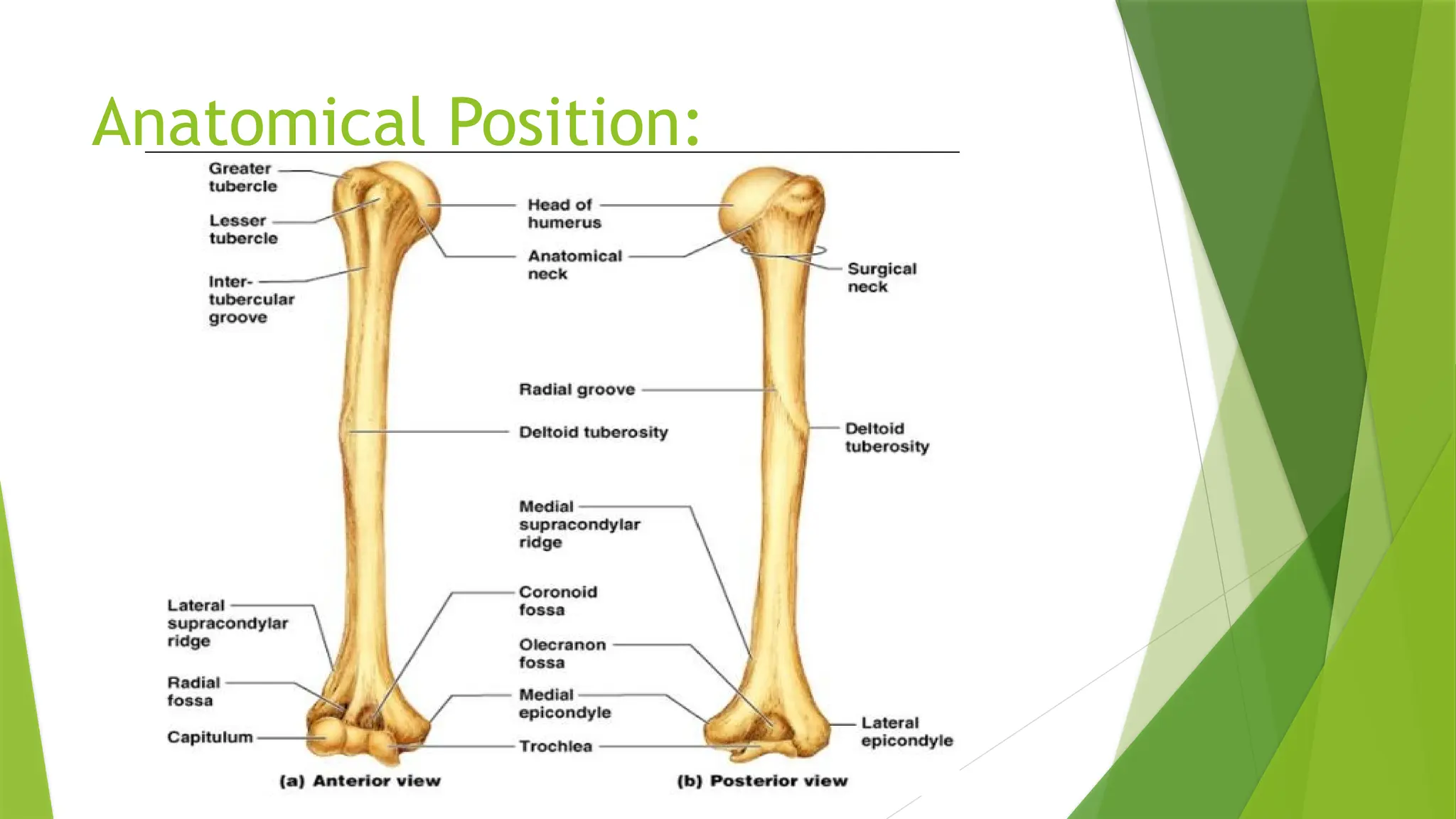
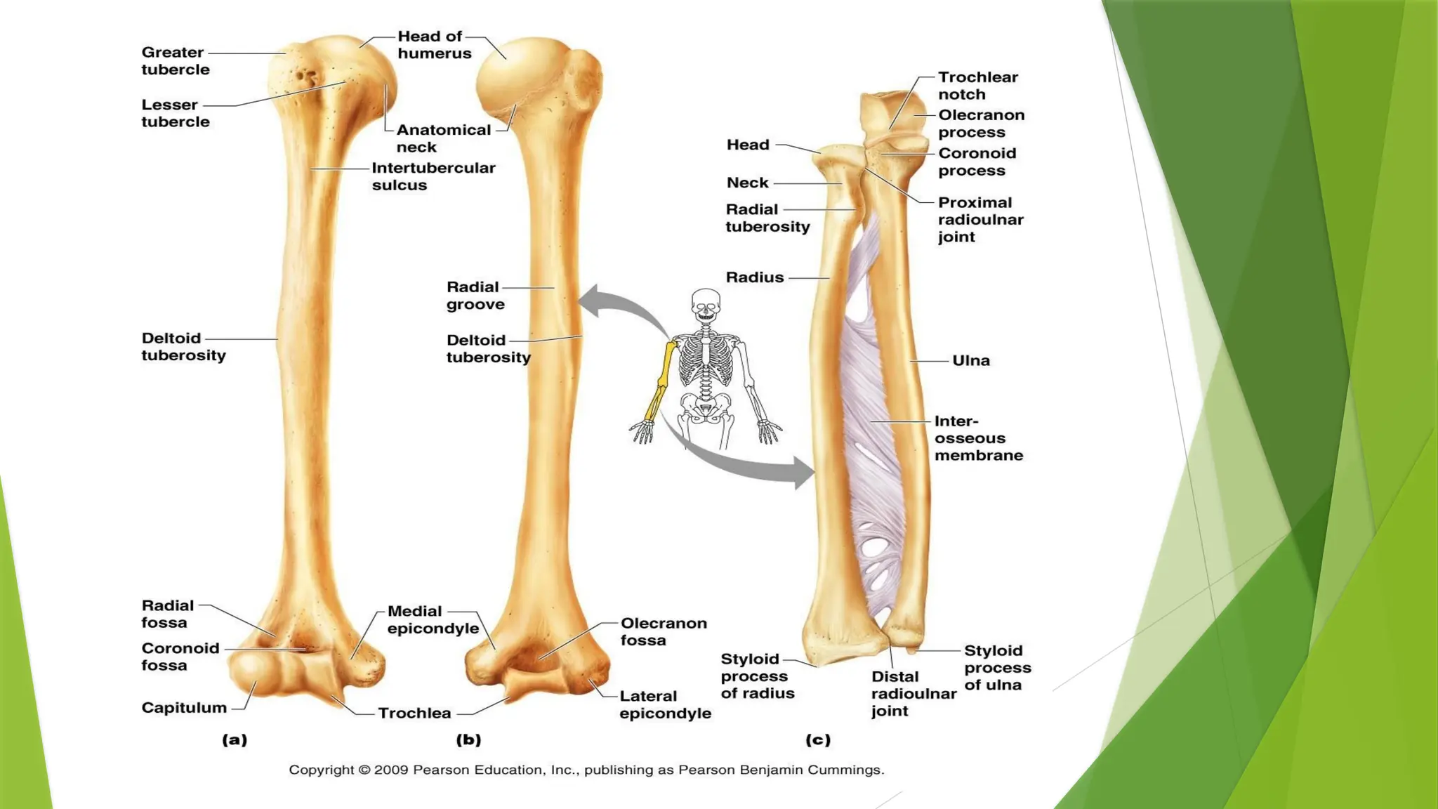
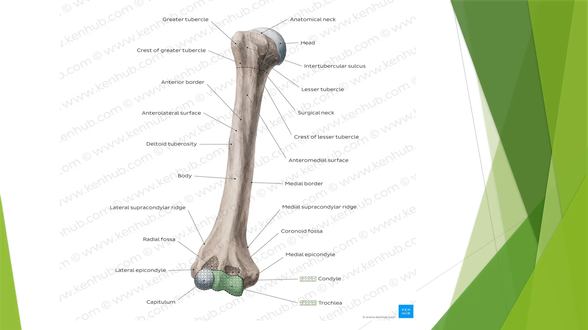
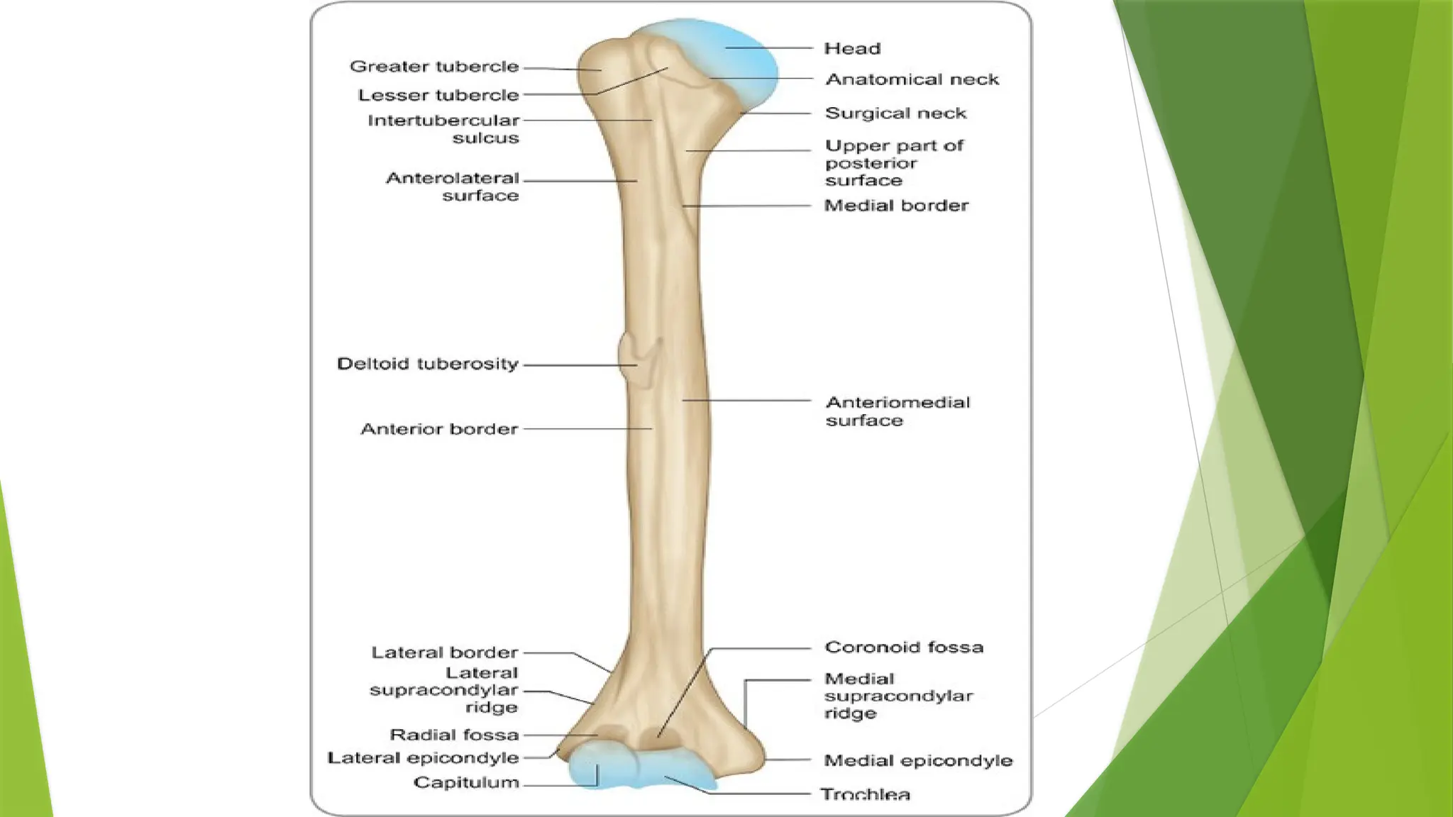
Parts of Humerus:
Proximal End: This is the upper part of
your humerus closest to your shoulder.
Body or shaft: This is the long, middle
portion of your humerus.
Distal End: This is the lower area of your
humerus that’s closest to your elbow.
6.
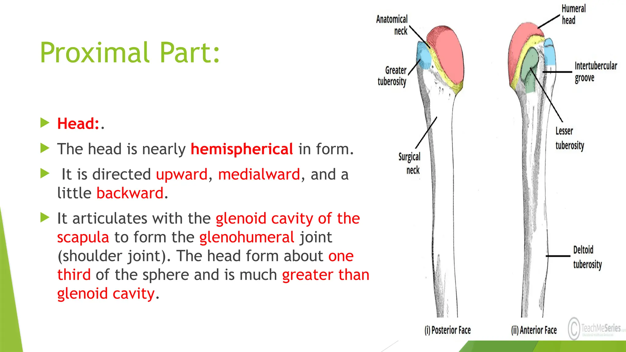
Proximal Part:
Head:.
The head is nearly hemispherical in form.
It is directed upward, medialward, and a
little backward.
It articulates with the glenoid cavity of the
scapula to form the glenohumeral joint
(shoulder joint). The head form about one
third of the sphere and is much greater than
glenoid cavity.
7.
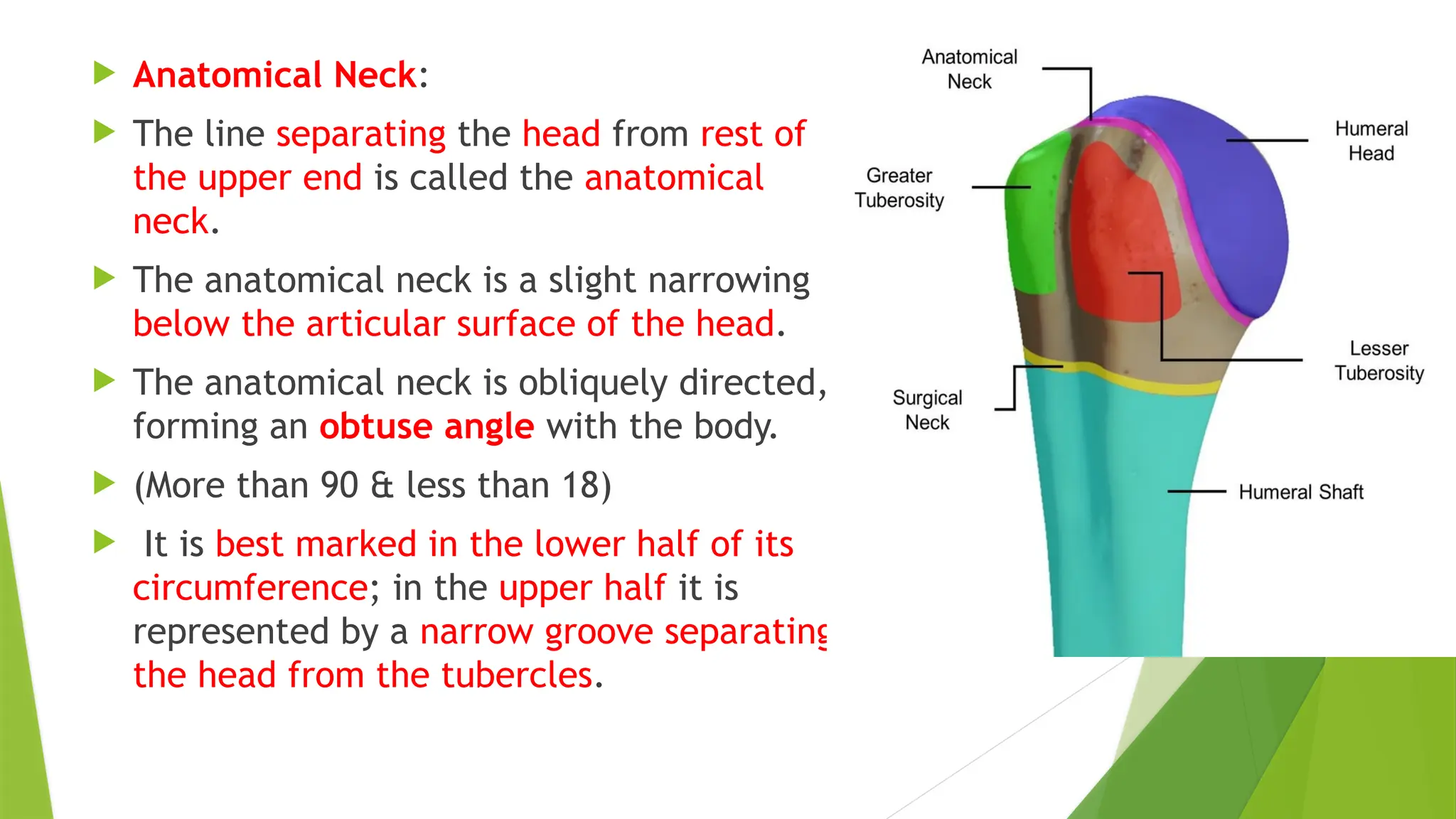
Anatomical Neck:
The line separating the head from rest of
the upper end is called the anatomical
neck.
The anatomical neck is a slight narrowing
below the articular surface of the head.
The anatomical neck is obliquely directed,
forming an obtuse angle with the body.
(More than 90 & less than 18)
It is best marked in the lower half of its
circumference; in the upper half it is
represented by a narrow groove separating
the head from the tubercles.
8.
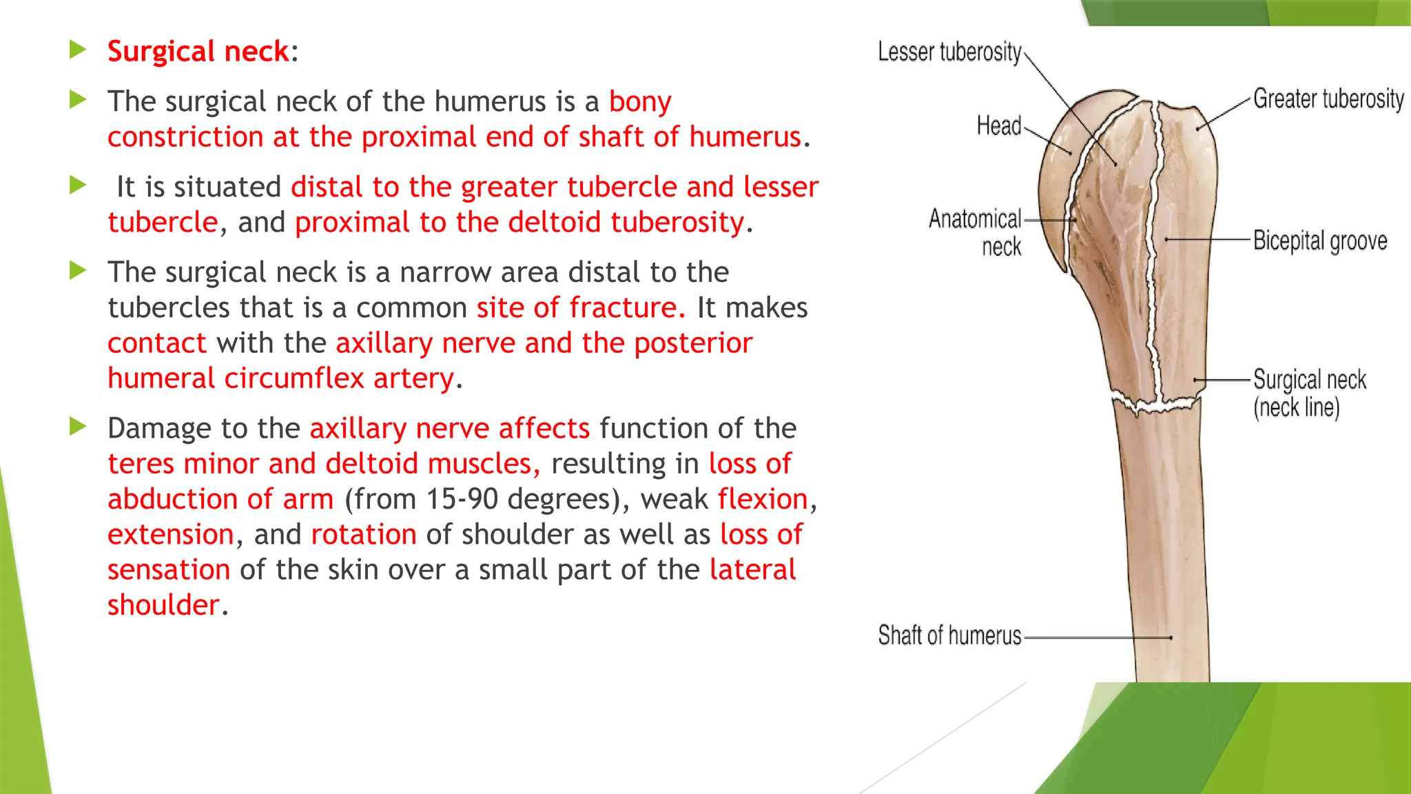
Surgical neck:
The surgical neck of the humerus is a bony
constriction at the proximal end of shaft of humerus.
It is situated distal to the greater tubercle and lesser
tubercle, and proximal to the deltoid tuberosity.
The surgical neck is a narrow area distal to the
tubercles that is a common site of fracture. It makes
contact with the axillary nerve and the posterior
humeral circumflex artery.
Damage to the axillary nerve affects function of the
teres minor and deltoid muscles, resulting in loss of
abduction of arm (from 15-90 degrees), weak flexion,
extension, and rotation of shoulder as well as loss of
sensation of the skin over a small part of the lateral
shoulder.
9.
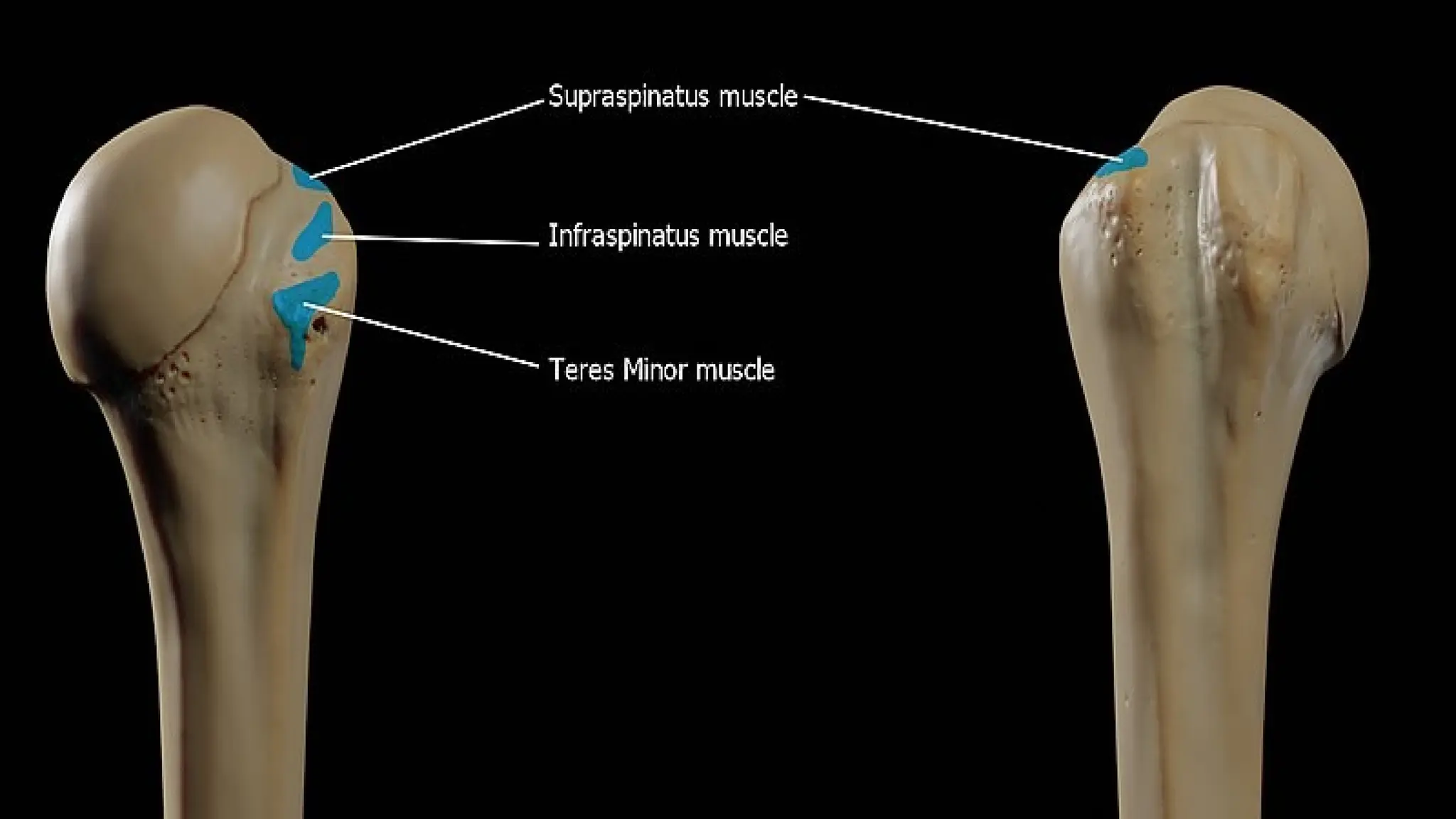
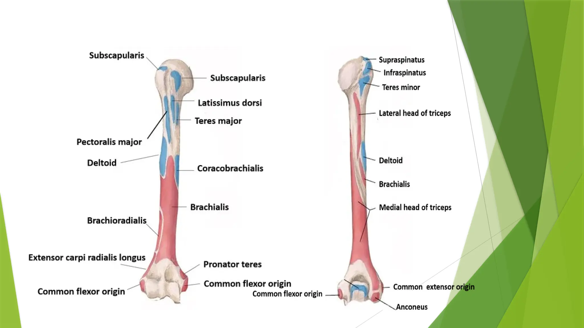
Greater tubercle:
The greater tubercle (greater tuberosity) is a large, posteriorly placed
projection that is placed laterally.
It provides attachment points for the supraspinatus, infraspinatus, and
teres minor muscles, three of the four muscles of the rotator cuff
(except subscapularis on lesser tubercle), a muscle group that stabilizes
the shoulder joint. (SIT)
The upper surface of the greater tubercle is rounded, and marked by
three flat impressions:
the highest ("superior facet") gives insertion to the supraspinatus
muscle.
the middle ("middle facet") gives insertion to the infraspinatus muscle.
the lowest ("inferior facet"), and the body of the bone for about 2.5 cm,
gives insertion to the teres minor muscle.
The lateral surface of the greater tubercle is convex, rough, and
continuous with the lateral surface of the body of the humerus. It can
be described as having a cranial and a caudal
11.
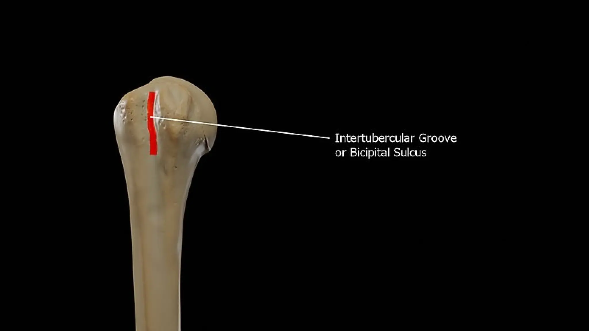
Between thegreater tubercle and the lesser
tubercle is the bicipital groove (intertubercular
sulcus).
12.
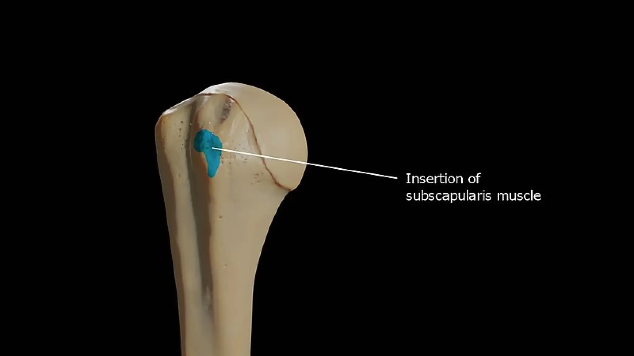
Lesser Tubercle:
The lesser tubercle of the humerus, although smaller, is more
prominent than the greater tubercle: it is situated in front, and is
directed medially and anteriorly.
The projection of the lesser tubercle is anterior from the junction
that is found between the anatomical neck and the shaft of the
humerus and easily identified due to the intertubercular sulcus
(Bicipital groove).
The crest of the lesser tubercle forms the medial lip of the bicipital
groove and is the site for insertion of teres major and latissimus dorsi
muscles.
Above and in front it presents an impression for the insertion of the
tendon of the subscapularis muscle.
14.
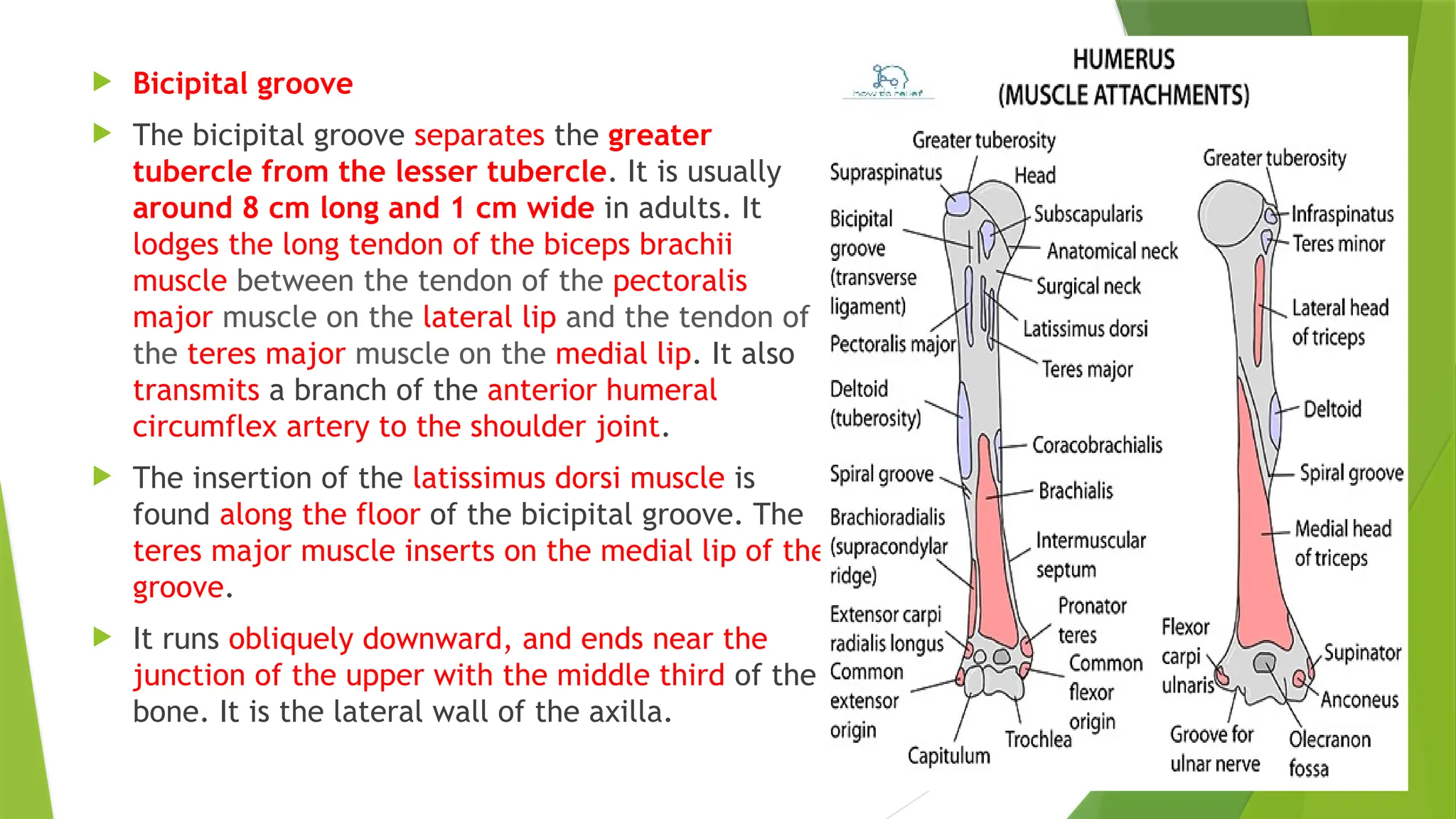
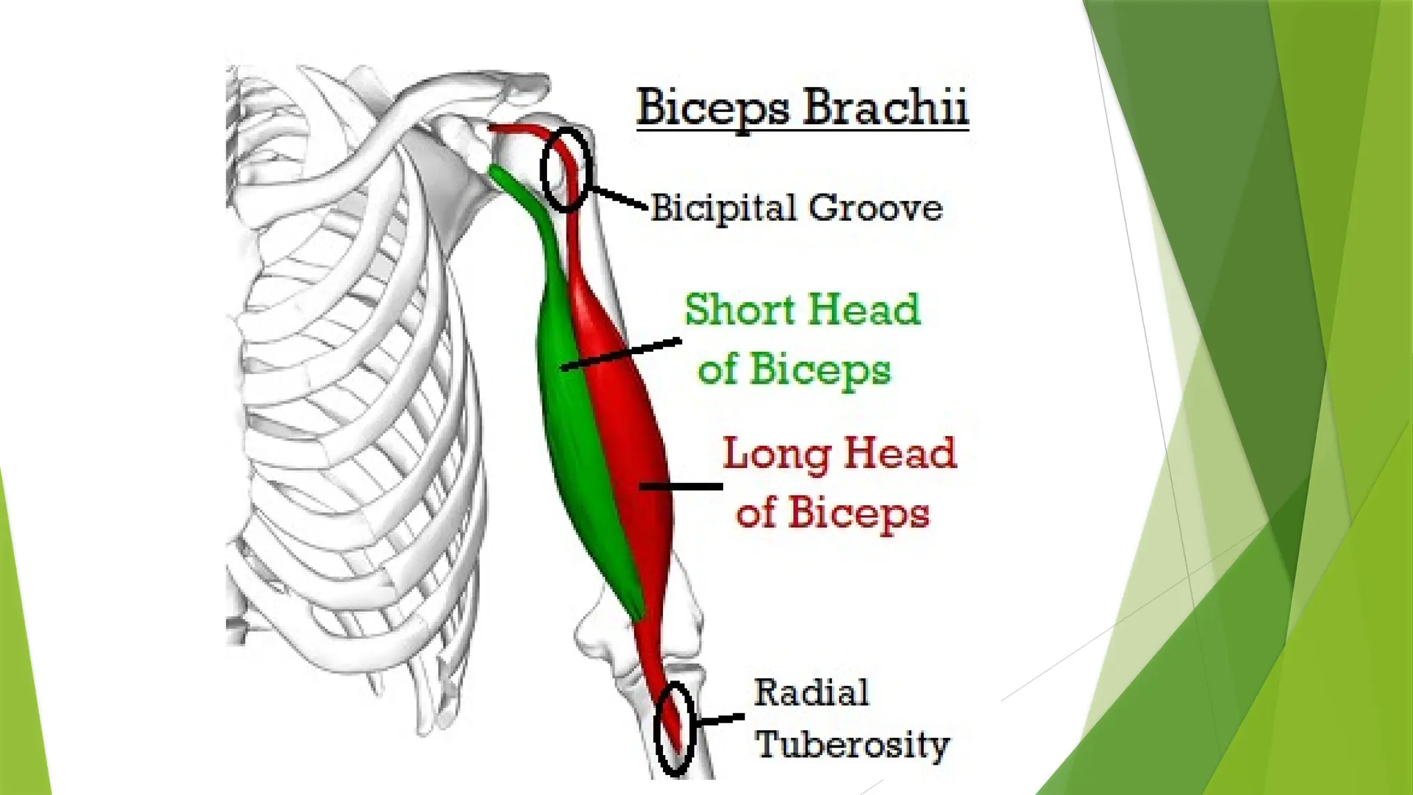
Bicipital groove
The bicipital groove separates the greater
tubercle from the lesser tubercle. It is usually
around 8 cm long and 1 cm wide in adults. It
lodges the long tendon of the biceps brachii
muscle between the tendon of the pectoralis
major muscle on the lateral lip and the tendon of
the teres major muscle on the medial lip. It also
transmits a branch of the anterior humeral
circumflex artery to the shoulder joint.
The insertion of the latissimus dorsi muscle is
found along the floor of the bicipital groove. The
teres major muscle inserts on the medial lip of the
groove.
It runs obliquely downward, and ends near the
junction of the upper with the middle third of the
bone. It is the lateral wall of the axilla.
17.
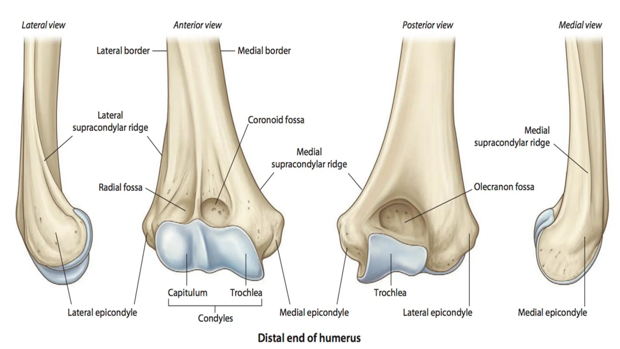
Distal humerus
Thedistal or lower extremity of the humerus
is flattened from before backward, and
curved slightly forward; it ends below in a
broad, articular surface, which is divided into
two parts by a slight ridge. Projecting on
either side are the lateral and medial
epicondyles.
19.
Articular surface
The articular surface extends a little lower than the
epicondyles, and is curved slightly forward; its medial
extremity occupies a lower level than the lateral.
The lateral portion of this surface consists of a smooth,
rounded eminence, named the capitulum little head of
the humerus; it articulates with the cup-shaped
depression on the head of the radius, and is limited to the
front and lower part of the bone.
The trochlea (pulley) is a pulley shaped surface. It
articulates with the trochlear notch of the ulna. The
medial edge of the trochlea projects down 6 mm more
than the lateral edge. This results in the formation of
carrying angle .
21.
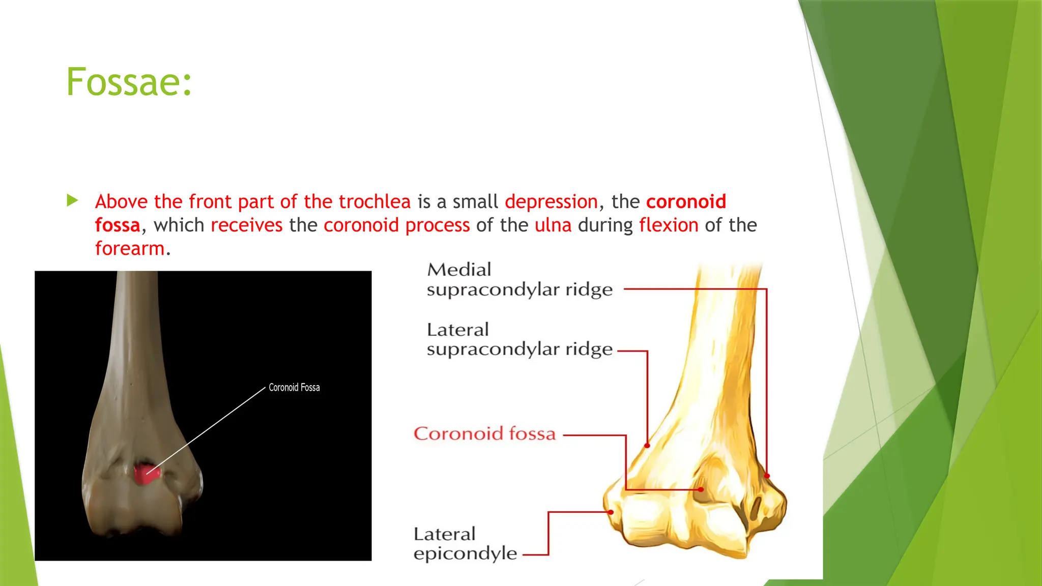
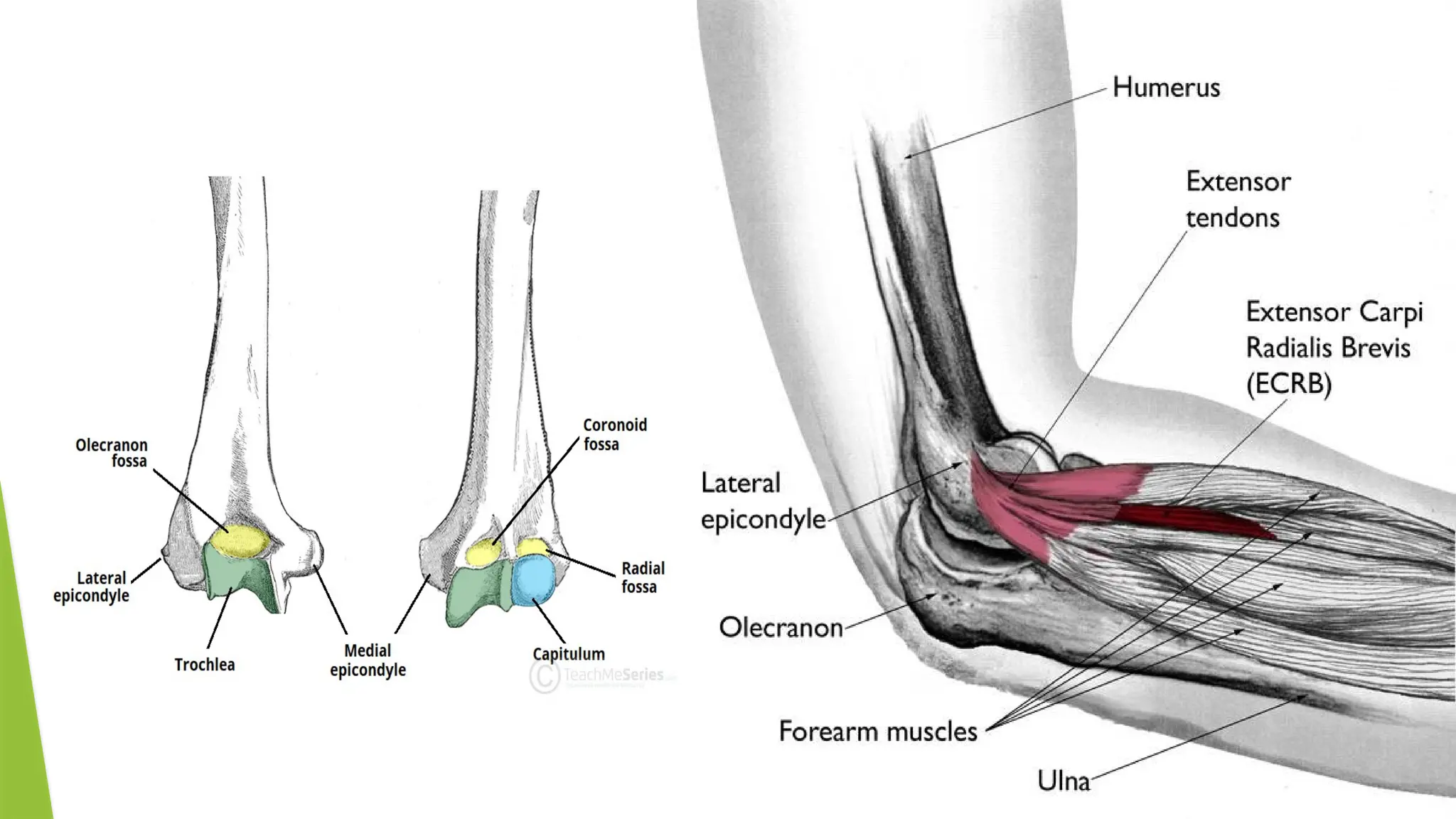
Fossae:
Above thefront part of the trochlea is a small depression, the coronoid
fossa, which receives the coronoid process of the ulna during flexion of the
forearm.
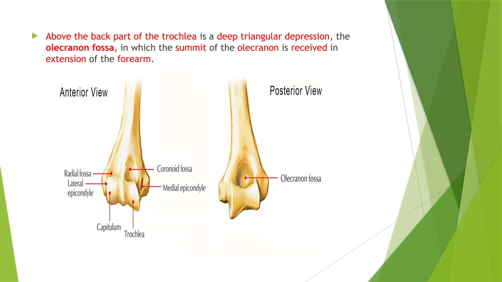
22.
Above theback part of the trochlea is a deep triangular depression, the
olecranon fossa, in which the summit of the olecranon is received in
extension of the forearm.
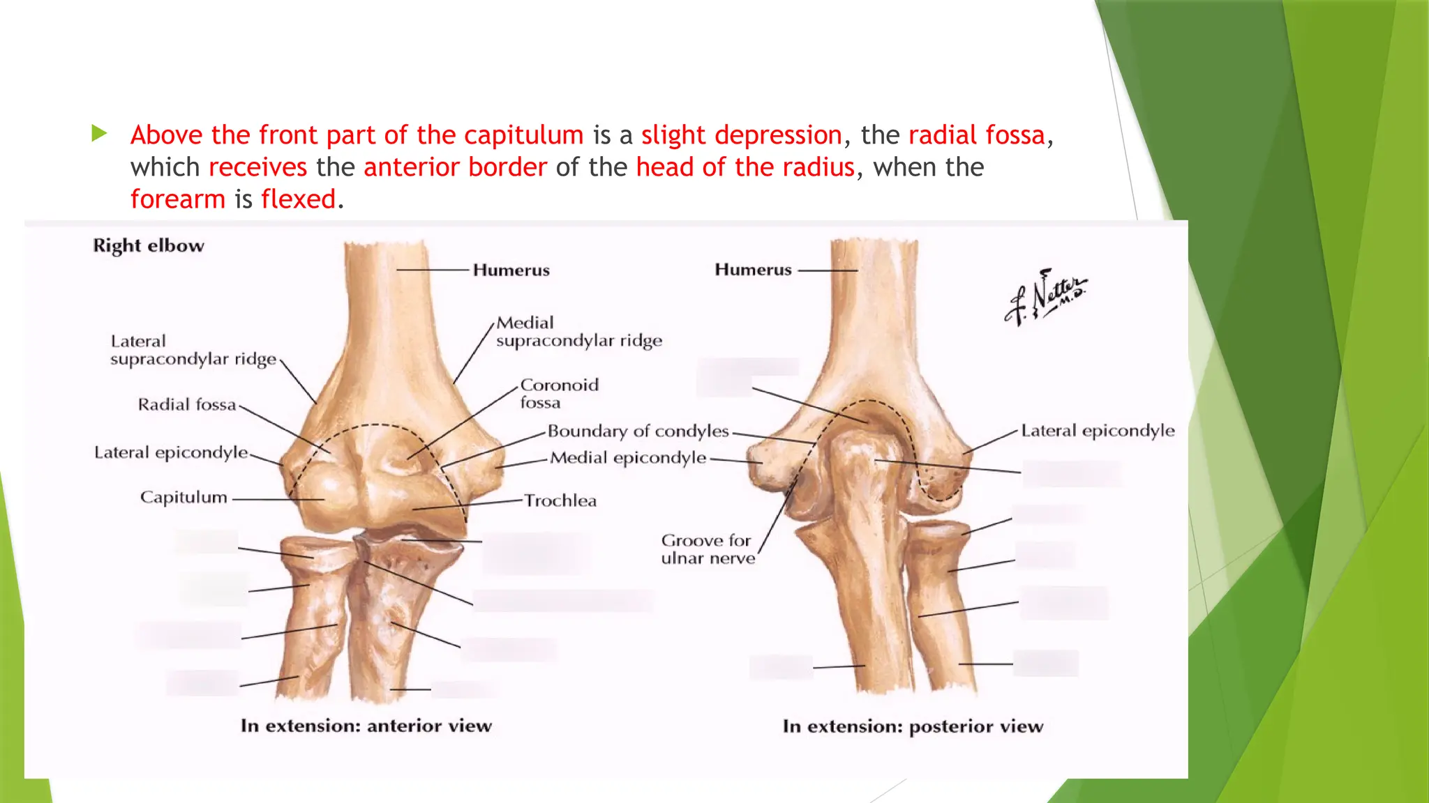
23.
Above thefront part of the capitulum is a slight depression, the radial fossa,
which receives the anterior border of the head of the radius, when the
forearm is flexed.
24.
Epicondyles
The epicondylesare continuous above with the supracondylar ridges.
The lateral epicondyle of the humerus is a large, tuberculated
eminence, curved a little forward, and giving attachment to the
radial collateral ligament of the elbow joint, and to a tendon common
to the origin of the supinator and some of the extensor muscles.
Specifically, these extensor muscles include the anconeus muscle, the
supinator, extensor carpi radialis brevis, extensor digitorum, extensor
digiti minimi, and extensor carpi ulnaris. ( SAD hu because of EX so
DUR raho)
26.
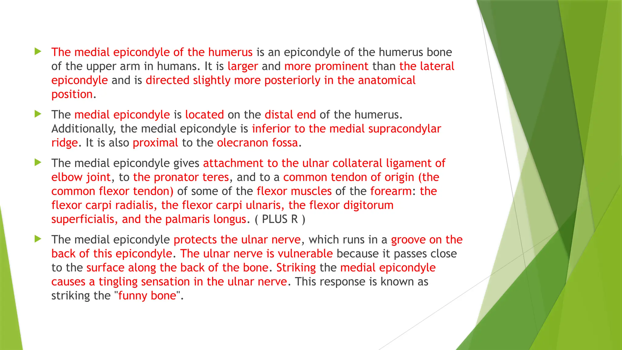
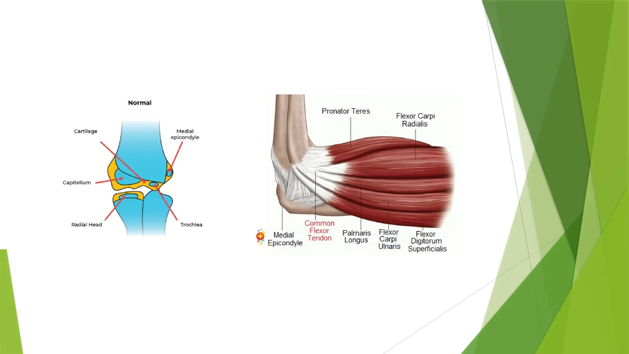
The medialepicondyle of the humerus is an epicondyle of the humerus bone
of the upper arm in humans. It is larger and more prominent than the lateral
epicondyle and is directed slightly more posteriorly in the anatomical
position.
The medial epicondyle is located on the distal end of the humerus.
Additionally, the medial epicondyle is inferior to the medial supracondylar
ridge. It is also proximal to the olecranon fossa.
The medial epicondyle gives attachment to the ulnar collateral ligament of
elbow joint, to the pronator teres, and to a common tendon of origin (the
common flexor tendon) of some of the flexor muscles of the forearm: the
flexor carpi radialis, the flexor carpi ulnaris, the flexor digitorum
superficialis, and the palmaris longus. ( PLUS R )
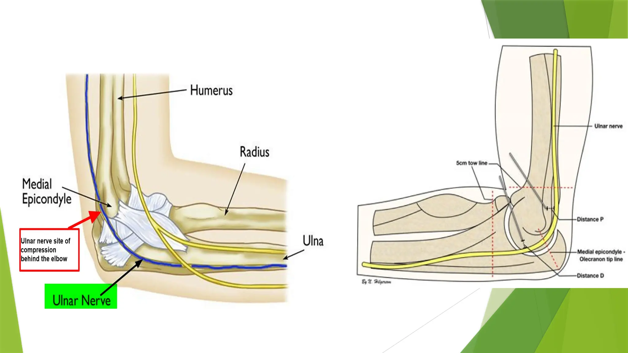
The medial epicondyle protects the ulnar nerve, which runs in a groove on the
back of this epicondyle. The ulnar nerve is vulnerable because it passes close
to the surface along the back of the bone. Striking the medial epicondyle
causes a tingling sensation in the ulnar nerve. This response is known as
striking the "funny bone".
29.
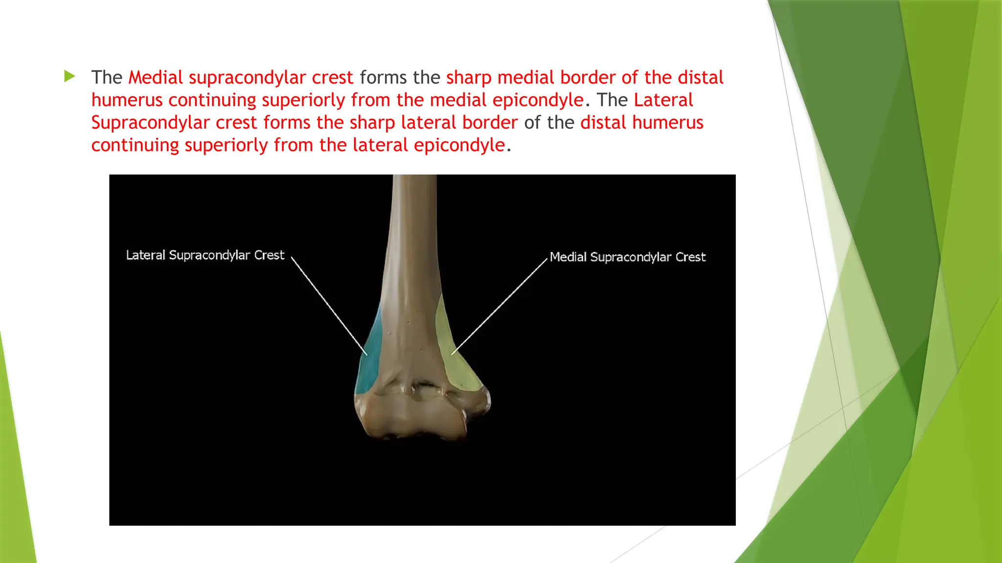
The Medialsupracondylar crest forms the sharp medial border of the distal
humerus continuing superiorly from the medial epicondyle. The Lateral
Supracondylar crest forms the sharp lateral border of the distal humerus
continuing superiorly from the lateral epicondyle.
30.
Shaft or Body:
Borders:
Its three borders are:
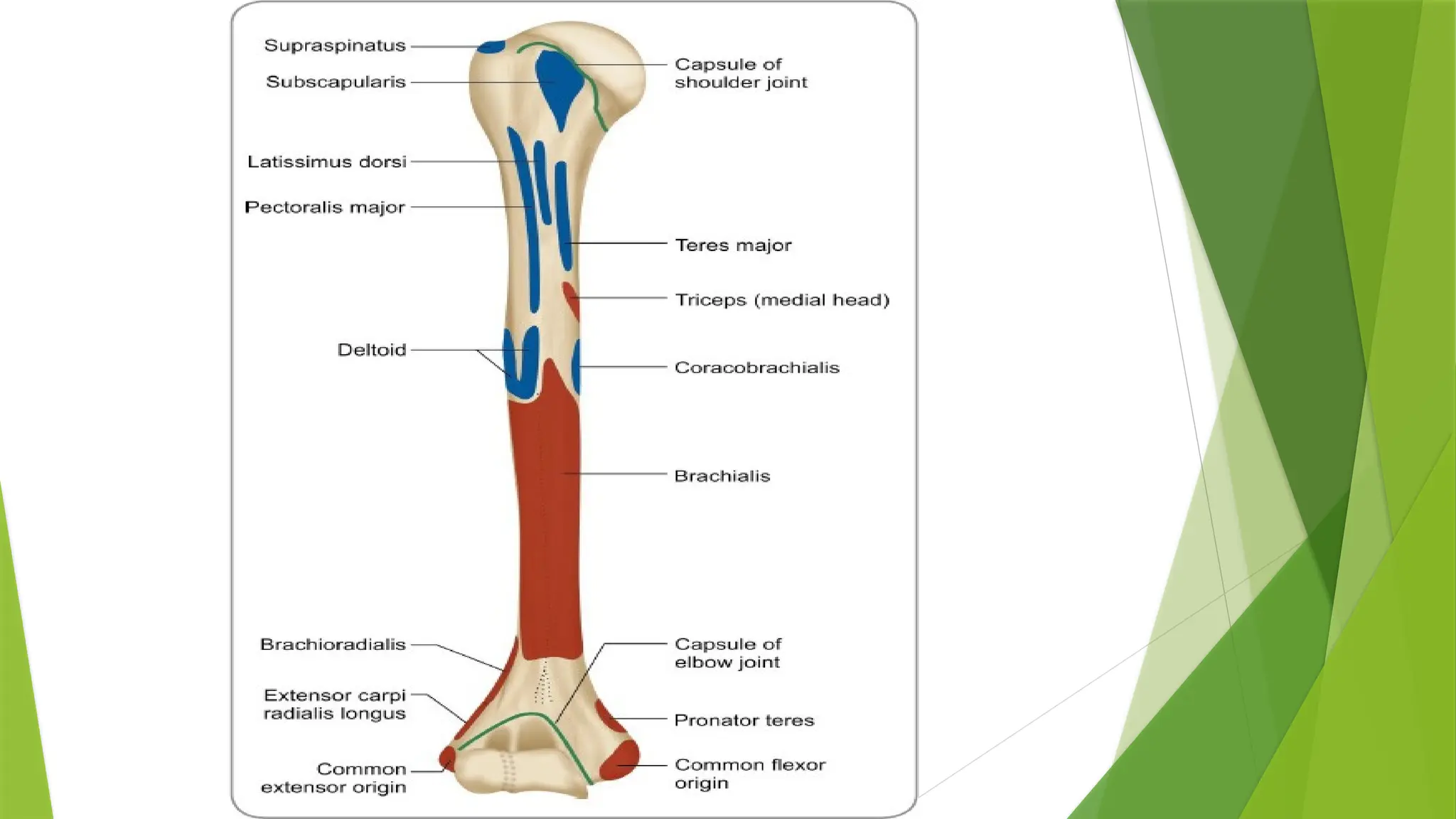
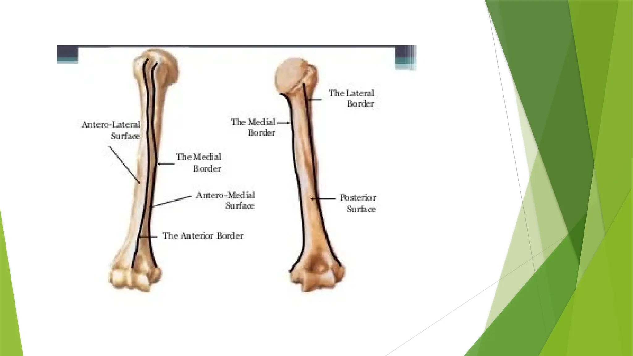
Anterior: the anterior border runs from the front of the greater
tubercle above to the coronoid fossa below, separating the antero-
medial from the antero-lateral surface. Its upper part is a prominent
ridge, the crest of the greater tubercle; it serves for the insertion of
the tendon of the pectoralis major muscle. About its center it forms
the anterior boundary of the deltoid tuberosity, on which the deltoid
muscle attaches; below, it is smooth and rounded, affording
attachment to the brachialis muscle.
34.
Lateral: thelateral border runs from the back part of the greater
tubercle to the lateral epicondyle, and separates the anterolateral
from the posterior surface. Its upper half is rounded and indistinctly
marked, serving for the attachment of the lower part of the insertion
of the teres minor muscle, and below this giving origin to the lateral
head of the triceps brachii muscle; its center is traversed by a broad
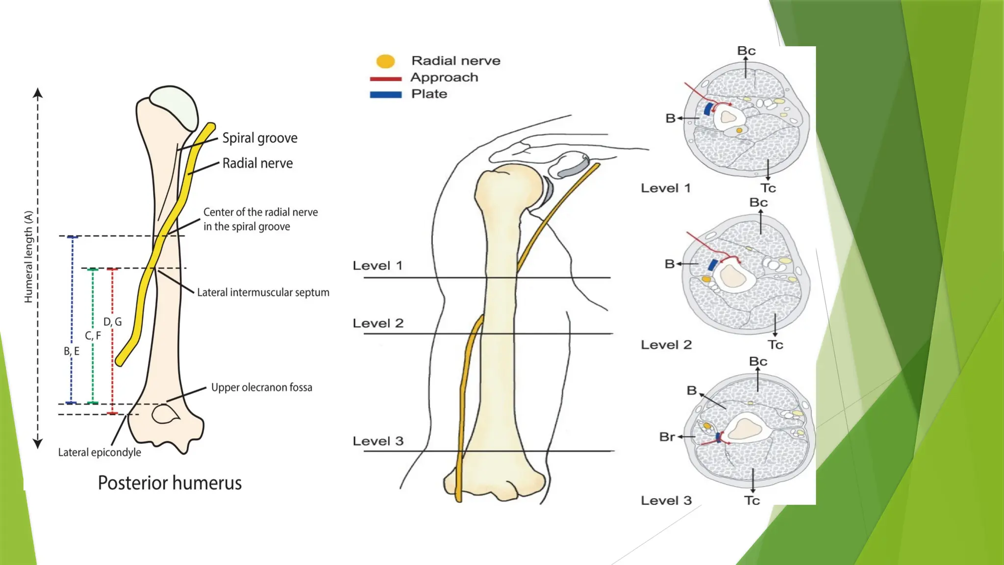
but shallow oblique depression, the spiral groove (musculospiral
groove). The radial nerve runs in the spiral groove. Its lower part
forms a prominent, rough margin, a little curved from backward,
forward the lateral supracondylar ridge, which presents an anterior
lip for the origin of the brachioradialis muscle above, and extensor
carpi radialis longus muscle above, a posterior lip for the triceps
brachii muscle, and an intermediate ridge for the attachment of the
lateral intermuscular septum.
39.
Medial:
themedial border extends from the lesser tubercle to the medial
epicondyle. Its upper third consists of a prominent ridge, the crest of
the lesser tubercle, which gives insertion to the tendon of the teres
major muscle. About its center is a slight impression for the insertion
of the coracobrachialis muscle, and just below this is the entrance of
the nutrient canal, directed downward; sometimes there is a second
nutrient canal at the commencement of the radial sulcus. The inferior
third of this border is raised into a slight ridge, the medial
supracondylar ridge, which became very prominent below; it presents
an anterior lip for the origins of the brachialis muscle and the
pronator teres muscle, a posterior lip for the medial head of the
triceps brachii muscle, and an intermediate ridge for the attachment
of the medial intermuscular septum.
41.
The bodyor shaft of the humerus is triangular to cylindrical in cut
section and is compressed anteroposterior. It has 3 surfaces, namely:
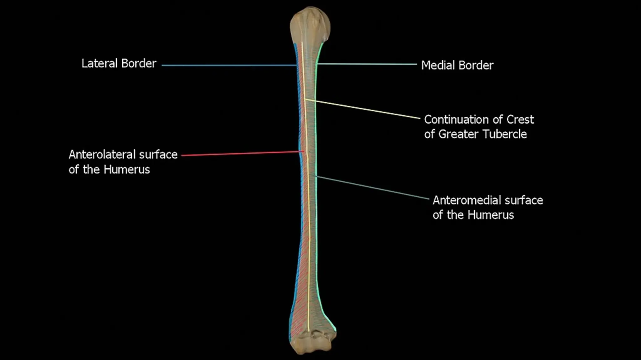
Anterolateral surface:
the area between the lateral border of the humerus to the line drawn
as a continuation of the crest of the greater tubercle.
The antero-lateral surface is directed lateralward above, where it is
smooth, rounded, and covered by the deltoid muscle; forward and
lateralward below, where it is slightly concave from above downward,
and gives origin to part of the Brachialis.
About the middle of this surface is a rough, rectangular elevation, the
deltoid tuberosity for the insertion of the deltoid muscle; below this
is the radial sulcus, directed obliquely from behind, forward, and
downward, and transmitting the radial nerve and profunda artery.
43.
Anteromedial surface:
the area between the medial border of the humerus to the
line drawn as a continuation of the crest of the greater
tubercle. The antero-medial surface, less extensive than
the antero-lateral, is directed medialward above, forward
and medialward below; its upper part is narrow, and
forms the floor of the intertubercular groove which gives
insertion to the tendon of the latissimus dorsi muscle; its
middle part is slightly rough for the attachment of some of
the fibers of the tendon of insertion of the
coracobrachialis muscle; its lower part is smooth, concave
from above downward, and gives origin to the brachialis
muscle.
46.
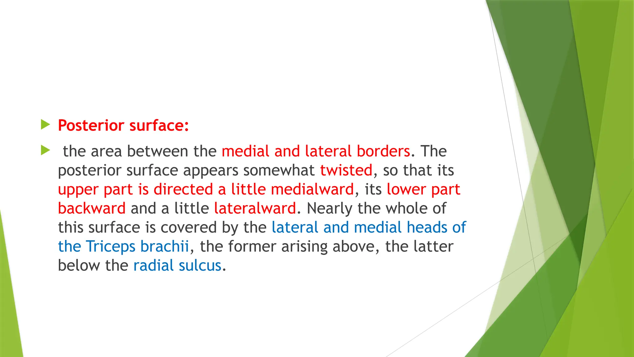
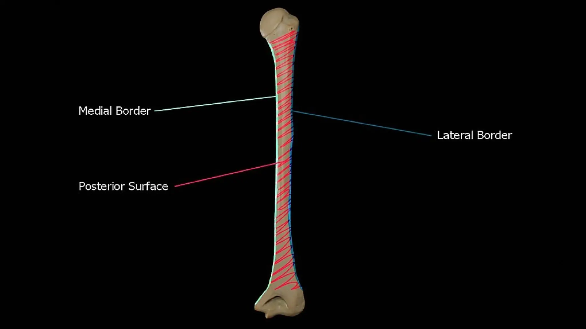
Posterior surface:
the area between the medial and lateral borders. The
posterior surface appears somewhat twisted, so that its
upper part is directed a little medialward, its lower part
backward and a little lateralward. Nearly the whole of
this surface is covered by the lateral and medial heads of
the Triceps brachii, the former arising above, the latter
below the radial sulcus.
48.
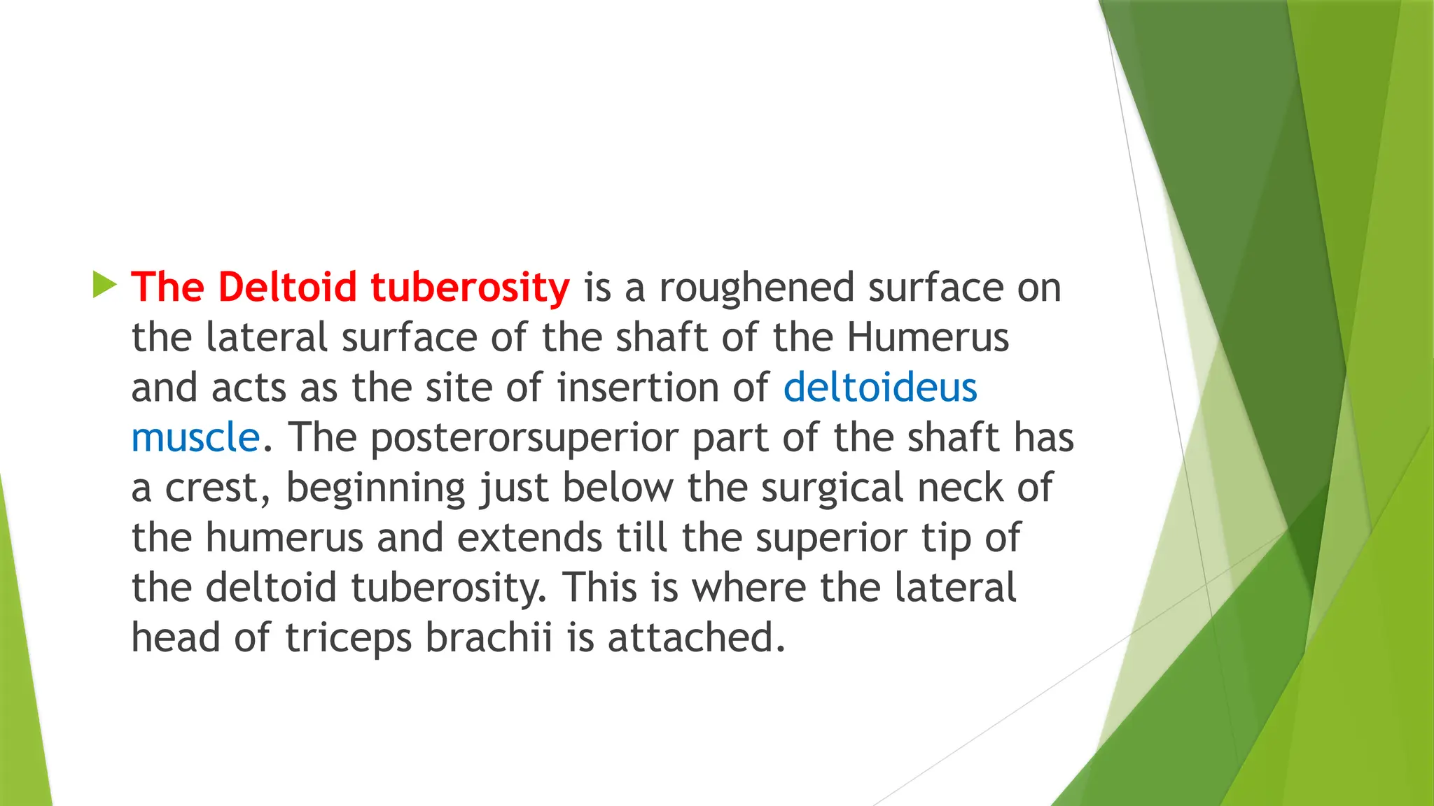
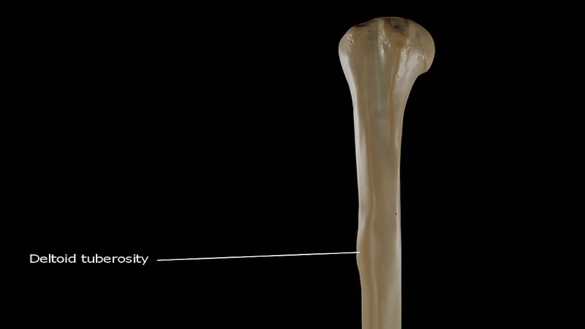
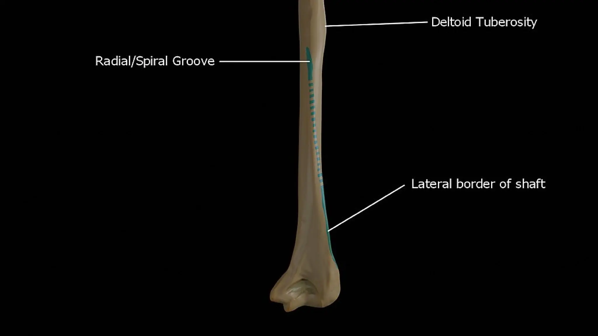
The Deltoidtuberosity is a roughened surface on
the lateral surface of the shaft of the Humerus
and acts as the site of insertion of deltoideus
muscle. The posterorsuperior part of the shaft has
a crest, beginning just below the surgical neck of
the humerus and extends till the superior tip of
the deltoid tuberosity. This is where the lateral
head of triceps brachii is attached.
50.
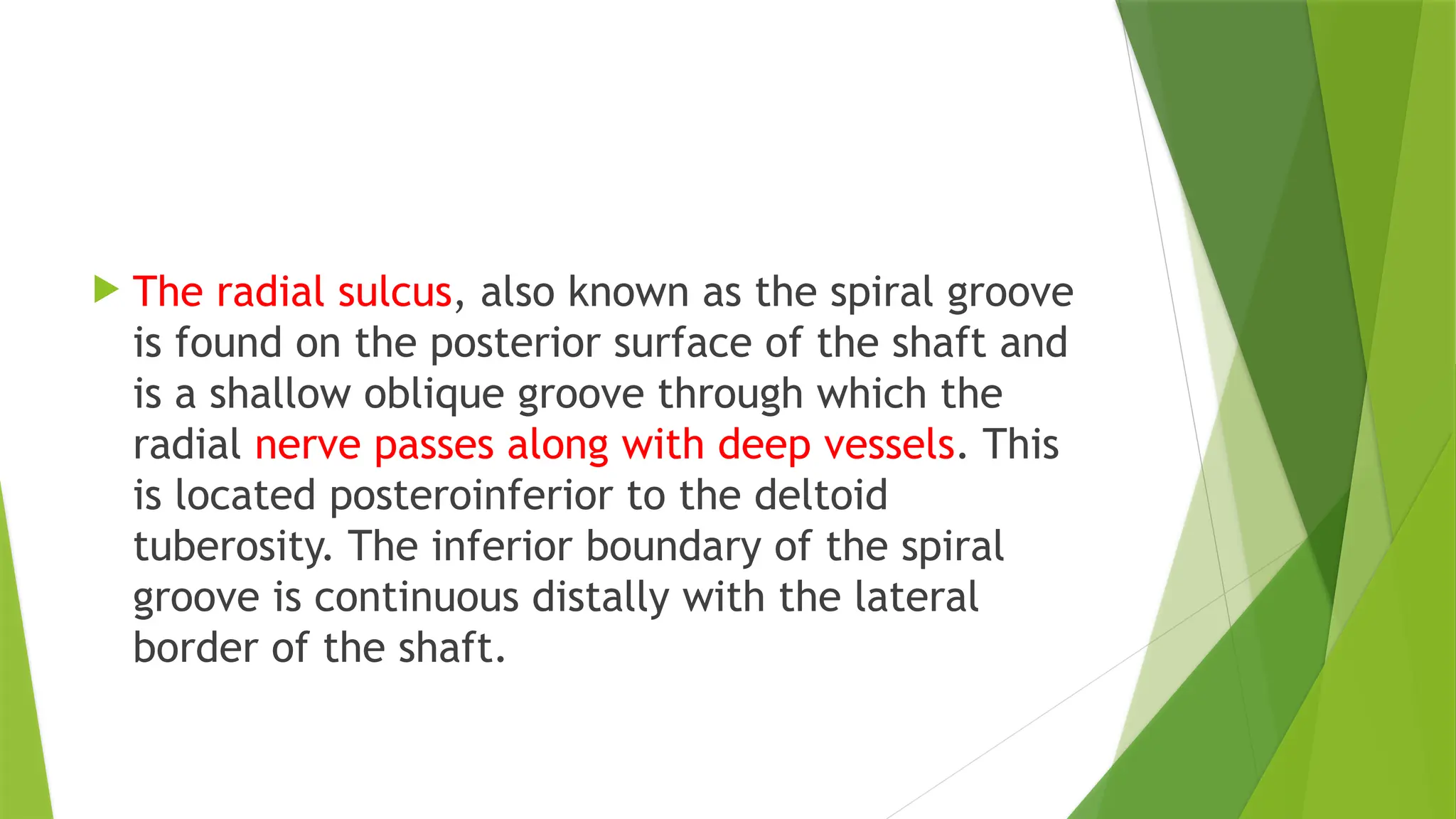
The radialsulcus, also known as the spiral groove
is found on the posterior surface of the shaft and
is a shallow oblique groove through which the
radial nerve passes along with deep vessels. This
is located posteroinferior to the deltoid
tuberosity. The inferior boundary of the spiral
groove is continuous distally with the lateral
border of the shaft.
52.
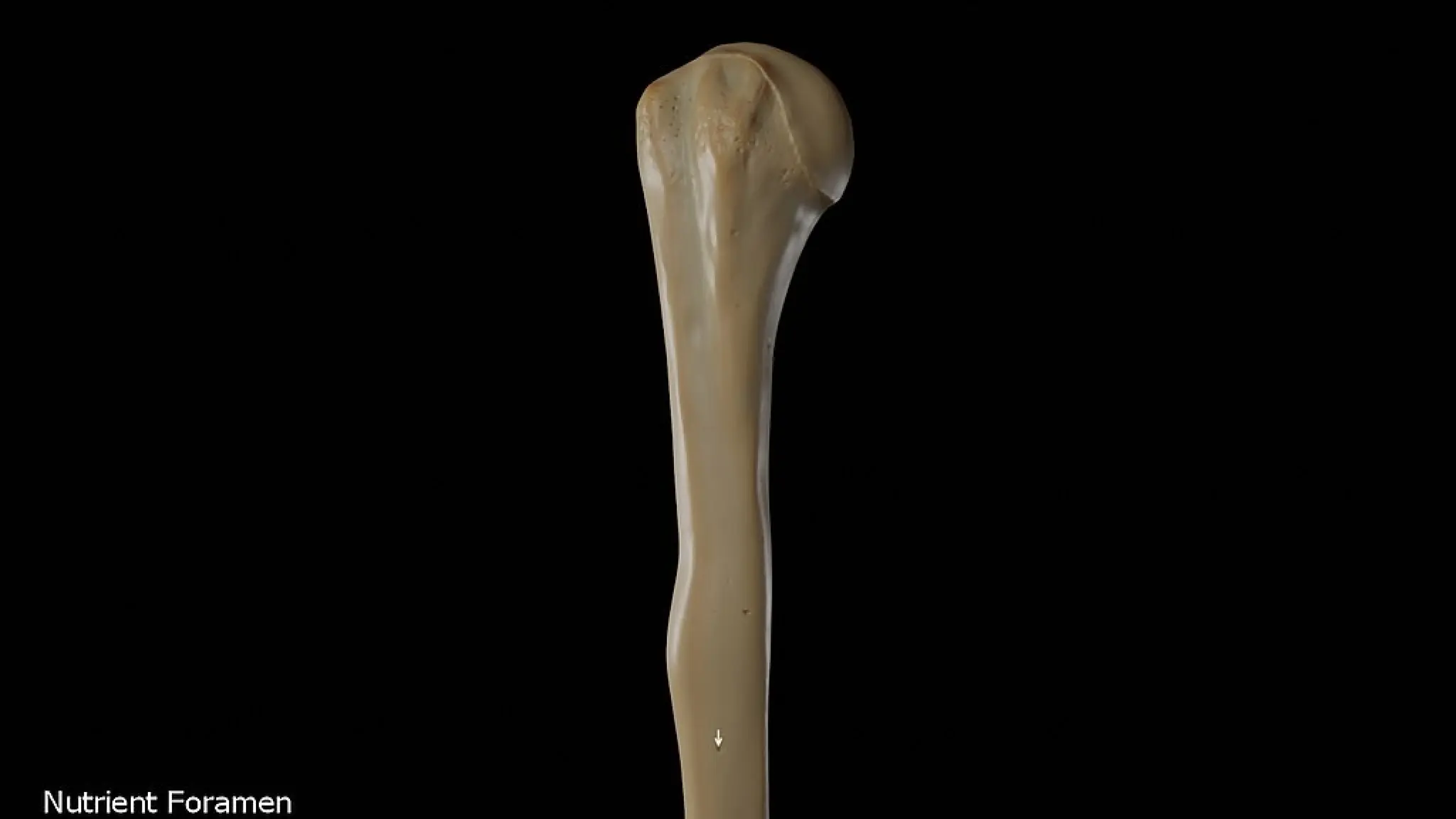
The nutrientforamen of the humerus is
located in the anteromedial surface of the
humerus. The nutrient arteries enter the
humerus through this foramen.