Download free for 30 days
Sign in
Upload
Language (EN)
Support
Business
Mobile
Social Media
Marketing
Technology
Art & Photos
Career
Design
Education
Presentations & Public Speaking
Government & Nonprofit
Healthcare
Internet
Law
Leadership & Management
Automotive
Engineering
Software
Recruiting & HR
Retail
Sales
Services
Science
Small Business & Entrepreneurship
Food
Environment
Economy & Finance
Data & Analytics
Investor Relations
Sports
Spiritual
News & Politics
Travel
Self Improvement
Real Estate
Entertainment & Humor
Health & Medicine
Devices & Hardware
Lifestyle
Change Language
Language
English
Español
Português
Français
Deutsche
Cancel
Save
EN
Uploaded by
Maqbool Salafi
4,412 views
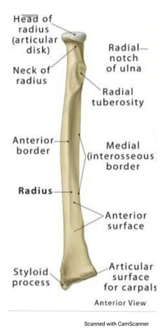
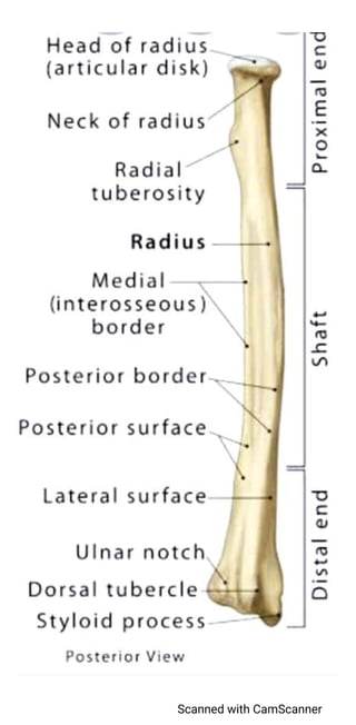
Radius bone structure
Lateral forearm bone
Education
◦
Read more
4
Save
Share
Embed
Embed presentation
Download
Downloaded 12 times
1
/ 7
2
/ 7
Most read
3
/ 7
Most read
4
/ 7
Most read
5
/ 7
6
/ 7
7
/ 7
More Related Content
PPTX
Tibia and Fibula p2.pptx . . . . . . . . .
by
ABIDOFFICIALCHANNEL
PDF
Ulna bone structure
by
Maqbool Salafi
PPTX
Lec 5 tibia bone
by
Eimaan Ktk
PPT
Slideshow: Gluteal Region
by
The Funky Professor
PPTX
The scapula
by
Kuldeep Jingar
PPT
Slideshow: Radius
by
The Funky Professor
PPTX
Purvi shah anatomy of fibula ppt
by
Purvi Shah
PPTX
osteology of clavicle
by
Kd Sudarsan
Tibia and Fibula p2.pptx . . . . . . . . .
by
ABIDOFFICIALCHANNEL
Ulna bone structure
by
Maqbool Salafi
Lec 5 tibia bone
by
Eimaan Ktk
Slideshow: Gluteal Region
by
The Funky Professor
The scapula
by
Kuldeep Jingar
Slideshow: Radius
by
The Funky Professor
Purvi shah anatomy of fibula ppt
by
Purvi Shah
osteology of clavicle
by
Kd Sudarsan
What's hot
PPTX
Purvi shah carpal bone anatomy ppt
by
Purvi Shah
PPTX
Radius bone anatomy
by
Shalu Thariwal
PPT
Slideshow: Scapula
by
The Funky Professor
PPT
Slideshow: Clavicle
by
The Funky Professor
PPTX
Tibia bone.pptx
by
Dr. Priyanka Poonia
PPTX
Humerus Bone and attachments
by
Dr Musadiq
PPTX
Fibula
by
Dr. Vibhash Kumar Vaidya
PPTX
Ulna Bone Anatomy
by
Dr Adnan Sami
PPTX
Bronchial tree
by
Medical Students
PPTX
ANATOMICAL SNUFF BOX.pptx
by
Mayowa19
PPT
Slideshow: Ulna
by
The Funky Professor
PPTX
Femur (Gross Anatomy)
by
AtifRaza11
PPTX
Patella
by
BipulBorthakur
PPT
Slideshow: Humerus
by
The Funky Professor
PPTX
Humerus bone osteology: Upper limb Anatomy
by
Priyanka Pundir
PPTX
Cubital fossa
by
Lucidante1
PPTX
Axillary artey ppt
by
Dr.Mayur Sayta
PPT
Anterior abdominal muscles
by
Dr. Armaan Singh
PDF
Vertebral Column
by
Sanjiv Haribhakti
PPTX
Radius and ulna.pptx
by
MahekPande
Purvi shah carpal bone anatomy ppt
by
Purvi Shah
Radius bone anatomy
by
Shalu Thariwal
Slideshow: Scapula
by
The Funky Professor
Slideshow: Clavicle
by
The Funky Professor
Tibia bone.pptx
by
Dr. Priyanka Poonia
Humerus Bone and attachments
by
Dr Musadiq
Fibula
by
Dr. Vibhash Kumar Vaidya
Ulna Bone Anatomy
by
Dr Adnan Sami
Bronchial tree
by
Medical Students
ANATOMICAL SNUFF BOX.pptx
by
Mayowa19
Slideshow: Ulna
by
The Funky Professor
Femur (Gross Anatomy)
by
AtifRaza11
Patella
by
BipulBorthakur
Slideshow: Humerus
by
The Funky Professor
Humerus bone osteology: Upper limb Anatomy
by
Priyanka Pundir
Cubital fossa
by
Lucidante1
Axillary artey ppt
by
Dr.Mayur Sayta
Anterior abdominal muscles
by
Dr. Armaan Singh
Vertebral Column
by
Sanjiv Haribhakti
Radius and ulna.pptx
by
MahekPande
More from Maqbool Salafi
PDF
Hip bone
by
Maqbool Salafi
PDF
Ankle joint
by
Maqbool Salafi
PDF
Knee joint
by
Maqbool Salafi
PDF
Hip joint
by
Maqbool Salafi
PDF
Elbow joint
by
Maqbool Salafi
PDF
Wrist joint
by
Maqbool Salafi
PDF
Shoulder joint
by
Maqbool Salafi
PDF
Joints and their types
by
Maqbool Salafi
PDF
Fibula
by
Maqbool Salafi
PDF
Tibia structure
by
Maqbool Salafi
PDF
Femur
by
Maqbool Salafi
PDF
Diagram scapula
by
Maqbool Salafi
PDF
Scapula
by
Maqbool Salafi
PDF
Clavicle(2)
by
Maqbool Salafi
PDF
Typical vertebra
by
Maqbool Salafi
PDF
Axial skeleton
by
Maqbool Salafi
PDF
Bone classification
by
Maqbool Salafi
PDF
Skeletal muscular system
by
Maqbool Salafi
PDF
Nervous tissue
by
Maqbool Salafi
PDF
Muscle tissue slides
by
Maqbool Salafi
Hip bone
by
Maqbool Salafi
Ankle joint
by
Maqbool Salafi
Knee joint
by
Maqbool Salafi
Hip joint
by
Maqbool Salafi
Elbow joint
by
Maqbool Salafi
Wrist joint
by
Maqbool Salafi
Shoulder joint
by
Maqbool Salafi
Joints and their types
by
Maqbool Salafi
Fibula
by
Maqbool Salafi
Tibia structure
by
Maqbool Salafi
Femur
by
Maqbool Salafi
Diagram scapula
by
Maqbool Salafi
Scapula
by
Maqbool Salafi
Clavicle(2)
by
Maqbool Salafi
Typical vertebra
by
Maqbool Salafi
Axial skeleton
by
Maqbool Salafi
Bone classification
by
Maqbool Salafi
Skeletal muscular system
by
Maqbool Salafi
Nervous tissue
by
Maqbool Salafi
Muscle tissue slides
by
Maqbool Salafi
Recently uploaded
PPTX
Types of counselling Directive, Non Directive, Eclectic Counselling
by
Priya Sush
PPTX
S.Y. B. Pharm Medicinal Chemistry I Unit-I
by
Ms. Ashatai Patil
PDF
BP801T BIOSTATISITCS AND RESEARCH METHODOLOGY (Theory) Unit 2 Part 1
by
Rushi Mandali
PDF
Art, Memory, and Modernity: A Study of W. H. Auden’s In Memory of W. B. Yeats
by
priyarathod315
PDF
Power, Propaganda, and Fear: A Study of W. H. Auden’s Epitaph on a Tyrant
by
priyarathod315
PDF
Intellectual Property Rights II Types (IPR)
by
Amit Gangwal
PPTX
Renal Physiology -Nephron.pptx
by
Disha Soriya
PPTX
Greengnorance Toolkit Module 3 Shopping and Food
by
Karl Donert
PPTX
Renal Physiology- Juxtaglomerular Apparatus.pptx
by
Disha Soriya
PDF
Intellectual Property Rights III Types (IPR)
by
Amit Gangwal
PDF
Biological source, chemical constituents, and therapeutic efficacy of the fol...
by
Sai Meer College of Pharmacy
PPTX
Methods & Applications of Enzyme Immobilization Technique.pptx
by
Anupkumar Sharma
PPTX
Surface tension is the force acting per unit lenth of thec surface
by
savithakps95
PPTX
Plant fibres used as surgical dressings & Sutures – Surgical Catgut and Ligat...
by
Sai Meer College of Pharmacy
PDF
A Study of W. H. Auden’s September 1, 1939
by
priyarathod315
PPTX
Return For Exchange in Odoo 18 Inventory
by
Celine George
PDF
"September 1, 1939": A Modern Reading of Auden in Times of Crisis
by
Kruti Vyas
PPTX
How to Create_Generate Engineering Change Orders ECOs in Odoo 18
by
Celine George
PDF
Intellectual Property Rights I Types (IPR)
by
Amit Gangwal
PDF
"Perfection of a Kind": A New Critical Reading of W.H. Auden’s Epitaph on a T...
by
Kruti Vyas
Types of counselling Directive, Non Directive, Eclectic Counselling
by
Priya Sush
S.Y. B. Pharm Medicinal Chemistry I Unit-I
by
Ms. Ashatai Patil
BP801T BIOSTATISITCS AND RESEARCH METHODOLOGY (Theory) Unit 2 Part 1
by
Rushi Mandali
Art, Memory, and Modernity: A Study of W. H. Auden’s In Memory of W. B. Yeats
by
priyarathod315
Power, Propaganda, and Fear: A Study of W. H. Auden’s Epitaph on a Tyrant
by
priyarathod315
Intellectual Property Rights II Types (IPR)
by
Amit Gangwal
Renal Physiology -Nephron.pptx
by
Disha Soriya
Greengnorance Toolkit Module 3 Shopping and Food
by
Karl Donert
Renal Physiology- Juxtaglomerular Apparatus.pptx
by
Disha Soriya
Intellectual Property Rights III Types (IPR)
by
Amit Gangwal
Biological source, chemical constituents, and therapeutic efficacy of the fol...
by
Sai Meer College of Pharmacy
Methods & Applications of Enzyme Immobilization Technique.pptx
by
Anupkumar Sharma
Surface tension is the force acting per unit lenth of thec surface
by
savithakps95
Plant fibres used as surgical dressings & Sutures – Surgical Catgut and Ligat...
by
Sai Meer College of Pharmacy
A Study of W. H. Auden’s September 1, 1939
by
priyarathod315
Return For Exchange in Odoo 18 Inventory
by
Celine George
"September 1, 1939": A Modern Reading of Auden in Times of Crisis
by
Kruti Vyas
How to Create_Generate Engineering Change Orders ECOs in Odoo 18
by
Celine George
Intellectual Property Rights I Types (IPR)
by
Amit Gangwal
"Perfection of a Kind": A New Critical Reading of W.H. Auden’s Epitaph on a T...
by
Kruti Vyas
Download