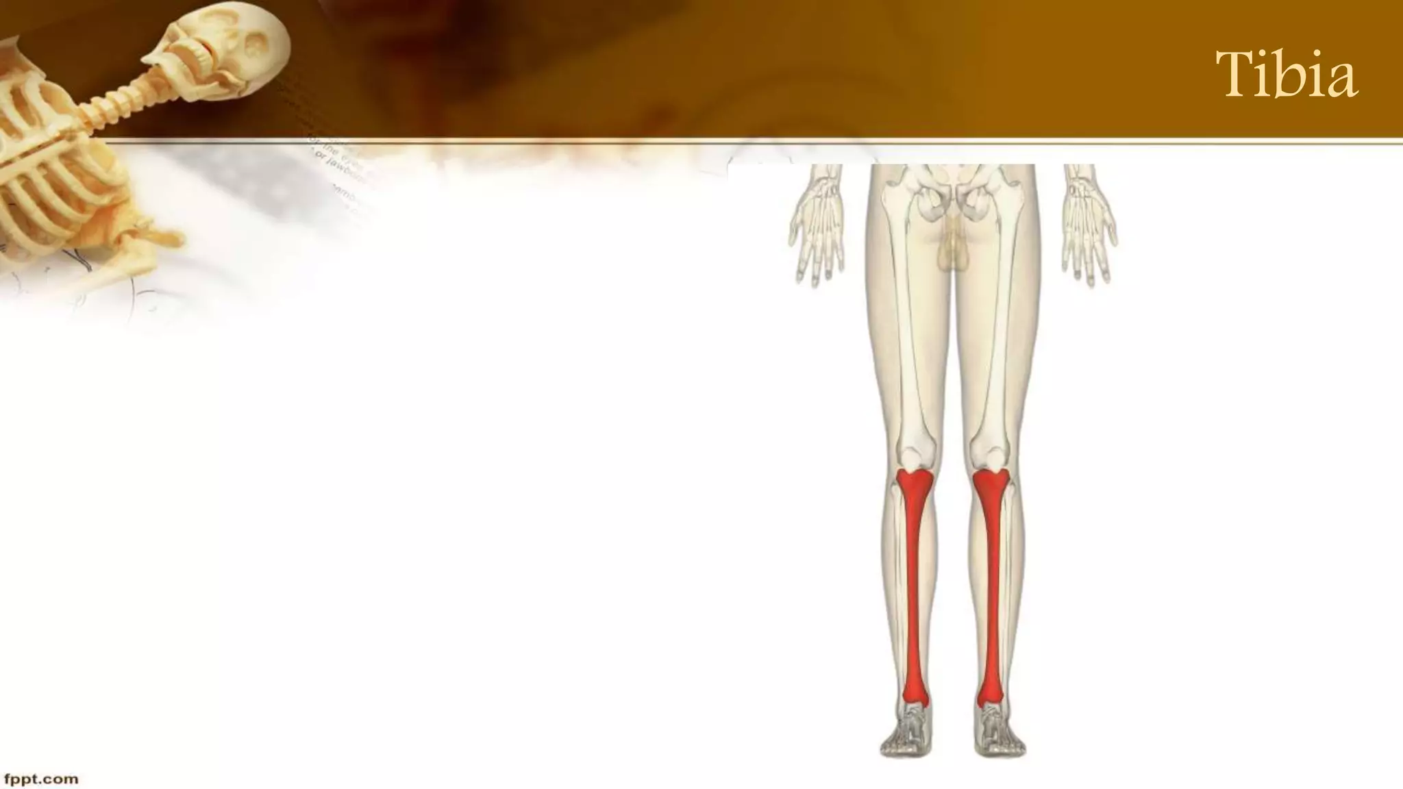
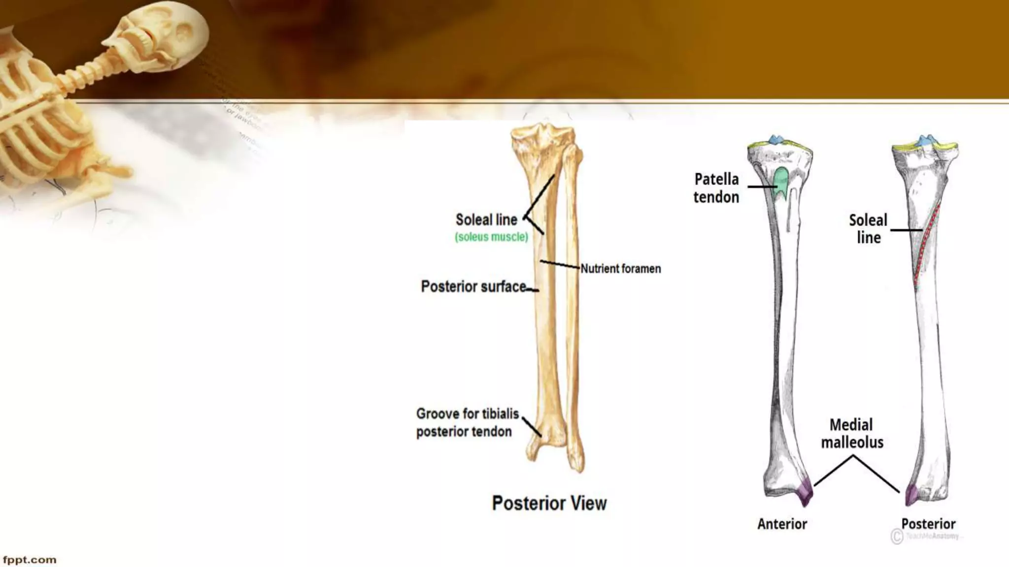
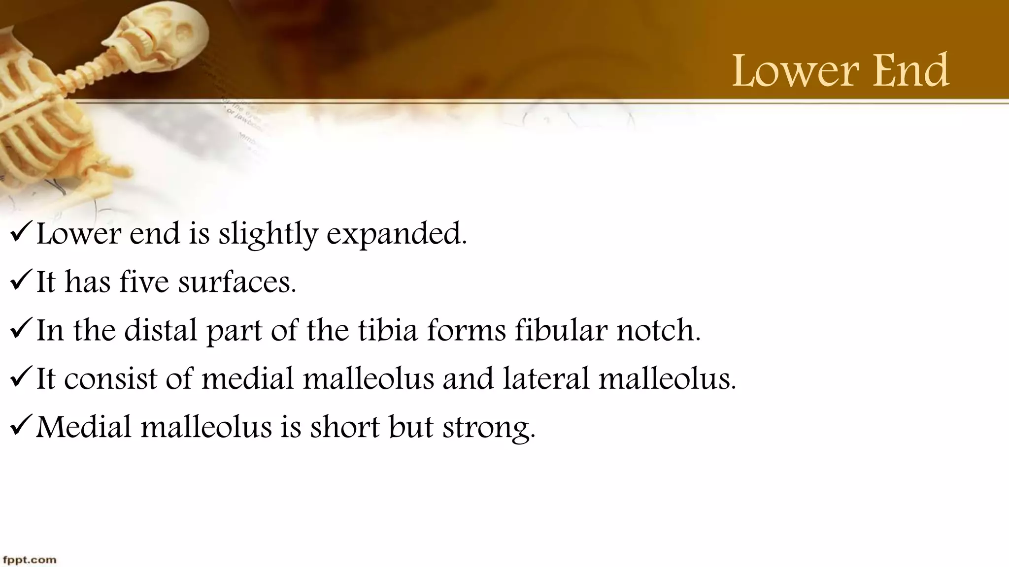
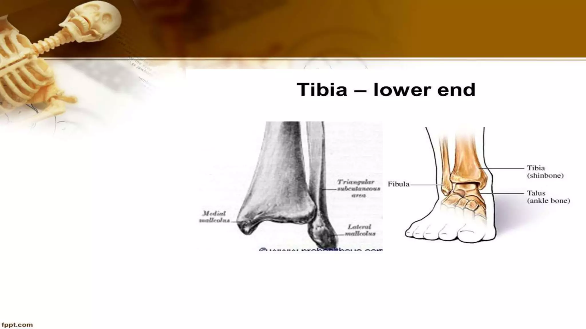
Anatomy of tibia in details by Purvi shah
it's clinical anatomy, side determination, features.
Tibia Lower Limb bone part-4
Like, share and comment
thank you!!
Femur Bone anatomy LL bone part 2
Details of femur Bone, attachments and clinical anatomy.
Femur thigh bone, longest bone
Like, share and comment.
Thank You!!
The document describes the anatomy of the posterior compartment of the leg. It contains two compartments - a superficial compartment with the gastrocnemius, plantaris, and soleus muscles, and a deep compartment with the popliteus, flexor digitorum longus, flexor hallucis longus, and tibialis posterior muscles. All of these muscles are flexors or invertors of the ankle and are supplied by the tibial nerve.
The clavicle has lateral and medial ends that articulate with the acromion and manubrium respectively. It ossifies from two primary centers and has attachments for muscles like the deltoid and pectoralis major. The scapula has surfaces for the subscapularis and infraspinatus muscles. It has processes like the acromion, coracoid and spine. Muscles like the trapezius and serratus anterior originate or insert on the scapula. Both bones ossify membraneously and have sex differences in shape and size.
Anatomy of fibula
LL bone part-5
fibula details, side determination, ossification and it's clinical anatomy. fibula is lateral and smaller bone.
Like, share and comment.
Thank You!!
The document discusses the anatomy of the carpal bones. It describes that the carpus is made up of 8 bones arranged in two rows, with the proximal row containing the scaphoid, lunate, triquetral, and pisiform bones and the distal row containing the trapezium, trapezoid, capitate, and hamate bones. Each bone has a distinct shape that aids in identification: the scaphoid is boat shaped, lunate is half moon shaped, etc. The document also notes clinical points like how a scaphoid fracture is common from a fall on an outstretched hand and may cause avascular necrosis if blood supply is deprived.
The ulna is one of the two long bones in the forearm, located on the medial side. It runs parallel to the radius. The proximal end has two processes - the olecranon and coronoid process. The olecranon fits into the elbow joint and the coronoid process fits into the humerus during flexion. The distal end has a head that articulates with the radius and triangular disk of the wrist. The body tapers from top to bottom and has borders for muscle attachments.
This document describes the anatomy of the front of the thigh. It details the cutaneous nerves, muscles in the anterior compartment including the pectineus, sartorius and quadriceps femoris. It describes the femoral triangle containing the femoral vessels and nerve. The adductor canal containing the continuation of the femoral artery and vein is also summarized. Finally, the courses and branches of the femoral artery and vein are outlined.
Slideshow: Hip Joint and Pelvic Gateways
The Funky Professor videos can be viewed here;
http://publishing.rcseng.ac.uk/journal/video?videoTaxonomy=FUNK
The tibia is the main bone of the leg located on the medial side. It has an upper end with medial and lateral condyles that form the tibial plateau and articulate with the femur. The shaft is triangular in shape with borders and surfaces. The lower end widens and has a medial malleolus and fibular notch. Muscles and ligaments attach along borders and surfaces to allow for movement and stability of the knee and ankle joints.
The clavicle is an S-shaped bone that transmits weight from the upper limb to the sternum. It has two ends - a lateral end that articulates with the acromion of the scapula to form the AC joint, and a medial end that articulates with the manubrium sterni to form the SC joint. The shaft of the clavicle is divided into a lateral one-third and a medial two-thirds, with various surfaces, borders, and tubercles that provide attachments for muscles and ligaments like the deltoid, trapezius, pectoralis major, and coracoclavicular ligaments. The clavicle is the only long bone that lies horizontally
There are two groups of muscles in the back of the forearm - a superficial group and a deep group. The superficial group contains 7 muscles that originate on the lateral epicondyle of the humerus and insert on the bones of the hand and fingers. They function to extend the wrist and fingers. The deep group contains 5 muscles that originate on the bones of the forearm and insert on the thumb and second finger. They function to supinate the forearm and extend the thumb.
This document describes the anatomy of the tibia bone. It notes that the tibia is the medial and larger bone of the leg, also known as the shin bone. It has an upper end with medial and lateral condyles that articulate with the femur to form the knee joint. The lower end has a medial malleolus and articulates with the talus bone to form the ankle joint. Key structures that attach to various parts of the tibia are also described.
The fibula is the lateral and smaller bone of the leg that forms part of the ankle joint. It has three parts - an upper end or head that is expanded in all directions, a shaft with anterior, posterior and interosseous borders, and a lower end or lateral malleolus. The upper end articulates with the lateral condyle of the tibia, and the lower end has a triangular articular facet that articulates with the talus bone. Various muscles like the peroneals and tibialis posterior attach along the shaft, and ligaments like the anterior and posterior tibiofibular ligaments attach the fibula to the tibia.
The scapula, also known as the shoulder blade, is a flat triangular bone located at the back of the trunk and resides over the posterior surface of ribs two to seven. ... It also articulates with the humerus and clavicle, forming the glenohumeral (shoulder) joint and acromioclavicular joint respectively.
The posterior compartment of the thigh contains the hamstring muscles (biceps femoris, semitendinosus, semimembranosus, and part of the adductor magnus), which are supplied by branches from the sciatic nerve. It also contains cutaneous nerves that innervate the skin (medial, posterior, and lateral cutaneous nerves) and veins that drain into the great and small saphenous veins. The blood supply comes from branches of the profunda femoris artery and drains into the profunda femoris vein.
The document discusses the arches of the foot, including the medial longitudinal arch, lateral longitudinal arch, anterior and posterior transverse arches. It describes the bones that make up each arch, their ends, pillars, and factors that help maintain the arches, such as ligaments, tendons, and muscles. Common foot deformities like flat foot, high arched foot, club foot, hallux vulgus, and others are also summarized.
The fibula is the lateral bone of the leg located next to the tibia. It is a long, thin bone that does not bear much weight. The fibula articulates proximally with the lateral tibia and distally at the ankle. Its main functions are to provide attachment points for muscles acting on the leg and to assist with ankle stability. It has three articulating surfaces at its ends and three borders along its shaft to which various muscles attach for origination.
The hip joint is a ball-and-socket synovial joint located between the femur and pelvis. It allows flexion, extension, abduction, and rotation of the thigh. The joint is stabilized by strong ligaments and surrounding muscles. It receives its main blood supply from the medial and lateral circumflex femoral arteries and is innervated by nerves from the lumbar plexus.
The document summarizes the anatomy of the hand and wrist. It describes that the hand consists of 27 bones divided into phalanges, metacarpals, and carpals. The carpals are further divided into the proximal and distal rows. The phalanges include proximal, middle, and distal bones in the fingers and two bones in the thumb. The forearm contains the radius and ulna bones. The wrist joint is formed where the carpal bones articulate with the radius proximally and metacarpals distally.
The document describes the muscles of the anterior, lateral, and posterior fascial compartments of the forearm. In the anterior compartment, there are superficial, intermediate, and deep groups of muscles including the flexor carpi radialis, palmaris longus, flexor digitorum superficialis, and flexor digitorum profundus. The muscles are supplied by the median and ulnar nerves except for the flexor carpi ulnaris. The lateral compartment includes the brachioradialis and extensors of the wrist and fingers. The posterior compartment comprises extensors of the wrist, fingers and thumb including the abductor pollicis longus, extensor pollicis brevis and extensor
The document summarizes the bones of the lower limb, including:
- The bones of the thigh (femur and patella)
- The bones of the leg (tibia and fibula)
- The bones of the foot (tarsals, metatarsals, and phalanges)
It describes the features and landmarks of each individual bone in detail. Key points include that the femur articulates with the acetabulum to form the hip joint and tibia/patella to form the knee joint. The tibia articulates with the femur and fibula proximally and talus distally. The fibula articulates with the tibia proximally and talus distally
The document describes the anatomy of the lower limb bones. It discusses the four regions of the lower limb - the thigh, leg, ankle, and foot. It provides details on the bones of the thigh (femur), leg (tibia and fibula), and knee joint. The femur is the longest bone and forms the hip and knee joints. The tibia and fibula form the leg, with the tibia bearing more weight and forming the ankle joint along with the talus bone. Key ligaments like the patellar ligament, collateral ligaments, and cruciate ligaments are described for the knee joint.
The tibia is the main bone of the leg. It has an upper end with medial and lateral condyles that form the tibial plateau and articulate with the femur. The lower end expands slightly and has a medial malleolus. The shaft is triangular with borders and surfaces that provide origins and insertions for muscles. Fractures commonly occur in the tibial shaft, especially the junction of the upper and middle thirds.
Tibia,fibula, patella print By M Thiru murugan.pptxthiru murugan
Tibia, Fibula & Patella
By,M. Thiru murugan
Tibia (Shinbone): The tibia is the main bone of the lower leg.
It expands at its proximal and distal ends; articulating at the knee and ankle joints respectively.
The tibia is the second largest bone in the body and it is a key weight-bearing structure.
Proximal: The proximal tibia is widened by the medial and lateral condyles, which aid in weight-bearing. The condyles form a flat surface, known as the tibial plateau.
This structure articulates with the femoral condyles to form the key articulation of the knee joint.
Located between the condyles is a region called the intercondylar eminence - this projects upwards on either side as the medial and lateral intercondylar tubercles. This area is the main site of attachment for the ligaments of the knee joint.
Shaft
The shaft of the tibia is prism-shaped, with 3 borders and 3 surfaces; anterior, posterior and lateral.
Anterior border: palpable subcutaneously down the anterior surface of the leg as the shin. The proximal aspect of the anterior border is marked by the tibial tuberosity; the attachment site for the patella ligament.
Posterior surface: marked by a ridge of bone known as soleal line. This line is the site of origin for part of the soleus muscle
Lateral border: also known as the interosseous border. It gives attachment to the interosseous membrane that binds the tibia and the fibula together.
Distal: The distal end of the tibia widens to assist with weight-bearing.
The medial malleolus is a bony projection continuing inferiorly on the medial aspect of the tibia.
It articulates with the tarsal bones to form part of the ankle joint.
On the posterior surface of the tibia, there is a groove for passage of tendon of tibialis posterior.
Laterally is the fibular notch, where the fibula is bound to the tibia - forming the distal tibiofibular joint.
Fibula (Calf Bone) The fibula is the second bone in the lower leg,
The fibula is a bone located within the lateral aspect of the leg. Its main function is to act as an attachment for muscles, and not as a weight-bearer.
Articulations:
Proximal tibiofibular joint: articulates with the lateral condyle of the tibia.
Distal tibiofibular joint: articulates with the fibular notch of the tibia.
Ankle joint: articulates with the talus bone of the foot
Proximal:
At the proximal end, the fibula has an enlarged head, which contains a facet for articulation with the tibia.
On the posterior and lateral surface of the fibular neck (fibular nerve located)
Shaft
The fibular shaft has 3 surfaces - anterior, lateral and posterior. The leg is split into 3 compartments, and each surface faces its respective compartment
Distal
Distally, the lateral surface is called the lateral malleolus.
The lateral malleolus is more prominent than the medial malleolus, and can be palpated at the ankle on the lateral side of the leg.
Patella: The patella (kneecap) is located at the front of the knee joint, within the patellofemora
The skeleton of the lower limb consists of the femur (thigh bone), tibia (medial leg bone), and fibula (lateral leg bone). The skeleton of the foot includes 7 tarsal bones, notably the calcaneum which forms the heel, 5 metatarsal bones, and 14 phalanges. Key subcutaneous bones include the patella at the knee, the anterior border of the tibia, the tibial tuberosity, and the medial and lateral malleoli of the tibia and fibula, respectively. The foot is a complex structure with 26 bones that must provide support and allow for locomotion.
The document provides an overview of the bones in the upper and lower extremities. It describes the femur bone of the thigh, including its head, neck, greater and lesser trochanters, and shaft. It then discusses the patella and tibia bones of the lower leg. Finally, it summarizes the bones of the foot, including the tarsals, metatarsals, and phalanges, and provides details on their structures and articulations.
The patella is a triangular sesamoid bone located in the quadriceps tendon in front of the knee joint. It has three borders and two surfaces. The posterior surface articulates with the femur and is divided into medial and lateral facets. The patella attaches proximally to the quadriceps tendon and distally to the patellar ligament. During knee movement, different facets of the patella articulate with the femur. Clinical issues involving the patella include lateral dislocation and patellofemoral syndrome which is pain from cartilage breakdown on the patellar surfaces.
Osseous system II , is the appendicular skeletal system , it representing about the bone of pectoral girdle , pelvic girdle and upper limbs and lower limbs. structure and function of humerus , radius ulna and structure and function of femur , tibia fibula.
The document discusses the bones of the lower limbs, including the thigh, leg, and foot. It provides objectives and terminology for classifying and identifying the main features of the femur, patella, tibia, fibula, and bones of the foot. Key points covered include the trochanters, condyles, tuberosities, and processes of the femur and features of the tibia, fibula, tarsals, metatarsals, and phalanges. MCQ questions are also provided to test comprehension.
The knee joint is the largest and most complex joint in the body. It consists of four joints: the patellofemoral joint, the medial tibiofemoral joint, the lateral tibiofemoral joint, and the superior tibiofibular joint. The knee joint involves three bones: the femur, tibia, and patella. It allows for flexion and extension through interactions between the femoral condyles and tibial plateaus. Various ligaments and muscles provide stability and mobility to the knee complex during movement.
The tibia is the larger medial bone of the lower leg. Its upper end has medial and lateral condyles that articulate with the femur. The lower end expands and has a medial malleolus that projects downward. Muscles and ligaments attach along borders and surfaces, including the patellar ligament at the tibial tuberosity. The tibia articulates proximally at the knee and distally at the ankle.
The bony pelvis consists of 4 bones: two hip bones, the sacrum, and the coccyx. Each hip bone is comprised of three parts - the ilium, pubis, and ischium. The ilium forms the fan-shaped upper portion and contributes to the acetabulum. The pubis forms the anterior portion and articulates with its counterpart via the pubic symphysis. The ischium forms the posteroinferior portion and contributes to the acetabulum. Together, the right and left hip bones form the pelvic girdle and provide attachment for muscles and support for the pelvic organs.
The document discusses the anatomy of the foot and common foot conditions. It begins with an outline of the anatomy, describing the bones, joints, arches and muscles of the foot. It then discusses common congenital anomalies like pes cavus and pes planus. Other conditions covered include clubfoot, tarsal tunnel syndrome. The take-home message emphasizes the role of the foot in force attenuation and the importance of both static and dynamic structures for efficient foot biomechanics.
The patella is a triangular bone located at the front of the knee joint. It protects the knee joint and enhances the leverage of the quadriceps muscle. The patella can dislocate laterally due to forces from the quadriceps. It can also fracture from direct impact or sudden quadriceps contraction. Patellar injuries are common in sports involving sudden twisting or impact to the knee.
The tibia is the thicker, inner bone of the lower leg that runs parallel to the fibula. It connects the femur above to the talus and fibula below. The tibia has expanded upper and lower ends connected by a triangular shaft. Common fractures of the tibia include breaks across the shaft from direct blows or at the upper end from knee injuries. Stress fractures can also occur from overuse. Treatment depends on the severity but may involve casting or surgery to stabilize broken bone fragments.
This document provides an overview of the temporomandibular joint (TMJ), including its:
- Types (synovial, bicondylar, ginglymoarthroidal)
- Anatomy (bones, articular disc, ligaments, muscles)
- Histology of the articular surfaces
- Biomechanics and functions like opening and closing the mouth
- Age-related changes like flattening of bones and thinning of tissues
The hip bone is made up of three parts - the ilium, pubis, and ischium - that come together at the cup-shaped acetabulum. The acetabulum articulates with the head of the femur to form the hip joint. Key features include the iliac crest, pubic crest, ischial tuberosity, and obturator foramen. The hip bones come together at the front to form the pelvic girdle and provide attachments for muscles and ligaments around the hip and pelvic region.
Mental Health Disorder and Physical therapy
Mental Health refers to Cognitive, behavioral, and emotional well-being. It is all how people think, feel and behave.
Tendonitis details and it's physiotherapy management.
It is define as inflammation of the tendon, tendonitis occur due to overuse and trauma. Depending upon involvement of tendon special test are used. it is treated with PRICE protocol.
Contusion, also known as a bruise, is an injury to tissue or bone caused by blood vessels rupturing or breaking and bleeding into the surrounding tissue. This causes discoloration that turns into a bruise. Contusions usually result from a hard hit, fall, or blow. The bleeding can put pressure on nerve endings, causing pain when the area is touched. Contusions are more common in elderly people due to more delicate blood vessels, and can also be caused by medications that interfere with blood clotting. Physiotherapy treatments for contusions include following the PRICE protocol of protection, rest, ice application, compression, and elevation.
Sprain is ref to Ligament injury.Depending upon severity of injury it is classified as mild, moderate & severe. For Grade 1 & 2require conservative treatment while for grade 3 require surgical intervention.
Charles Bell describe Bell's Palsy. It's LNM lesion. paralysis of the facial muscle; loss of facial expression. Idiopathic in nature. Most of Patient recover from Bell's Palsy 20 to 25 days. #purvishah #Bell'sPalsyICIDH2Format.
ECG: In 1895 first described by Einthoven, known as 'FATHER OF ECG'. It is a non invasive method. recording of electrical impulses generated in the heart. Important Investigated tool, cardiac activity. #PURVISHAH
This document provides information about stroke, including its definition, types, risk factors, and the role of physiotherapy in treatment. It discusses the two main types of stroke - ischemic and hemorrhagic - and describes some common syndromes associated with different areas of brain injury, such as the middle cerebral artery syndrome. It then outlines physiotherapy techniques used in both the acute and rehabilitation stages, including positioning, improving range of motion, strengthening, balance training, and gait re-education.
Introduction to AI for Nonprofits with Tapp NetworkTechSoup
Dive into the world of AI! Experts Jon Hill and Tareq Monaur will guide you through AI's role in enhancing nonprofit websites and basic marketing strategies, making it easy to understand and apply.
This presentation was provided by Steph Pollock of The American Psychological Association’s Journals Program, and Damita Snow, of The American Society of Civil Engineers (ASCE), for the initial session of NISO's 2024 Training Series "DEIA in the Scholarly Landscape." Session One: 'Setting Expectations: a DEIA Primer,' was held June 6, 2024.
it describes the bony anatomy including the femoral head , acetabulum, labrum . also discusses the capsule , ligaments . muscle that act on the hip joint and the range of motion are outlined. factors affecting hip joint stability and weight transmission through the joint are summarized.
Main Java[All of the Base Concepts}.docxadhitya5119
This is part 1 of my Java Learning Journey. This Contains Custom methods, classes, constructors, packages, multithreading , try- catch block, finally block and more.
A workshop hosted by the South African Journal of Science aimed at postgraduate students and early career researchers with little or no experience in writing and publishing journal articles.
Macroeconomics- Movie Location
This will be used as part of your Personal Professional Portfolio once graded.
Objective:
Prepare a presentation or a paper using research, basic comparative analysis, data organization and application of economic information. You will make an informed assessment of an economic climate outside of the United States to accomplish an entertainment industry objective.
Executive Directors Chat Leveraging AI for Diversity, Equity, and InclusionTechSoup
Let’s explore the intersection of technology and equity in the final session of our DEI series. Discover how AI tools, like ChatGPT, can be used to support and enhance your nonprofit's DEI initiatives. Participants will gain insights into practical AI applications and get tips for leveraging technology to advance their DEI goals.
Thinking of getting a dog? Be aware that breeds like Pit Bulls, Rottweilers, and German Shepherds can be loyal and dangerous. Proper training and socialization are crucial to preventing aggressive behaviors. Ensure safety by understanding their needs and always supervising interactions. Stay safe, and enjoy your furry friends!
Biological screening of herbal drugs: Introduction and Need for
Phyto-Pharmacological Screening, New Strategies for evaluating
Natural Products, In vitro evaluation techniques for Antioxidants, Antimicrobial and Anticancer drugs. In vivo evaluation techniques
for Anti-inflammatory, Antiulcer, Anticancer, Wound healing, Antidiabetic, Hepatoprotective, Cardio protective, Diuretics and
Antifertility, Toxicity studies as per OECD guidelines
How to Manage Your Lost Opportunities in Odoo 17 CRMCeline George
Odoo 17 CRM allows us to track why we lose sales opportunities with "Lost Reasons." This helps analyze our sales process and identify areas for improvement. Here's how to configure lost reasons in Odoo 17 CRM
3. Tibia
It is the medial and larger bone of the leg.
It is homologous with the radius of UL.
t supports most of your weight and is an important part of both
the knee joint and Ankle joint.
It carries weight of body from the femur to foot.
4. Side Determination
Upper end is much larger than lower end.
The medial side of the lower end projects downwards beyond the
rest of the bone is called medial malleolus.
The anterior border of the shaft is most prominent and crest like.
5. Features
The Tibia has an upper end , a shaft and a lower end.
UPPER END: Expanded from side to side,
Upper end includes; a medial condyle, a lateral
condyle, intercondylar area,a tuberosity.
6.
7.
8. Features
Medial Condyle: It is larger than lateral condyle.
Articular surface is oval and it’s long axis is anteroposterior.
Central part of the surface is concave and comes into contact with
femoral condyle.
Posterior surface of the medial condyle has a groove.
The anterior and medial surfaces are marked by a numerous vascular
foramina.
9. Features
Lateral Epicondyle: It is smaller than medial condyle.
The superior surface articulates with lateral condyle of femur.
Articular surface is nearly circular.
The articular surface has a raised medial margin which covers the
lateral intercondylar tubercle.
10. Intercondylar area
It is the roughened area on the superior surface.
The area is narrowest in its middle part.
This part is elevated to form the intercondylar eminence which is
flanked by the medial and lateral intercondylar tubercles.
11. Tuberosity Of The Tibia
Tuberosity of the tibia is a prominence located on the anterior
aspect of the upper end of the tibia.
Tuberosity divided into an upper smooth area and a lower rough
area.
The epiphyseal line for the upper end of the tibia passes through
the junction of these two parts.
12. Shaft
The shaft of tibia is PRISMOID in shape.
Three borders Anterior (sharp and s shaped)
Medial (rounded)
Interosseous.
Three surfaces Lateral
Medial
Posterior(it is crossed obliquely by a rough ridge
called soleal line.)
13.
14. Lower End
Lower end is slightly expanded.
It has five surfaces.
In the distal part of the tibia forms fibular notch.
It consist of medial malleolus and lateral malleolus.
Medial malleolus is short but strong.
15.
16. Attachments On The Tibia
Attachments on the MEDIAL CONDYLE; semimembranous,
capsular ligaments of knee joint, medial patellar retinaculum.
Attachments on the LATERAL CONDYLE; iliotibial band, capsular
ligaments of the superior tibiofibular joint, EDL, tendon of
politeus.
Attachments on the INTERCONDYLAR AREA; ACL and PCL.
17. Attachments On Tibia
Attachments On The Tibial Tuberosity; ligamentum patellae.
Attachments On The Shaft; Tibialis anterior, Sartorius, gracilis and
semitendinous.
Soleal line give attachments to soleus, fascia covering popliteus
and transverse fascial septum,FDL,interosseous tibiofibular
ligaments,capsular ligament of ankle joint.
18.
19.
20. Blood Supply
Nutrient artery to the tibia is the largest nutrient artery in the
body.
It is a branch of the posterior tibial artery which enters the bone
on its posterior surface.
21. Articulation
The tibia is a part of four joints; the knee,
ankle, superior and inferior tibiofibular joint.
22. Ossification
Tibia ossifies from one primary and two secondary centres.
The primary centre appears in the shaft during 7th week of
intrauterine life.
A secondary centre for upper end appears just before birth, and
fuse with the shaft at 16-18 years.
23. Clinical Anatomy
Upper end of tibia is one of the commonest sites for ACUTE
OSTEOMYELITIS (knee joint remain safe because of capsular
attachment).
The tibia is commonly fractured at the junction of the upper two
thirds lower one third of shaft.
(fracture unite slowly, or may not unite at all as the blood supply to
this part of the bone is poor, tearing of nutrient artery.
24.
25. Clinical Anatomy
Forward dislocation of the tibia on the talus produce characteristic
prominence of the heel in this injury(this is commonest type of the
fracture at the ankle).
Pott’s fracture: spiral fracture of lateral malleolus, avulsion of the
tibial collateral ligament and posterior margin of the lower end of
the tibia shears off against the talus.
26. Important Points
The tibia has been modeled as taking an axial force during
walking that is up to 4.7 bodyweight.
Its bending moment in the sagittal plane in the late stance phase is
up to 71.6 bodyweight times mm.
Gerdy's tubercle is a lateral tubercle of the tibia, located where the
iliotibial tract inserts.