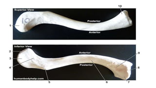
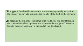
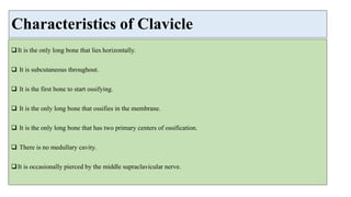
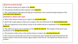
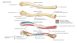
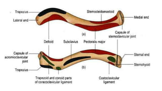
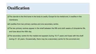
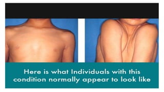
Clavicle bone: Osteology, Clavicle bone anatomy, Features of Clavicle bone, Side determination, Function of Clavicle, Characteristics of Clavicle Bone, Muscle Attachments, Ossification, Clinical Anatomy.
VIP Call Girls Hyderabad Megha 9907093804 Independent Escort Service Hyderabad
CLAVICLE Bone Osteology: Appendicular skeleton
1. CLAVICLE: Bone Study
Dr. Priyanka(PT)
BPT,MPT(Neurological Disorders)
Assistant Professor(SIMS College, Dehradun)
2. Introduction
Modified long bone.
Placed horizontally in the body.
Presents shaft and two ends.
Shaft:- Cylindrical part and shows a gentle S-shaped curve.
Two ends:- Medial and Lateral.
Medial end:- Quadrilateral in shape.
Lateral end:- Flattened.
6. SIDE DETERMINATION
Medial:- Quadrilateral sternal end.
Anterior:- Convexity medial two-thirds of the shaft.
Inferior:- Groove for subclavius, conoid tubercle and trapezoid line.
The lateral end is flat, and the medial end is large and quadrilateral.
The shaft is slightly curved, so that it is convex forward in its medial two-thirds, and concave
forwards in its lateral one-third.
The inferior surface is grooved longitudinally in its middle one-third.
7.
8. It supports the shoulder so that the arm can swing clearly away from
the trunk. The clavicle transmits the weight of the limb to the sternum.
It receives the weight of the upper limb via lateral one-third through
the coracoclavicular r ligament and transmits the weight of the upper
limb to the axial skeleton via the medial two-thirds part.
9. Characteristics of Clavicle
It is the only long bone that lies horizontally.
It is subcutaneous throughout.
It is the first bone to start ossifying.
It is the only long bone that ossifies in the membrane.
It is the only long bone that has two primary centers of ossification.
There is no medullary cavity.
It is occasionally pierced by the middle supraclavicular nerve.
12. Shaft:- Lateral 1/3
1. Flat above downward.
2. Two borders
• Anterior border:- Concave forward.
• Posterior Border:- Convex forward.
3. Two Surfaces
• Superior Surface:- Smooth
• Inferior surface:- Presents an elevation called Conoid tubercle.
The ridge is called Trapezoid Ridge.
13.
14.
15. Shaft:- Medial 2/3
• Rounded and has 4 surfaces.
• Four surfaces:-
1. Superior Surface:-
• Smooth laterally
• Rough medially
2. Posterior Surface:- Smooth
3. Inferior surface:-
• Medially:- Rough oval impression.
• Laterally: The longitudinal groove is known as the Subclavian groove, where
subclavius muscle attaches.
4. Anterior Surface
16. Lateral End/ Acromial End
Flattened.
Bears a facet that articulates with the
acromion process making the
Acromioclavicular joint.
17. Medial End/Sternal End
Quadrangular.
Articulates with the clavicular notch of manubrium sterni
making the Sternoclavicular Joint.
Articular surface extends to the inferior aspect for
articulation with the first costal cartilage.
18.
19. Attachments
At the lateral end, the margin of the articular surface for its acromioclavicular joint gives attachment to the
joint capsule.
At the medial end, the margin of the articular surface for the sternum gives attachment to:
a. Fibrous capsule of sternoclavicular joint all around.
b. Articular disc posterosuperiorly.
c. Interclavicular ligament superiorly.
20. Lateral one-third of shaft
a. The anterior border gives origin to the deltoid.
b. The posterior border provides insertion to the trapezius.
c. The conoid tubercle and trapezoid ridge give attachment to the conoid and trapezoid parts of the
coracoclavicular ligament.
Medial two-thirds of the shaft
a. Most of the anterior surface gives origin to the pectoralis major.
b. Half of the rough superior surface gives origin to the clavicular head of the sternocleidomastoid.
c. The oval impression on the inferior surface at the medial end gives attachment to the costoclavicular
ligament.
d. The subclavian groove gives insertion to the subclavius muscle. The margins of the groove give
attachment to the clavipectoral fascia.
e. The posterior surface close to the medial end gives origin to the sternohyoid muscle.
f. The subclavian vessels and cords of the brachial plexus pass towards the axilla lying between the
inferior surface of the clavicle and the upper surface of the first rib. The Subclavius muscle acts as a
cushion.
21.
22.
23. Ossification
The clavicle is the first bone in the body to ossify. Except for its medial end, it ossifies in the
membrane.
It ossifies from two primary centres and one secondary centre.
The two primary centres appear in the shaft between the fifth and sixth weeks of intrauterine life,
and fuse about the 45th day.
The secondary centre for the medial end appears during 15-17 years and fuses with the shaft
during 21- 22 years. Occasionally, there may be a secondary centre for the acromial end.
24.
25. Clinical Anatomy
The clavicle is commonly fractured by falling on the outstretched hand (indirect violence).
The most common site of fracture is the junction between the two curvatures of the bone, which is
the weakest point. The lateral fragment is displaced downwards by the weight of the limb as trapezius
muscle alone is unable to support the weight of the upper limb.
The clavicles may be congenitally absent, or imperfectly developed in a disease called cleidocranial
dysostosis.
In this condition, the shoulders droop, and can be approximated anteriorly in front of the chest.