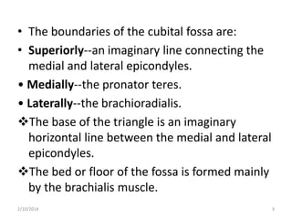
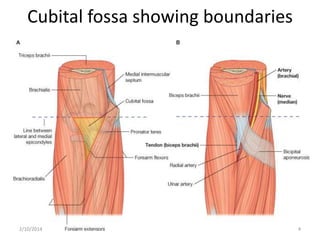
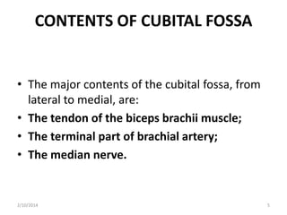
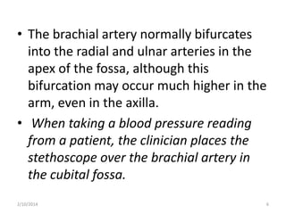
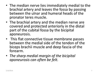
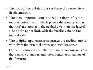
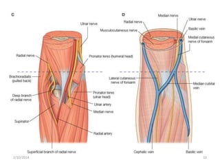
CUBITAL FOSSA
2/10/2014

1
• The cubital fossa is an important area of
transition between the arm and the forearm.
• It is located anterior to the elbow joint and is a
triangular depression formed between two
forearm muscles:
• The brachioradialis muscle originating from
the lateral supraepicondylar ridge of the
humerus;
• The pronator teres muscle originating from
the medial epicondyle of the humerus
2/10/2014

2
• The boundaries of the cubital fossa are:
• Superiorly--an imaginary line connecting the
medial and lateral epicondyles.
• Medially--the pronator teres.
• Laterally--the brachioradialis.
The base of the triangle is an imaginary
horizontal line between the medial and lateral
epicondyles.
The bed or floor of the fossa is formed mainly
by the brachialis muscle.
2/10/2014

3
Cubital fossa showing boundaries

2/10/2014

4
CONTENTS OF CUBITAL FOSSA

• The major contents of the cubital fossa, from
lateral to medial, are:
• The tendon of the biceps brachii muscle;
• The terminal part of brachial artery;
• The median nerve.

2/10/2014

5
• The brachial artery normally bifurcates
into the radial and ulnar arteries in the
apex of the fossa, although this
bifurcation may occur much higher in the
arm, even in the axilla.
• When taking a blood pressure reading
from a patient, the clinician places the
stethoscope over the brachial artery in
the cubital fossa.
2/10/2014

6
• The median nerve lies immediately medial to the
brachial artery and leaves the fossa by passing
between the ulnar and humeral heads of the
pronator teres muscle.
• The brachial artery and the median nerve are
covered and protected anteriorly in the distal
part of the cubital fossa by the bicipital
aponeurosis.
• This flat connective tissue membrane passes
between the medial side of the tendon of the
biceps brachii muscle and deep fascia of the
forearm.
• The sharp medial margin of the bicipital
aponeurosis can often be felt.
2/10/2014

7
• The radial nerve lies just under the lip of the
brachioradialis muscle, which forms the lateral
margin of the fossa.
• In this position, the radial nerve divides into
superficial and deep branches: the superficial
branch continues into the forearm just deep
to the brachioradialis muscle;
• the deep branch passes between the two
heads of the supinator muscle to access the
posterior compartment of the forearm.
2/10/2014

8
• The roof of the cubital fossa is formed by superficial
fascia and skin.
• The most important structure within the roof is the
median cubital vein, which passes diagonally across
the roof and connects the cephalic vein on the lateral
side of the upper limb with the basilic vein on the
medial side.
• The bicipital aponeurosis separates the median cubital
vein from the brachial artery and median nerve.
• Other structures within the roof are cutaneous nervesthe medial cutaneous and lateral cutaneous nerves of
the forearm.
2/10/2014

9
2/10/2014

10
Favour & Didong
2/10/2014

11
2/10/2014

12

Cubital fossa

  • 1.
  • 2.
    • The cubitalfossa is an important area of transition between the arm and the forearm. • It is located anterior to the elbow joint and is a triangular depression formed between two forearm muscles: • The brachioradialis muscle originating from the lateral supraepicondylar ridge of the humerus; • The pronator teres muscle originating from the medial epicondyle of the humerus 2/10/2014 2
  • 3.
    • The boundariesof the cubital fossa are: • Superiorly--an imaginary line connecting the medial and lateral epicondyles. • Medially--the pronator teres. • Laterally--the brachioradialis. The base of the triangle is an imaginary horizontal line between the medial and lateral epicondyles. The bed or floor of the fossa is formed mainly by the brachialis muscle. 2/10/2014 3
  • 4.
    Cubital fossa showingboundaries 2/10/2014 4
  • 5.
    CONTENTS OF CUBITALFOSSA • The major contents of the cubital fossa, from lateral to medial, are: • The tendon of the biceps brachii muscle; • The terminal part of brachial artery; • The median nerve. 2/10/2014 5
  • 6.
    • The brachialartery normally bifurcates into the radial and ulnar arteries in the apex of the fossa, although this bifurcation may occur much higher in the arm, even in the axilla. • When taking a blood pressure reading from a patient, the clinician places the stethoscope over the brachial artery in the cubital fossa. 2/10/2014 6
  • 7.
    • The mediannerve lies immediately medial to the brachial artery and leaves the fossa by passing between the ulnar and humeral heads of the pronator teres muscle. • The brachial artery and the median nerve are covered and protected anteriorly in the distal part of the cubital fossa by the bicipital aponeurosis. • This flat connective tissue membrane passes between the medial side of the tendon of the biceps brachii muscle and deep fascia of the forearm. • The sharp medial margin of the bicipital aponeurosis can often be felt. 2/10/2014 7
  • 8.
    • The radialnerve lies just under the lip of the brachioradialis muscle, which forms the lateral margin of the fossa. • In this position, the radial nerve divides into superficial and deep branches: the superficial branch continues into the forearm just deep to the brachioradialis muscle; • the deep branch passes between the two heads of the supinator muscle to access the posterior compartment of the forearm. 2/10/2014 8
  • 9.
    • The roofof the cubital fossa is formed by superficial fascia and skin. • The most important structure within the roof is the median cubital vein, which passes diagonally across the roof and connects the cephalic vein on the lateral side of the upper limb with the basilic vein on the medial side. • The bicipital aponeurosis separates the median cubital vein from the brachial artery and median nerve. • Other structures within the roof are cutaneous nervesthe medial cutaneous and lateral cutaneous nerves of the forearm. 2/10/2014 9
  • 10.
  • 11.
  • 12.

Editor's Notes

  • #10 The ulnar nerve does not pass through the cubitalfossa. Instead, it passes posterior to the medial epicondyle.