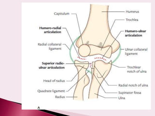
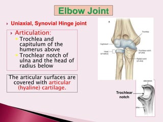
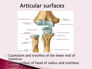
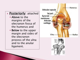
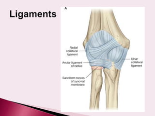
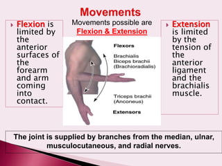
Dr.Nandhini V
 At the end of this session, students should be
able to:
 DEMONSTRATE the articulating bones, capsule, lateral
& medial collateral ligaments and synovial membrane
of the elbow joint
 DEMONSTRATE the movements (flexion and extension)
of the elbow. LIST the main muscles producing the
above movements
Two articulations –
1. Humero-ulnar
2. Humero-radial
the complexity of elbow joint is increased by its
continuity with superior radio ulnar joint.
humero-radial
+
humero-ulnar = cubital articulation
+
superior radioulnar
- synovial joint
- hinge variety
- compound
- typical
 Articulation:
 Trochlea and
capitulum of the
humerus above
 Trochlear notch of
ulna and the head of
radius below
The articular surfaces are
covered with articular
(hyaline) cartilage.
 Uniaxial, Synovial Hinge joint
Trochlear
notch
 Capitulum and trochlea of the lower end of
humerus
 Upper surface of head of radius and trochlear
notch of ulna
 Anteriorly: attached
 Above to the humerus
along the upper
margins of the
coronoid and radial
fossae and to the front
of the medial and
lateral epicondyles and
 Below to the margin of
the coronoid process of
the ulna and to the
anular ligament, which
surrounds the head of
the radius.
 Posteriorly: attached
 Above to the
margins of the
olecranon fossa of
the humerus and
 Below to the upper
margin and sides of
the olecranon
process of the ulna
and to the anular
ligament.
 This lines the
capsule and covers
fatty pads in the
floors of the
coronoid, radial, and
olecranon fossae
 Is continuous below
with synovial
membrane of the
superior radioulnar
joint
 Triangular in
shape:
 Apex attached to
the lateral
epicondyle of
humerus
 Base attached to
the upper margin
of annular
ligament.
Lateral (radial collateral) ligament
Composed of three
parts (bands)
Medial (ulnar collateral) ligament
• Anterior strong cord-like band: between medial
epicondyle and the coronoid process of ulna
• Posterior weaker fan-like band: between medial
epicondyle and the olecranon process of ulna
• Transverse band: passes between the anterior and
posterior bands
1. Subtendinous
olecranon bursa
2. Subcutaneous
olecranon bursa
3. Bicipitoradial bursa
4. Bursa separating
biceps tendon from
oblique cord
 Anterior: Brachialis,
tendon of biceps,
median nerve,
brachial artery
 Posterior: Triceps
muscle, small bursa
intervening
 Lateral: Common
extensor tendon & the
supinator
 Medial: Ulnar nerve
Bursae around the elbow joint:
Subcutaneous olecranon bursa
Subtendinous olecranon bursa
 Flexion is
limited by
the
anterior
surfaces of
the
forearm
and arm
coming
into
contact.
 Extension
is limited
by the
tension of
the
anterior
ligament
and the
brachialis
muscle.
Movements possible are
Flexion & Extension
The joint is supplied by branches from the median, ulnar,
musculocutaneous, and radial nerves.
1. Trochlea of humerus fits into trochlear notch of
ulna
2. Ulnar collateral ligament
3. Radial collateral ligament
 -brachial artery
 -radial artery
 -ulnar artery
Anastomosis
around elbow joint
 Angle between the long axis
of the extended forearm and
the long axis of the arm
 Opens laterally
 Is about 5-15 degrees
 Disappears when the elbow
joint is flexed
 Permits the forearms to clear
the hips in swinging
movements during walking,
and is important when
carrying objects
 Cubitus valgus
 Cubitus varus
 Posterior dislocation of elbow
 The elbow joint is a stable joint because of the:
 Wrench-shaped articular surface of the
olecranon and the pulley-shaped trochlea of the
humerus
 Strong medial and lateral ligaments.
 Elbow dislocations are common, and most are
posterior.
 Posterior dislocation usually follows falling on
the outstretched hand.
 Posterior dislocations of the joint are common
in children because the parts of the bones that
stabilize the joint are incompletely developed.
Avulsion of the epiphysis of the medial epicondyle
is also common in childhood because the medial
ligament is much stronger than the bond of union
between the epiphysis and the diaphysis.
 GOLFER’S ELBOW (Medial epicondylitis)
(Lateral epicondylitis)
 Nerve entrapment
- median nerve
- ulnar nerve
-posterior interosseous nerve
elbow joint .ppt

elbow joint .ppt

  • 1.
  • 2.
     At theend of this session, students should be able to:  DEMONSTRATE the articulating bones, capsule, lateral & medial collateral ligaments and synovial membrane of the elbow joint  DEMONSTRATE the movements (flexion and extension) of the elbow. LIST the main muscles producing the above movements
  • 3.
    Two articulations – 1.Humero-ulnar 2. Humero-radial the complexity of elbow joint is increased by its continuity with superior radio ulnar joint. humero-radial + humero-ulnar = cubital articulation + superior radioulnar
  • 5.
    - synovial joint -hinge variety - compound - typical
  • 6.
     Articulation:  Trochleaand capitulum of the humerus above  Trochlear notch of ulna and the head of radius below The articular surfaces are covered with articular (hyaline) cartilage.  Uniaxial, Synovial Hinge joint Trochlear notch
  • 7.
     Capitulum andtrochlea of the lower end of humerus  Upper surface of head of radius and trochlear notch of ulna
  • 8.
     Anteriorly: attached Above to the humerus along the upper margins of the coronoid and radial fossae and to the front of the medial and lateral epicondyles and  Below to the margin of the coronoid process of the ulna and to the anular ligament, which surrounds the head of the radius.
  • 9.
     Posteriorly: attached Above to the margins of the olecranon fossa of the humerus and  Below to the upper margin and sides of the olecranon process of the ulna and to the anular ligament.
  • 11.
     This linesthe capsule and covers fatty pads in the floors of the coronoid, radial, and olecranon fossae  Is continuous below with synovial membrane of the superior radioulnar joint
  • 14.
     Triangular in shape: Apex attached to the lateral epicondyle of humerus  Base attached to the upper margin of annular ligament. Lateral (radial collateral) ligament
  • 15.
    Composed of three parts(bands) Medial (ulnar collateral) ligament • Anterior strong cord-like band: between medial epicondyle and the coronoid process of ulna • Posterior weaker fan-like band: between medial epicondyle and the olecranon process of ulna • Transverse band: passes between the anterior and posterior bands
  • 18.
    1. Subtendinous olecranon bursa 2.Subcutaneous olecranon bursa 3. Bicipitoradial bursa 4. Bursa separating biceps tendon from oblique cord
  • 19.
     Anterior: Brachialis, tendonof biceps, median nerve, brachial artery  Posterior: Triceps muscle, small bursa intervening  Lateral: Common extensor tendon & the supinator  Medial: Ulnar nerve Bursae around the elbow joint: Subcutaneous olecranon bursa Subtendinous olecranon bursa
  • 22.
     Flexion is limitedby the anterior surfaces of the forearm and arm coming into contact.  Extension is limited by the tension of the anterior ligament and the brachialis muscle. Movements possible are Flexion & Extension The joint is supplied by branches from the median, ulnar, musculocutaneous, and radial nerves.
  • 23.
    1. Trochlea ofhumerus fits into trochlear notch of ulna 2. Ulnar collateral ligament 3. Radial collateral ligament
  • 24.
     -brachial artery -radial artery  -ulnar artery Anastomosis around elbow joint
  • 25.
     Angle betweenthe long axis of the extended forearm and the long axis of the arm  Opens laterally  Is about 5-15 degrees  Disappears when the elbow joint is flexed  Permits the forearms to clear the hips in swinging movements during walking, and is important when carrying objects
  • 27.
  • 28.
  • 29.
     The elbowjoint is a stable joint because of the:  Wrench-shaped articular surface of the olecranon and the pulley-shaped trochlea of the humerus  Strong medial and lateral ligaments.  Elbow dislocations are common, and most are posterior.  Posterior dislocation usually follows falling on the outstretched hand.  Posterior dislocations of the joint are common in children because the parts of the bones that stabilize the joint are incompletely developed. Avulsion of the epiphysis of the medial epicondyle is also common in childhood because the medial ligament is much stronger than the bond of union between the epiphysis and the diaphysis.
  • 31.
     GOLFER’S ELBOW(Medial epicondylitis)
  • 32.
  • 35.
     Nerve entrapment -median nerve - ulnar nerve -posterior interosseous nerve