Downloaded 415 times
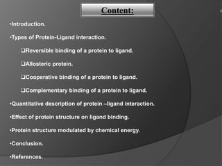
![Reversible binding of a protein to ligand.
Allosteric protein.
Cooperative binding of a protein to ligand.
Complementary binding of a protein to ligand.
Types of Protein-Ligand interaction:
There are many ways through which a protein can bind to a protein or ligand. Some of them
have been enlisted down here. They are:
[Reference: Lehninger, textbook for biochemistry]
3](https://image.slidesharecdn.com/proteinligandinteraction-151214091912/85/Protein-ligand-interaction-3-320.jpg)
![•Proteins are dynamic molecules whose functions almost invariably depend on interactions
with other molecules, and these interactions are affected in physiologically important ways
by sometimes subtle, sometimes striking changes in protein conformation.
•The functions of many proteins involve the reversible binding of other molecules. A
molecule bound reversibly by a protein is called a ligand.
•A ligand binds at a site on the protein called the binding site, which is
complementary to the ligand in size, shape, charge, and hydrophobic or hydrophilic
character.
Introduction:
[Reference: Lehninger, Principles for biochemistry]
4](https://image.slidesharecdn.com/proteinligandinteraction-151214091912/85/Protein-ligand-interaction-4-320.jpg)
![Reversible binding of a protein to ligand:
Oxygen-Binding Proteins:
Myoglobin and hemoglobin may be the most-studied and best understood proteins.
They were the first proteins for which three-dimensional structures were determined.
a. 0xygen Can Bind to a Heme Prosthetic Group:
Figure 1 Heme. (a) Porphyrins, of which protoporphyrin 1X is only one
example, consist of four pyrrole rings linked by methene bridges, with
substitutions denoted X.
[Ref: Lehninger, http://biology.kenyon.edu]
5](https://image.slidesharecdn.com/proteinligandinteraction-151214091912/85/Protein-ligand-interaction-5-320.jpg)
![Fig. 1 (b) The iron atom of heme has six coordination bonds: four in
the plane of, and bonded to, the flat porphyrin ring system
6
[Reference: Lehninger, textbook for biochemistry]](https://image.slidesharecdn.com/proteinligandinteraction-151214091912/85/Protein-ligand-interaction-6-320.jpg)
![•O2 is transported in blood by haemoglobin:
Fig.2 Binding Of oxygen to myoglobin.
Oxygen Transport:
7
[Reference: Lehninger, textbook for biochemistry]](https://image.slidesharecdn.com/proteinligandinteraction-151214091912/85/Protein-ligand-interaction-7-320.jpg)
![Effect of Ph on O2 Binding to Hb:
Fig.3 Effect of pH on oxygen binding to
hemoglobin
8
[Reference: Lehninger, textbook for biochemistry]](https://image.slidesharecdn.com/proteinligandinteraction-151214091912/85/Protein-ligand-interaction-8-320.jpg)
![b. Myoglobin has a sigle binding site for O2:
Fig.4 Structure of Myoglobin
9
[Reference: Lehninger, textbook for biochemistry]](https://image.slidesharecdn.com/proteinligandinteraction-151214091912/85/Protein-ligand-interaction-9-320.jpg)
![Allosteric Proteins:
•An allosteric protein is one in which the binding of a ligand to one site affects the
binding properties of another site on the same protein.
•The term "allosteric" derives from the Greek word, "other” and stereos, “solid” or
"shape”.
•The main function of allosteric protein is the regulation of oxygen binding to protein.
0xygen binding to Haemoglobin ls regulated by 2,3-Bisphosphoglycerate:
10
[Reference: Lehninger, textbook for biochemistry]](https://image.slidesharecdn.com/proteinligandinteraction-151214091912/85/Protein-ligand-interaction-10-320.jpg)
![Therefore describe another binding process for hemoglobin:
HbBPG + 02 HbO2 + BPG
BPG binds at a site distant from the oxygen-binding site and regulates the O2-
binding afflnity of hemoglobin in relation to the pO2 in the lungs.
Fig. 5 Effect of BPG on oxygen binding to hemoglobin
11
[Reference: Lehninger, textbook for biochemistry]](https://image.slidesharecdn.com/proteinligandinteraction-151214091912/85/Protein-ligand-interaction-11-320.jpg)
![Sickle cell anaemia ls a molecular disease of hemoglobin:
FIG. 6 Normal and sickle-cell hemoglobin .
12
[Reference: Lehninger, textbook for biochemistry]](https://image.slidesharecdn.com/proteinligandinteraction-151214091912/85/Protein-ligand-interaction-12-320.jpg)
![Cooperative binding of a protein to ligand:
• Cooperative binding of oxygen by hemoglobin was first analyzed by Archibald Hill
in 1910. From this work came a general approach to the study of cooperative ligand
binding to multi-subunit proteins.
• Cooperative conformational changes depend on variations in the structural stability of
different parts of a protein
Fig. 7 A sigmoid (cooperative) binding curve.
13
[Reference: Lehninger, textbook for biochemistry]](https://image.slidesharecdn.com/proteinligandinteraction-151214091912/85/Protein-ligand-interaction-13-320.jpg)
![FIG. 8 Structural changes in a multisubunit protein undergoing cooperative binding to
ligand
14
[Reference: Lehninger, textbook for biochemistry]](https://image.slidesharecdn.com/proteinligandinteraction-151214091912/85/Protein-ligand-interaction-14-320.jpg)
![Quantitative Description of Cooperative Ligand Binding:
For a protein within binding sites, the equilibrium of Equation 1 becomes
P + nL PL (1)
and the expression for the association constant becomes,
(2)
The expression for Ɵ is:
(3)
Rearranging, then taking the log of both sides, yield :
(4)
(5)
15
[Reference: Lehninger, textbook for biochemistry]](https://image.slidesharecdn.com/proteinligandinteraction-151214091912/85/Protein-ligand-interaction-15-320.jpg)
![Where Kd : [L]n
0.5
• Equation 5 is the Hill equation, and a plot of log [Ɵ(1-Ɵ)] versus log [L] is
called a Hill plot.
Fig. A sigmoid (cooperative) binding curve
16
[Reference: Lehninger, textbook for biochemistry]](https://image.slidesharecdn.com/proteinligandinteraction-151214091912/85/Protein-ligand-interaction-16-320.jpg)
![•To adapt the Hill equation to the binding of oxygen to hemoglobin we must
again substitute pO2 for [L] and for Ka:
Mechanisms for cooperative Binding:
FIG. 9 Two general models for the interconversion of inactive
and active forms of a protein during cooperative ligand
binding
17
[Reference: Lehninger, textbook for biochemistry]](https://image.slidesharecdn.com/proteinligandinteraction-151214091912/85/Protein-ligand-interaction-17-320.jpg)
![• Conformations of oxygen-binding proteins affect and are affected by the binding of
small ligands (O2 or CO) to the heme group.
• However, most Protein ligand interactions do not involve a prosthetic group. Instead, the
binding site for a Iigand is more often Iike the hemoglobin binding site for BPG-a cleft in
the protein lined with amino acid residues, arranged to make the binding interaction
highly specific.
Complementary Interactions between Proteins and Ligand:
•Immunity is brought about by a variety of leukocytes(white blood cells), including
macrophages and lymphocytes, all of which develop from undifferentiated stem cells in
the bone marrow.
•Leukocytes can leave the bloodstream and patrol the tissues, each cell producing one or
more proteins capable of recognizing and binding to molecules that might signal an
infection.
The immune response features a specialized array of cells and proteins:
18
[Reference: Lehninger, textbook for biochemistry, www.wikipedia.org]](https://image.slidesharecdn.com/proteinligandinteraction-151214091912/85/Protein-ligand-interaction-18-320.jpg)
![•The immune response consists of two complementary systems, the humoral and
cellular immune systems.
•The humoral immune system (Latin humor, “fluid”) is directed at bacterial infections
and extracellular viruses (those found in the body fluids), but can also respond to
individual foreign proteins.
•The cellular immune system destroys host cells infected by viruses and also destroys
some parasites and foreign tissues.
Some types of leukocytes associated with immune response:
19
[Reference: Lehninger, textbook for biochemistry]](https://image.slidesharecdn.com/proteinligandinteraction-151214091912/85/Protein-ligand-interaction-19-320.jpg)
![Fig. 10 Imunoglobulin
20
[Reference: Lehninger, textbook for biochemistry]](https://image.slidesharecdn.com/proteinligandinteraction-151214091912/85/Protein-ligand-interaction-20-320.jpg)
![•The function of myoglobin depends on the protein's ability not only to bind oxygen
but also to release it when and where it is needed.
•
•A quantitative description of this interaction is therefore a central part of many
biochemical investigations.
•
•In general, the reversible binding of a protein (P) to a ligand (L) can be described by
a simple equilibrium expression:
P + L PL 1
•The reaction is characterized by equilibrium constant, Ka, such that,
2
P + L PL
3
Quantitative description of a protein – ligand Interaction: 21
[Reference: Lehninger, textbook for biochemistry]](https://image.slidesharecdn.com/proteinligandinteraction-151214091912/85/Protein-ligand-interaction-21-320.jpg)
![4
5
Fig 11 Graphical representations of ligand binding. A hypothetical binding curve
for a ligand L
The value of Ka can be determined from a plot of g versus the concentration of
free ligand, [L] (F'ig. 11 ).
22
[Reference: Lehninger, textbook for biochemistry]](https://image.slidesharecdn.com/proteinligandinteraction-151214091912/85/Protein-ligand-interaction-22-320.jpg)

![The expression for Ɵ is:
Rearranging, then taking the log of both sides, yield:
3
4
5
2
24
[Reference: Lehninger, textbook for biochemistry]](https://image.slidesharecdn.com/proteinligandinteraction-151214091912/85/Protein-ligand-interaction-24-320.jpg)
![FIG. 12 Hill plots for oxygen binding to myoglobin and haemoglobin
25
[Reference: Lehninger, textbook for biochemistry]](https://image.slidesharecdn.com/proteinligandinteraction-151214091912/85/Protein-ligand-interaction-25-320.jpg)
![•The binding of a ligand to a protein is rarely as simple as the above equations
would suggest. The interaction is greatly affected by protein structure and is often
accompanied by conformational changes.
Effect of protein structure on ligand binding:
FIG. 13. Steric effects caused by ligand binding to the heme of myoglobin.
(a) Oxygen binds to heme with the O, axis at an angle, a binding conformation readily
accommodated by myoglobin.
(b) Carbon monoxide binds to free heme with the CO axis perpendicular to the plane of
the porphyrin ring
26
[Reference: Lehninger, textbook for biochemistry, www.amzon.com]](https://image.slidesharecdn.com/proteinligandinteraction-151214091912/85/Protein-ligand-interaction-26-320.jpg)


Proteins interact with other molecules through various types of binding. Reversible binding allows proteins to transport molecules like oxygen, with hemoglobin binding oxygen through a heme group. Binding can be allosteric, affecting other sites; or cooperative, where ligand binding causes conformational changes influencing additional binding. The protein structure complements the ligand, precisely matching its shape and chemistry. Quantitative analyses describe these interactions through equilibrium constants and binding curves.


![[Brief]Structure and functions of hemoglobin and myglobin (Bio-Inorganic chem...](https://cdn.slidesharecdn.com/ss_thumbnails/briefstructureandfunctionsofhb-mb-180511052541-thumbnail.jpg?width=640&height=640&fit=bounds)