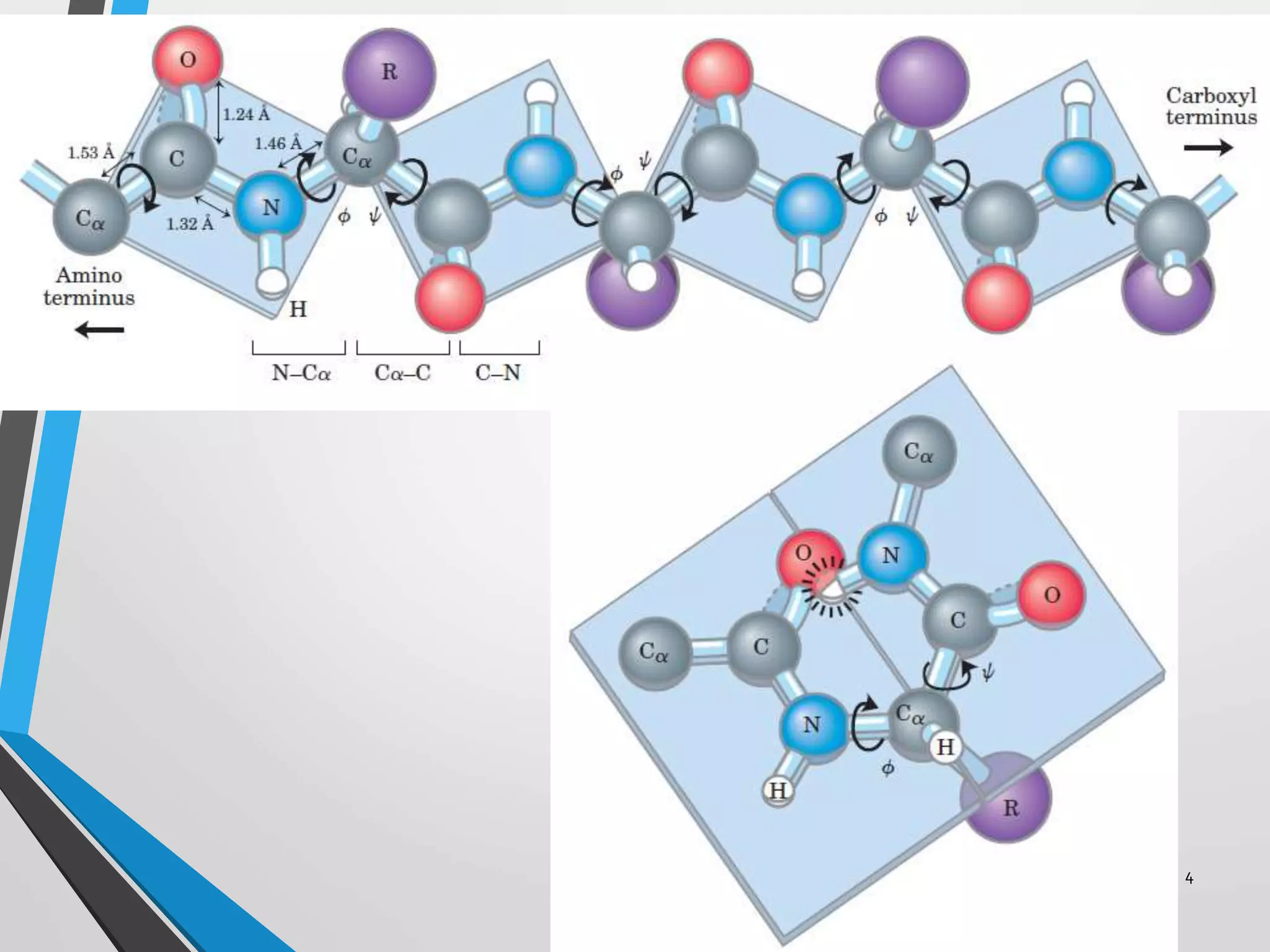
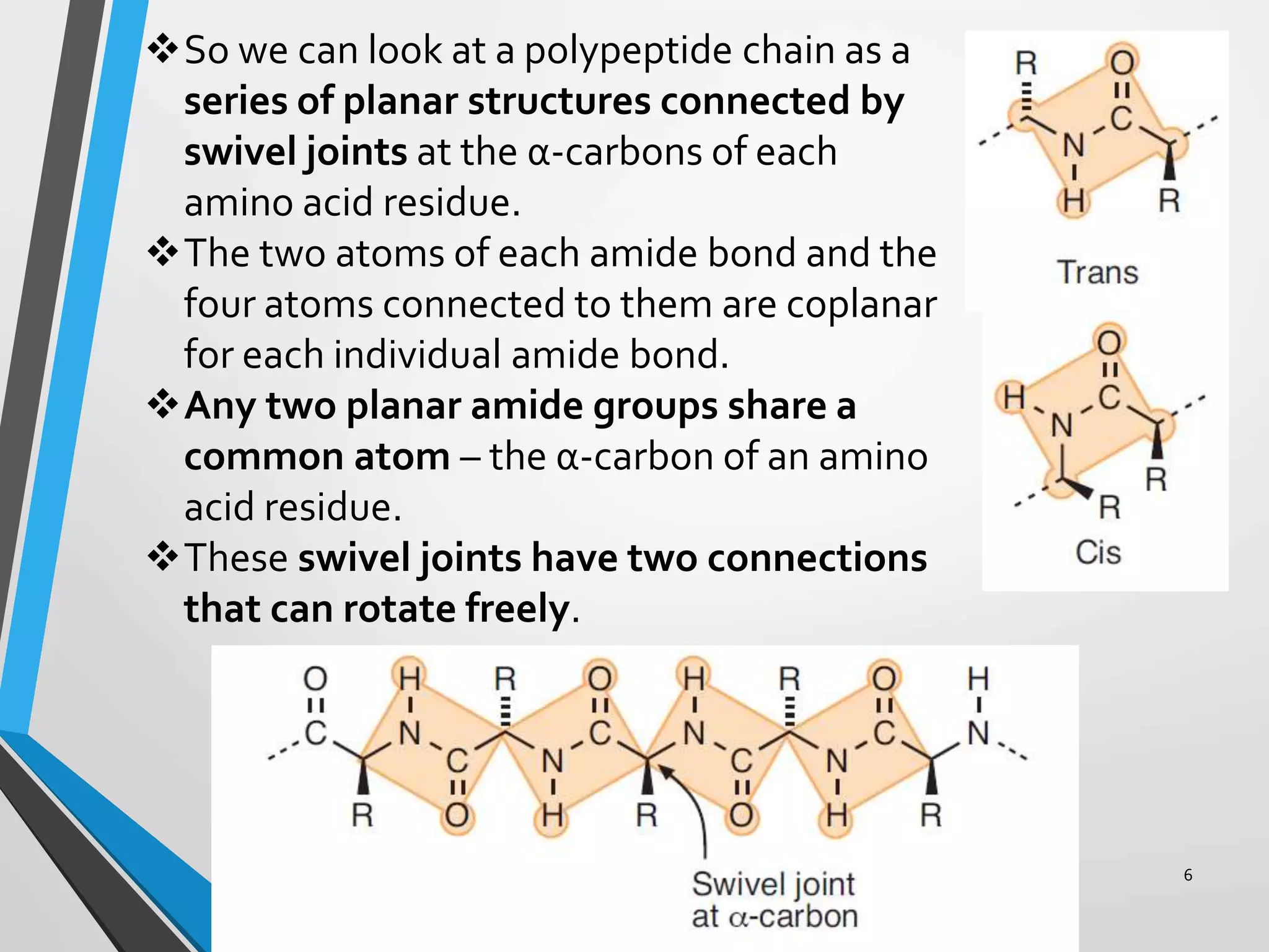
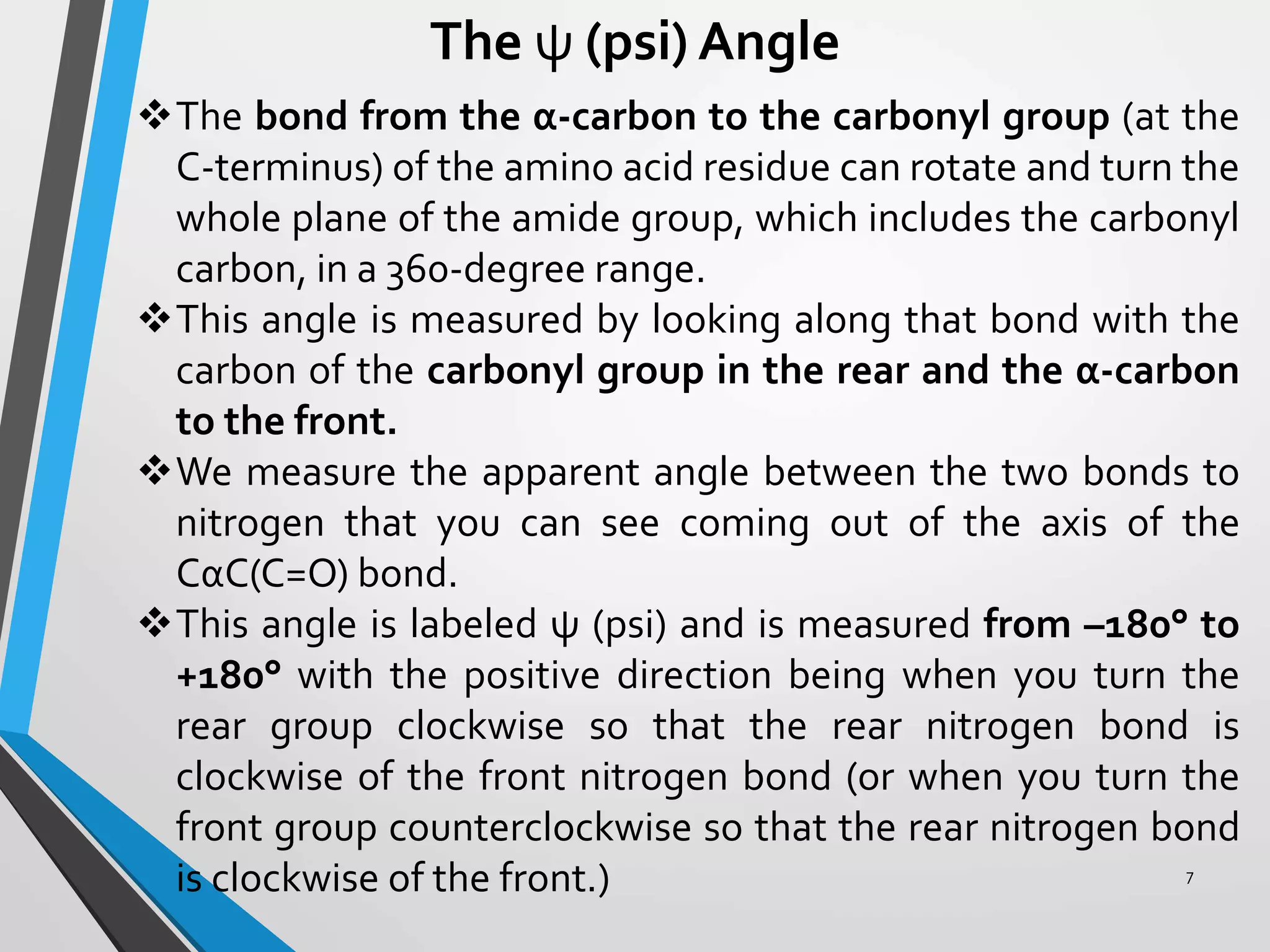
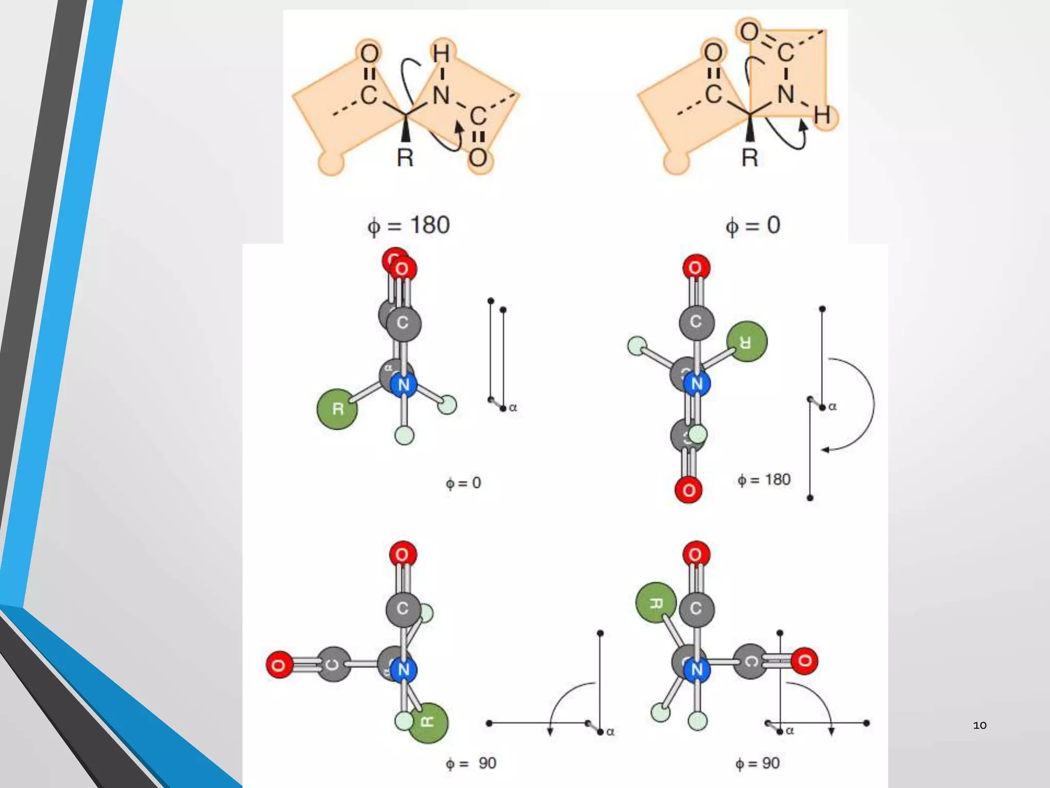
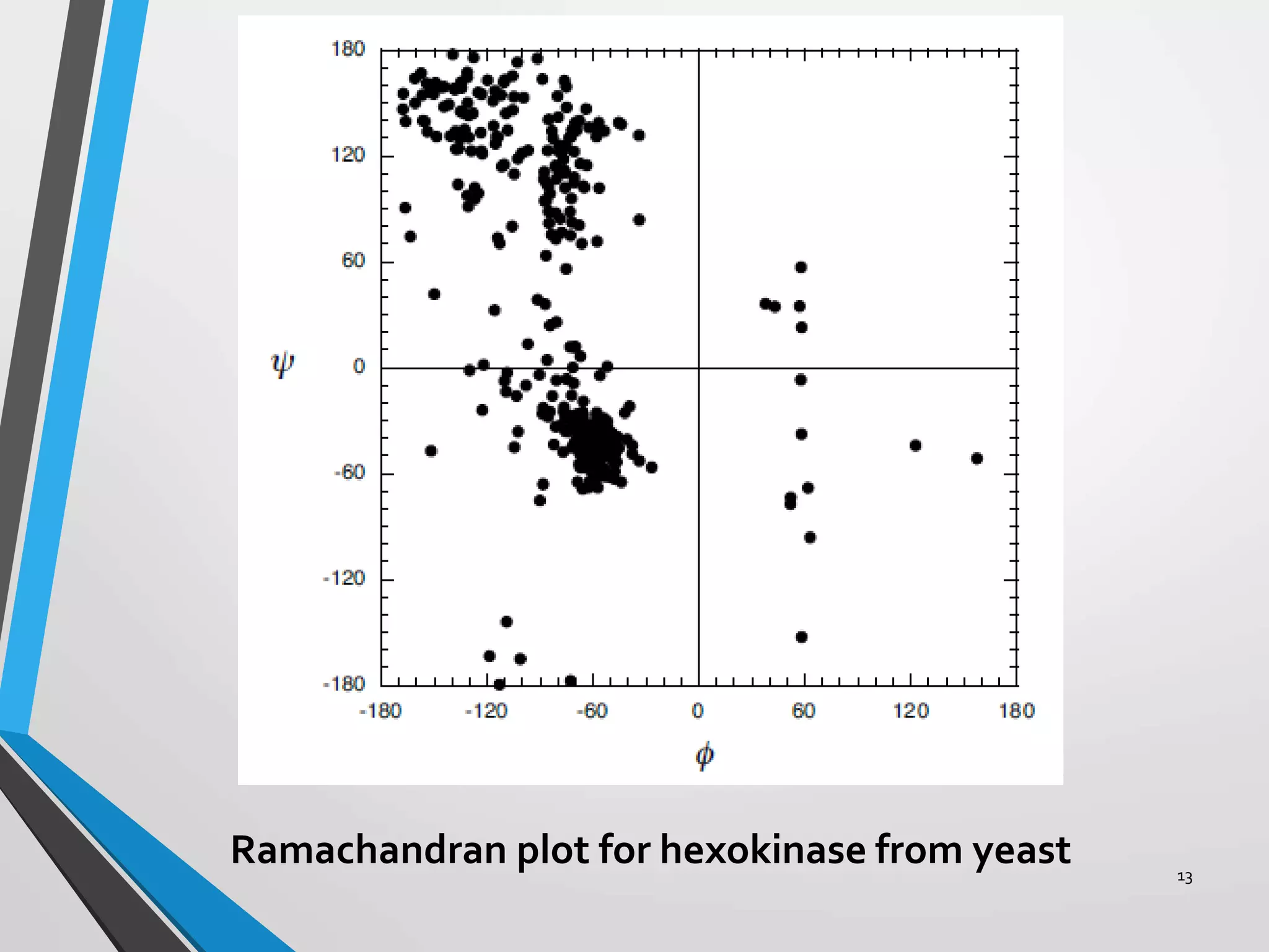
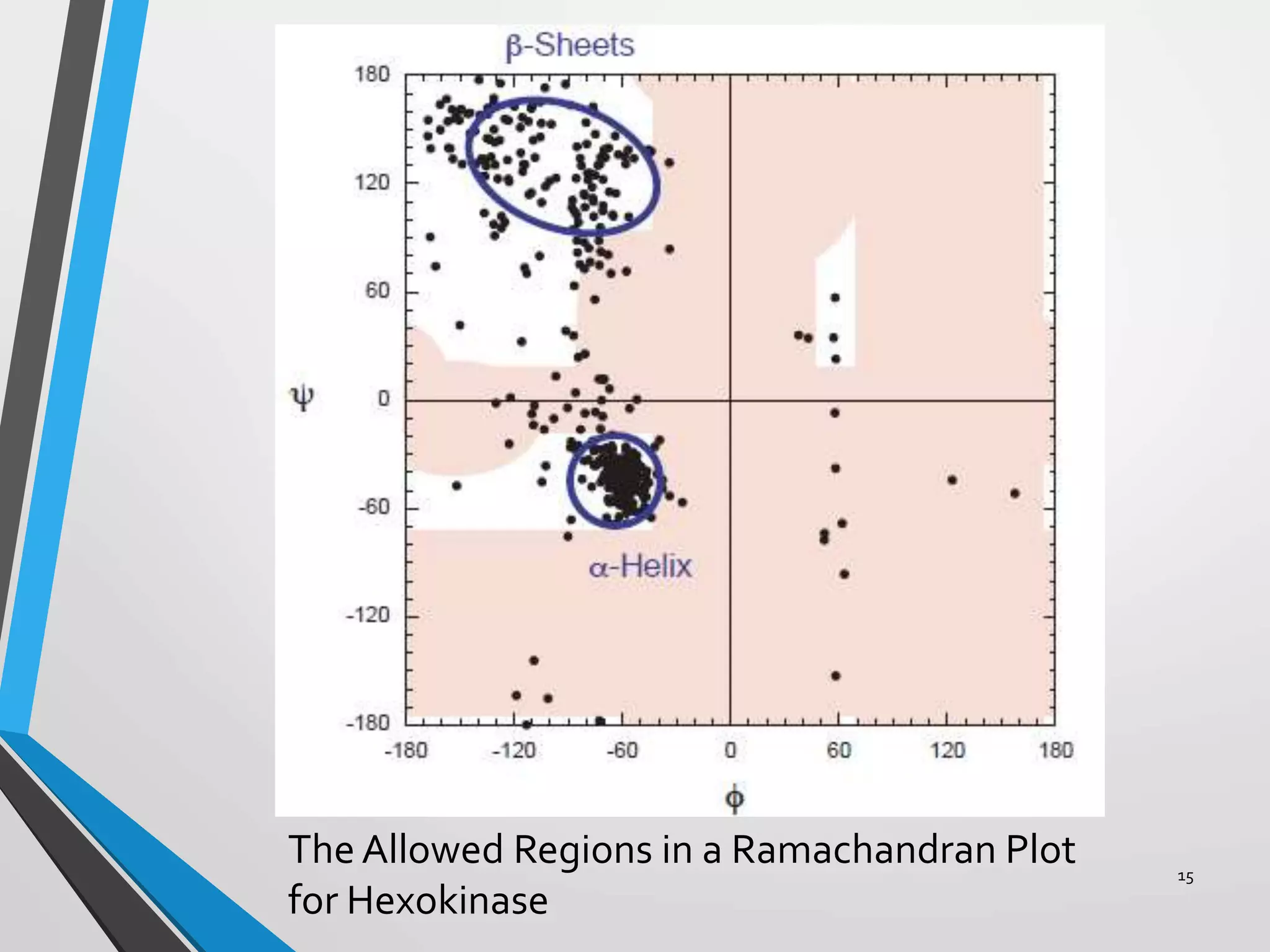
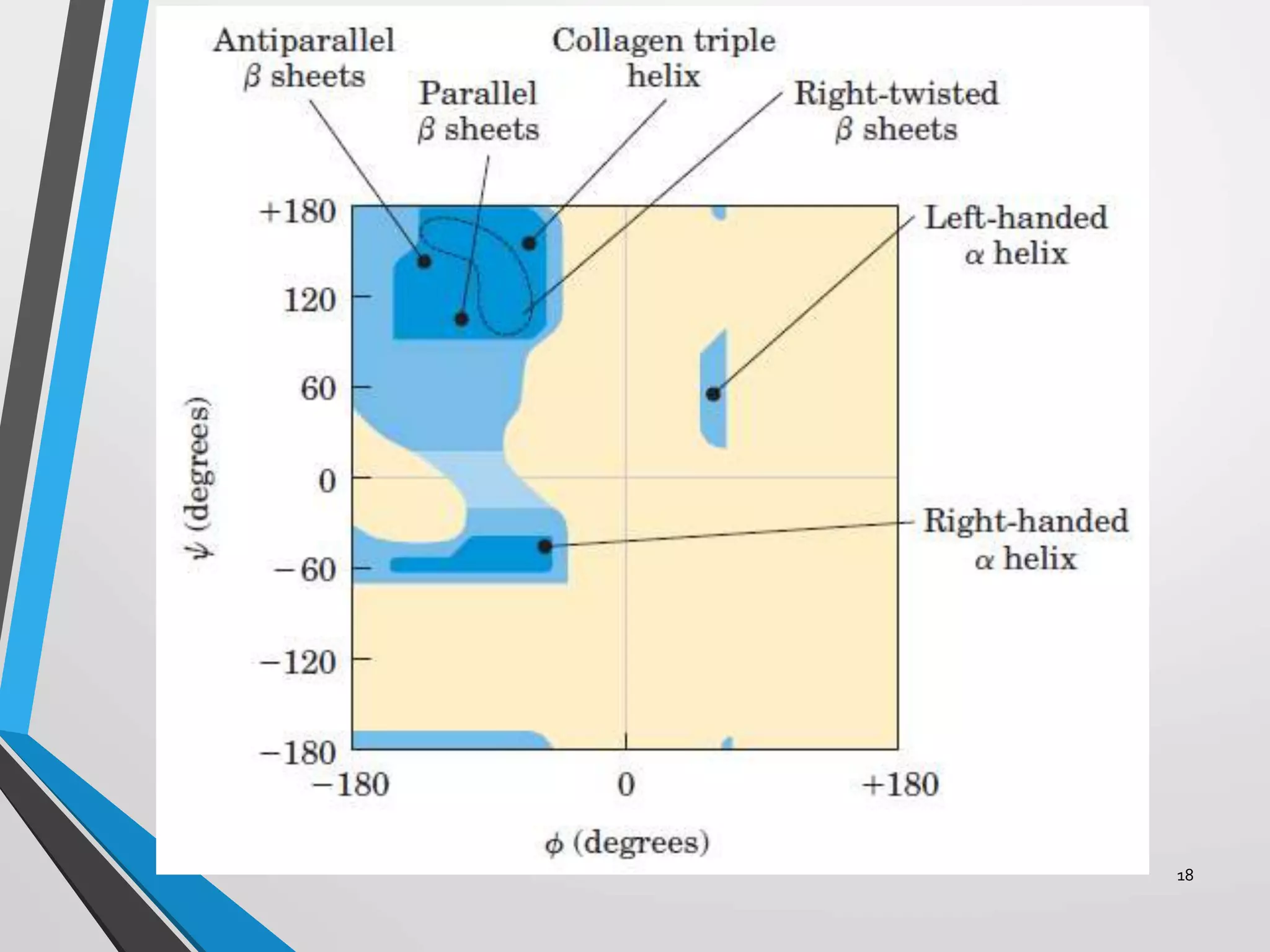
1
Presented by-
Mr. Sushant Balasaheb Jadhav
Roll no. – 18PBT206
M.Tech. Pharmaceutical Biotechnology
Institute of ChemicalTechnology
2
CONTENTS
 Introduction
 Limits on Rotation in Amides
 The ψ (psi) Angle
 The φ (phi) Angle
 The Ramachandran Plot
 Steric Limits of ψ and φ
 Conclusion
 References
INTRODUCTION
The secondary structures that polypeptides can adopt in
proteins are governed by hydrogen bonding interactions
between the electronegative carbonyl oxygen atoms and
the electropositive amide hydrogen atoms in the backbone
chain of the molecule.
These hydrogen-bonding interactions can form the
framework that stabilizes the secondary structure.
Many secondary structures with reasonable hydrogen
bonding networks could be proposed but we see only a few
possibilities in polypeptides composed of L-amino acids
(proteins).
Most of the possible secondary structures are not possible
due to limits on the configuration of the backbone of each
amino acid residue.
Understanding these limitations will help you to understand
the secondary structures of proteins. 3
4
Limits on Rotation in Amides
 We do not have as many degrees of freedom as we have
bonds.
 The amide bonds are held in a planar orientation due to
orbital overlap between the π-orbitals of the SP2
hybridized atoms of the carbonyl group and the π-orbital of
the SP2 hybridized amine group.
 This distributed π-orbital system resists rotation, as the
orbital overlap would have to be broken.
 The distributed nature of the system also makes the amide
bond resistant to hydrolysis.
 The amide group will prefer to have all atoms connected to it
in the same plane as the amide bond (no rotation).
 This can result in a ‘cis’ or ‘trans’ configuration.
 Due to steric and electronic reasons the ‘trans’
configuration is the predominant form for the amide bond.
 Note the steric clash between the side chain groups in the
‘cis’ form.
5
So we can look at a polypeptide chain as a
series of planar structures connected by
swivel joints at the α-carbons of each
amino acid residue.
The two atoms of each amide bond and the
four atoms connected to them are coplanar
for each individual amide bond.
Any two planar amide groups share a
common atom – the α-carbon of an amino
acid residue.
These swivel joints have two connections
that can rotate freely.
6
The ψ (psi) Angle
The bond from the α-carbon to the carbonyl group (at the
C-terminus) of the amino acid residue can rotate and turn the
whole plane of the amide group, which includes the carbonyl
carbon, in a 360-degree range.
This angle is measured by looking along that bond with the
carbon of the carbonyl group in the rear and the α-carbon
to the front.
We measure the apparent angle between the two bonds to
nitrogen that you can see coming out of the axis of the
CαC(C=O) bond.
This angle is labeled ψ (psi) and is measured from –180° to
+180° with the positive direction being when you turn the
rear group clockwise so that the rear nitrogen bond is
clockwise of the front nitrogen bond (or when you turn the
front group counterclockwise so that the rear nitrogen bond
is clockwise of the front.) 7
8
The φ (phi) Angle
The bond from the nitrogen (at the N-terminus) to the α-
carbon of the amino acid residue can rotate and turn the
whole plane of the other amide group, which includes the
nitrogen, in a 360-degree range.
This angle is measured by looking along that bond with the
nitrogen atom in front and the α-carbon to the rear.
We measure the apparent angle between the two bonds to
the carbonyl carbons that you can see coming out of the axis
of the NCα bond.
This angle is labeled φ (phi) and is measured from –180° to
+180° with the positive direction being when you turn the
rear group clockwise so that the rear carbonyl bond is
clockwise of the front carbonyl bond (or when you turn the
front group counterclockwise so that the rear carbonyl bond
is clockwise of the front.)
9
10
The Ramachandran Plot
We can vary ψ from –180° to 180° and we can vary φ from –
180° to 180° (that is 360° of rotation for each).
But many combinations of these angles are almost never
seen and others are very, very common in proteins.
We will obtain a data set for the positions of each atom in
space. We can get such data from one of the several
depositories of protein structural data.
We will use X-ray crystallography data, as it is the most
precise. (Although it may not be completely accurate, as
crystal packing forces usually distort proteins slightly.
11
12
The Ramachandran Plot
NMR data for proteins in solution are not very precise but
are considered to be more accurate as the protein is in its
native environment.
We can take this data and detect the amino acid residues
(this is usually done for us as the X-ray crystallography
“.PDB” data format labels all atoms and assigns them to
residues).
A computer program can take a “.PDB” file and report the of
ψ and φ angles for each and every residue.
A plot of ψ vs. φ is called a Ramachandran plot.
Ramachandran plot for hexokinase from yeast 13
Steric Limits of ψ and φ
Atoms take up space and cannot occupy the same
space at the same time.
Covalent bonds connect them and these bonds cannot
be broken.
So the movements that we are concerned with involve
rotations only.
There are only two angles that rotate in a given
residue, ψ and φ.
But not all combinations are possible due to physical
clashes of atoms in 3-dimensional space.
These physical clashes are called steric interactions
and the limit the available values for ψ and φ.
14
The Allowed Regions in a Ramachandran Plot
for Hexokinase
15
CONCLUSION
Understanding the steric limitations in individual amino acid
residues reveals how these limitations result in the observed
types of secondary structures found in nature.
The complex folding of proteins is controlled, in a large
part, by the limitations on the ψ and φ angles available to
each amino acid residue.
If one inspects the ψ and φ angle combinations that exist in
idealized secondary structures we can place the sites in a
Ramachandran plot where we would expect to see data
related to these structures.
In a real protein we would expect data points from these
defined secondary structures to appear near the ideal
location.
16
17
Dihedral angles, translation distances and number
of residues per turn for regular secondary structure
conformations
18
19
REFERENCES
http://www.greeley.org/~hod/papers/Unsorte
d/Ramachandran.doc.pdf
Biochemistry by Jeremy M. Berg, John L.
Tymoczko and Lubert Stryer
Lehninger PRINCIPLES OF BIOCHEMISTRY
by David L. Nelson and Michael M. Cox
20

Ramachandran plot

  • 1.
    1 Presented by- Mr. SushantBalasaheb Jadhav Roll no. – 18PBT206 M.Tech. Pharmaceutical Biotechnology Institute of ChemicalTechnology
  • 2.
    2 CONTENTS  Introduction  Limitson Rotation in Amides  The ψ (psi) Angle  The φ (phi) Angle  The Ramachandran Plot  Steric Limits of ψ and φ  Conclusion  References
  • 3.
    INTRODUCTION The secondary structuresthat polypeptides can adopt in proteins are governed by hydrogen bonding interactions between the electronegative carbonyl oxygen atoms and the electropositive amide hydrogen atoms in the backbone chain of the molecule. These hydrogen-bonding interactions can form the framework that stabilizes the secondary structure. Many secondary structures with reasonable hydrogen bonding networks could be proposed but we see only a few possibilities in polypeptides composed of L-amino acids (proteins). Most of the possible secondary structures are not possible due to limits on the configuration of the backbone of each amino acid residue. Understanding these limitations will help you to understand the secondary structures of proteins. 3
  • 4.
  • 5.
    Limits on Rotationin Amides  We do not have as many degrees of freedom as we have bonds.  The amide bonds are held in a planar orientation due to orbital overlap between the π-orbitals of the SP2 hybridized atoms of the carbonyl group and the π-orbital of the SP2 hybridized amine group.  This distributed π-orbital system resists rotation, as the orbital overlap would have to be broken.  The distributed nature of the system also makes the amide bond resistant to hydrolysis.  The amide group will prefer to have all atoms connected to it in the same plane as the amide bond (no rotation).  This can result in a ‘cis’ or ‘trans’ configuration.  Due to steric and electronic reasons the ‘trans’ configuration is the predominant form for the amide bond.  Note the steric clash between the side chain groups in the ‘cis’ form. 5
  • 6.
    So we canlook at a polypeptide chain as a series of planar structures connected by swivel joints at the α-carbons of each amino acid residue. The two atoms of each amide bond and the four atoms connected to them are coplanar for each individual amide bond. Any two planar amide groups share a common atom – the α-carbon of an amino acid residue. These swivel joints have two connections that can rotate freely. 6
  • 7.
    The ψ (psi)Angle The bond from the α-carbon to the carbonyl group (at the C-terminus) of the amino acid residue can rotate and turn the whole plane of the amide group, which includes the carbonyl carbon, in a 360-degree range. This angle is measured by looking along that bond with the carbon of the carbonyl group in the rear and the α-carbon to the front. We measure the apparent angle between the two bonds to nitrogen that you can see coming out of the axis of the CαC(C=O) bond. This angle is labeled ψ (psi) and is measured from –180° to +180° with the positive direction being when you turn the rear group clockwise so that the rear nitrogen bond is clockwise of the front nitrogen bond (or when you turn the front group counterclockwise so that the rear nitrogen bond is clockwise of the front.) 7
  • 8.
  • 9.
    The φ (phi)Angle The bond from the nitrogen (at the N-terminus) to the α- carbon of the amino acid residue can rotate and turn the whole plane of the other amide group, which includes the nitrogen, in a 360-degree range. This angle is measured by looking along that bond with the nitrogen atom in front and the α-carbon to the rear. We measure the apparent angle between the two bonds to the carbonyl carbons that you can see coming out of the axis of the NCα bond. This angle is labeled φ (phi) and is measured from –180° to +180° with the positive direction being when you turn the rear group clockwise so that the rear carbonyl bond is clockwise of the front carbonyl bond (or when you turn the front group counterclockwise so that the rear carbonyl bond is clockwise of the front.) 9
  • 10.
  • 11.
    The Ramachandran Plot Wecan vary ψ from –180° to 180° and we can vary φ from – 180° to 180° (that is 360° of rotation for each). But many combinations of these angles are almost never seen and others are very, very common in proteins. We will obtain a data set for the positions of each atom in space. We can get such data from one of the several depositories of protein structural data. We will use X-ray crystallography data, as it is the most precise. (Although it may not be completely accurate, as crystal packing forces usually distort proteins slightly. 11
  • 12.
    12 The Ramachandran Plot NMRdata for proteins in solution are not very precise but are considered to be more accurate as the protein is in its native environment. We can take this data and detect the amino acid residues (this is usually done for us as the X-ray crystallography “.PDB” data format labels all atoms and assigns them to residues). A computer program can take a “.PDB” file and report the of ψ and φ angles for each and every residue. A plot of ψ vs. φ is called a Ramachandran plot.
  • 13.
    Ramachandran plot forhexokinase from yeast 13
  • 14.
    Steric Limits ofψ and φ Atoms take up space and cannot occupy the same space at the same time. Covalent bonds connect them and these bonds cannot be broken. So the movements that we are concerned with involve rotations only. There are only two angles that rotate in a given residue, ψ and φ. But not all combinations are possible due to physical clashes of atoms in 3-dimensional space. These physical clashes are called steric interactions and the limit the available values for ψ and φ. 14
  • 15.
    The Allowed Regionsin a Ramachandran Plot for Hexokinase 15
  • 16.
    CONCLUSION Understanding the stericlimitations in individual amino acid residues reveals how these limitations result in the observed types of secondary structures found in nature. The complex folding of proteins is controlled, in a large part, by the limitations on the ψ and φ angles available to each amino acid residue. If one inspects the ψ and φ angle combinations that exist in idealized secondary structures we can place the sites in a Ramachandran plot where we would expect to see data related to these structures. In a real protein we would expect data points from these defined secondary structures to appear near the ideal location. 16
  • 17.
    17 Dihedral angles, translationdistances and number of residues per turn for regular secondary structure conformations
  • 18.
  • 19.
    19 REFERENCES http://www.greeley.org/~hod/papers/Unsorte d/Ramachandran.doc.pdf Biochemistry by JeremyM. Berg, John L. Tymoczko and Lubert Stryer Lehninger PRINCIPLES OF BIOCHEMISTRY by David L. Nelson and Michael M. Cox
  • 20.