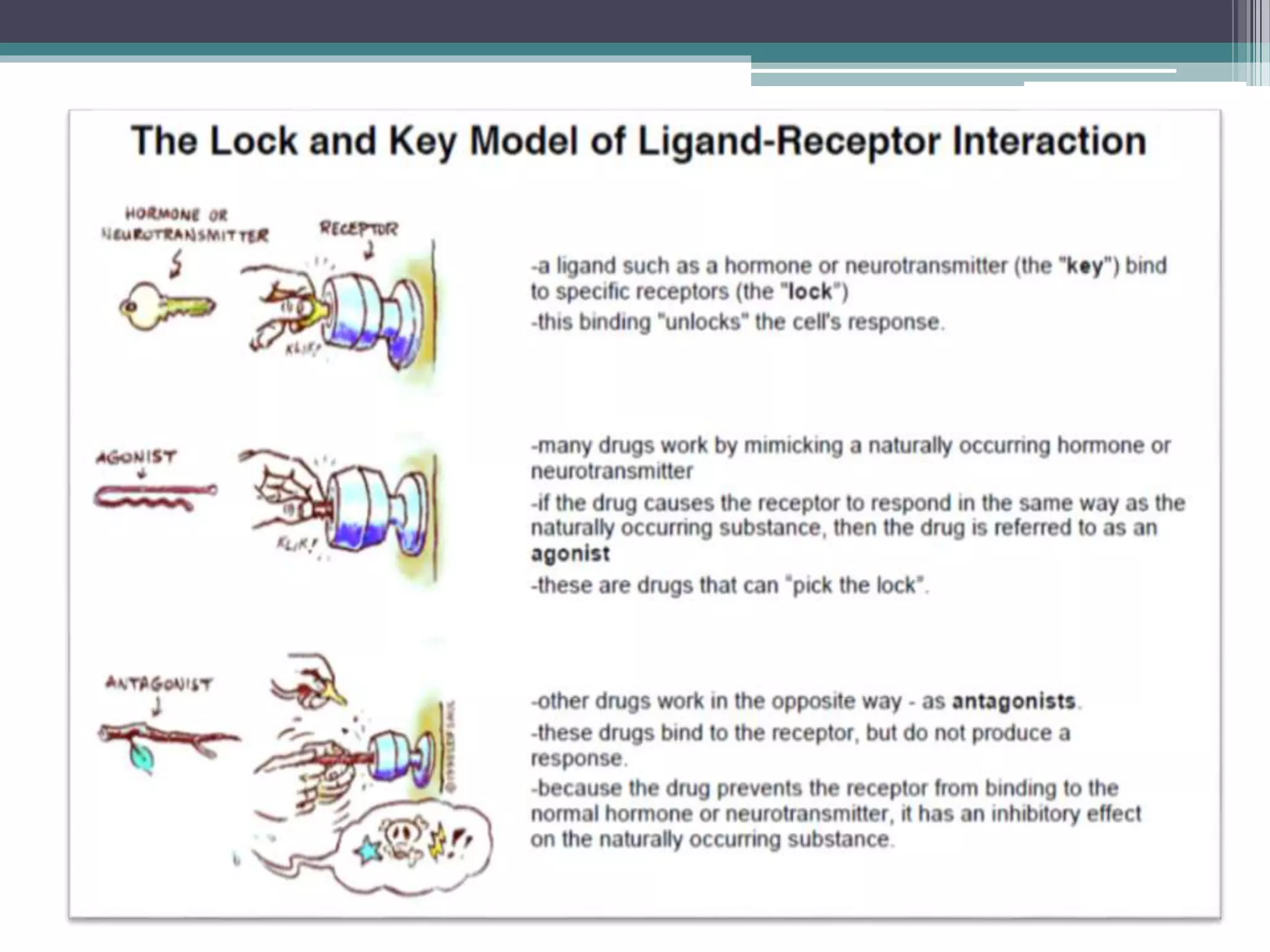
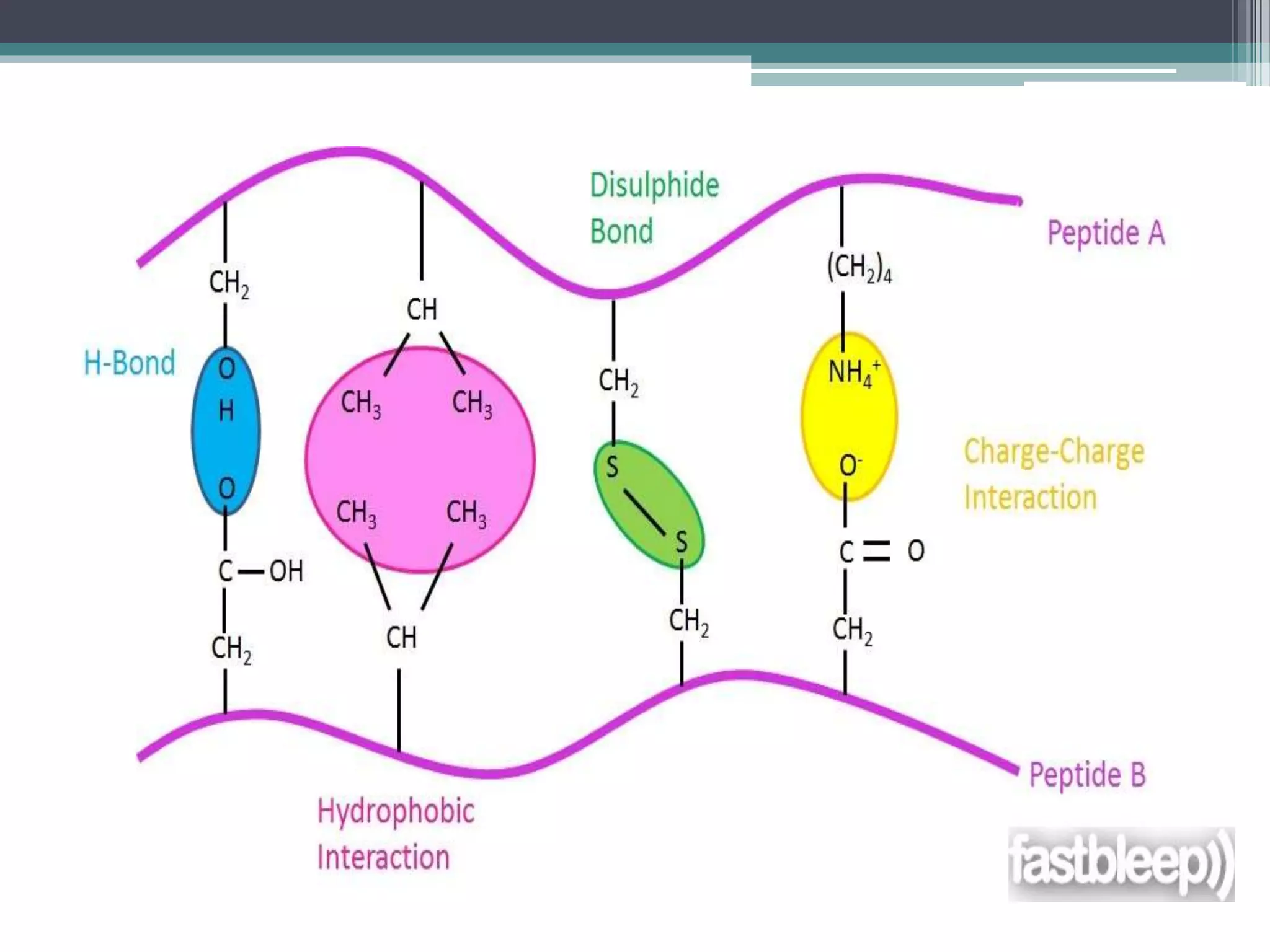
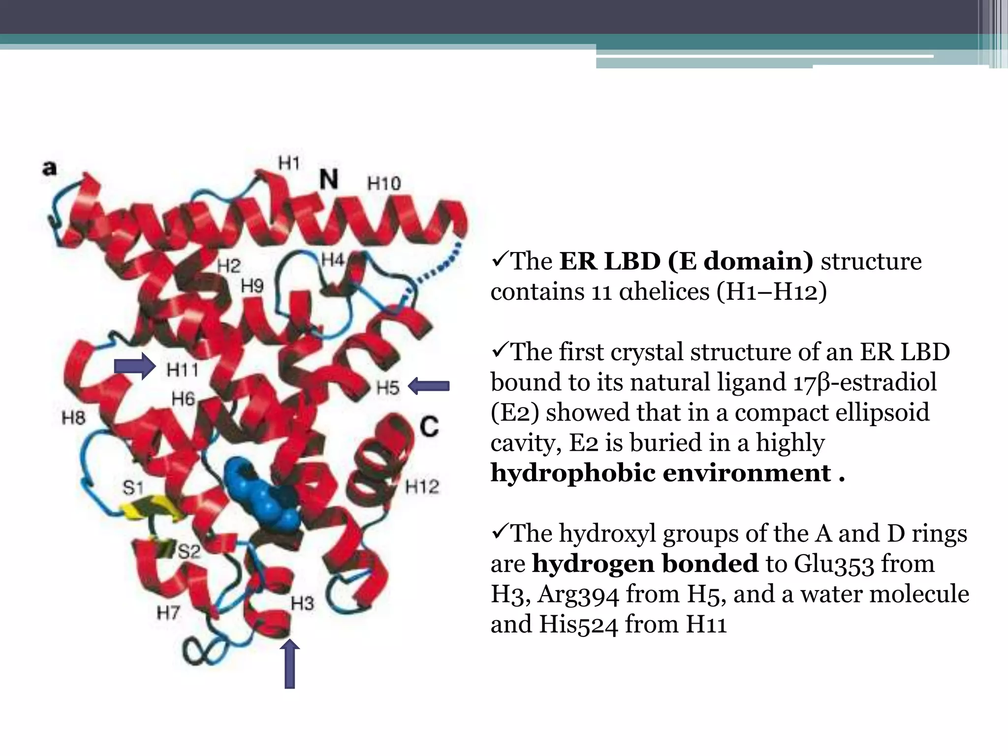
This document discusses ligand-receptor interactions, using estrogen receptor and estrogen as an example. It describes how ligands bind to specific receptors on target cells, inducing a conformational change and cellular response. It then details the domains of estrogen receptors, how estrogen binds and activates the receptors, and the role of the receptor domains in transcription. The discussion includes examples of covalent and non-covalent ligand-receptor interactions.
Prediction of the three dimensional structure of a given protein sequence i.e. target protein from the amino acid sequence of a homologous (template) protein for which an X-ray or NMR structure is available based on an alignment to one or more known protein structures
molecular docking its types and de novo drug design and application and softw...GAUTAM KHUNE
This ppt deals with all the aspects related to molecular docking ,its types(rigid ,flexible and manual) and screening based on it and also deals with de novo drug design , various softwares available for docking methodologies and applications for molecular docking in new drug design
INTRODUCTION
A PERFECT THERAPEUTIC DRUG
DRUG DISCOVERY- HISTORY
MODERN DRUG DISCOVERY
BIOINFORATICS IN DRUG DISCOVERY
DRUG DISCOVERY BASED ON BIOINFORMATIC TOOLS
BIOINFORMATICS IN COMPUTER-AIDED DRUG DISCOVERY
ECONOMICS OF DRUG DISCOVERY
CONCLUSION
REFERENCES
Drug discovery take years to decade for discovering a new drug and very costly
Effort to cut down the research timeline and cost by reducing wet-lab experiment use computer modeling
Others have done the work. Some have used the work. I have spoken only on behalf of their behalf.
introduction of Lipids,Chemistry,Fuctions of lipids,Classification of lipids Structural elucidation of Essential Fatty acid,Prostaglandins, Vitamin A, Phospolipids,Cholesterol,Lanosterol,
A receptor is a protein molecule usually found embedded within the plasma membrane surface of a cell that receives chemical signals from outside the cell and when such chemical signals bind to a receptor, they cause some form of cellular/tissue response.
Receptor is a macromolecule whose function is to recognize and respond to chemical signal
There are 3 types of receptors. Those are:
i. Internal /Intracellular/Cytoplasmic receptors:
found in the cytoplasm of the cell
respond to hydrophobic ligand molecules
ii. Cell-surface / transmembrane receptors/ cell specific proteins
performs signal transduction, converting an extracellular signal into an intracellular signal.
iii. Nuclear receptor
Located in the nucleus of the cell
i. Internal /Intracellular/Cytoplasmic receptors
Steriod receptor
ii. Cell-surface / transmembrane receptors/ cell specific proteins
G-protein- coupled receptor
Enzyme- linked receptor/ tyrosin kinase receptor
Ion channel- linked receptor/ ligand gated receptor
iii. Nuclear receptor
Thyroid receptor
Advanced Medicinal Chemistry of GPCR Receptorsaurabh gupta
Contents:-
Introduction
Structure of G-protein
Signal Molecules / Ligands of GPCRs
G- Protein Mediated Pathways
Receptor Site Theories
Forces involved in drug receptor interactions
Introduction
Common terms used
Protein ligand interaction
1)in oxygen binding protein
Heme
Structure of hemoglobin
Ligand binding effected by protein structure
Oxygenation &deoxygenation
Cooperative binding of oxygen
Models for cooperative binding
Hb also transports H+&co2
2)complementary interaction immune system &immunoglobulin
Introduction
Structure of antibodies
Binding of antigen to antibody
Applications
Conclusion
Reference
The binding of ligand and receptors has been a
key component in the understanding o the
complex signalling pathway in our cells.
tow types of receptors 1.Intracellular receptors
2.Cell surface receptors
Diagramatic representation of the binding
of ligand and receptor
Earliest Galaxies in the JADES Origins Field: Luminosity Function and Cosmic ...Sérgio Sacani
We characterize the earliest galaxy population in the JADES Origins Field (JOF), the deepest
imaging field observed with JWST. We make use of the ancillary Hubble optical images (5 filters
spanning 0.4−0.9µm) and novel JWST images with 14 filters spanning 0.8−5µm, including 7 mediumband filters, and reaching total exposure times of up to 46 hours per filter. We combine all our data
at > 2.3µm to construct an ultradeep image, reaching as deep as ≈ 31.4 AB mag in the stack and
30.3-31.0 AB mag (5σ, r = 0.1” circular aperture) in individual filters. We measure photometric
redshifts and use robust selection criteria to identify a sample of eight galaxy candidates at redshifts
z = 11.5 − 15. These objects show compact half-light radii of R1/2 ∼ 50 − 200pc, stellar masses of
M⋆ ∼ 107−108M⊙, and star-formation rates of SFR ∼ 0.1−1 M⊙ yr−1
. Our search finds no candidates
at 15 < z < 20, placing upper limits at these redshifts. We develop a forward modeling approach to
infer the properties of the evolving luminosity function without binning in redshift or luminosity that
marginalizes over the photometric redshift uncertainty of our candidate galaxies and incorporates the
impact of non-detections. We find a z = 12 luminosity function in good agreement with prior results,
and that the luminosity function normalization and UV luminosity density decline by a factor of ∼ 2.5
from z = 12 to z = 14. We discuss the possible implications of our results in the context of theoretical
models for evolution of the dark matter halo mass function.
Introduction:
RNA interference (RNAi) or Post-Transcriptional Gene Silencing (PTGS) is an important biological process for modulating eukaryotic gene expression.
It is highly conserved process of posttranscriptional gene silencing by which double stranded RNA (dsRNA) causes sequence-specific degradation of mRNA sequences.
dsRNA-induced gene silencing (RNAi) is reported in a wide range of eukaryotes ranging from worms, insects, mammals and plants.
This process mediates resistance to both endogenous parasitic and exogenous pathogenic nucleic acids, and regulates the expression of protein-coding genes.
What are small ncRNAs?
micro RNA (miRNA)
short interfering RNA (siRNA)
Properties of small non-coding RNA:
Involved in silencing mRNA transcripts.
Called “small” because they are usually only about 21-24 nucleotides long.
Synthesized by first cutting up longer precursor sequences (like the 61nt one that Lee discovered).
Silence an mRNA by base pairing with some sequence on the mRNA.
Discovery of siRNA?
The first small RNA:
In 1993 Rosalind Lee (Victor Ambros lab) was studying a non- coding gene in C. elegans, lin-4, that was involved in silencing of another gene, lin-14, at the appropriate time in the
development of the worm C. elegans.
Two small transcripts of lin-4 (22nt and 61nt) were found to be complementary to a sequence in the 3' UTR of lin-14.
Because lin-4 encoded no protein, she deduced that it must be these transcripts that are causing the silencing by RNA-RNA interactions.
Types of RNAi ( non coding RNA)
MiRNA
Length (23-25 nt)
Trans acting
Binds with target MRNA in mismatch
Translation inhibition
Si RNA
Length 21 nt.
Cis acting
Bind with target Mrna in perfect complementary sequence
Piwi-RNA
Length ; 25 to 36 nt.
Expressed in Germ Cells
Regulates trnasposomes activity
MECHANISM OF RNAI:
First the double-stranded RNA teams up with a protein complex named Dicer, which cuts the long RNA into short pieces.
Then another protein complex called RISC (RNA-induced silencing complex) discards one of the two RNA strands.
The RISC-docked, single-stranded RNA then pairs with the homologous mRNA and destroys it.
THE RISC COMPLEX:
RISC is large(>500kD) RNA multi- protein Binding complex which triggers MRNA degradation in response to MRNA
Unwinding of double stranded Si RNA by ATP independent Helicase
Active component of RISC is Ago proteins( ENDONUCLEASE) which cleave target MRNA.
DICER: endonuclease (RNase Family III)
Argonaute: Central Component of the RNA-Induced Silencing Complex (RISC)
One strand of the dsRNA produced by Dicer is retained in the RISC complex in association with Argonaute
ARGONAUTE PROTEIN :
1.PAZ(PIWI/Argonaute/ Zwille)- Recognition of target MRNA
2.PIWI (p-element induced wimpy Testis)- breaks Phosphodiester bond of mRNA.)RNAse H activity.
MiRNA:
The Double-stranded RNAs are naturally produced in eukaryotic cells during development, and they have a key role in regulating gene expression .
A brief information about the SCOP protein database used in bioinformatics.
The Structural Classification of Proteins (SCOP) database is a comprehensive and authoritative resource for the structural and evolutionary relationships of proteins. It provides a detailed and curated classification of protein structures, grouping them into families, superfamilies, and folds based on their structural and sequence similarities.
Richard's aventures in two entangled wonderlandsRichard Gill
Since the loophole-free Bell experiments of 2020 and the Nobel prizes in physics of 2022, critics of Bell's work have retreated to the fortress of super-determinism. Now, super-determinism is a derogatory word - it just means "determinism". Palmer, Hance and Hossenfelder argue that quantum mechanics and determinism are not incompatible, using a sophisticated mathematical construction based on a subtle thinning of allowed states and measurements in quantum mechanics, such that what is left appears to make Bell's argument fail, without altering the empirical predictions of quantum mechanics. I think however that it is a smoke screen, and the slogan "lost in math" comes to my mind. I will discuss some other recent disproofs of Bell's theorem using the language of causality based on causal graphs. Causal thinking is also central to law and justice. I will mention surprising connections to my work on serial killer nurse cases, in particular the Dutch case of Lucia de Berk and the current UK case of Lucy Letby.
Slide 1: Title Slide
Extrachromosomal Inheritance
Slide 2: Introduction to Extrachromosomal Inheritance
Definition: Extrachromosomal inheritance refers to the transmission of genetic material that is not found within the nucleus.
Key Components: Involves genes located in mitochondria, chloroplasts, and plasmids.
Slide 3: Mitochondrial Inheritance
Mitochondria: Organelles responsible for energy production.
Mitochondrial DNA (mtDNA): Circular DNA molecule found in mitochondria.
Inheritance Pattern: Maternally inherited, meaning it is passed from mothers to all their offspring.
Diseases: Examples include Leber’s hereditary optic neuropathy (LHON) and mitochondrial myopathy.
Slide 4: Chloroplast Inheritance
Chloroplasts: Organelles responsible for photosynthesis in plants.
Chloroplast DNA (cpDNA): Circular DNA molecule found in chloroplasts.
Inheritance Pattern: Often maternally inherited in most plants, but can vary in some species.
Examples: Variegation in plants, where leaf color patterns are determined by chloroplast DNA.
Slide 5: Plasmid Inheritance
Plasmids: Small, circular DNA molecules found in bacteria and some eukaryotes.
Features: Can carry antibiotic resistance genes and can be transferred between cells through processes like conjugation.
Significance: Important in biotechnology for gene cloning and genetic engineering.
Slide 6: Mechanisms of Extrachromosomal Inheritance
Non-Mendelian Patterns: Do not follow Mendel’s laws of inheritance.
Cytoplasmic Segregation: During cell division, organelles like mitochondria and chloroplasts are randomly distributed to daughter cells.
Heteroplasmy: Presence of more than one type of organellar genome within a cell, leading to variation in expression.
Slide 7: Examples of Extrachromosomal Inheritance
Four O’clock Plant (Mirabilis jalapa): Shows variegated leaves due to different cpDNA in leaf cells.
Petite Mutants in Yeast: Result from mutations in mitochondrial DNA affecting respiration.
Slide 8: Importance of Extrachromosomal Inheritance
Evolution: Provides insight into the evolution of eukaryotic cells.
Medicine: Understanding mitochondrial inheritance helps in diagnosing and treating mitochondrial diseases.
Agriculture: Chloroplast inheritance can be used in plant breeding and genetic modification.
Slide 9: Recent Research and Advances
Gene Editing: Techniques like CRISPR-Cas9 are being used to edit mitochondrial and chloroplast DNA.
Therapies: Development of mitochondrial replacement therapy (MRT) for preventing mitochondrial diseases.
Slide 10: Conclusion
Summary: Extrachromosomal inheritance involves the transmission of genetic material outside the nucleus and plays a crucial role in genetics, medicine, and biotechnology.
Future Directions: Continued research and technological advancements hold promise for new treatments and applications.
Slide 11: Questions and Discussion
Invite Audience: Open the floor for any questions or further discussion on the topic.
Nutraceutical market, scope and growth: Herbal drug technologyLokesh Patil
As consumer awareness of health and wellness rises, the nutraceutical market—which includes goods like functional meals, drinks, and dietary supplements that provide health advantages beyond basic nutrition—is growing significantly. As healthcare expenses rise, the population ages, and people want natural and preventative health solutions more and more, this industry is increasing quickly. Further driving market expansion are product formulation innovations and the use of cutting-edge technology for customized nutrition. With its worldwide reach, the nutraceutical industry is expected to keep growing and provide significant chances for research and investment in a number of categories, including vitamins, minerals, probiotics, and herbal supplements.
Nutraceutical market, scope and growth: Herbal drug technology
Interaction between ligand and receptor
1. A DISCUSSION ON LIGAND
RECEPTOR INTERACTION WITH
CERTAIN EXAMPLE.
PRESENTED BY
SWADHA GUPTA
CUG 2014 1065
III SEM M.Sc. LIFE SCIENCES
CENTRAL UNIVERSITY OF GUJARAT.
2. Interaction between a molecule (usually of an
extracellular origin) and a protein on or within a target
cell.
One type of ligand-receptor interaction can be between
steroid hormones and their cytoplasmic or nuclear
receptors.
Another can be between secreted polypeptide ligands
and transmembrane receptors.
INTRODUCTION
3.
4. EXTRACELLULAR
SIGNALLING
Signaling molecules are released by signaling cells
The signal is called the ligand , any molecule that is bound by a
protein , from the Latin 'ligare' meaning 'to bind'.
The ligand binds to its specific receptor on a target cell
This ligand-receptor interaction induces a conformational or shape-
change in the receptor
Produces a specific response - called the cellular response
Can include a vast array of compounds
e.g. small amino acid derivatives, small peptides, proteins
5. HISTORY
Some of the signalling components inside the cell
were described in molecular detail before the receptors
were.
These include the second messenger cyclic AMP
(cAMP) and the enzyme adenylate cyclase . (Nobel
Prize in Physiology or Medicine 1971 to Earl W.
Sutherland)
6. • The ability of a protein to bind to or interact with a ligand
depends on the formation of weak, non-covalent bonds
between them.
• This process relies on the sequence of amino acids in
each protein and the way in which their side chains (or R
groups) interact with each other.
• Different side chains can form different bonds. Because
of this, protein interactions can be very specific.
9. Figure 1. Theoretical energy landscape of a GPCR. (a) Conformational states of an unbound GPCR. (b)
Binding of an agonist or partial agonist lowers the energy barrier and/
or reduces the energy of the more active conformation relative to the inactive conformation. (c)
Binding of an inverse agonist increases the energy barrier and/or reduces
the energy of the inactive state conformation relative to the active conformation
10. Types of chemical interaction between
ligand and receptor
1. Covalent interaction-Form when two different atoms share
electrons in the outer atomic orbitals Irreversible interaction
2. Non-covalent interaction-
▫ Ionic (Electrostatic) bonds
▫ Hydrogen bonds
▫ Vander Waals interaction
11.
12. • Oestrogens are involved in the growth, development and
homeostasis of a number of tissues
• The physiological effects of these steroids are mediated by a ligand-
inducible nuclear transcription factor, the oestrogen receptor (ER)2
• Hormone binding to the ligand-binding domain (LBD) of the ER
initiates a series of molecular events culminating in the activation or
repression of target genes.
• 17β-estradiol (E2) -Natural oestrogen.
Oestrogen Receptor Interaction
13.
14.
15. Estrogen receptors are a group of proteins found inside cells. They
are receptors that are activated by the hormone estrogen (17β-estradiol).
• ERα and ERβ have six domains named A–F from N- to C-terminus,
encoded by 8–9 exons .
• The functional domains of the ER are:
N-terminus (domains A and B)
modulates transcription in a gene- and cell-specific manner through
Activation Function-1 (AF-1)
Hinge region (“D” domain)
contains nuclear localization signal which gets unmasked upon ligand,
binding serves as a flexible region connecting DBD and LBD,
Hinge regions of ER and ER share only 36% homology .
.
16. C-terminus (F Domain)
In ERα, the F domain plays a role in distinguishing estrogen
agonists versus antagonists
Contains 42 amino acids
The “F” domain was found to modulate gene transcription in a ligand
specific manner.
It is also known to impact receptor dimerization
17. The ER LBD (E domain) structure
contains 11 αhelices (H1–H12)
The first crystal structure of an ER LBD
bound to its natural ligand 17β-estradiol
(E2) showed that in a compact ellipsoid
cavity, E2 is buried in a highly
hydrophobic environment .
The hydroxyl groups of the A and D rings
are hydrogen bonded to Glu353 from
H3, Arg394 from H5, and a water molecule
and His524 from H11
18. Hydrophobic core is
formed by parts of
H3 (Met 342 to Leu
354),
H6 (Trp 383 to Arg 394),
H8 and the preceding
loop (Val 418 to
Leu 428),
H11 (Met 517 to Met
528),
H12 (Leu 539 to His
547) and
the S1/S2 hairpin (Leu
402 to Leu 410).
19. • ER DBD ( C Domain)
A highly conserved ,
Comprised of two functionally distinct zinc fingers through which ER
interacts directly with the DNA helix; and
ER DNA binding domain usually binds to the estrogen response
element (ERE) composed of a palindromic hexanucleotide
5′AGGTCAnnnTGACCT3
20.
21.
22.
23. REFERENCES
• The Cell – G. M Cooper, fourth edition
• Principle of biochemistry – Lehninger 5th edition
• Molecular basis of agonism and antagonism in the
oestrogen receptor- Andrzej M. Brzozowski*k, Ashley C. W.
Pike*k,
NATURE | VOL 389 | 16 OCTOBER 1997
• The Dynamic Structure of the Estrogen Receptor- Raj
Kumar, Mikhail N. Zakharov,
Journal of Amino Acids Volume 2011 (2011), Article ID 812540
• Estrogen receptor interaction with estrogen response
elements- Carolyn M. Klinge
Nucleic Acids Res. 2001 Jul 15; 29(14): 2905–2919.