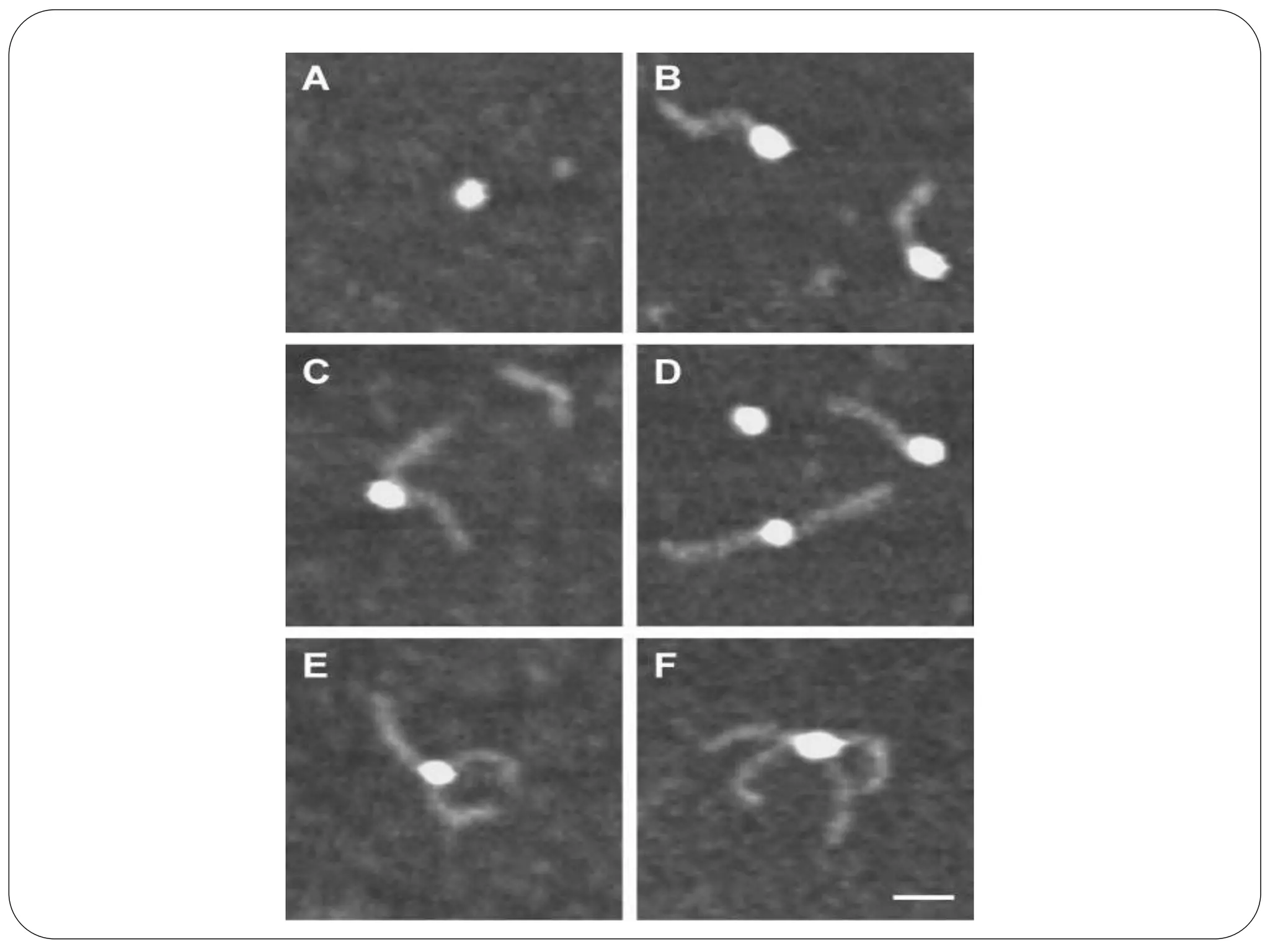
1. Atomic force microscopy was used to image streptavidin protein particles alone and in complexes with biotinylated DNA ligands.
2. Streptavidin appeared as globular particles, while biotinylated DNA ligands appeared as 50nm rods. When incubated together, streptavidin particles were observed with single or multiple DNA rods protruding, indicating ligand binding.
3. Analysis of streptavidin particles bound with two DNA ligands found an underrepresentation of acute angles between the ligands compared to expected probabilities, suggesting steric hindrance disfavors adjacent ligand binding.