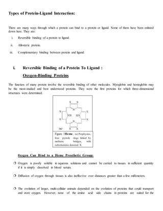
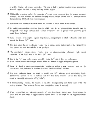
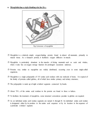
This is based on protein-ligand interaction physical method, which gives us knowledge about how our body protein interacts with other molecule and protein function.
Proteins are dynamic molecules whose functions almost invariably depend on interactions with other molecules.
A molecule bound reversibly by a protein is called a ligand.
A ligand binds at a site on the protein called the binding site, which is complementary to the ligand in size, shape, charge, and hydrophobic or hydrophilic character.
Introduction
Common terms used
Protein ligand interaction
1)in oxygen binding protein
Heme
Structure of hemoglobin
Ligand binding effected by protein structure
Oxygenation &deoxygenation
Cooperative binding of oxygen
Models for cooperative binding
Hb also transports H+&co2
2)complementary interaction immune system &immunoglobulin
Introduction
Structure of antibodies
Binding of antigen to antibody
Applications
Conclusion
Reference
DNA Protein interaction occur when a protein binds a molecule of DNA, often to regulate the biological function of DNA, usually the expression of a gene. DNA Protein interactions play very vital roles in any living cell. It controls various cellular processes which are very essential for living beings, viz. replication, transcription, recombination, DNA repair etc. There are several types of proteins found in a cell.Direct recognition occurs when the amino acid side chains of a protein interact with specific DNA bases.
Most protein-DNA interactions are mediated by direct physical interaction (hydrogen bonding or hydrophobic interactions) between the protein and the DNA base pairs.
DNA-binding proteins can be identified by many experimental techniques such as chromatin immunoprecipitation on microarrays, X-ray crystallography and nuclear magnetic resonance (NMR).
Protein Folding-biophysical and cellular aspects, protein denaturationAnishaMukherjee5
Protein folding is the physical process by which a protein chain acquires its native 3-dimensional structure, a conformation that is usually biologically functional, in an expeditious and reproducible manner.
In this presentation i have explained about all the super secondary structure their types and their functions . The ppt has been made in such a way that it will clear out our basic concepts first and then it will go higher. I hope you like it
Protein ligand interaction by KK Sahu sirKAUSHAL SAHU
Introduction
Historical Aspects
What are proteins
- Structure
- Functions
What are ligands
- Binding Site
Protein ligand Interaction
In oxygen binding proteins(Reversible Binding) * Hemoglobin
* Structure of Hemoglobin
* Structure of myoglobin
* Quantitative description of Protein ligand interaction
* Ligand binding effected by protein structure
* Oxygenation and deoxygenation of hemoglobin
* Co-operative binding of oxygen
* Model for co-operative binding
* Hemoglobin also transports H+ and CO2
2. Complementary interaction :- The Immune system and immunoglobulins
*Introduction
*Structure of Antibodies
*Binding of Antigen to Antibody
Importance
Application
Conclusion
References
Prediction of the three dimensional structure of a given protein sequence i.e. target protein from the amino acid sequence of a homologous (template) protein for which an X-ray or NMR structure is available based on an alignment to one or more known protein structures
Mismatch Repair Mechanism Is One Of The Important DNA Repair Mechanism Which Recognizes And Replaces The Wrong Nucleotides. DNA Repair Is Important Since Its Failure Leads To Deadly Diseases Like Cancer. In This Presentation, You Will Learn About DNA Repair, Mismatch Repair, Proteins Involved In Prokaryotic And Eukaryotic MMR, Diagrams, Biological Importance Of MMR And References For Further Study.
Folding depends upon sequence of Amino Acids not the Composition. Folding starts with the secondary structure and ends at quaternary structure.
Denaturation occur at secondary, tertiary & quaternary level but not at primary level.
Sequence alig Sequence Alignment Pairwise alignment:-naveed ul mushtaq
Sequence Alignment Pairwise alignment:- Global Alignment and Local AlignmentTwo types of alignment Progressive Programs for multiple sequence alignment BLOSUM Point accepted mutation (PAM)PAM VS BLOSUM
[Brief]Structure and functions of hemoglobin and myglobin (Bio-Inorganic chem...Anim60
This ppt is made from the bio-inorganic point of view for those who are having difficulty in finding the correct type and quality of information. This ppt has all the important points which one needs to know about this topic.
HEMOGLOBIN - STRUCTURE IN RELATION TO FUNCTIONMuunda Mudenda
Hemoglobin (Haemoglobin) is one of the most widely studied proteins in biology. It also stands as the first protein to have been crystallized and its structure elucidated. Apart from that, hemoglobin gave scientists a better understanding of the structure to function phenomenon in proteomics. This document discusses hemoglobin including the types and mechanism of action in its roles of oxygen and carbon dioxide transportation. Please note that this document is for academic purposes and does not, in any way, claim to be exhaustive. However, if you would like to understand the subject in more depth, write me an email and we can discuss the subject together. Enjoy the read.
Of the O2 molecules to each one of the four subunits of hemoglobin al.pdfinfo824691
Of the O2 molecules to each one of the four subunits of hemoglobin allosterically regulated.
Which of the following is the best explanation for this phenomenon? Affinity of hemoglobin for
O2 sensitive to pH. Conformational changes in hemoglobin alter the affinity for O2 the home is
positioned in a hydrophobic pocket. Hemoglobin consists mainly of alpha-helices. Change the
oxidation state of Fe atoms. What are the Structural levels that are the most different between
myoglobin and hemoglobin? Primary and quaternary structure Second and tertiary structure
Secondary and tertiary structure Type of prosthetic group and tertiary structure Glycosylation
and secondary structure No structural difference where are membranes created in a cell?
Phospholipids are added to the outer leaflet of the plasma membrane. Phospholipids are added to
the cytoplasmic leaflet of the of the smooth ER membrane. Phospholipids are added to vesicles
from lipid droplets. Proteins targeted to the nucleus contain a nuclear localization signal (NLS).
What does it do? The NLS is recognized by a translucent at the nuclear membrane that allows
the protein to be translocated into the nucleus. The NLS is recognized by soluble \"shuttle\'
protein in the cytosol which brings nuclear proteins into the nucleus. The NLS binds the nuclear
pore to allow transport into the nucleus. The NLS brings the ribosome .0 dock at the nuclear
membrane and the protein is directly synthesized in the nucleus. A triglyceride (three double
bonds) A monounsaturated fatty acid An Omega-3 fatty acid A trans fat Cholesterol
Solution
The iron is in ferous state in haemoglobin. Oxygen temporary and reversibly oxidized ferous into
ferric and oxygen temporarily turn into superoxide ion. Ferrioxyhaemoglobin cannot bind
oxygen . The enzyme methemoglobin reductase will be able to reducing the iron center.
answer... E
Myoglobin consists a single polypeptide chain haemoglobin consists several polypeptide chain
Myoglobin occurs as monomeric protein haemoglobin occurs as terameric protein.
The tertiary structure of myoglobin is similar to a typical water soluble protein structure. Each
protein molecule contain one heme prosthetic group and each heme residue contain one central
coordinately bound iron atom.
answer ...C
Cell membrane contain variety of biological molecules including protein and lipid. Fusion of
inter cellular vescicals with the membrane incorporate the vesicle membran\'s component into
the cell membrane
Answer C
Nuclear localization signals bind the nuclear pore to allow transport into the nucleus
Answer...C
The given structure is structure of omega 3fatty acid.
Earliest Galaxies in the JADES Origins Field: Luminosity Function and Cosmic ...Sérgio Sacani
We characterize the earliest galaxy population in the JADES Origins Field (JOF), the deepest
imaging field observed with JWST. We make use of the ancillary Hubble optical images (5 filters
spanning 0.4−0.9µm) and novel JWST images with 14 filters spanning 0.8−5µm, including 7 mediumband filters, and reaching total exposure times of up to 46 hours per filter. We combine all our data
at > 2.3µm to construct an ultradeep image, reaching as deep as ≈ 31.4 AB mag in the stack and
30.3-31.0 AB mag (5σ, r = 0.1” circular aperture) in individual filters. We measure photometric
redshifts and use robust selection criteria to identify a sample of eight galaxy candidates at redshifts
z = 11.5 − 15. These objects show compact half-light radii of R1/2 ∼ 50 − 200pc, stellar masses of
M⋆ ∼ 107−108M⊙, and star-formation rates of SFR ∼ 0.1−1 M⊙ yr−1
. Our search finds no candidates
at 15 < z < 20, placing upper limits at these redshifts. We develop a forward modeling approach to
infer the properties of the evolving luminosity function without binning in redshift or luminosity that
marginalizes over the photometric redshift uncertainty of our candidate galaxies and incorporates the
impact of non-detections. We find a z = 12 luminosity function in good agreement with prior results,
and that the luminosity function normalization and UV luminosity density decline by a factor of ∼ 2.5
from z = 12 to z = 14. We discuss the possible implications of our results in the context of theoretical
models for evolution of the dark matter halo mass function.
What is greenhouse gasses and how many gasses are there to affect the Earth.moosaasad1975
What are greenhouse gasses how they affect the earth and its environment what is the future of the environment and earth how the weather and the climate effects.
THE IMPORTANCE OF MARTIAN ATMOSPHERE SAMPLE RETURN.Sérgio Sacani
The return of a sample of near-surface atmosphere from Mars would facilitate answers to several first-order science questions surrounding the formation and evolution of the planet. One of the important aspects of terrestrial planet formation in general is the role that primary atmospheres played in influencing the chemistry and structure of the planets and their antecedents. Studies of the martian atmosphere can be used to investigate the role of a primary atmosphere in its history. Atmosphere samples would also inform our understanding of the near-surface chemistry of the planet, and ultimately the prospects for life. High-precision isotopic analyses of constituent gases are needed to address these questions, requiring that the analyses are made on returned samples rather than in situ.
Nutraceutical market, scope and growth: Herbal drug technologyLokesh Patil
As consumer awareness of health and wellness rises, the nutraceutical market—which includes goods like functional meals, drinks, and dietary supplements that provide health advantages beyond basic nutrition—is growing significantly. As healthcare expenses rise, the population ages, and people want natural and preventative health solutions more and more, this industry is increasing quickly. Further driving market expansion are product formulation innovations and the use of cutting-edge technology for customized nutrition. With its worldwide reach, the nutraceutical industry is expected to keep growing and provide significant chances for research and investment in a number of categories, including vitamins, minerals, probiotics, and herbal supplements.
(May 29th, 2024) Advancements in Intravital Microscopy- Insights for Preclini...Scintica Instrumentation
Intravital microscopy (IVM) is a powerful tool utilized to study cellular behavior over time and space in vivo. Much of our understanding of cell biology has been accomplished using various in vitro and ex vivo methods; however, these studies do not necessarily reflect the natural dynamics of biological processes. Unlike traditional cell culture or fixed tissue imaging, IVM allows for the ultra-fast high-resolution imaging of cellular processes over time and space and were studied in its natural environment. Real-time visualization of biological processes in the context of an intact organism helps maintain physiological relevance and provide insights into the progression of disease, response to treatments or developmental processes.
In this webinar we give an overview of advanced applications of the IVM system in preclinical research. IVIM technology is a provider of all-in-one intravital microscopy systems and solutions optimized for in vivo imaging of live animal models at sub-micron resolution. The system’s unique features and user-friendly software enables researchers to probe fast dynamic biological processes such as immune cell tracking, cell-cell interaction as well as vascularization and tumor metastasis with exceptional detail. This webinar will also give an overview of IVM being utilized in drug development, offering a view into the intricate interaction between drugs/nanoparticles and tissues in vivo and allows for the evaluation of therapeutic intervention in a variety of tissues and organs. This interdisciplinary collaboration continues to drive the advancements of novel therapeutic strategies.
Observation of Io’s Resurfacing via Plume Deposition Using Ground-based Adapt...Sérgio Sacani
Since volcanic activity was first discovered on Io from Voyager images in 1979, changes
on Io’s surface have been monitored from both spacecraft and ground-based telescopes.
Here, we present the highest spatial resolution images of Io ever obtained from a groundbased telescope. These images, acquired by the SHARK-VIS instrument on the Large
Binocular Telescope, show evidence of a major resurfacing event on Io’s trailing hemisphere. When compared to the most recent spacecraft images, the SHARK-VIS images
show that a plume deposit from a powerful eruption at Pillan Patera has covered part
of the long-lived Pele plume deposit. Although this type of resurfacing event may be common on Io, few have been detected due to the rarity of spacecraft visits and the previously low spatial resolution available from Earth-based telescopes. The SHARK-VIS instrument ushers in a new era of high resolution imaging of Io’s surface using adaptive
optics at visible wavelengths.
Lateral Ventricles.pdf very easy good diagrams comprehensive
Protein ligand interaction
1. A
SEMINAR
ON
PROTEIN LIGAND INTERACTION PHYSICAL METHOD
DIRECTOR PRESENTEDBY
R.K.RAO (PRINCIPAL) NIKITA DEWANGAN
GUIDED BY - RACHANA TIWARI M.SC. 2nd
SEMESTER
BIOTECHNOLOGY
G.D. RUNGTA COLLEGE OF SCIENCE & TECHNOLOGY
KOHKA-KURUD,BHILAI DURG (C.G.)
2. PROTEIN LIGAND INTERACTION
SYSNOPSIS
Introduction
Types of Protein Ligand interaction
Reversible binding of a protein to ligand
Allosteric protein
Complementary binding of a protein to ligand
Effect of protein structure on ligand binding
Conclusion
References
3. Introduction:
Proteins are dynamic molecules whose functions almost invariably depend on interactions with other
molecules, and these interactions are affected in physiologically important ways by sometimes subtle,
sometimes striking changes in protein conformation.
The function of many other protein involve interactions with a variety of different molecules. Most of
these interactions are fleeting, though they may be the basis of complex physiological processes such as
oxygen transport, immune function and muscle contraction.
A ligand binds at a site on the protein called the binding site, which is complementary to the
ligand in size, shape, charge, and hydrophobic or hydrophilic character.
The interaction is specific the protein can discriminate among the thousands of different molecules
in its environment and selectively bind only one or a few.
A given protein may have separate binding sites for several different ligands.
Specific conformational changes are frequently essential to a protein’s function. Changes in
conformation may be subtle, reflecting molecular vibrations and small movements of amino acid
residues throughout the protein.
The structural adaptation that occurs between protein and ligand is called induced fit.
In a multi-subunit protein, a conformational change in one subunit often affects the conformation of other
subunits.
The specific molecular interactions are crucial in maintaining the high degree of order in a living system.
4. Types of Protein-Ligand Interaction:
There are many ways through which a protein can bind to a protein or ligand. Some of them have been enlisted
down here. They are:
i. Reversible binding of a protein to ligand.
ii. Allosteric protein.
iii. Complementary binding between protein and ligand.
i. Reversible Binding of a Protein To Ligand :
Oxygen-Binding Proteins
The function of many protein involve the reversible binding of other molecules. Myoglobin and hemoglobin may
be the most-studied and best understood proteins. They were the first proteins for which three-dimensional
structures were determined.
0xygen Can Bind to a Heme Prosthetic Group:
Oxygen is poorly soluble in aqueous solutions and cannot be carried to tissues in sufficient quantity
if it is simply dissolved in blood serum.
Diffusion of oxygen through tissues is also ineffective over distances greater than a few millimeters.
The evolution of larger, multi-cellular animals depended on the evolution of proteins that could transport
and store oxygen. However, none of the amino acid side chains in proteins are suited for the
Figure 1Heme. (a) Porphyrins,
four pyrrole rings linked by
methene bridges, with
substitutions denoted X.
5. reversible binding of oxygen molecules. This role is filled by certain transition metals, among them
iron and copper, that have a strong tendency to bind oxygen.
Multi-cellular organisms exploit the properties of metals, most commonly iron, for oxygen transport.
However, free iron promotes the formation of highly reactive oxygen species such as hydroxyl radicals
that can damage DNA and other macromolecules.
Iron used in cells is therefore bound in forms that sequester it and/or make it less reactive.
In multi-cellular organisms, especially those in which iron, in its oxygen-carrying capacity, must be
transported over large distances-iron is often incorporated into a protein-bound prosthetic group
called heme (or haem)
Heme consists of a complex organic ring structure, protoporphyrin, to which is bound a single iron
atom in its ferrous (Fe2+) state.
The iron atom has six coordination bonds, four to nitrogen atoms that are part of the flat porphyrin
ring system and two perpendicular to the porphyrin.
The coordinated nitrogen atoms (which have an electron-donating character) help prevent
conversion of the heme iron to the ferric (Fe3+) state.
Iron in the Fe2+ state binds oxygen reversibly; in the Fe3+ state it does not bind oxygen.
In Fe3+ state it does not binds oxygen heme is found in a number of oxygen transporting protein.
Heme is found in many oxygen-transporting proteins, as well as in some proteins, such as the
cytochromes, that participate in oxidation-reduction (electron-transfer) reactions.
Free heme molecules (heme not bound to protein) leave Fe2+ with two "open" coordination bonds.
Simultaneous reaction of one o2 molecule with two free heme molecules (or two free Fe2+) can
result in irreversible conversion of Fe2+ to Fe3+.
ln heme-containing proteins, this reaction is prevented by sequestering each heme deep within the
protein structure. Thus, access to the two open coordination bonds is restricted.
When oxygen binds, the electronic properties of heme iron change; this accounts for the change in
color from the dark purple of oxygen-depleted venous blood to the bright red of oxygen rich arterial
blood.
6. Effect of pH on O2 Binding to Hb:
The binding of H+ and CO2 is inversely related to the binding of oxygen.
At the relatively low pH and high CO2 concentration of peripheral tissues, the affinity of
hemoglobin for oxygen decreases as H+ and CO2 are bound, and o2 is released to the tissues.
Conversely, in the capillaries of the lung, as CO2 is excreted and the blood pH consequently rises,
the affinity of hemoglobin for oxygen increases and the protein binds more O2 for transport to the
peripheral tissues.
This effect of pH and CO2 concentration on the binding and release of oxygen by hemoglobin is called
the Bohr effect, after Christian Bohr, the Danish physiologist (and father of physicist Niels Bohr) who
discovered it in 1904.
The binding equilibrium for hemoglobin and one molecule of oxygen can be designated by the
reaction:
Hb + 02 HbO2
but this is not a complete statement. To account for the effect of H+ concentration on this binding
equilibrium, the reaction is:
HHb+ + O2 HbO2 + H+
where Hb+ denotes a protonated form of hemoglobin.
Fig: -2, Heme group with iron
7. a. Myoglobin has a sigle binding site for O2:-
Myoglobin is a relatively simple oxygen-binding protein found in almost all mammals, primarily in
muscle tissue. As a transport protein, it facilitates oxygen diffusion in muscle.
Myoglobin is particularly abundant in the muscles of diving mammals such as seals and whales,
where it also has an oxygen storage function for prolonged excursions undersea.
Proteins very similar to myoglobin are widely distributed, occurring even in some single-celled
organisms.
Myoglobin is a single polypeptide of 153 amino acid residues with one molecule of heme. It is typical of
the family of proteins called globins, all of which have similar primary and tertiary structures.
The polypeptide is made up of eight α-helical segments connected by bends.
About 78o/o of the amino acid residues in the protein are found in these α- helices.
To facilitate the treatment of myoglobin, some structural conventions peculiar to globins are required.
As an individual amino acid residue segments are named A through H. An individual amino acid residue
is designated either by its position in the amino acid sequence or by its location in the sequence of
a particular α-helical segment.
Fig:-3,structure of myoglobin
8. for example, the His residue coordinated to the heme in myoglobin, His93 (the 93rd residue from
the amino-terminal end of the myoglobin polypeptide sequence), is also called His F8 (the 8th residue
in α- helix F).
The bends in the structure are designated AB, CD, EF, FG, and so forth, reflecting the α-helical
segments they connect.
ii. Allosteric Proteins:-
An allosteric protein is one in which the binding of a ligand to one site affects the binding properties
of another site on the same protein.
The term "allosteric" derives from the Greek word, "other," and stereos, "solid" or "shape." Allosteric
proteins are those having "other shapes," or conformations, induced by the binding of ligands
referred to as modulators.
The conformational changes induced by the modulator(s) interconvert more active and less active
forms of the protein.
The modulators for allosteric proteins may be either inhibitors or activators.
When the normal ligand and modulator are identical, the interaction is termed homotropic. When the
modulator is a molecule other than the normal ligand, the interaction is heterotropic.
Some proteins have two or more modulators and therefore can have both homotropic and
heterotropic interactions
0xygen binding to Haemoglobin ls regulated by 2,3-Bisphosphoglycerate:
The interaction of 2,3-bisphosphoglycerate (BPG) with hemoglobin molecules further refines the
function of hemoglobin, and provides an example of heterotropic allosteric modulation.
BPG is present in relatively high concentrations in erythrocytes.
When hemoglobin is isolated, it contains substantial amounts of bound BPG, which can be difficult
to remove completely.
2,3-Bisphosphoglycerate is known to greatly reduce the affinity of
hemoglobin for oxygen-there is an inverse relationship between the
binding of O2 and the binding of BPG.
Therefore describe another binding process for hemoglobin:
9. HbBPG + O2 HbO2 + BPG
BPG binds at a site distant from the oxygen-binding site and regulates the O2-binding afflnity of
hemoglobin in relation to the pO2 in the lungs.
Regulation of oxygen binding to hemoglobin by BPG has an important role in fetal development
Because a fetus must extract oxygen from its mother's blood, fetal hemoglobin must have greater
affinity than the maternal hemoglobin for O2.
Sickle Cell Anaemia ls a Molecular Disease of Hemoglobin:
The hereditary human disease sickle-cell anemia demonstrates strikingly the importance of amino
acid sequence in determining the secondary tertiary, and quaternary structures of globular proteins,
and thus their biological functions.
Almost 500 genetic variants of hemoglobin are known to occur in the human population; all but a few
are quite rare.
Most variations consist of differences in a single amino acid residue.
Each hemogiobin variation is the product of an altered gene. The variant genes are called alleles.
Because humans generally have two copies of each gene, an individual may have two copies of one
allele (thus being homozygous for that gene) or one copy of each of two different alleles (thus
heterozygous).
Sickle-cell anemia occurs in individuals who inherit the allele for sickle-cell hemoglobin from both
parents.
The erythrocytes of these individuals are fewer and also abnormal. In addition to an unusually large
number of immature cells, the blood contains many long, thin, sickle shaped erythrocytes.
When hemoglobin from sickle cells (called hemoglobin S) is deoxygenated, it becomes insoluble and
forms polymers that aggregate into tubular fibers.
Normal hemoglobin (heamoglobin A) remains soluble on deoxygenation. The insoluble fibers of
deoxygenated hemoglobin S cause the deformed, sickle shape of the erythrocytes, and the proportion
of sickled cells increases greatly as blood is deoxygenated.
10. The altered properties of hemoglobin S result from a single
amino acid substitution, a Val instead of a Glu residue at
position 6 in the two β chains. The R group of valine has no
electric charge, whereas glutamate has a negative charge at pH
7.4.
Hemoglobin S therefore has two fewer negative charges than
hemoglobin A (one fewer on each β chain). Replacement of
the GIu residue by Val creates a "sticky" hydrophobic contact
point at position 6 of the β chain, which is on the outer
surface of the molecule. These sticky spots cause
deoxyhemoglobin S molecules to associate abnormally with
each other, forming the long, fibrous aggregates
characteristic of this disorder.
Sickle-cell anemia is Life-threatening and painful. people
with this disease suffer repeated crises brought on by
physical exertion. They become weak, dizzy, and short of
breath, and they also experience heart murmurs and an
increased pulse rate.
The hemoglobin content of their blood is only about half
the normal value of 15 to 16 g/100 mL, ally high in certain
parts of Africa.
Complementary Interactions between Proteins and Ligand:-
Conformations of oxygen-binding proteins affect and are affected by the binding of small ligands
(O2 or CO) to the heme group.
However, most Protein ligand interactions do not involve a prosthetic group. Instead, the binding site
for a Iigand is more often Iike the hemoglobin binding site for BPG-a cleft in the protein lined with
amino acid residues, arranged to make the binding interaction highly specific.
FIG. 6 Normal and sickle-cell
hemoglobin. (a) Subtle differences
between the conformations of
hemoglobin A and hemoglobin S result
froma single amino acid change in the
βchains. (b) As a result of this change,
deoxyhemoglobin S has a hydrophobic
patch on its surface, which causesthe
molecules toaggregate into strands
that align into insoluble fibers.
11. Effective discrimination between ligands is the norm at binding sites, even when the ligands have only
minor structural differences.
On a physiological level , the response of the immune system to an invader is an intricate and coordinated
set of interactions among many classes of protein, molecules and cell types.
At the level of individual proteins, the immune response demonstrates how an acutely sensitive and
specific biochemical system is built upon the reversible binding of ligands to proteins.
The immune response features a specialized array of cells
and proteins:
Immunity is brought about by a variety of leukocytes(white
blood cells), including macrophages and lymphocytes, all of
which develop from undifferentiated stem cells in the bone
marrow.
Leukocytes can leave the bloodstream and patrol the tissues,
each cell producing one or more proteins capable of recognizing
and binding to molecules that might signal an infection.
The immune response consists of two complementary systems,
the humoral and cellular immune systems.
The humoral immune system (Latin humor, “fluid”) is directed
at bacterial infections and extracellular viruses (those found in
the body fluids), but can also respond to individual foreign
proteins.
The cellular immune system destroys host cells infected by viruses and also destroys some parasites
and foreign tissues.
At the heart of the humoral immune response are soluble proteins called antibodies or
immunoglobulin, often abbreviated Ig. Immunoglobulin bind bacteria, viruses, or large molecules
identified as foreign and target them for destruction.
Making up 20% of blood protein, the immunoglobulin’s are produced by B lymphocytes, or B cells,
so named because they complete their development in the bone marrow.
The cell mediated respons is contributed by T cells or T lymphocytes so called because development
occurs in thymus.
Each recognition protein of the immune system, either an antibody produced by a B cell or a receptor on
the surface of a Tcell, specifically binds some particular chemical structure, distinguishing it from
Fig;-4, immunoglobulin
12. virtually all other. Humans are capable of producing more than 108 different antibodies with distinct
binding specificities.
Any molecule or pathogen (may be a virus, bacterial cell, protein and other molecule) capable of eliciting
an immune response is called an antigen.
A antibody have a binding site for antigen. A specific antigen binds to specific antibody, antigen binding
site called epitope.
Effect of protein structure on ligand binding:
The interaction is greatly affected by protein structure and is often accompanied by conformational
changes.
For example, the specificity with which heme binds its various ligands is altered when the heme is a
component of myoglobin. Carbon monoxide binds to free heme molecules more than 20,000 times
better than does 02 , but it binds only about 200 times better than 02 when the heme is bound in
myoglobin.
The difference may be partly explained by steric hindrance. When O2 binds to free heme ,the axis of the
oxygen molecule is positioned at an angle to Fe-O bond. In contrast, when CO binds to free heme, the Fe,
C, O atoms lie in a strainght line.
Fig:- 5, steric effects caused by ligand binding to the heme of myoglobin. (a) oxygen binds to heme with the O,
axis at an angle, a binding conformation readily accommondated by myoglobin. (b) carbon monoxide binds to
free heme with the CO axis perpendicular to the plane of the p[orphyrin ring.
13. Conclusion
Thereby I conclude up this topic with a quick review about Protein-Ligand Interaction. Proteins are molecules
whose functions almost invariably depend on interactions with other molecules and its conformational change.
Protein binds up with the other molecule through different ways. Its important for the transport and regulation of
essential molecules like oxygen.
Reference:
1. Nelson David L., Cox Michael M., 2012, Protein- Ligand Interaction, Fundamental of
biochemistry, W.H. Freeman and Company, Fifth Edition, Pg. 153- 158.
2. Net source –
http://www.wikipedia.org
www.softarchive.com
www.amzon.com