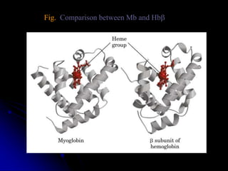
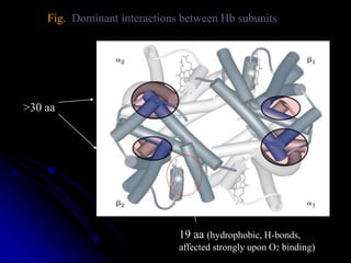
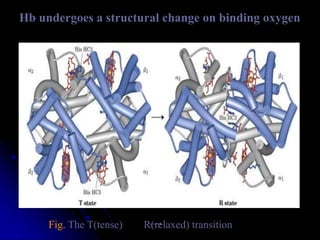
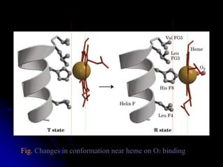
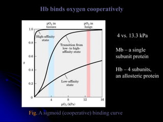
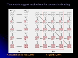
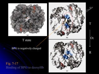
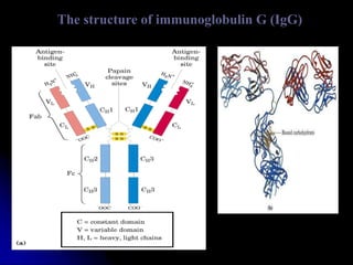
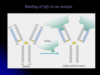
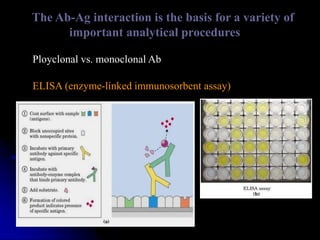
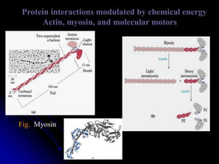
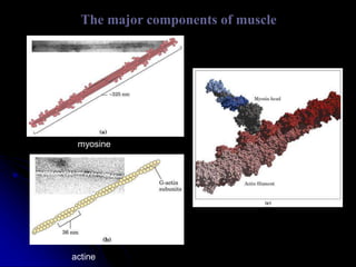
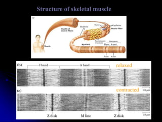
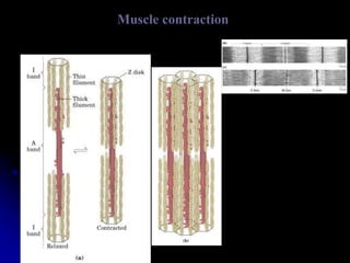
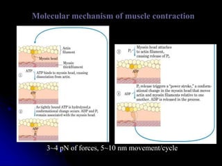
PROTEIN LIGAND INTERACTION
&
PROTEIN PROTEIN INTERACTION
By
KAUSHAL KUMAR SAHU
Assistant Professor (Ad Hoc)
Department of Biotechnology
Govt. Digvijay Autonomous P. G. College
Raj-Nandgaon ( C. G. )
CONTENT
 INTRODUCTION
 Protein – ligand Intraction
Oxygen binding
Ag – Ab Intraction
 Protein – Protein Intraction
Muscle Contraction
• CONCLUSION
• Reference
INTRODUCTION
 Protein–protein interactions occur when two or more protein bind together,
often to carry out their biological function.
 Many of the most important molecular processes in the cell such as DNA
replication are carried out by large molecular machines that are built from a large
number of protein components organised by their protein–protein interactions.
 Interactions between proteins are important for the majority of biological
functions.
 For example, signals from the exterior of a cell are mediated to the inside
of that cell by protein– protein interactions of the signaling molecules.
 Indeed, protein–protein interactions are at the core of the entire interatomic
system of any living cell.
OXYGEN BINDING
 Heme consists of a complex organic ring structure,
protoporphynrin, to which is bound a single iron atom in
its ferrous (Fe2+ ) state.
 The iron atom has six coordination bonds, four to nitrogen
atoms that are part of the flat porphyrin ring system and
two perpendicular to the porphyrin state.
 Iron in the Fe 2+ state binds oxygen reversibly; in the Fe 3+
state it does not bind oxygen.
Protein structure affects how ligands bind
1. Steric effects
2. Molecular motions/breathing
in the structure
1: 20,000 1: 200
The structure of myoglobulin
*a single binding site for O2
*78% a helices
*His93 or HisF8 (the 8th
residue in a helix F) binds
to heme
*Bends between a helices
Fig. Comparison between Mb and Hbb
Fig. Dominant interactions between Hb subunits
>30 aa
19 aa (hydrophobic, H-bonds,
affected strongly upon O2 binding)
Hb undergoes a structural change on binding oxygen
Fig. The T(tense) R(relaxed) transition
Fig. Changes in conformation near heme on O2 binding
Hb binds oxygen cooperatively
Fig. A sigmoid (cooperative) binding curve
4 vs. 13.3 kPa
Mb – a single
subunit protein
Hb – 4 subunits,
an allosteric protein
Two models suggest mechanisms for cooperative binding
Concerted (all-or-none), 1965 Sequential, 1966
Fig. 7-17
Binding of BPG to deoxyHb
T state
T
R
O2
++
BPG is negatively charged
The structure of immunoglobulin G (IgG)
Binding of IgG to an antigen
The Ab-Ag interaction is the basis for a variety of
important analytical procedures
Ployclonal vs. monoclonal Ab
ELISA (enzyme-linked immunosorbent assay)
Protein interactions modulated by chemical energy
Actin, myosin, and molecular motors
Fig. Myosin
The major components of muscle
myosine
actine
Structure of skeletal muscle
relaxed
contracted
Muscle contraction
Molecular mechanism of muscle contraction
3~4 pN of forces, 5~10 nm movement/cycle
REFERANCE
LEHNINGER PRINCIPLES OF
BIOCHEMISTRY
FIFTH EDITION

Protein protein interaction

  • 1.
    PROTEIN LIGAND INTERACTION & PROTEINPROTEIN INTERACTION By KAUSHAL KUMAR SAHU Assistant Professor (Ad Hoc) Department of Biotechnology Govt. Digvijay Autonomous P. G. College Raj-Nandgaon ( C. G. )
  • 2.
    CONTENT  INTRODUCTION  Protein– ligand Intraction Oxygen binding Ag – Ab Intraction  Protein – Protein Intraction Muscle Contraction • CONCLUSION • Reference
  • 3.
    INTRODUCTION  Protein–protein interactionsoccur when two or more protein bind together, often to carry out their biological function.  Many of the most important molecular processes in the cell such as DNA replication are carried out by large molecular machines that are built from a large number of protein components organised by their protein–protein interactions.  Interactions between proteins are important for the majority of biological functions.  For example, signals from the exterior of a cell are mediated to the inside of that cell by protein– protein interactions of the signaling molecules.  Indeed, protein–protein interactions are at the core of the entire interatomic system of any living cell.
  • 5.
    OXYGEN BINDING  Hemeconsists of a complex organic ring structure, protoporphynrin, to which is bound a single iron atom in its ferrous (Fe2+ ) state.  The iron atom has six coordination bonds, four to nitrogen atoms that are part of the flat porphyrin ring system and two perpendicular to the porphyrin state.  Iron in the Fe 2+ state binds oxygen reversibly; in the Fe 3+ state it does not bind oxygen.
  • 7.
    Protein structure affectshow ligands bind 1. Steric effects 2. Molecular motions/breathing in the structure 1: 20,000 1: 200
  • 8.
    The structure ofmyoglobulin *a single binding site for O2 *78% a helices *His93 or HisF8 (the 8th residue in a helix F) binds to heme *Bends between a helices
  • 9.
  • 10.
    Fig. Dominant interactionsbetween Hb subunits >30 aa 19 aa (hydrophobic, H-bonds, affected strongly upon O2 binding)
  • 11.
    Hb undergoes astructural change on binding oxygen Fig. The T(tense) R(relaxed) transition
  • 12.
    Fig. Changes inconformation near heme on O2 binding
  • 13.
    Hb binds oxygencooperatively Fig. A sigmoid (cooperative) binding curve 4 vs. 13.3 kPa Mb – a single subunit protein Hb – 4 subunits, an allosteric protein
  • 14.
    Two models suggestmechanisms for cooperative binding Concerted (all-or-none), 1965 Sequential, 1966
  • 15.
    Fig. 7-17 Binding ofBPG to deoxyHb T state T R O2 ++ BPG is negatively charged
  • 16.
    The structure ofimmunoglobulin G (IgG)
  • 17.
    Binding of IgGto an antigen
  • 18.
    The Ab-Ag interactionis the basis for a variety of important analytical procedures Ployclonal vs. monoclonal Ab ELISA (enzyme-linked immunosorbent assay)
  • 20.
    Protein interactions modulatedby chemical energy Actin, myosin, and molecular motors Fig. Myosin
  • 21.
    The major componentsof muscle myosine actine
  • 22.
    Structure of skeletalmuscle relaxed contracted
  • 23.
  • 24.
    Molecular mechanism ofmuscle contraction 3~4 pN of forces, 5~10 nm movement/cycle
  • 25.