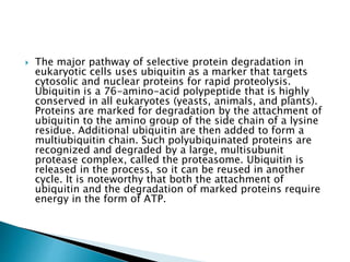
SlideShare a Scribd company logo
 Protein folding is a process in which a
polypeptide folds into a specific, stable,
functional, three-dimensional structure. It is
the process by which a protein structure
assumes its functional shape or
conformation.
 Proteins are much more complicated than just a chain of amino-
acids because proteins fold spontaneously depending on the R
groups in their amino-acid sequence. The structure of proteins is
very important for their function. The protein folding process can
be divided up into four stages, which we can think of as stages in
putting together a book:
 Primary structure: a chain of amino-acids joined together (letters
coming together to form words)
 Secondary structure: the chains fold into sheets or coils
(sentences)
 Tertiary structure: the sheets or coils fold in on one another
(chapters of a book) 4. Quaternary structure: amino-acid chains
folded in their tertiary structure interact with one another to give
the final functional protein (e.g. haemoglobin has 4 chains) (a
book)
 The primary structure of a protein, its linear amino-acid
sequence, determines its native conformation. The specific
amino acid residues and their position in the polypeptide
chain are the determining factors for which portions of the
protein fold closely together and form its three
dimensional conformation. The amino acid composition is
not as important as the sequence. The essential fact of
folding, however, remains that the amino acid sequence of
each protein contains the information that specifies both
the native structure and the pathway to attain that state.
This is not to say that nearly identical amino acid
sequences always fold similarly. Conformations differ
based on environmental factors as well; similar proteins
fold differently based on where they are found.
 Formation of a secondary structure is the first step in the folding process that a
protein takes to assume its native structure. Characteristic of secondary structure
is the structures known as alpha helices and beta sheets that fold rapidly because
they are stabilized by intramolecular hydrogen bonds, as was first characterized
by Linus Pauling. Formation of intramolecular hydrogen bonds provides another
important contribution to protein stability. Alpha helices are formed by hydrogen
bonding of the backbone to form a spiral shape (refer to figure on the right).[8]
The beta pleated sheet is a structure that forms with the backbone bending over
itself to form the hydrogen bonds (as displayed in the figure to the left). The
hydrogen bonds are between the amide hydrogen and carbonyl carbon of the
peptide bond. There exists anti-parallel beta pleated sheets and parallel beta
pleated sheets where the stability of the hydrogen bonds is stronger in the anti-
parallel beta sheet as it hydrogen bonds with the ideal 180 degree angle
compared to the slanted hydrogen bonds formed by parallel sheets.The alpha
helices and beta pleated sheets can be amphipathic in nature, or contain a
hydrophilic portion and a hydrophobic portion. This property of these secondary
structures aids in the folding of the protein as it aligns the helices and sheets in
such a way where the hydrophilic sides are facing the aqueous environment
surrounding the protein and the hydrophobic sides are facing the hydrophobic
core of the protein. Secondary structure hierarchically gives way to tertiary
structure formation.
 Tertiary structure
 Once the protein's tertiary structure is formed and
stabilized by the hydrophobic interactions, there may also
be covalent bonding in the form of disulfide bridges
formed between two cysteine residues. Tertiary structure
of a protein involves a single polypeptide chain; however,
additional interactions of folded polypeptide chains give
rise to quaternary structure formation.
 Quaternary structure
 Tertiary structure may give way to the formation of
quaternary structure in some proteins, which usually
involves the "assembly" or "coassembly" of subunits that
have already folded; in other words, multiple polypeptide
chains could interact to form a fully functional quaternary
protein.
 Folding is a spontaneous process independent of energy inputs
from nucleoside triphosphates. The passage of the folded state
is mainly guided by hydrophobic interactions, formation of
intramolecular hydrogen bonds, van der Waals forces, and it is
opposed by conformational entropy.[13] The process of folding
often begins co-translationally, so that the N-terminus of the
protein begins to fold while the C-terminal portion of the protein
is still being synthesized by the ribosome; however, a protein
molecule may fold spontaneously during or after biosynthesis.
While these macromolecules may be regarded as "folding
themselves", the process also depends on the solvent (water or
lipid bilayer),[14] the concentration of salts, the pH, the
temperature, the possible presence of cofactors and of molecular
chaperones. Proteins will have limitations on their folding
abilities by the restricted bending angles or conformations that
are possible. These allowable angles of protein folding are
described with a two-dimensional plot known as the
Ramachandran plot, depicted with psi and phi angles of
allowable rotation
 Protein folding must be thermodynamically favorable within a cell in order for it to be a
spontaneous reaction. Since it is known that protein folding is a spontaneous reaction, then it
must assume a negative Gibbs free energy value. Gibbs free energy in protein folding is
directly related to enthalpy and entropy.[8] For a negative delta G to arise and for protein
folding to become thermodynamically favorable, then either enthalpy, entropy, or both terms
must be favorable.
 Entropy is decreased as the water molecules become more orderly near the hydrophobic
solute.
 Minimizing the number of hydrophobic side-chains exposed to water is an important driving
force behind the folding process.[16] The hydrophobic effect is the phenomenon in which the
hydrophobic chains of a protein collapse into the core of the protein (away from the
hydrophilic environment).[8] In an aqueous environment, the water molecules tend to
aggregate around the hydrophobic regions or side chains of the protein, creating water shells
of ordered water molecules.[17] An ordering of water molecules around a hydrophobic region
increases order in a system and therefore contributes a negative change in entropy (less
entropy in the system). The water molecules are fixed in these water cages which drives the
hydrophobic collapse, or the inward folding of the hydrophobic groups. The hydrophobic
collapse introduces entropy back to the system via the breaking of the water cages which frees
the ordered water molecules.[8] The multitude of hydrophobic groups interacting within the
core of the globular folded protein contributes a significant amount to protein stability after
folding, because of the vastly accumulated van der Waals forces (specifically London Dispersion
forces).[8] The hydrophobic effect exists as a driving force in thermodynamics only if there is
the presence of an aqueous medium with an amphiphilic molecule containing a large
hydrophobic region.[18] The strength of hydrogen bonds depends on their environment; thus,
H-bonds enveloped in a hydrophobic core contribute more than H-bonds exposed to the
aqueous environment to the stability of the native state.
 Chaperones are a class of proteins that aid in the correct folding of other proteins in vivo.
Chaperones exist in all cellular compartments and interact with the polypeptide chain in order
to allow the native three-dimensional conformation of the protein to form; however,
chaperones themselves are not included in the final structure of the protein they are assisting
in. Chaperones may assist in folding even when the nascent polypeptide is being synthesized
by the ribosome. Molecular chaperones operate by binding to stabilize an otherwise unstable
structure of a protein in its folding pathway, but chaperones do not contain the necessary
information to know the correct native structure of the protein they are aiding; rather,
chaperones work by preventing incorrect folding conformations. In this way, chaperones do
not actually increase the rate of individual steps involved in the folding pathway toward the
native structure; instead, they work by reducing possible unwanted aggregations of the
polypeptide chain that might otherwise slow down the search for the proper intermediate and
they provide a more efficient pathway for the polypeptide chain to assume the correct
conformations.[20] Chaperones are not to be confused with folding catalysts, which actually do
catalyze the otherwise slow steps in the folding pathway. Examples of folding catalysts are
protein disulfide isomerases and peptidyl-prolyl isomerases that may be involved in formation
of disulfide bonds or interconversion between cis and trans stereoisomers, respectively.[21]
Chaperones are shown to be critical in the process of protein folding in vivo because they
provide the protein with the aid needed to assume its proper alignments and conformations
efficiently enough to become "biologically relevant". This means that the polypeptide chain
could theoretically fold into its native structure without the aid of chaperones, as demonstrated
by protein folding experiments conducted in vitro; however, this process proves to be too
inefficient or too slow to exist in biological systems; therefore, chaperones are necessary for
protein folding in vivo. Along with its role in aiding native structure formation, chaperones are
shown to be involved in various roles such as protein transport, degradation, and even allow
denatured proteins exposed to certain external denaturant factors an opportunity to refold into
their correct native structures.
 A fully denatured protein lacks both tertiary and secondary structure, and exists as a so-called
random coil. Under certain conditions some proteins can refold; however, in many cases,
denaturation is irreversible. Cells sometimes protect their proteins against the denaturing
influence of heat with enzymes known as heat shock proteins (a type of chaperone), which
assist other proteins both in folding and in remaining folded. Some proteins never fold in cells
at all except with the assistance of chaperones which either isolate individual proteins so that
their folding is not interrupted by interactions with other proteins or help to unfold misfolded
proteins, allowing them to refold into the correct native structure. This function is crucial to
prevent the risk of precipitation into insoluble amorphous aggregates. The external factors
involved in protein denaturation or disruption of the native state include temperature, external
fields (electric, magnetic),[26] molecular crowding, and even the limitation of space, which can
have a big influence on the folding of proteins. High concentrations of solutes, extremes of pH,
mechanical forces, and the presence of chemical denaturants can contribute to protein
denaturation, as well. These individual factors are categorized together as stresses.
Chaperones are shown to exist in increasing concentrations during times of cellular stress and
help the proper folding of emerging proteins as well as denatured or misfolded ones.
 Under some conditions proteins will not fold into their biochemically functional forms.
Temperatures above or below the range that cells tend to live in will cause thermally unstable
proteins to unfold or denature (this is why boiling makes an egg white turn opaque). Protein
thermal stability is far from constant, however; for example, hyperthermophilic bacteria have
been found that grow at temperatures as high as 122 °C,which of course requires that their full
complement of vital proteins and protein assemblies be stable at that temperature or above.
 Protein aggregation is a biological
phenomenon in which misfolded proteins
aggregate (i.e., accumulate and clump
together) either intra- or extracellularly.
 These protein aggregates are often
correlated with diseases. In fact, protein
aggregates have been implicated in a wide
variety of disease known as amyloidoses,
including ALS, Alzheimer's, Parkinson's and
prion disease.
 After synthesis, proteins typically fold into a particular three-dimensional conformation that is
the most thermodynamically favorable: their native state.[5] This folding process is driven by
the hydrophobic effect: a tendency for hydrophobic (water-fearing) portions of the protein to
shield itself from the hydrophilic (water-loving) environment of the cell by burying into the
interior of the protein. Thus, the exterior of a protein is typically hydrophilic, whereas the
interior is typically hydrophobic.
 Protein structures are stabilized by non-covalent interactions and disulfide bonds between two
cysteine residues. The non-covalent interactions include ionic interactions and weak van der
waals interactions. Ionic interactions form between an anion and a cation and form salt bridges
that help stabilize the protein. Van der waals interactions include nonpolar interactions (i.e.
London dispersion forces) and polar interactions (i.e. hydrogen bonds, dipole-dipole bond).
These play an important role in a protein's secondary structure, such as forming an alpha helix
or a beta sheet, and tertiary structure. Interactions between amino acid residues in a specific
protein are very important in that protein's final structure.
 When there are changes in the non-covalent interactions, as may happen with a change in the
amino acid sequence, the protein is susceptible to misfolding or unfolding. In these cases, if
the cell does not assist the protein in re-folding, or degrade the unfolded protein, the
unfolded/misfolded protein may aggregate, in which the exposed hydrophobic portions of the
protein may interact with the exposed hydrophobic patches of other proteins.[6][7] There are
three main types of protein aggregates that may form: amorphous aggregates, oligomers, and
amyloid fibrils
 Protein aggregation can occur due to a variety of causes. There are four classes that
these causes can be categorized into, which are detailed below.
 Mutations[edit]
 Mutations that occur in the DNA sequence may or may not affect the amino acid
sequence of the protein. When the sequence is affected, a different amino acid may
change the interactions between the side chains that affect the folding of the protein.
This can lead to exposed hydrophobic regions of the protein that aggregate with the
same misfolded/unfolded protein or a different protein.
 In addition to mutations in the affected proteins themselves, protein aggregation could
also be caused indirectly through mutations in proteins in regulatory pathways such as
the refolding pathway (molecular chaperones) or the ubiquitin-proteasome pathway
(ubiquitin ligases).[9] Chaperones help with protein refolding by providing a safe
environment for the protein to fold. Ubiquitin ligases target proteins for degradation
through ubiquitin modification.
 Problems with protein synthesis[edit]
 Protein aggregation can be caused by problems that occur during transcription or
translation. During transcription, DNA is copied into mRNA, forming a strand of pre-
mRNA that undergoes RNA processing to form mRNA.[10] During translation, ribosomes
and tRNA help translate the mRNA sequence into an amino acid sequence.[10] If
problems arise during either step, making an incorrect mRNA strand and/or an incorrect
amino acid sequence, this can cause the protein to misfold, leading to protein
aggregation.
 Environmental stresses
Environmental stresses such as extreme temperatures and pH or oxidative stress can also lead to protein
aggregation.[11] One such disease is cryoglobulinemia.
Extreme temperatures can weaken and destabilize the non-covalent interactions between the amino acid
residues. pHs outside of the protein's pH range can change the protonation state of the amino acids, which
can increase or decrease the non-covalent interactions. This can also lead to less stable interactions and
result in protein unfolding.
Oxidative stress can be caused by radicals such as reactive oxygen species (ROS). These unstable radicals
can attack the amino acid residues, leading to oxidation of side chains (e.g. aromatic side chains,
methionine side chains) and/or cleavage of the polypeptide bonds.[12] This can affect the non-covalent
interactions that hold the protein together correctly, which can cause protein destabilization, and may
cause the protein to unfold.
 Aging
Cells have mechanisms that can refold or degrade protein aggregates. However, as cells age, these control
mechanisms are weakened and the cell is less able to resolve the aggregates.
The hypothesis that protein aggregation is a causative process in aging is testable now since some models
of delayed aging are in hand. If the development of protein aggregates was an aging independent process,
slowing down aging will show no effect on the rate of proteotoxicity over time. However, if aging is
associated with decline in the activity of protective mechanisms against proteotoxicity, the slow aging
models would show reduced aggregation and proteotoxicity. To address this problem several toxicity
assays have been done in C. elegans. These studies indicated that reducing the activity of insulin/IGF
signaling (IIS), a prominent aging regulatory pathway protects from neurodegeneration-linked toxic
protein aggregation. The validity of this approach has been tested and confirmed in mammals as reducing
the activity of the IGF-1 signaling pathway protected Alzheimer's model mice from the behavioral and
biochemical impairments associated with the disease.
 There are two main protein quality control systems in the cell that are responsible
for eliminating protein aggregates. Misfolded proteins can get refolded by the bi-
chaperone system or degraded by the ubiquitin proteasome system or autophagy.
 Refolding[edit]
 The bi-chaperone system utilizes the Hsp70 (DnaK-DnaJ-GrpE in E. coli and Ssa1-
Ydj1/Sis1-Sse1/Fe1 in yeast) and Hsp100 (ClpB in E. coli and Hsp104 in yeast)
chaperones for protein disaggregation and refolding.
 Hsp70 interacts with the protein aggregates and recruits Hsp100. Hsp70 stabilizes
an activated Hsp100. Hsp100 proteins have aromatic pore loops that are used for
threading activity to disentangle single polypeptides. This threading activity can
be initiated at the N-terminus, C-terminus or in the middle of the polypeptide.
The polypeptide gets translocated through Hsp100 in a series of steps, utilizing
an ATP at each step. The polypeptide unfolds and is then allowed to refold either
by itself or with the help of heat shock proteins.
 Degradation[edit]
 Misfolded proteins can be eliminated through the ubiquitin-proteasome system
(UPS). This consists of an E1-E2-E3 pathway that ubiquinates proteins to mark
them for degradation. In eukaryotes, the proteins get degraded by the 26S
proteasome. In mammalian cells, the E3 ligase, carboxy-terminal Hsp70
interacting protein (CHIP), targets Hsp70-bound proteins. In yeast, the E3 ligases
Doa10 and Hrd1 have similar functions on endoplasmic reticulum proteins.
 Proteolysis is the breakdown of proteins into
smaller polypeptides or amino acids.
Uncatalysed, the hydrolysis of peptide bonds
is extremely slow, taking hundreds of years.
Proteolysis is typically catalysed by cellular
enzymes called proteases, but may also occur
by intra-molecular digestion. Low pH or high
temperatures can also cause proteolysis non-
enzymatically.
 The levels of proteins within cells are
determined not only by rates of synthesis, but
also by rates of degradation
 In eukaryotic cells, two major pathways—the
ubiquitin-proteasome pathway and lysosomal
proteolysis—mediate protein degradation.
 The major pathway of selective protein degradation in
eukaryotic cells uses ubiquitin as a marker that targets
cytosolic and nuclear proteins for rapid proteolysis.
Ubiquitin is a 76-amino-acid polypeptide that is highly
conserved in all eukaryotes (yeasts, animals, and plants).
Proteins are marked for degradation by the attachment of
ubiquitin to the amino group of the side chain of a lysine
residue. Additional ubiquitin are then added to form a
multiubiquitin chain. Such polyubiquinated proteins are
recognized and degraded by a large, multisubunit
protease complex, called the proteasome. Ubiquitin is
released in the process, so it can be reused in another
cycle. It is noteworthy that both the attachment of
ubiquitin and the degradation of marked proteins require
energy in the form of ATP.
 Proteins are marked for rapid degradation by the covalent attachment of
several molecules of ubiquitin. Ubiquitin is first activated by the enzyme
E1. Activated ubiquitin is then transferred to one of several different
ubiquitin-conjugating enzymes (E2). In most cases, the ubiquitin is then
transferred to a ubiquitin ligase (E3) and then to a specific target
protein. Multiple ubiquitins are then added, and the polyubiquinated
proteins are degraded by a protease complex (the proteasome).
 Since the attachment of ubiquitin marks proteins for rapid
degradation, the stability of many proteins is determined by
whether they become ubiquitinated. Ubiquitination is a multistep
process. First, ubiquitin is activated by being attached to the
ubiquitin-activating enzyme, E1. The ubiquitin is then
transferred to a second enzyme, called ubiquitin-conjugating
enzyme (E2). The final transfer of ubiquitin to the target protein
is then mediated by a third enzyme, called ubiquitin ligase or E3,
which is responsible for the selective recognition of appropriate
substrate proteins. In some cases, the ubiquitin is first
transferred from E2 to E3 and then to the target protein (see
Figure 7.39). In other cases, the ubiquitin may be transferred
directly from E2 to the target protein in a complex with E3. Most
cells contain a single E1, but have many E2s and multiple
families of E3 enzymes. Different members of the E2 and E3
families recognize different substrate proteins, and the
specificity of these enzymes is what selectively targets cellular
proteins for degradation by the ubiquitin-proteasome pathway.
 A number of proteins that control fundamental cellular processes, such
as gene expression and cell proliferation, are targets for regulated
ubiquitination and proteolysis. An interesting example of such
controlled degradation is provided by proteins (known as cyclins) that
regulate progression through the division cycle of eukaryotic cells. The
entry of all eukaryotic cells into mitosis is controlled in part by cyclin B,
which is a regulatory subunit of a protein kinase called Cdc2 . The
association of cyclin B with Cdc2 is required for activation of the Cdc2
kinase, which initiates the events of mitosis (including chromosome
condensation and nuclear envelope breakdown) by phosphorylating
various cellular proteins. Cdc2 also activates a ubiquitin-mediated
proteolysis system that degrades cyclin B toward the end of mitosis. This
degradation of cyclin B inactivates Cdc2, allowing the cell to exit mitosis
and progress to interphase of the next cell cycle. The ubiquitination of
cyclin B is a highly selective reaction, targeted by a 9-amino-acid cyclin
B sequence called the destruction box. Mutations of this sequence
prevent cyclin B proteolysis and lead to the arrest of dividing cells in
mitosis, demonstrating the importance of regulated protein degradation
in controlling the fundamental process of cell division.
The progression of eukaryotic cells
through the division cycle is controlled
in part by the synthesis and
degradation of cyclin B, which is a
regulatory subunit of the Cdc2 protein
kinase. Synthesis of cyclin B during
interphase leads to the formation of an
active cyclin B–Cdc2 complex, which
induces entry into mitosis. Rapid
degradation of cyclin B then leads to
inactivation of the Cdc2 kinase,
allowing the cell to exit mitosis and
return to interphase of the next cell
cycle.
 The other major pathway of protein degradation in eukaryotic cells
involves the uptake of proteins by lysosomes. Lysosomes are
membrane-enclosed organelles that contain an array of digestive
enzymes, including several proteases (see Chapter 9). They have several
roles in cell metabolism, including the digestion of extracellular proteins
taken up by endocytosis as well as the gradual turnover of cytoplasmic
organelles and cytosolic proteins.
 The containment of proteases and other digestive enzymes within
lysosomes prevents uncontrolled degradation of the contents of the cell.
Therefore, in order to be degraded by lysosomal proteolysis, cellular
proteins must first be taken up by lysosomes. One pathway for this
uptake of cellular proteins, autophagy, involves the formation of vesicles
(autophagosomes) in which small areas of cytoplasm or cytoplasmic
organelles are enclosed in membranes derived from the endoplasmic
reticulum (Figure 7.41). These vesicles then fuse with lysosomes, and
the degradative lysosomal enzymes digest their contents. The uptake of
proteins into autophagosomes appears to be nonselective, so it results
in the eventual slow degradation of long-lived cytoplasmic proteins.
Lysosomes contain various digestive
enzymes, including proteases.
Lysosomes take up cellular proteins by
fusion with autophagosomes, which
are formed by the enclosure of areas of
cytoplasm or organelles (e.g., a
mitochondrion) in fragments of the
endoplasmic reticulum. This fusion
yields a phagolysosome, which digests
the contents of the autophagosome.
 However, not all protein uptake by lysosomes is
nonselective. For example, lysosomes are able to take up
and degrade certain cytosolic proteins in a selective
manner as a response to cellular starvation. The proteins
degraded by lysosomal proteases under these conditions
contain amino acid sequences similar to the broad
consensus sequence Lys-Phe-Glu-Arg-Gln, which
presumably targets them to lysosomes. A member of the
Hsp70 family of molecular chaperones is also required for
the lysosomal degradation of these proteins, presumably
acting to unfold the polypeptide chains during their
transport across the lysosomal membrane. The proteins
susceptible to degradation by this pathway are thought to
be normally long-lived but dispensable proteins. Under
starvation conditions, these proteins are sacrificed to
provide amino acids and energy, allowing some basic
metabolic processes to continue.

More Related Content

What's hot

Protein Folding-biophysical and cellular aspects, protein denaturation
Protein Folding-biophysical and cellular aspects, protein denaturationProtein Folding-biophysical and cellular aspects, protein denaturation
Protein Folding-biophysical and cellular aspects, protein denaturation
AnishaMukherjee5
 
Ramachandran plot
Ramachandran plotRamachandran plot
Ramachandran plot
Radhakrishna Gopala Pillai
 
Protein – DNA interactions, an overview
Protein – DNA interactions, an overviewProtein – DNA interactions, an overview
Protein – DNA interactions, an overview
Dariyus Kabraji
 
Molecular chaperones
Molecular chaperonesMolecular chaperones
Molecular chaperones
anju vs
 
Supersecondary structure ppt
Supersecondary structure pptSupersecondary structure ppt
Supersecondary structure ppt
Mary Theresa
 
Post translational modification
Post translational modificationPost translational modification
Post translational modification
Bahauddin Zakariya University lahore
 
protein stability
protein stabilityprotein stability
protein stability
Deepak Rohilla
 
Tertiary structure of proteins
Tertiary structure of proteinsTertiary structure of proteins
Tertiary structure of proteinsKinza Ayub
 
Protein folding slids
Protein folding slidsProtein folding slids
Protein folding slids
anam tariq
 
Ramachandran plot
Ramachandran plotRamachandran plot
Ramachandran plot
Radhakrishna Gopala Pillai
 
Protein protein interactions
Protein protein interactionsProtein protein interactions
Protein protein interactions
SHRIKANT YANKANCHI
 
Scoring matrices
Scoring matricesScoring matrices
Scoring matrices
Ashwini
 
Dna binding proteins
Dna binding proteinsDna binding proteins
Dna binding proteins
Hari Sharan Makaju
 
Multiple sequence alignment
Multiple sequence alignmentMultiple sequence alignment
Multiple sequence alignment
Ramya S
 
The mechanism of protein folding
The mechanism of protein foldingThe mechanism of protein folding
The mechanism of protein folding
Prasanthperceptron
 
Protein structure, Protein unfolding and misfolding
Protein structure, Protein unfolding and misfoldingProtein structure, Protein unfolding and misfolding
Protein structure, Protein unfolding and misfolding
Namrata Chhabra
 
Protein targeting or translocation of proteins
Protein targeting or translocation of proteinsProtein targeting or translocation of proteins
Protein targeting or translocation of proteins
Haider Ali Malik
 
Dna binding motiffs
Dna binding motiffsDna binding motiffs
Dna binding motiffs
IndrajaDoradla
 
Ramchand plot By KK Sahu Sir
Ramchand plot By KK Sahu SirRamchand plot By KK Sahu Sir
Ramchand plot By KK Sahu Sir
KAUSHAL SAHU
 
Abzymes
AbzymesAbzymes
Abzymes
jeeva raj
 

What's hot (20)

Protein Folding-biophysical and cellular aspects, protein denaturation
Protein Folding-biophysical and cellular aspects, protein denaturationProtein Folding-biophysical and cellular aspects, protein denaturation
Protein Folding-biophysical and cellular aspects, protein denaturation
 
Ramachandran plot
Ramachandran plotRamachandran plot
Ramachandran plot
 
Protein – DNA interactions, an overview
Protein – DNA interactions, an overviewProtein – DNA interactions, an overview
Protein – DNA interactions, an overview
 
Molecular chaperones
Molecular chaperonesMolecular chaperones
Molecular chaperones
 
Supersecondary structure ppt
Supersecondary structure pptSupersecondary structure ppt
Supersecondary structure ppt
 
Post translational modification
Post translational modificationPost translational modification
Post translational modification
 
protein stability
protein stabilityprotein stability
protein stability
 
Tertiary structure of proteins
Tertiary structure of proteinsTertiary structure of proteins
Tertiary structure of proteins
 
Protein folding slids
Protein folding slidsProtein folding slids
Protein folding slids
 
Ramachandran plot
Ramachandran plotRamachandran plot
Ramachandran plot
 
Protein protein interactions
Protein protein interactionsProtein protein interactions
Protein protein interactions
 
Scoring matrices
Scoring matricesScoring matrices
Scoring matrices
 
Dna binding proteins
Dna binding proteinsDna binding proteins
Dna binding proteins
 
Multiple sequence alignment
Multiple sequence alignmentMultiple sequence alignment
Multiple sequence alignment
 
The mechanism of protein folding
The mechanism of protein foldingThe mechanism of protein folding
The mechanism of protein folding
 
Protein structure, Protein unfolding and misfolding
Protein structure, Protein unfolding and misfoldingProtein structure, Protein unfolding and misfolding
Protein structure, Protein unfolding and misfolding
 
Protein targeting or translocation of proteins
Protein targeting or translocation of proteinsProtein targeting or translocation of proteins
Protein targeting or translocation of proteins
 
Dna binding motiffs
Dna binding motiffsDna binding motiffs
Dna binding motiffs
 
Ramchand plot By KK Sahu Sir
Ramchand plot By KK Sahu SirRamchand plot By KK Sahu Sir
Ramchand plot By KK Sahu Sir
 
Abzymes
AbzymesAbzymes
Abzymes
 

Viewers also liked

Functional proteins 2 (1)
Functional proteins 2 (1)Functional proteins 2 (1)
Functional proteins 2 (1)Aafaq Malik
 
Nu fsp chapter 6 protein
Nu fsp chapter 6   proteinNu fsp chapter 6   protein
Nu fsp chapter 6 proteinhuynhdavid
 
Bacterial magnetosomes. microbiology, biomineralization and biotechnological ...
Bacterial magnetosomes. microbiology, biomineralization and biotechnological ...Bacterial magnetosomes. microbiology, biomineralization and biotechnological ...
Bacterial magnetosomes. microbiology, biomineralization and biotechnological ...CAS0609
 
Pro fold
Pro foldPro fold
Pro fold
Facebook
 
Isolation and characterization of an extracellular antifungal protein from an...
Isolation and characterization of an extracellular antifungal protein from an...Isolation and characterization of an extracellular antifungal protein from an...
Isolation and characterization of an extracellular antifungal protein from an...
Maulik Kamdar
 
Presentation1
Presentation1Presentation1
Presentation1
firesea
 
Microbial Biocorrosion - An Introduction...
Microbial Biocorrosion - An Introduction...Microbial Biocorrosion - An Introduction...
Microbial Biocorrosion - An Introduction...
KANTHARAJAN GANESAN
 
Nucleic Acid Purification
Nucleic Acid Purification Nucleic Acid Purification
Nucleic Acid Purification
Sai Ram
 
Denaturation of protein
Denaturation of protein Denaturation of protein
Denaturation of protein
Dr. Savy P. Minal
 
Isolation precautions
Isolation precautionsIsolation precautions
Isolation precautions
MEEQAT HOSPITAL
 
DNA Extraction and Isolation
DNA Extraction and IsolationDNA Extraction and Isolation
DNA Extraction and Isolation
binderline
 
DNA Extraction
DNA ExtractionDNA Extraction
Protein structure
Protein structure Protein structure
Protein structure
harini challapalli
 
Bio synthesis of nano particles using bacteria
Bio synthesis of nano particles using bacteriaBio synthesis of nano particles using bacteria
Bio synthesis of nano particles using bacteriaudhay roopavath
 
Structure of proteins
Structure of proteinsStructure of proteins
Structure of proteins
Devyani Joshi
 
Isolation and standard precautions
Isolation and standard precautionsIsolation and standard precautions
Isolation and standard precautionswcmc
 

Viewers also liked (20)

Functional proteins 2 (1)
Functional proteins 2 (1)Functional proteins 2 (1)
Functional proteins 2 (1)
 
Nu fsp chapter 6 protein
Nu fsp chapter 6   proteinNu fsp chapter 6   protein
Nu fsp chapter 6 protein
 
Bacterial magnetosomes. microbiology, biomineralization and biotechnological ...
Bacterial magnetosomes. microbiology, biomineralization and biotechnological ...Bacterial magnetosomes. microbiology, biomineralization and biotechnological ...
Bacterial magnetosomes. microbiology, biomineralization and biotechnological ...
 
Transcription notes
Transcription notesTranscription notes
Transcription notes
 
Chapter 17.4
Chapter 17.4Chapter 17.4
Chapter 17.4
 
Pro fold
Pro foldPro fold
Pro fold
 
Isolation and characterization of an extracellular antifungal protein from an...
Isolation and characterization of an extracellular antifungal protein from an...Isolation and characterization of an extracellular antifungal protein from an...
Isolation and characterization of an extracellular antifungal protein from an...
 
Presentation1
Presentation1Presentation1
Presentation1
 
Microbial Biocorrosion - An Introduction...
Microbial Biocorrosion - An Introduction...Microbial Biocorrosion - An Introduction...
Microbial Biocorrosion - An Introduction...
 
Nucleic Acid Purification
Nucleic Acid Purification Nucleic Acid Purification
Nucleic Acid Purification
 
Denaturation of protein
Denaturation of protein Denaturation of protein
Denaturation of protein
 
Isolation Precaution
Isolation PrecautionIsolation Precaution
Isolation Precaution
 
Isolation precautions
Isolation precautionsIsolation precautions
Isolation precautions
 
DNA Extraction and Isolation
DNA Extraction and IsolationDNA Extraction and Isolation
DNA Extraction and Isolation
 
DNA Extraction
DNA ExtractionDNA Extraction
DNA Extraction
 
Protein structure
Protein structure Protein structure
Protein structure
 
Bio synthesis of nano particles using bacteria
Bio synthesis of nano particles using bacteriaBio synthesis of nano particles using bacteria
Bio synthesis of nano particles using bacteria
 
Dna extraction
Dna extractionDna extraction
Dna extraction
 
Structure of proteins
Structure of proteinsStructure of proteins
Structure of proteins
 
Isolation and standard precautions
Isolation and standard precautionsIsolation and standard precautions
Isolation and standard precautions
 

Similar to Protein folding

proteinfolding-170226165229.pptx12345747
proteinfolding-170226165229.pptx12345747proteinfolding-170226165229.pptx12345747
proteinfolding-170226165229.pptx12345747
alizain9604
 
Protein folding
Protein foldingProtein folding
Protein folding
ANANT MOHAN SHAMA
 
presentation. (1).pptx
presentation. (1).pptxpresentation. (1).pptx
presentation. (1).pptx
MISSCOM1
 
Protein folding
Protein foldingProtein folding
Protein folding
Yogesh Joshi
 
Protein structure
Protein structure  Protein structure
Protein structure
Sailee Gurav
 
Protein folding by KK Sahu
Protein folding by KK SahuProtein folding by KK Sahu
Protein folding by KK Sahu
KAUSHAL SAHU
 
219103 lecture 8
219103 lecture 8219103 lecture 8
219103 lecture 8
mohamedseyam13
 
Protein structural organisation
Protein structural organisationProtein structural organisation
Protein structural organisation
Dr.M.Prasad Naidu
 
Structural organization of proteins (Chemistry of Proteins (Part - III)
Structural organization of proteins (Chemistry of Proteins (Part - III)Structural organization of proteins (Chemistry of Proteins (Part - III)
Structural organization of proteins (Chemistry of Proteins (Part - III)
Ashok Katta
 
Protein folding by KK Sahu sir
Protein folding by KK Sahu sirProtein folding by KK Sahu sir
Protein folding by KK Sahu sir
KAUSHAL SAHU
 
Protein Folding
Protein Folding Protein Folding
Protein Folding
Halavath Ramesh
 
PROTEIN STRUCTURE PRESENTATION
PROTEIN STRUCTURE PRESENTATIONPROTEIN STRUCTURE PRESENTATION
PROTEIN STRUCTURE PRESENTATION
devadevi666
 
Levels of protein structure.pptx
Levels of protein structure.pptxLevels of protein structure.pptx
Levels of protein structure.pptx
Om Prakash Shah
 
Protien folding by kk sahu 2
Protien folding by kk sahu 2Protien folding by kk sahu 2
Protien folding by kk sahu 2
KAUSHAL SAHU
 
Proteins-Classification ,Structure of protein, properties and biological impo...
Proteins-Classification ,Structure of protein, properties and biological impo...Proteins-Classification ,Structure of protein, properties and biological impo...
Proteins-Classification ,Structure of protein, properties and biological impo...
SoniaBajaj10
 
Proteins as drug target
Proteins as drug targetProteins as drug target
Proteins as drug target
Ravish Yadav
 
Structural level of organization of proteins
Structural level of organization of proteinsStructural level of organization of proteins
Structural level of organization of proteins
IndrajaDoradla
 
Proteins.pdf
Proteins.pdfProteins.pdf
Proteins.pdf
zainulabideen762825
 
Effect of physical parameters on the properties of PROTEINS
Effect of physical parameters on the properties of PROTEINSEffect of physical parameters on the properties of PROTEINS
Effect of physical parameters on the properties of PROTEINS
Neeraj Kumar
 

Similar to Protein folding (20)

proteinfolding-170226165229.pptx12345747
proteinfolding-170226165229.pptx12345747proteinfolding-170226165229.pptx12345747
proteinfolding-170226165229.pptx12345747
 
Protein folding
Protein foldingProtein folding
Protein folding
 
presentation. (1).pptx
presentation. (1).pptxpresentation. (1).pptx
presentation. (1).pptx
 
Protein folding
Protein foldingProtein folding
Protein folding
 
Protein structure
Protein structure  Protein structure
Protein structure
 
Protein folding by KK Sahu
Protein folding by KK SahuProtein folding by KK Sahu
Protein folding by KK Sahu
 
219103 lecture 8
219103 lecture 8219103 lecture 8
219103 lecture 8
 
report
reportreport
report
 
Protein structural organisation
Protein structural organisationProtein structural organisation
Protein structural organisation
 
Structural organization of proteins (Chemistry of Proteins (Part - III)
Structural organization of proteins (Chemistry of Proteins (Part - III)Structural organization of proteins (Chemistry of Proteins (Part - III)
Structural organization of proteins (Chemistry of Proteins (Part - III)
 
Protein folding by KK Sahu sir
Protein folding by KK Sahu sirProtein folding by KK Sahu sir
Protein folding by KK Sahu sir
 
Protein Folding
Protein Folding Protein Folding
Protein Folding
 
PROTEIN STRUCTURE PRESENTATION
PROTEIN STRUCTURE PRESENTATIONPROTEIN STRUCTURE PRESENTATION
PROTEIN STRUCTURE PRESENTATION
 
Levels of protein structure.pptx
Levels of protein structure.pptxLevels of protein structure.pptx
Levels of protein structure.pptx
 
Protien folding by kk sahu 2
Protien folding by kk sahu 2Protien folding by kk sahu 2
Protien folding by kk sahu 2
 
Proteins-Classification ,Structure of protein, properties and biological impo...
Proteins-Classification ,Structure of protein, properties and biological impo...Proteins-Classification ,Structure of protein, properties and biological impo...
Proteins-Classification ,Structure of protein, properties and biological impo...
 
Proteins as drug target
Proteins as drug targetProteins as drug target
Proteins as drug target
 
Structural level of organization of proteins
Structural level of organization of proteinsStructural level of organization of proteins
Structural level of organization of proteins
 
Proteins.pdf
Proteins.pdfProteins.pdf
Proteins.pdf
 
Effect of physical parameters on the properties of PROTEINS
Effect of physical parameters on the properties of PROTEINSEffect of physical parameters on the properties of PROTEINS
Effect of physical parameters on the properties of PROTEINS
 

Recently uploaded

Large scale production of streptomycin.pptx
Large scale production of streptomycin.pptxLarge scale production of streptomycin.pptx
Large scale production of streptomycin.pptx
Cherry
 
Orion Air Quality Monitoring Systems - CWS
Orion Air Quality Monitoring Systems - CWSOrion Air Quality Monitoring Systems - CWS
Orion Air Quality Monitoring Systems - CWS
Columbia Weather Systems
 
Richard's aventures in two entangled wonderlands
Richard's aventures in two entangled wonderlandsRichard's aventures in two entangled wonderlands
Richard's aventures in two entangled wonderlands
Richard Gill
 
GBSN- Microbiology (Lab 3) Gram Staining
GBSN- Microbiology (Lab 3) Gram StainingGBSN- Microbiology (Lab 3) Gram Staining
GBSN- Microbiology (Lab 3) Gram Staining
Areesha Ahmad
 
EY - Supply Chain Services 2018_template.pptx
EY - Supply Chain Services 2018_template.pptxEY - Supply Chain Services 2018_template.pptx
EY - Supply Chain Services 2018_template.pptx
AlguinaldoKong
 
In silico drugs analogue design: novobiocin analogues.pptx
In silico drugs analogue design: novobiocin analogues.pptxIn silico drugs analogue design: novobiocin analogues.pptx
In silico drugs analogue design: novobiocin analogues.pptx
AlaminAfendy1
 
ESR_factors_affect-clinic significance-Pathysiology.pptx
ESR_factors_affect-clinic significance-Pathysiology.pptxESR_factors_affect-clinic significance-Pathysiology.pptx
ESR_factors_affect-clinic significance-Pathysiology.pptx
muralinath2
 
Anemia_ different types_causes_ conditions
Anemia_ different types_causes_ conditionsAnemia_ different types_causes_ conditions
Anemia_ different types_causes_ conditions
muralinath2
 
Predicting property prices with machine learning algorithms.pdf
Predicting property prices with machine learning algorithms.pdfPredicting property prices with machine learning algorithms.pdf
Predicting property prices with machine learning algorithms.pdf
binhminhvu04
 
Hemostasis_importance& clinical significance.pptx
Hemostasis_importance& clinical significance.pptxHemostasis_importance& clinical significance.pptx
Hemostasis_importance& clinical significance.pptx
muralinath2
 
Viksit bharat till 2047 India@2047.pptx
Viksit bharat till 2047  India@2047.pptxViksit bharat till 2047  India@2047.pptx
Viksit bharat till 2047 India@2047.pptx
rakeshsharma20142015
 
Observation of Io’s Resurfacing via Plume Deposition Using Ground-based Adapt...
Observation of Io’s Resurfacing via Plume Deposition Using Ground-based Adapt...Observation of Io’s Resurfacing via Plume Deposition Using Ground-based Adapt...
Observation of Io’s Resurfacing via Plume Deposition Using Ground-based Adapt...
Sérgio Sacani
 
Citrus Greening Disease and its Management
Citrus Greening Disease and its ManagementCitrus Greening Disease and its Management
Citrus Greening Disease and its Management
subedisuryaofficial
 
Multi-source connectivity as the driver of solar wind variability in the heli...
Multi-source connectivity as the driver of solar wind variability in the heli...Multi-source connectivity as the driver of solar wind variability in the heli...
Multi-source connectivity as the driver of solar wind variability in the heli...
Sérgio Sacani
 
Lateral Ventricles.pdf very easy good diagrams comprehensive
Lateral Ventricles.pdf very easy good diagrams comprehensiveLateral Ventricles.pdf very easy good diagrams comprehensive
Lateral Ventricles.pdf very easy good diagrams comprehensive
silvermistyshot
 
Earliest Galaxies in the JADES Origins Field: Luminosity Function and Cosmic ...
Earliest Galaxies in the JADES Origins Field: Luminosity Function and Cosmic ...Earliest Galaxies in the JADES Origins Field: Luminosity Function and Cosmic ...
Earliest Galaxies in the JADES Origins Field: Luminosity Function and Cosmic ...
Sérgio Sacani
 
SCHIZOPHRENIA Disorder/ Brain Disorder.pdf
SCHIZOPHRENIA Disorder/ Brain Disorder.pdfSCHIZOPHRENIA Disorder/ Brain Disorder.pdf
SCHIZOPHRENIA Disorder/ Brain Disorder.pdf
SELF-EXPLANATORY
 
PRESENTATION ABOUT PRINCIPLE OF COSMATIC EVALUATION
PRESENTATION ABOUT PRINCIPLE OF COSMATIC EVALUATIONPRESENTATION ABOUT PRINCIPLE OF COSMATIC EVALUATION
PRESENTATION ABOUT PRINCIPLE OF COSMATIC EVALUATION
ChetanK57
 
NuGOweek 2024 Ghent - programme - final version
NuGOweek 2024 Ghent - programme - final versionNuGOweek 2024 Ghent - programme - final version
NuGOweek 2024 Ghent - programme - final version
pablovgd
 
Astronomy Update- Curiosity’s exploration of Mars _ Local Briefs _ leadertele...
Astronomy Update- Curiosity’s exploration of Mars _ Local Briefs _ leadertele...Astronomy Update- Curiosity’s exploration of Mars _ Local Briefs _ leadertele...
Astronomy Update- Curiosity’s exploration of Mars _ Local Briefs _ leadertele...
NathanBaughman3
 

Recently uploaded (20)

Large scale production of streptomycin.pptx
Large scale production of streptomycin.pptxLarge scale production of streptomycin.pptx
Large scale production of streptomycin.pptx
 
Orion Air Quality Monitoring Systems - CWS
Orion Air Quality Monitoring Systems - CWSOrion Air Quality Monitoring Systems - CWS
Orion Air Quality Monitoring Systems - CWS
 
Richard's aventures in two entangled wonderlands
Richard's aventures in two entangled wonderlandsRichard's aventures in two entangled wonderlands
Richard's aventures in two entangled wonderlands
 
GBSN- Microbiology (Lab 3) Gram Staining
GBSN- Microbiology (Lab 3) Gram StainingGBSN- Microbiology (Lab 3) Gram Staining
GBSN- Microbiology (Lab 3) Gram Staining
 
EY - Supply Chain Services 2018_template.pptx
EY - Supply Chain Services 2018_template.pptxEY - Supply Chain Services 2018_template.pptx
EY - Supply Chain Services 2018_template.pptx
 
In silico drugs analogue design: novobiocin analogues.pptx
In silico drugs analogue design: novobiocin analogues.pptxIn silico drugs analogue design: novobiocin analogues.pptx
In silico drugs analogue design: novobiocin analogues.pptx
 
ESR_factors_affect-clinic significance-Pathysiology.pptx
ESR_factors_affect-clinic significance-Pathysiology.pptxESR_factors_affect-clinic significance-Pathysiology.pptx
ESR_factors_affect-clinic significance-Pathysiology.pptx
 
Anemia_ different types_causes_ conditions
Anemia_ different types_causes_ conditionsAnemia_ different types_causes_ conditions
Anemia_ different types_causes_ conditions
 
Predicting property prices with machine learning algorithms.pdf
Predicting property prices with machine learning algorithms.pdfPredicting property prices with machine learning algorithms.pdf
Predicting property prices with machine learning algorithms.pdf
 
Hemostasis_importance& clinical significance.pptx
Hemostasis_importance& clinical significance.pptxHemostasis_importance& clinical significance.pptx
Hemostasis_importance& clinical significance.pptx
 
Viksit bharat till 2047 India@2047.pptx
Viksit bharat till 2047  India@2047.pptxViksit bharat till 2047  India@2047.pptx
Viksit bharat till 2047 India@2047.pptx
 
Observation of Io’s Resurfacing via Plume Deposition Using Ground-based Adapt...
Observation of Io’s Resurfacing via Plume Deposition Using Ground-based Adapt...Observation of Io’s Resurfacing via Plume Deposition Using Ground-based Adapt...
Observation of Io’s Resurfacing via Plume Deposition Using Ground-based Adapt...
 
Citrus Greening Disease and its Management
Citrus Greening Disease and its ManagementCitrus Greening Disease and its Management
Citrus Greening Disease and its Management
 
Multi-source connectivity as the driver of solar wind variability in the heli...
Multi-source connectivity as the driver of solar wind variability in the heli...Multi-source connectivity as the driver of solar wind variability in the heli...
Multi-source connectivity as the driver of solar wind variability in the heli...
 
Lateral Ventricles.pdf very easy good diagrams comprehensive
Lateral Ventricles.pdf very easy good diagrams comprehensiveLateral Ventricles.pdf very easy good diagrams comprehensive
Lateral Ventricles.pdf very easy good diagrams comprehensive
 
Earliest Galaxies in the JADES Origins Field: Luminosity Function and Cosmic ...
Earliest Galaxies in the JADES Origins Field: Luminosity Function and Cosmic ...Earliest Galaxies in the JADES Origins Field: Luminosity Function and Cosmic ...
Earliest Galaxies in the JADES Origins Field: Luminosity Function and Cosmic ...
 
SCHIZOPHRENIA Disorder/ Brain Disorder.pdf
SCHIZOPHRENIA Disorder/ Brain Disorder.pdfSCHIZOPHRENIA Disorder/ Brain Disorder.pdf
SCHIZOPHRENIA Disorder/ Brain Disorder.pdf
 
PRESENTATION ABOUT PRINCIPLE OF COSMATIC EVALUATION
PRESENTATION ABOUT PRINCIPLE OF COSMATIC EVALUATIONPRESENTATION ABOUT PRINCIPLE OF COSMATIC EVALUATION
PRESENTATION ABOUT PRINCIPLE OF COSMATIC EVALUATION
 
NuGOweek 2024 Ghent - programme - final version
NuGOweek 2024 Ghent - programme - final versionNuGOweek 2024 Ghent - programme - final version
NuGOweek 2024 Ghent - programme - final version
 
Astronomy Update- Curiosity’s exploration of Mars _ Local Briefs _ leadertele...
Astronomy Update- Curiosity’s exploration of Mars _ Local Briefs _ leadertele...Astronomy Update- Curiosity’s exploration of Mars _ Local Briefs _ leadertele...
Astronomy Update- Curiosity’s exploration of Mars _ Local Briefs _ leadertele...
 

Protein folding

  • 1.
  • 2.  Protein folding is a process in which a polypeptide folds into a specific, stable, functional, three-dimensional structure. It is the process by which a protein structure assumes its functional shape or conformation.
  • 3.  Proteins are much more complicated than just a chain of amino- acids because proteins fold spontaneously depending on the R groups in their amino-acid sequence. The structure of proteins is very important for their function. The protein folding process can be divided up into four stages, which we can think of as stages in putting together a book:  Primary structure: a chain of amino-acids joined together (letters coming together to form words)  Secondary structure: the chains fold into sheets or coils (sentences)  Tertiary structure: the sheets or coils fold in on one another (chapters of a book) 4. Quaternary structure: amino-acid chains folded in their tertiary structure interact with one another to give the final functional protein (e.g. haemoglobin has 4 chains) (a book)
  • 4.  The primary structure of a protein, its linear amino-acid sequence, determines its native conformation. The specific amino acid residues and their position in the polypeptide chain are the determining factors for which portions of the protein fold closely together and form its three dimensional conformation. The amino acid composition is not as important as the sequence. The essential fact of folding, however, remains that the amino acid sequence of each protein contains the information that specifies both the native structure and the pathway to attain that state. This is not to say that nearly identical amino acid sequences always fold similarly. Conformations differ based on environmental factors as well; similar proteins fold differently based on where they are found.
  • 5.  Formation of a secondary structure is the first step in the folding process that a protein takes to assume its native structure. Characteristic of secondary structure is the structures known as alpha helices and beta sheets that fold rapidly because they are stabilized by intramolecular hydrogen bonds, as was first characterized by Linus Pauling. Formation of intramolecular hydrogen bonds provides another important contribution to protein stability. Alpha helices are formed by hydrogen bonding of the backbone to form a spiral shape (refer to figure on the right).[8] The beta pleated sheet is a structure that forms with the backbone bending over itself to form the hydrogen bonds (as displayed in the figure to the left). The hydrogen bonds are between the amide hydrogen and carbonyl carbon of the peptide bond. There exists anti-parallel beta pleated sheets and parallel beta pleated sheets where the stability of the hydrogen bonds is stronger in the anti- parallel beta sheet as it hydrogen bonds with the ideal 180 degree angle compared to the slanted hydrogen bonds formed by parallel sheets.The alpha helices and beta pleated sheets can be amphipathic in nature, or contain a hydrophilic portion and a hydrophobic portion. This property of these secondary structures aids in the folding of the protein as it aligns the helices and sheets in such a way where the hydrophilic sides are facing the aqueous environment surrounding the protein and the hydrophobic sides are facing the hydrophobic core of the protein. Secondary structure hierarchically gives way to tertiary structure formation.
  • 6.  Tertiary structure  Once the protein's tertiary structure is formed and stabilized by the hydrophobic interactions, there may also be covalent bonding in the form of disulfide bridges formed between two cysteine residues. Tertiary structure of a protein involves a single polypeptide chain; however, additional interactions of folded polypeptide chains give rise to quaternary structure formation.  Quaternary structure  Tertiary structure may give way to the formation of quaternary structure in some proteins, which usually involves the "assembly" or "coassembly" of subunits that have already folded; in other words, multiple polypeptide chains could interact to form a fully functional quaternary protein.
  • 7.  Folding is a spontaneous process independent of energy inputs from nucleoside triphosphates. The passage of the folded state is mainly guided by hydrophobic interactions, formation of intramolecular hydrogen bonds, van der Waals forces, and it is opposed by conformational entropy.[13] The process of folding often begins co-translationally, so that the N-terminus of the protein begins to fold while the C-terminal portion of the protein is still being synthesized by the ribosome; however, a protein molecule may fold spontaneously during or after biosynthesis. While these macromolecules may be regarded as "folding themselves", the process also depends on the solvent (water or lipid bilayer),[14] the concentration of salts, the pH, the temperature, the possible presence of cofactors and of molecular chaperones. Proteins will have limitations on their folding abilities by the restricted bending angles or conformations that are possible. These allowable angles of protein folding are described with a two-dimensional plot known as the Ramachandran plot, depicted with psi and phi angles of allowable rotation
  • 8.  Protein folding must be thermodynamically favorable within a cell in order for it to be a spontaneous reaction. Since it is known that protein folding is a spontaneous reaction, then it must assume a negative Gibbs free energy value. Gibbs free energy in protein folding is directly related to enthalpy and entropy.[8] For a negative delta G to arise and for protein folding to become thermodynamically favorable, then either enthalpy, entropy, or both terms must be favorable.  Entropy is decreased as the water molecules become more orderly near the hydrophobic solute.  Minimizing the number of hydrophobic side-chains exposed to water is an important driving force behind the folding process.[16] The hydrophobic effect is the phenomenon in which the hydrophobic chains of a protein collapse into the core of the protein (away from the hydrophilic environment).[8] In an aqueous environment, the water molecules tend to aggregate around the hydrophobic regions or side chains of the protein, creating water shells of ordered water molecules.[17] An ordering of water molecules around a hydrophobic region increases order in a system and therefore contributes a negative change in entropy (less entropy in the system). The water molecules are fixed in these water cages which drives the hydrophobic collapse, or the inward folding of the hydrophobic groups. The hydrophobic collapse introduces entropy back to the system via the breaking of the water cages which frees the ordered water molecules.[8] The multitude of hydrophobic groups interacting within the core of the globular folded protein contributes a significant amount to protein stability after folding, because of the vastly accumulated van der Waals forces (specifically London Dispersion forces).[8] The hydrophobic effect exists as a driving force in thermodynamics only if there is the presence of an aqueous medium with an amphiphilic molecule containing a large hydrophobic region.[18] The strength of hydrogen bonds depends on their environment; thus, H-bonds enveloped in a hydrophobic core contribute more than H-bonds exposed to the aqueous environment to the stability of the native state.
  • 9.  Chaperones are a class of proteins that aid in the correct folding of other proteins in vivo. Chaperones exist in all cellular compartments and interact with the polypeptide chain in order to allow the native three-dimensional conformation of the protein to form; however, chaperones themselves are not included in the final structure of the protein they are assisting in. Chaperones may assist in folding even when the nascent polypeptide is being synthesized by the ribosome. Molecular chaperones operate by binding to stabilize an otherwise unstable structure of a protein in its folding pathway, but chaperones do not contain the necessary information to know the correct native structure of the protein they are aiding; rather, chaperones work by preventing incorrect folding conformations. In this way, chaperones do not actually increase the rate of individual steps involved in the folding pathway toward the native structure; instead, they work by reducing possible unwanted aggregations of the polypeptide chain that might otherwise slow down the search for the proper intermediate and they provide a more efficient pathway for the polypeptide chain to assume the correct conformations.[20] Chaperones are not to be confused with folding catalysts, which actually do catalyze the otherwise slow steps in the folding pathway. Examples of folding catalysts are protein disulfide isomerases and peptidyl-prolyl isomerases that may be involved in formation of disulfide bonds or interconversion between cis and trans stereoisomers, respectively.[21] Chaperones are shown to be critical in the process of protein folding in vivo because they provide the protein with the aid needed to assume its proper alignments and conformations efficiently enough to become "biologically relevant". This means that the polypeptide chain could theoretically fold into its native structure without the aid of chaperones, as demonstrated by protein folding experiments conducted in vitro; however, this process proves to be too inefficient or too slow to exist in biological systems; therefore, chaperones are necessary for protein folding in vivo. Along with its role in aiding native structure formation, chaperones are shown to be involved in various roles such as protein transport, degradation, and even allow denatured proteins exposed to certain external denaturant factors an opportunity to refold into their correct native structures.
  • 10.  A fully denatured protein lacks both tertiary and secondary structure, and exists as a so-called random coil. Under certain conditions some proteins can refold; however, in many cases, denaturation is irreversible. Cells sometimes protect their proteins against the denaturing influence of heat with enzymes known as heat shock proteins (a type of chaperone), which assist other proteins both in folding and in remaining folded. Some proteins never fold in cells at all except with the assistance of chaperones which either isolate individual proteins so that their folding is not interrupted by interactions with other proteins or help to unfold misfolded proteins, allowing them to refold into the correct native structure. This function is crucial to prevent the risk of precipitation into insoluble amorphous aggregates. The external factors involved in protein denaturation or disruption of the native state include temperature, external fields (electric, magnetic),[26] molecular crowding, and even the limitation of space, which can have a big influence on the folding of proteins. High concentrations of solutes, extremes of pH, mechanical forces, and the presence of chemical denaturants can contribute to protein denaturation, as well. These individual factors are categorized together as stresses. Chaperones are shown to exist in increasing concentrations during times of cellular stress and help the proper folding of emerging proteins as well as denatured or misfolded ones.  Under some conditions proteins will not fold into their biochemically functional forms. Temperatures above or below the range that cells tend to live in will cause thermally unstable proteins to unfold or denature (this is why boiling makes an egg white turn opaque). Protein thermal stability is far from constant, however; for example, hyperthermophilic bacteria have been found that grow at temperatures as high as 122 °C,which of course requires that their full complement of vital proteins and protein assemblies be stable at that temperature or above.
  • 11.  Protein aggregation is a biological phenomenon in which misfolded proteins aggregate (i.e., accumulate and clump together) either intra- or extracellularly.  These protein aggregates are often correlated with diseases. In fact, protein aggregates have been implicated in a wide variety of disease known as amyloidoses, including ALS, Alzheimer's, Parkinson's and prion disease.
  • 12.  After synthesis, proteins typically fold into a particular three-dimensional conformation that is the most thermodynamically favorable: their native state.[5] This folding process is driven by the hydrophobic effect: a tendency for hydrophobic (water-fearing) portions of the protein to shield itself from the hydrophilic (water-loving) environment of the cell by burying into the interior of the protein. Thus, the exterior of a protein is typically hydrophilic, whereas the interior is typically hydrophobic.  Protein structures are stabilized by non-covalent interactions and disulfide bonds between two cysteine residues. The non-covalent interactions include ionic interactions and weak van der waals interactions. Ionic interactions form between an anion and a cation and form salt bridges that help stabilize the protein. Van der waals interactions include nonpolar interactions (i.e. London dispersion forces) and polar interactions (i.e. hydrogen bonds, dipole-dipole bond). These play an important role in a protein's secondary structure, such as forming an alpha helix or a beta sheet, and tertiary structure. Interactions between amino acid residues in a specific protein are very important in that protein's final structure.  When there are changes in the non-covalent interactions, as may happen with a change in the amino acid sequence, the protein is susceptible to misfolding or unfolding. In these cases, if the cell does not assist the protein in re-folding, or degrade the unfolded protein, the unfolded/misfolded protein may aggregate, in which the exposed hydrophobic portions of the protein may interact with the exposed hydrophobic patches of other proteins.[6][7] There are three main types of protein aggregates that may form: amorphous aggregates, oligomers, and amyloid fibrils
  • 13.  Protein aggregation can occur due to a variety of causes. There are four classes that these causes can be categorized into, which are detailed below.  Mutations[edit]  Mutations that occur in the DNA sequence may or may not affect the amino acid sequence of the protein. When the sequence is affected, a different amino acid may change the interactions between the side chains that affect the folding of the protein. This can lead to exposed hydrophobic regions of the protein that aggregate with the same misfolded/unfolded protein or a different protein.  In addition to mutations in the affected proteins themselves, protein aggregation could also be caused indirectly through mutations in proteins in regulatory pathways such as the refolding pathway (molecular chaperones) or the ubiquitin-proteasome pathway (ubiquitin ligases).[9] Chaperones help with protein refolding by providing a safe environment for the protein to fold. Ubiquitin ligases target proteins for degradation through ubiquitin modification.  Problems with protein synthesis[edit]  Protein aggregation can be caused by problems that occur during transcription or translation. During transcription, DNA is copied into mRNA, forming a strand of pre- mRNA that undergoes RNA processing to form mRNA.[10] During translation, ribosomes and tRNA help translate the mRNA sequence into an amino acid sequence.[10] If problems arise during either step, making an incorrect mRNA strand and/or an incorrect amino acid sequence, this can cause the protein to misfold, leading to protein aggregation.
  • 14.  Environmental stresses Environmental stresses such as extreme temperatures and pH or oxidative stress can also lead to protein aggregation.[11] One such disease is cryoglobulinemia. Extreme temperatures can weaken and destabilize the non-covalent interactions between the amino acid residues. pHs outside of the protein's pH range can change the protonation state of the amino acids, which can increase or decrease the non-covalent interactions. This can also lead to less stable interactions and result in protein unfolding. Oxidative stress can be caused by radicals such as reactive oxygen species (ROS). These unstable radicals can attack the amino acid residues, leading to oxidation of side chains (e.g. aromatic side chains, methionine side chains) and/or cleavage of the polypeptide bonds.[12] This can affect the non-covalent interactions that hold the protein together correctly, which can cause protein destabilization, and may cause the protein to unfold.  Aging Cells have mechanisms that can refold or degrade protein aggregates. However, as cells age, these control mechanisms are weakened and the cell is less able to resolve the aggregates. The hypothesis that protein aggregation is a causative process in aging is testable now since some models of delayed aging are in hand. If the development of protein aggregates was an aging independent process, slowing down aging will show no effect on the rate of proteotoxicity over time. However, if aging is associated with decline in the activity of protective mechanisms against proteotoxicity, the slow aging models would show reduced aggregation and proteotoxicity. To address this problem several toxicity assays have been done in C. elegans. These studies indicated that reducing the activity of insulin/IGF signaling (IIS), a prominent aging regulatory pathway protects from neurodegeneration-linked toxic protein aggregation. The validity of this approach has been tested and confirmed in mammals as reducing the activity of the IGF-1 signaling pathway protected Alzheimer's model mice from the behavioral and biochemical impairments associated with the disease.
  • 15.  There are two main protein quality control systems in the cell that are responsible for eliminating protein aggregates. Misfolded proteins can get refolded by the bi- chaperone system or degraded by the ubiquitin proteasome system or autophagy.  Refolding[edit]  The bi-chaperone system utilizes the Hsp70 (DnaK-DnaJ-GrpE in E. coli and Ssa1- Ydj1/Sis1-Sse1/Fe1 in yeast) and Hsp100 (ClpB in E. coli and Hsp104 in yeast) chaperones for protein disaggregation and refolding.  Hsp70 interacts with the protein aggregates and recruits Hsp100. Hsp70 stabilizes an activated Hsp100. Hsp100 proteins have aromatic pore loops that are used for threading activity to disentangle single polypeptides. This threading activity can be initiated at the N-terminus, C-terminus or in the middle of the polypeptide. The polypeptide gets translocated through Hsp100 in a series of steps, utilizing an ATP at each step. The polypeptide unfolds and is then allowed to refold either by itself or with the help of heat shock proteins.  Degradation[edit]  Misfolded proteins can be eliminated through the ubiquitin-proteasome system (UPS). This consists of an E1-E2-E3 pathway that ubiquinates proteins to mark them for degradation. In eukaryotes, the proteins get degraded by the 26S proteasome. In mammalian cells, the E3 ligase, carboxy-terminal Hsp70 interacting protein (CHIP), targets Hsp70-bound proteins. In yeast, the E3 ligases Doa10 and Hrd1 have similar functions on endoplasmic reticulum proteins.
  • 16.  Proteolysis is the breakdown of proteins into smaller polypeptides or amino acids. Uncatalysed, the hydrolysis of peptide bonds is extremely slow, taking hundreds of years. Proteolysis is typically catalysed by cellular enzymes called proteases, but may also occur by intra-molecular digestion. Low pH or high temperatures can also cause proteolysis non- enzymatically.
  • 17.  The levels of proteins within cells are determined not only by rates of synthesis, but also by rates of degradation  In eukaryotic cells, two major pathways—the ubiquitin-proteasome pathway and lysosomal proteolysis—mediate protein degradation.
  • 18.  The major pathway of selective protein degradation in eukaryotic cells uses ubiquitin as a marker that targets cytosolic and nuclear proteins for rapid proteolysis. Ubiquitin is a 76-amino-acid polypeptide that is highly conserved in all eukaryotes (yeasts, animals, and plants). Proteins are marked for degradation by the attachment of ubiquitin to the amino group of the side chain of a lysine residue. Additional ubiquitin are then added to form a multiubiquitin chain. Such polyubiquinated proteins are recognized and degraded by a large, multisubunit protease complex, called the proteasome. Ubiquitin is released in the process, so it can be reused in another cycle. It is noteworthy that both the attachment of ubiquitin and the degradation of marked proteins require energy in the form of ATP.
  • 19.  Proteins are marked for rapid degradation by the covalent attachment of several molecules of ubiquitin. Ubiquitin is first activated by the enzyme E1. Activated ubiquitin is then transferred to one of several different ubiquitin-conjugating enzymes (E2). In most cases, the ubiquitin is then transferred to a ubiquitin ligase (E3) and then to a specific target protein. Multiple ubiquitins are then added, and the polyubiquinated proteins are degraded by a protease complex (the proteasome).
  • 20.  Since the attachment of ubiquitin marks proteins for rapid degradation, the stability of many proteins is determined by whether they become ubiquitinated. Ubiquitination is a multistep process. First, ubiquitin is activated by being attached to the ubiquitin-activating enzyme, E1. The ubiquitin is then transferred to a second enzyme, called ubiquitin-conjugating enzyme (E2). The final transfer of ubiquitin to the target protein is then mediated by a third enzyme, called ubiquitin ligase or E3, which is responsible for the selective recognition of appropriate substrate proteins. In some cases, the ubiquitin is first transferred from E2 to E3 and then to the target protein (see Figure 7.39). In other cases, the ubiquitin may be transferred directly from E2 to the target protein in a complex with E3. Most cells contain a single E1, but have many E2s and multiple families of E3 enzymes. Different members of the E2 and E3 families recognize different substrate proteins, and the specificity of these enzymes is what selectively targets cellular proteins for degradation by the ubiquitin-proteasome pathway.
  • 21.  A number of proteins that control fundamental cellular processes, such as gene expression and cell proliferation, are targets for regulated ubiquitination and proteolysis. An interesting example of such controlled degradation is provided by proteins (known as cyclins) that regulate progression through the division cycle of eukaryotic cells. The entry of all eukaryotic cells into mitosis is controlled in part by cyclin B, which is a regulatory subunit of a protein kinase called Cdc2 . The association of cyclin B with Cdc2 is required for activation of the Cdc2 kinase, which initiates the events of mitosis (including chromosome condensation and nuclear envelope breakdown) by phosphorylating various cellular proteins. Cdc2 also activates a ubiquitin-mediated proteolysis system that degrades cyclin B toward the end of mitosis. This degradation of cyclin B inactivates Cdc2, allowing the cell to exit mitosis and progress to interphase of the next cell cycle. The ubiquitination of cyclin B is a highly selective reaction, targeted by a 9-amino-acid cyclin B sequence called the destruction box. Mutations of this sequence prevent cyclin B proteolysis and lead to the arrest of dividing cells in mitosis, demonstrating the importance of regulated protein degradation in controlling the fundamental process of cell division.
  • 22. The progression of eukaryotic cells through the division cycle is controlled in part by the synthesis and degradation of cyclin B, which is a regulatory subunit of the Cdc2 protein kinase. Synthesis of cyclin B during interphase leads to the formation of an active cyclin B–Cdc2 complex, which induces entry into mitosis. Rapid degradation of cyclin B then leads to inactivation of the Cdc2 kinase, allowing the cell to exit mitosis and return to interphase of the next cell cycle.
  • 23.  The other major pathway of protein degradation in eukaryotic cells involves the uptake of proteins by lysosomes. Lysosomes are membrane-enclosed organelles that contain an array of digestive enzymes, including several proteases (see Chapter 9). They have several roles in cell metabolism, including the digestion of extracellular proteins taken up by endocytosis as well as the gradual turnover of cytoplasmic organelles and cytosolic proteins.  The containment of proteases and other digestive enzymes within lysosomes prevents uncontrolled degradation of the contents of the cell. Therefore, in order to be degraded by lysosomal proteolysis, cellular proteins must first be taken up by lysosomes. One pathway for this uptake of cellular proteins, autophagy, involves the formation of vesicles (autophagosomes) in which small areas of cytoplasm or cytoplasmic organelles are enclosed in membranes derived from the endoplasmic reticulum (Figure 7.41). These vesicles then fuse with lysosomes, and the degradative lysosomal enzymes digest their contents. The uptake of proteins into autophagosomes appears to be nonselective, so it results in the eventual slow degradation of long-lived cytoplasmic proteins.
  • 24. Lysosomes contain various digestive enzymes, including proteases. Lysosomes take up cellular proteins by fusion with autophagosomes, which are formed by the enclosure of areas of cytoplasm or organelles (e.g., a mitochondrion) in fragments of the endoplasmic reticulum. This fusion yields a phagolysosome, which digests the contents of the autophagosome.
  • 25.  However, not all protein uptake by lysosomes is nonselective. For example, lysosomes are able to take up and degrade certain cytosolic proteins in a selective manner as a response to cellular starvation. The proteins degraded by lysosomal proteases under these conditions contain amino acid sequences similar to the broad consensus sequence Lys-Phe-Glu-Arg-Gln, which presumably targets them to lysosomes. A member of the Hsp70 family of molecular chaperones is also required for the lysosomal degradation of these proteins, presumably acting to unfold the polypeptide chains during their transport across the lysosomal membrane. The proteins susceptible to degradation by this pathway are thought to be normally long-lived but dispensable proteins. Under starvation conditions, these proteins are sacrificed to provide amino acids and energy, allowing some basic metabolic processes to continue.