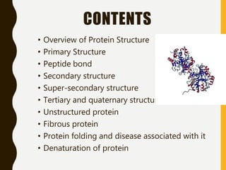
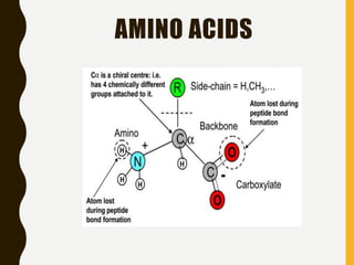
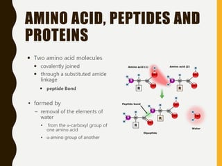
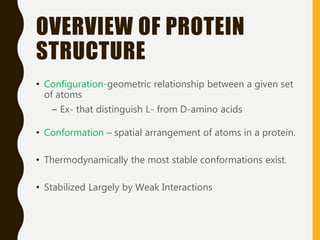
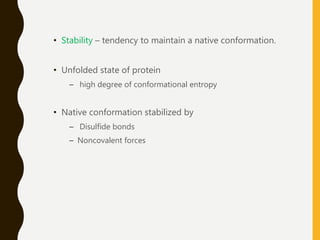
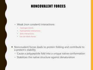
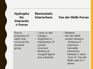
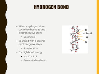
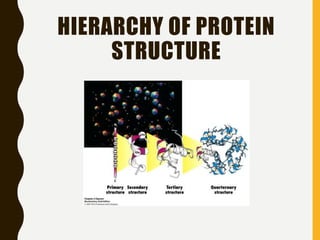
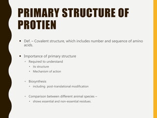
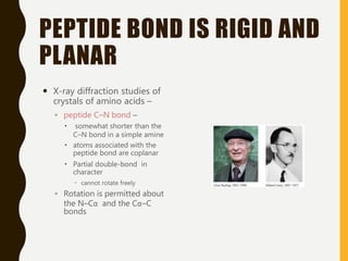
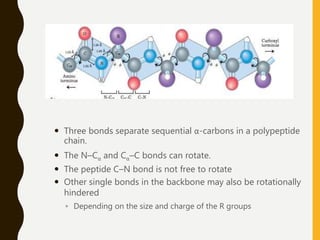
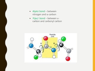
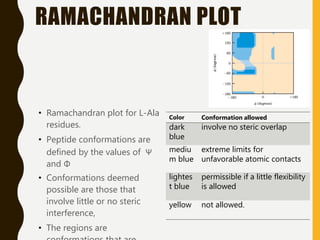
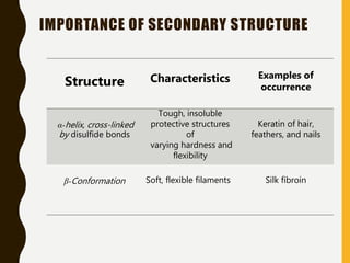
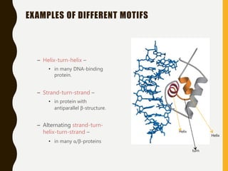
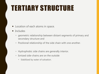
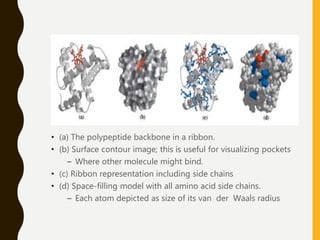
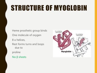
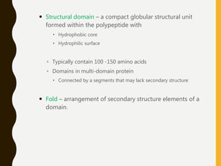
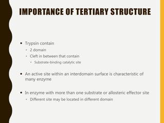
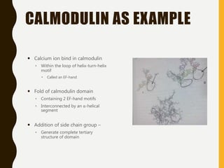
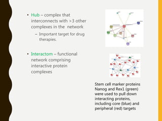
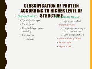
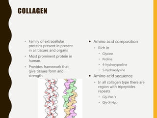
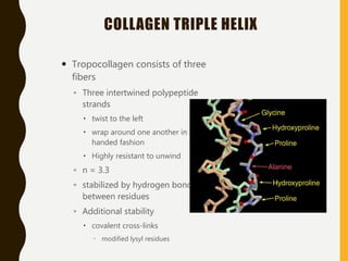
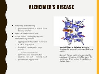
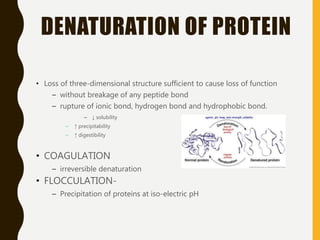
STRUCTURE OF
PROTEINS
CONTENTS
• Overview of Protein Structure
• Primary Structure
• Peptide bond
• Secondary structure
• Super-secondary structure
• Tertiary and quaternary structure
• Unstructured protein
• Fibrous protein
• Protein folding and disease associated with it
• Denaturation of protein
AMINO ACIDS
AMINO ACID, PEPTIDES AND
PROTEINS
 Two amino acid molecules
 covalently joined
 through a substituted amide
linkage
 peptide Bond
• formed by
– removal of the elements of
water
• from the α-carboxyl group of
one amino acid
• α-amino group of another
 When a few amino acids are joined in this fashion, the structure is
called an oligopeptide
 When many (10 to 50) amino acids are joined, the product is
called a polypeptide
 molecules referred to as polypeptides generally have molecular
weights below 10,000
 Those called proteins (having >50 amino acids) have higher
molecular weights.
OVERVIEW OF PROTEIN
STRUCTURE
• Configuration-geometric relationship between a given set
of atoms
– Ex- that distinguish L- from D-amino acids
• Conformation – spatial arrangement of atoms in a protein.
• Thermodynamically the most stable conformations exist.
• Stabilized Largely by Weak Interactions
• Stability – tendency to maintain a native conformation.
• Unfolded state of protein
– high degree of conformational entropy
• Native conformation stabilized by
– Disulfide bonds
– Noncovalent forces
NONCOVALENT FORCES
◦ Weak (non covalent) interactions
 Hydrogen bonds
 Hydrophobic interactions
 Ionic interactions
 Van der Walls forces
 Noncovalent forces leads to protein folding and contribute to
a protein’s stability
◦ Cause a polypeptide fold into a unique native conformation
◦ Stabilizes the native structure against denaturation
Hydropho
bic
Interactio
n Forces
Electrostatic
Interactions Van der Walls Forces
Due to
properties of
water that
surround the
nonpolar
group
◦ ( ionic or salt
linkage )
◦ Important in
stabilization of
protein
structure
◦ Binding of
charged ligand
and substrate
◦ Weakest
◦ Van der Walls
contact distance
Distance of
maximum
favorable
interaction
between 2 atom
Sum of Van der
Walls radii of 2
atoms
HYDROGEN BOND
– When a hydrogen atom
covalently bound to and
electronegative atom
• Donor atom
– is shared with a second
electronegative atom
• Acceptor atom
– For high bond energy
• d= 2.7 – 3.1Å
• Geometrically collinear
H- bond
O
H
N
HIERARCHY OF PROTEIN
STRUCTURE
PRIMARY STRUCTURE OF
PROTIEN
 Def. – Covalent structure, which includes number and sequence of amino
acids.
 Importance of primary structure
◦ Required to understand
 its structure
 Mechanism of action
◦ Biosynthesis
 including post-translational modification
◦ Comparison between different animal species –
 shows essential and non-essential residues.
SEQUENCE COMPARISON
• Used to predict the similarity in structure and function of protein. 2
sequences are –
– Homologous - when their sequence highly alignable
• Protein that evolved from same gene
– Analogy - structurally similar protein sequence, but no evolutionary
relationship found.
– Invariant residue - particular amino acid regularly found at same
position.
– Conservative substitution - by another amino acid with similar polarity
– Nonconservative substitution – by another amino acid with different
polarity
CLINICAL CORRELATION
• Sickle cell anemia
– HbS – a variant of normal hemoglobin
• Nonconservative mutation
– 6th position of β-globin gene
– Glutamic acid(polar) → Valine(nonpolar)
• So deoxy-HbS molecules get polymerized
– Precipitation of Hb within RBC
• Sickle shape and hemolysis
– HbC – Lysine is substituted for glutamic acid at
6th position (A3 helix) in β chain
– HbD – β121 (GH4) – Glu → Gln
PROTEOLYTIC CLEAVAGE OF INSULIN
 Proinsulin –
◦ Produced in pancreatic islet cells.
◦ Single polypeptide chain
containing
 86 amino acids
 3 intra-chain cystine disulfide bond
◦ Cleaved by proteases present in
islet cells
◦ Releases 2 molecule
 C-peptide – 35-residue fragments
 Insulin
 Insulin contain
◦ 2 polypeptide chain
 A – 21 AA
 B – 30 AA
◦ Covalently joined by the same
disulfide bond
DIFFERENT TYPE OF INSULIN USED
 Porcine insulin
◦ More acceptable than bovine insulin
 As sequence is more similar to human insulin
 Bovine insulin
 Human insulin –
◦ Primary insulin in developed country
◦ Prepared from
 Genetically engineered bacteria
 Modifying pork insulin
 Majority of the population are able to utilize animal insulin without
complication
◦ Because in amino acid sequence
 Small number and conservative nature of change
 They do not significantly change the 3-dimensional structure
PEPTIDE BOND IS RIGID AND
PLANAR
 X-ray diffraction studies of
crystals of amino acids –
◦ peptide C⎯N bond –
 somewhat shorter than the
C⎯N bond in a simple amine
 atoms associated with the
peptide bond are coplanar
 Partial double-bond in
character
 cannot rotate freely
◦ Rotation is permitted about
the N⎯Cα and the Cα⎯C
bonds
 Three bonds separate sequential α-carbons in a polypeptide
chain.
 The N⎯Cα and Cα⎯C bonds can rotate.
 The peptide C⎯N bond is not free to rotate
 Other single bonds in the backbone may also be rotationally
hindered
◦ Depending on the size and charge of the R groups
 Φ(phi) bond – between
nitrogen and α-carbon
 Ψ(psi) bond – between α-
carbon and carbonyl carbon
RAMACHANDRAN PLOT
• Ramachandran plot for L-Ala
residues.
• Peptide conformations are
defined by the values of Ψ
and Φ
• Conformations deemed
possible are those that
involve little or no steric
interference,
• The regions are
Color Conformation allowed
dark
blue
involve no steric overlap
mediu
m blue
extreme limits for
unfavorable atomic contacts
lightes
t blue
permissible if a little flexibility
is allowed
yellow not allowed.
SECONDARY STRUCTURE OF
PROTIEN
 particularly stable arrangements of amino
acid residues giving rise to recurring
structural patterns
– Local spatial arrangement of its main-chain atoms
– Confifurational relationship between tesidues which are
about 3-4 amino acids apart in linear sequence.
 Contributed by
◦ each amino acids.
◦ Φ-bond
◦ Ψ-bond
◦ α-helix and β-strand conformations – most thermodynamically stable.
REGULAR SECONDARY STRUCTURE
– Occurs in segments of a
polypeptide chain in which
– All Φ angles are equal.
– All Ψ angles are equal.
 Helical structures
 are characterized by
◦ n – number of residues per
turn of helix
◦ d – distance between α-carbon
atoms of adjacent amino acids
◦ pitch – distance between
repeating turn of helix on a
line drawn parallel to helix axis
 p = n × d
d
p
Α- HELICAL STRUCTURE
◦ Right-handed.
◦ n = 3.6
◦ Peptide bond plane –
parallel to the axis of helix.
◦ Each peptide form 2
hydrogen bonds
 peptide bond of 4th residue
above and below
◦ Optimum geometry and
distance for maximum
hydrogen bond strength.
H-bond
Β- STRUCTURE
– A polypeptide is hydrogen
bonded to another
polypeptide chain aligned
in a parallel or antiparallel
direction.
– Hydrogen-bonded β-
strands appear like a
pleated sheet.
Anti-parallel
Parallel
IMPORTANCE OF SECONDARY STRUCTURE
Structure Characteristics Examples of
occurrence
α-helix, cross-linked
by disulfide bonds
Tough, insoluble
protective structures
of
varying hardness and
flexibility
Keratin of hair,
feathers, and nails
β-Conformation Soft, flexible filaments Silk fibroin
SUPER-SECONDARY STRUCTURE
 Motif – recognizable folding pattern involving
◦ two or more elements of secondary structure
◦ connection(s) between them
 May or may not be independently stable
 Any advantageous folding pattern
 not a hierarchical structural element falling between secondary
and tertiary structure
EXAMPLES OF DIFFERENT MOTIFS
– Helix-turn-helix –
• in many DNA-binding
protein.
– Strand-turn-strand –
• in protein with
antiparallel β-structure.
– Alternating strand-turn-
helix-turn-strand –
• in many α/β-proteins
Helix
Helix
turn
TERTIARY STRUCTURE
 Location of each atoms in space.
 Includes
◦ geometric relationship between distant segments of primary and
secondary structure and
◦ Positional relationship of the side chain with one another.
◦ Hydrophobic side chains are generally interior.
◦ Ionized side-chains are on the outside
 Stabilized by water of solvation.
• (a) The polypeptide backbone in a ribbon.
• (b) Surface contour image; this is useful for visualizing pockets
– Where other molecule might bind.
• (c) Ribbon representation including side chains
• (d) Space-filling model with all amino acid side chains.
– Each atom depicted as size of its van der Waals radius
STRUCTURE OF MYOGLOBIN
Heme prosthetic group binds
One molecule of oxygen
8 α helices,
Rest forms turns and loops
due to
proline
No β sheets
 Structural domain – a compact globular structural unit
formed within the polypeptide with
 Hydrophobic core
 Hydrophilic surface
◦ Typically contain 100 -150 amino acids
◦ Domains in multi-domain protein
 Connected by a segments that may lack secondary structure
 Fold – arrangement of secondary structure elements of a
domain.
IMPORTANCE OF TERTIARY STRUCTURE
 Trypsin contain
◦ 2 domain
◦ Cleft in between that contain
 Substrate-binding catalytic site
 An active site within an interdomain surface is characteristic of
many enzyme
 In enzyme with more than one substrate or allosteric effector site
◦ Different site may be located in different domain
CALMODULIN AS EXAMPLE
 Calcium ion bind in calmodulin
◦ Within the loop of helix-turn-helix
motif
 Called an EF-hand
 Fold of calmodulin domain
◦ Containing 2 EF-hand motifs
◦ Interconnected by an α-helical
segment
 Addition of side chain group –
◦ Generate complete tertiary
structure of domain
QUATERNARY STRUCTURE
 Arrangement of polypeptide chain in multi-chain protein.
◦ Subunits in a quaternary structure are associated non-
covalently.
Quaternary structure of de-oxy
hemoglobin.
• X-ray diffraction-analysis of de-
oxy hemoglobin
• (a) A ribbon
representation.
• (b) A surface contour
model.
• The α-subunits are shown in
shades of gray
• The β-subunits in shades of
blue.
• Heme groups (red) are
relatively far apart.
STRUCTURE OF
HEMOGLOBIN
• Tetrameric Protein
• 2 α Globin Chains and 2 β
globin chains held by non-
covalent interactions
• Bind 4 molecules of oxygen
• Cooperative binding
UNSTRUCTURED PROTEIN
 Intrinsically unstructured protein – protein with a non-folded
conformation
 Partially unfolded conformation
 Ex –scaffold proteins, hormones, cyclin-dependant kinase and
their inhibitors
 Functions –by binding to other protein or to DNA and RNA
◦ The property of weak binding often advantageous
NEWER ADVANCES
 Protein Complexes
◦ Protein molecules in the
cellular milieu are present in
protein complexes
containing multiple protein
subunits.
◦ The complexes typically have
5-10 proteins.
 Network – proteins present
in 2 or more different
complexes can move
between complexes to
connect them into network.
• Hub – complex that
interconnects with >3 other
complexes in the network
– Important target for drug
therapies.
• Interactom – functional
network comprising
interactive protein
complexes
Stem cell marker proteins
Nanog and Rex1 (green)
were used to pull down
interacting proteins,
including core (blue) and
peripheral (red) targets
CLASSIFICATION OF PROTEIN
ACCORDING TO HIGHER LEVEL OF
STRUCTURE• Globular Protein –
– Spheroidal shape
– Vary in size
– Relatively high water
solubility.
– Function as
• catalyst
• Non-globular protein–
• Low water solubility
– Fibrous protein
• Larger amount of regular
secondary structure
• Long cylindrical shape
– Membranous protein
– Lipoprotein
– Glycoprotein
COLLAGEN
◦ Family of extracellular
proteins present in present
in all tissues and organs
◦ Most prominent protein in
human.
◦ Provides framework that
give tissues form and
strength.
 Amino acid composition
◦ Rich in
 Glycine
 Proline
 4-hydroxyproline
 5-hydroxylysine
 Amino acid sequence
◦ In all collagen type there are
region with tripeptides
repeats
 Gly-Pro-Y
 Gly-X-Hyp
COLLAGEN TRIPLE HELIX
 Tropocollagen consists of three
fibers
◦ Three intertwined polypeptide
strands
 twist to the left
 wrap around one another in a right-
handed fashion
 Highly resistant to unwind
◦ n = 3.3
◦ stabilized by hydrogen bonds
between residues
◦ Additional stability
 covalent cross-links
 modified lysyl residues
Disease Menkes'
syndrome
Ehlers-Danlos
syndrome
scurvy
etiology
dietary deficiency
of the copper
Genetic disease dietary deficiency of
vitamin C
defect
required by
lysyl oxidase
catalyzes
a key
step in
formatio
n of the
covalent
cross-
links
◦ defects in
the genes
that encode
collagen-
1
procollag
en N-
peptidase
 lysyl
hydroxyla
se
•prolyl and lysyl
hydroxylases
•deficit in the
number of
hydroxyproline and
hydroxylysine
residues
•undermines the
conformational
stability of collagen
fibers
Clinical
feature
kinky hair and
growth
retardation
◦ mobile joints
◦ skin
abnormalitie
s
bleeding gums
swelling joints
poor wound
healing
ultimately
death
ELASTIN
 Gives tissue and organ
capacity to stretch without
tearing
 Abundant in
◦ Ligaments
◦ Lungs
◦ Wall of arteries
◦ Skin
 Unordered coiled structure
◦ Amino acid residue within
folded structure highly
mobile
◦ Allysine form cross-links in
elastin
◦ Form the heterocyclic
structure of desmosine or
hemidesmosine
KERATIN
– in which each polypeptide is α-
helical
– Sequences in both proteins
shows tandem repetition of 7
residue segments
– Super twisted Coiled coil
– Rich in hydrophobic residues
– Left handed opposite in sense
to α helix
• Epidermal layer of skin
• Nails
• Hair
PROTEIN FOLDING
 Polypeptide sequence contain information for spontaneous
folding
 Folding is under
◦ thermodynamic
◦ kinetic control
 Initiated by short-range noncovalent interaction.
 Partially folded structure intermediate
◦ Interact with each other to form a molten-globule state
CHAPERON PROTEIN
 Also called ‘Heat shock protein’
 Synthesis increased at high temperature
 Prevent protein aggregation prior to completion of folding
 Bind to polypeptides shielding the hydrophobic surface
 Also required for refolding of protein after they cross cellular
membranes
PRION PROTEIN DISEASES
 Prion protein
◦ Infectious agent in absence of
DNA or RNA
◦ Can occur
 Spontaneously
 Inheritance of mutated Prion protein
◦ Clinical features
 Ataxia
 Dementia
 Paralysis
 Almost always fatal.
 deposition of insoluble protein
aggregates in neural cells
 include
◦ Creutzfeldt–Jakob disease in
humans
◦ scrapie in sheep
◦ bovine spongiform
encephalopathy (mad cow disease)
in cattle
◦ vCJD
 younger patients
 PrPc - highly soluble cellular
conformation of prion protein
◦ glycoprotein
◦ short arm of chromosome 20
◦ 3 α-helical
◦ 2 small β-strand
◦ rich in α-helix
 PrPsc - insoluble toxic conformation
◦ Conversion of α-helix → β-strand
◦ many hydrophobic aminoacyl side chains
exposed to solvent
◦ accumulating PrPsc units coalesce
 insoluble protease-resistant aggregates
◦ serve as template for
 conformational transformation of PrPc
molecules
 Many times of its number
BETA-THALASSEMIAS
 genetic defects
◦ impair the synthesis of one
of the polypeptide
subunits of hemoglobin
 α-hemoglobin-stabilizing
protein (AHSP)
◦ specific chaperone
◦ binds to free hemoglobin -
subunits
 awaiting incorporation into
the hemoglobin multimer
 absence of this chaperone
◦ free α-hemoglobin
subunits aggregate
◦ role for AHSP in
modulating the severity of
-thalassemia in human
subjects.
ALZHEIMER'S DISEASE
 Refolding or misfolding
◦ protein endogenous to human brain
tissue, β-amyloid
 Main cause remains elusive
 characteristic senile plaques and
neurofibrillary bundles
◦ aggregates of the protein β-amyloid
◦ 4.3-kDa polypeptide
◦ Proteolytic cleavage of a larger
protein
 amyloid precursor protein
◦ conformational transformation
soluble α-helix–rich → rich in β-sheet
◦ prone to self-aggregation
amyloid fibers in Alzheimer's - Crystal
structure of a segment from the amyloid-beta
protein
Normally the two protein sheets are tightly
associated in the spine of the fiber but in this
case orange-G has wedged its way between
the two sheets.
DENATURATION OF PROTEIN
• Loss of three-dimensional structure sufficient to cause loss of function
– without breakage of any peptide bond
– rupture of ionic bond, hydrogen bond and hydrophobic bond.
– ↓ solubility
– ↑ precipitability
– ↑ digestibility
• COAGULATION
– irreversible denaturation
• FLOCCULATION-
– Precipitation of proteins at iso-electric pH
Structure of protiens and the applied aspects

Structure of protiens and the applied aspects

  • 1.
  • 2.
    CONTENTS • Overview ofProtein Structure • Primary Structure • Peptide bond • Secondary structure • Super-secondary structure • Tertiary and quaternary structure • Unstructured protein • Fibrous protein • Protein folding and disease associated with it • Denaturation of protein
  • 3.
  • 4.
    AMINO ACID, PEPTIDESAND PROTEINS  Two amino acid molecules  covalently joined  through a substituted amide linkage  peptide Bond • formed by – removal of the elements of water • from the α-carboxyl group of one amino acid • α-amino group of another
  • 5.
     When afew amino acids are joined in this fashion, the structure is called an oligopeptide  When many (10 to 50) amino acids are joined, the product is called a polypeptide  molecules referred to as polypeptides generally have molecular weights below 10,000  Those called proteins (having >50 amino acids) have higher molecular weights.
  • 6.
    OVERVIEW OF PROTEIN STRUCTURE •Configuration-geometric relationship between a given set of atoms – Ex- that distinguish L- from D-amino acids • Conformation – spatial arrangement of atoms in a protein. • Thermodynamically the most stable conformations exist. • Stabilized Largely by Weak Interactions
  • 7.
    • Stability –tendency to maintain a native conformation. • Unfolded state of protein – high degree of conformational entropy • Native conformation stabilized by – Disulfide bonds – Noncovalent forces
  • 8.
    NONCOVALENT FORCES ◦ Weak(non covalent) interactions  Hydrogen bonds  Hydrophobic interactions  Ionic interactions  Van der Walls forces  Noncovalent forces leads to protein folding and contribute to a protein’s stability ◦ Cause a polypeptide fold into a unique native conformation ◦ Stabilizes the native structure against denaturation
  • 9.
    Hydropho bic Interactio n Forces Electrostatic Interactions Vander Walls Forces Due to properties of water that surround the nonpolar group ◦ ( ionic or salt linkage ) ◦ Important in stabilization of protein structure ◦ Binding of charged ligand and substrate ◦ Weakest ◦ Van der Walls contact distance Distance of maximum favorable interaction between 2 atom Sum of Van der Walls radii of 2 atoms
  • 10.
    HYDROGEN BOND – Whena hydrogen atom covalently bound to and electronegative atom • Donor atom – is shared with a second electronegative atom • Acceptor atom – For high bond energy • d= 2.7 – 3.1Å • Geometrically collinear H- bond O H N
  • 11.
  • 12.
    PRIMARY STRUCTURE OF PROTIEN Def. – Covalent structure, which includes number and sequence of amino acids.  Importance of primary structure ◦ Required to understand  its structure  Mechanism of action ◦ Biosynthesis  including post-translational modification ◦ Comparison between different animal species –  shows essential and non-essential residues.
  • 13.
    SEQUENCE COMPARISON • Usedto predict the similarity in structure and function of protein. 2 sequences are – – Homologous - when their sequence highly alignable • Protein that evolved from same gene – Analogy - structurally similar protein sequence, but no evolutionary relationship found. – Invariant residue - particular amino acid regularly found at same position. – Conservative substitution - by another amino acid with similar polarity – Nonconservative substitution – by another amino acid with different polarity
  • 14.
    CLINICAL CORRELATION • Sicklecell anemia – HbS – a variant of normal hemoglobin • Nonconservative mutation – 6th position of β-globin gene – Glutamic acid(polar) → Valine(nonpolar) • So deoxy-HbS molecules get polymerized – Precipitation of Hb within RBC • Sickle shape and hemolysis – HbC – Lysine is substituted for glutamic acid at 6th position (A3 helix) in β chain – HbD – β121 (GH4) – Glu → Gln
  • 15.
    PROTEOLYTIC CLEAVAGE OFINSULIN  Proinsulin – ◦ Produced in pancreatic islet cells. ◦ Single polypeptide chain containing  86 amino acids  3 intra-chain cystine disulfide bond ◦ Cleaved by proteases present in islet cells ◦ Releases 2 molecule  C-peptide – 35-residue fragments  Insulin  Insulin contain ◦ 2 polypeptide chain  A – 21 AA  B – 30 AA ◦ Covalently joined by the same disulfide bond
  • 16.
    DIFFERENT TYPE OFINSULIN USED  Porcine insulin ◦ More acceptable than bovine insulin  As sequence is more similar to human insulin  Bovine insulin  Human insulin – ◦ Primary insulin in developed country ◦ Prepared from  Genetically engineered bacteria  Modifying pork insulin  Majority of the population are able to utilize animal insulin without complication ◦ Because in amino acid sequence  Small number and conservative nature of change  They do not significantly change the 3-dimensional structure
  • 17.
    PEPTIDE BOND ISRIGID AND PLANAR  X-ray diffraction studies of crystals of amino acids – ◦ peptide C⎯N bond –  somewhat shorter than the C⎯N bond in a simple amine  atoms associated with the peptide bond are coplanar  Partial double-bond in character  cannot rotate freely ◦ Rotation is permitted about the N⎯Cα and the Cα⎯C bonds
  • 18.
     Three bondsseparate sequential α-carbons in a polypeptide chain.  The N⎯Cα and Cα⎯C bonds can rotate.  The peptide C⎯N bond is not free to rotate  Other single bonds in the backbone may also be rotationally hindered ◦ Depending on the size and charge of the R groups
  • 19.
     Φ(phi) bond– between nitrogen and α-carbon  Ψ(psi) bond – between α- carbon and carbonyl carbon
  • 20.
    RAMACHANDRAN PLOT • Ramachandranplot for L-Ala residues. • Peptide conformations are defined by the values of Ψ and Φ • Conformations deemed possible are those that involve little or no steric interference, • The regions are Color Conformation allowed dark blue involve no steric overlap mediu m blue extreme limits for unfavorable atomic contacts lightes t blue permissible if a little flexibility is allowed yellow not allowed.
  • 21.
    SECONDARY STRUCTURE OF PROTIEN particularly stable arrangements of amino acid residues giving rise to recurring structural patterns – Local spatial arrangement of its main-chain atoms – Confifurational relationship between tesidues which are about 3-4 amino acids apart in linear sequence.  Contributed by ◦ each amino acids. ◦ Φ-bond ◦ Ψ-bond ◦ α-helix and β-strand conformations – most thermodynamically stable.
  • 22.
    REGULAR SECONDARY STRUCTURE –Occurs in segments of a polypeptide chain in which – All Φ angles are equal. – All Ψ angles are equal.  Helical structures  are characterized by ◦ n – number of residues per turn of helix ◦ d – distance between α-carbon atoms of adjacent amino acids ◦ pitch – distance between repeating turn of helix on a line drawn parallel to helix axis  p = n × d d p
  • 23.
    Α- HELICAL STRUCTURE ◦Right-handed. ◦ n = 3.6 ◦ Peptide bond plane – parallel to the axis of helix. ◦ Each peptide form 2 hydrogen bonds  peptide bond of 4th residue above and below ◦ Optimum geometry and distance for maximum hydrogen bond strength. H-bond
  • 24.
    Β- STRUCTURE – Apolypeptide is hydrogen bonded to another polypeptide chain aligned in a parallel or antiparallel direction. – Hydrogen-bonded β- strands appear like a pleated sheet. Anti-parallel Parallel
  • 25.
    IMPORTANCE OF SECONDARYSTRUCTURE Structure Characteristics Examples of occurrence α-helix, cross-linked by disulfide bonds Tough, insoluble protective structures of varying hardness and flexibility Keratin of hair, feathers, and nails β-Conformation Soft, flexible filaments Silk fibroin
  • 26.
    SUPER-SECONDARY STRUCTURE  Motif– recognizable folding pattern involving ◦ two or more elements of secondary structure ◦ connection(s) between them  May or may not be independently stable  Any advantageous folding pattern  not a hierarchical structural element falling between secondary and tertiary structure
  • 27.
    EXAMPLES OF DIFFERENTMOTIFS – Helix-turn-helix – • in many DNA-binding protein. – Strand-turn-strand – • in protein with antiparallel β-structure. – Alternating strand-turn- helix-turn-strand – • in many α/β-proteins Helix Helix turn
  • 28.
    TERTIARY STRUCTURE  Locationof each atoms in space.  Includes ◦ geometric relationship between distant segments of primary and secondary structure and ◦ Positional relationship of the side chain with one another. ◦ Hydrophobic side chains are generally interior. ◦ Ionized side-chains are on the outside  Stabilized by water of solvation.
  • 29.
    • (a) Thepolypeptide backbone in a ribbon. • (b) Surface contour image; this is useful for visualizing pockets – Where other molecule might bind. • (c) Ribbon representation including side chains • (d) Space-filling model with all amino acid side chains. – Each atom depicted as size of its van der Waals radius
  • 30.
    STRUCTURE OF MYOGLOBIN Hemeprosthetic group binds One molecule of oxygen 8 α helices, Rest forms turns and loops due to proline No β sheets
  • 31.
     Structural domain– a compact globular structural unit formed within the polypeptide with  Hydrophobic core  Hydrophilic surface ◦ Typically contain 100 -150 amino acids ◦ Domains in multi-domain protein  Connected by a segments that may lack secondary structure  Fold – arrangement of secondary structure elements of a domain.
  • 32.
    IMPORTANCE OF TERTIARYSTRUCTURE  Trypsin contain ◦ 2 domain ◦ Cleft in between that contain  Substrate-binding catalytic site  An active site within an interdomain surface is characteristic of many enzyme  In enzyme with more than one substrate or allosteric effector site ◦ Different site may be located in different domain
  • 33.
    CALMODULIN AS EXAMPLE Calcium ion bind in calmodulin ◦ Within the loop of helix-turn-helix motif  Called an EF-hand  Fold of calmodulin domain ◦ Containing 2 EF-hand motifs ◦ Interconnected by an α-helical segment  Addition of side chain group – ◦ Generate complete tertiary structure of domain
  • 34.
    QUATERNARY STRUCTURE  Arrangementof polypeptide chain in multi-chain protein. ◦ Subunits in a quaternary structure are associated non- covalently. Quaternary structure of de-oxy hemoglobin. • X-ray diffraction-analysis of de- oxy hemoglobin • (a) A ribbon representation. • (b) A surface contour model. • The α-subunits are shown in shades of gray • The β-subunits in shades of blue. • Heme groups (red) are relatively far apart.
  • 35.
    STRUCTURE OF HEMOGLOBIN • TetramericProtein • 2 α Globin Chains and 2 β globin chains held by non- covalent interactions • Bind 4 molecules of oxygen • Cooperative binding
  • 36.
    UNSTRUCTURED PROTEIN  Intrinsicallyunstructured protein – protein with a non-folded conformation  Partially unfolded conformation  Ex –scaffold proteins, hormones, cyclin-dependant kinase and their inhibitors  Functions –by binding to other protein or to DNA and RNA ◦ The property of weak binding often advantageous
  • 37.
    NEWER ADVANCES  ProteinComplexes ◦ Protein molecules in the cellular milieu are present in protein complexes containing multiple protein subunits. ◦ The complexes typically have 5-10 proteins.  Network – proteins present in 2 or more different complexes can move between complexes to connect them into network.
  • 38.
    • Hub –complex that interconnects with >3 other complexes in the network – Important target for drug therapies. • Interactom – functional network comprising interactive protein complexes Stem cell marker proteins Nanog and Rex1 (green) were used to pull down interacting proteins, including core (blue) and peripheral (red) targets
  • 39.
    CLASSIFICATION OF PROTEIN ACCORDINGTO HIGHER LEVEL OF STRUCTURE• Globular Protein – – Spheroidal shape – Vary in size – Relatively high water solubility. – Function as • catalyst • Non-globular protein– • Low water solubility – Fibrous protein • Larger amount of regular secondary structure • Long cylindrical shape – Membranous protein – Lipoprotein – Glycoprotein
  • 40.
    COLLAGEN ◦ Family ofextracellular proteins present in present in all tissues and organs ◦ Most prominent protein in human. ◦ Provides framework that give tissues form and strength.  Amino acid composition ◦ Rich in  Glycine  Proline  4-hydroxyproline  5-hydroxylysine  Amino acid sequence ◦ In all collagen type there are region with tripeptides repeats  Gly-Pro-Y  Gly-X-Hyp
  • 41.
    COLLAGEN TRIPLE HELIX Tropocollagen consists of three fibers ◦ Three intertwined polypeptide strands  twist to the left  wrap around one another in a right- handed fashion  Highly resistant to unwind ◦ n = 3.3 ◦ stabilized by hydrogen bonds between residues ◦ Additional stability  covalent cross-links  modified lysyl residues
  • 42.
    Disease Menkes' syndrome Ehlers-Danlos syndrome scurvy etiology dietary deficiency ofthe copper Genetic disease dietary deficiency of vitamin C defect required by lysyl oxidase catalyzes a key step in formatio n of the covalent cross- links ◦ defects in the genes that encode collagen- 1 procollag en N- peptidase  lysyl hydroxyla se •prolyl and lysyl hydroxylases •deficit in the number of hydroxyproline and hydroxylysine residues •undermines the conformational stability of collagen fibers Clinical feature kinky hair and growth retardation ◦ mobile joints ◦ skin abnormalitie s bleeding gums swelling joints poor wound healing ultimately death
  • 43.
    ELASTIN  Gives tissueand organ capacity to stretch without tearing  Abundant in ◦ Ligaments ◦ Lungs ◦ Wall of arteries ◦ Skin  Unordered coiled structure ◦ Amino acid residue within folded structure highly mobile ◦ Allysine form cross-links in elastin ◦ Form the heterocyclic structure of desmosine or hemidesmosine
  • 44.
    KERATIN – in whicheach polypeptide is α- helical – Sequences in both proteins shows tandem repetition of 7 residue segments – Super twisted Coiled coil – Rich in hydrophobic residues – Left handed opposite in sense to α helix • Epidermal layer of skin • Nails • Hair
  • 45.
    PROTEIN FOLDING  Polypeptidesequence contain information for spontaneous folding  Folding is under ◦ thermodynamic ◦ kinetic control  Initiated by short-range noncovalent interaction.  Partially folded structure intermediate ◦ Interact with each other to form a molten-globule state
  • 46.
    CHAPERON PROTEIN  Alsocalled ‘Heat shock protein’  Synthesis increased at high temperature  Prevent protein aggregation prior to completion of folding  Bind to polypeptides shielding the hydrophobic surface  Also required for refolding of protein after they cross cellular membranes
  • 47.
    PRION PROTEIN DISEASES Prion protein ◦ Infectious agent in absence of DNA or RNA ◦ Can occur  Spontaneously  Inheritance of mutated Prion protein ◦ Clinical features  Ataxia  Dementia  Paralysis  Almost always fatal.  deposition of insoluble protein aggregates in neural cells  include ◦ Creutzfeldt–Jakob disease in humans ◦ scrapie in sheep ◦ bovine spongiform encephalopathy (mad cow disease) in cattle ◦ vCJD  younger patients
  • 48.
     PrPc -highly soluble cellular conformation of prion protein ◦ glycoprotein ◦ short arm of chromosome 20 ◦ 3 α-helical ◦ 2 small β-strand ◦ rich in α-helix  PrPsc - insoluble toxic conformation ◦ Conversion of α-helix → β-strand ◦ many hydrophobic aminoacyl side chains exposed to solvent ◦ accumulating PrPsc units coalesce  insoluble protease-resistant aggregates ◦ serve as template for  conformational transformation of PrPc molecules  Many times of its number
  • 49.
    BETA-THALASSEMIAS  genetic defects ◦impair the synthesis of one of the polypeptide subunits of hemoglobin  α-hemoglobin-stabilizing protein (AHSP) ◦ specific chaperone ◦ binds to free hemoglobin - subunits  awaiting incorporation into the hemoglobin multimer  absence of this chaperone ◦ free α-hemoglobin subunits aggregate ◦ role for AHSP in modulating the severity of -thalassemia in human subjects.
  • 50.
    ALZHEIMER'S DISEASE  Refoldingor misfolding ◦ protein endogenous to human brain tissue, β-amyloid  Main cause remains elusive  characteristic senile plaques and neurofibrillary bundles ◦ aggregates of the protein β-amyloid ◦ 4.3-kDa polypeptide ◦ Proteolytic cleavage of a larger protein  amyloid precursor protein ◦ conformational transformation soluble α-helix–rich → rich in β-sheet ◦ prone to self-aggregation amyloid fibers in Alzheimer's - Crystal structure of a segment from the amyloid-beta protein Normally the two protein sheets are tightly associated in the spine of the fiber but in this case orange-G has wedged its way between the two sheets.
  • 51.
    DENATURATION OF PROTEIN •Loss of three-dimensional structure sufficient to cause loss of function – without breakage of any peptide bond – rupture of ionic bond, hydrogen bond and hydrophobic bond. – ↓ solubility – ↑ precipitability – ↑ digestibility • COAGULATION – irreversible denaturation • FLOCCULATION- – Precipitation of proteins at iso-electric pH